Published by Oriental Scientific Publishing Company © 2019
This is an Open Access article licensed under a Creative Commons license: Attribution 4.0 International (CC-BY).
Evaluation of Potentiation Effect of Omega-3 Fatty Acid on the
Antidepressant Efficacy of Escitalopram in Albino Rats
Amberkar Mohanbabu Vittalrao1, Saurabh Agarwal2, Meena Kumari Kamalkishore1* and Basavaraj Poojar3
1Department of Pharmacology, Kasturba Medical College, Manipal, Manipal Academy of Higher Education, Manipal, Karnataka - 576104, India. 2Medical Science Liaison,Sanofi Pasteur India Pvt.Ltd,Dalibagh,Lucknow-226001, India. 3Department of Pharmacology, Kasturba Medical College, Mangaluru, Manipal Academy
of Higher Education, Manipal, Karnataka - 575001, India. *Corresponding author E-mail: mini41178@yahoo.co.in
http://dx.doi.org/10.13005/bpj/1777
(Received: 20 June 2019; accepted: 16 September 2019)
Depression is a common problem worldwide since the ages. Mostly it is treated with Selective serotonin reuptake inhibitors (SSRI) but they are not effective in each and every patient. Hence other methods for better effective ways to treat depression are needed. This study was divided as acute and chronic study. Each containing five groups control, escitalopram (standard),omega -3 fatty acid (FA),escitalopram + omega -3 FA (2 doses).A 15 min pretest was done followed 24h later by a 5 min test.Various models of depression were used and biochemical analysis was done. In acute study there was no significant potentiation effect seen .In chronic study there was significant potentiation effect of omega 3 FA. Based on the results we conclude omega 3 fatty acid can be considered as a part of therapeutic use along with escitalopram. Further clinical studies may be required to validate the results of this study.
Keywords: Selective serotonin reuptake inhibitor; Forced swimming test; Open-field test; Splash test; depression.
Depression is a mental disorder which is common , manifesting with depressed mood, anhedonia, lethargy, feelings of guilt or low self-esteem, disturbed sleep or appetite and poor concentration.It is estimated by the World Health Organisation that depression affects approximately 350 million people worldwide is associated with increasing morbidity1. A recent study has shown
that depression is one of the ten leading causes of disabilities affecting up to 21% of the world population and will be the second leading cause of disability adjusted life years (DALY) by 20202.
It has high impact on social and economic aspect, due to massive decrease in work productivity and increased usage of health care3. Enhancement
current pharmacological treatment of depression. However, despite its widespread use in clinical practice, it has challenges like onset of action, poor efficacy and presence of side effects4,5. Contrary
to expectations, existing therapeutic options are effective in only one-third of depressed patients. Moreover, the time required for its maximal antidepressant activity is approximately 3–4 weeks6. Furthermore, single agent does not have
more than 30% of remission rates,7.
Majority of patients have low compliance due to side effects and refuse to take antidepressants in appropriate doses.Therefore, the identification of novel drug or novel treatment combination with drugs that augment the efficacy of antidepressant is still needed. Role of nutrition has found greater role in depressive disorder as good amount of nutrients are essential for healthy mood8-10.
Nutrients are essential for optimal production of neurotransmitters influencing mood11. There
are reports mentioning lower concentration of Eicosapentaenoic acid(EPA) and Docosahexaenoic acid (DHA) in depressed individuals as compared to non-depressed individuals12,13.
Meta-analytic reviews14,15 and several
clinical trials16-19 have reported an antidepressant
effect of Polyunsaturated fatty acids(PUFAs). There is increased evidence from animal as well as human studies showing omega-3 fatty acids may play an etiological role in several inflammatory, autoimmune and neuropsychiatric disorders20,21.
Hence this study was conducted with an aim to explore the possible effect of omega-3 fatty acids in depression animal models.
Materials and Method
animals
Sixty Albino Wistar rats of either sex (from Central animal house,KMC,Manipal) were used in the study. The rats used were 3-4 months old and weight 150–250 gm. Animals were housed in cages and were provided with pellet diet and water ad libitum throughout the study except during testing. Rats were maintained on a 12 h
light (0700–1900 h)-12 h dark cycle at temperature of 25±2◦C and relative controlled humidity. They were allowed to acclimatize for a week before the onset of the experiment. Experiment was conducted in accordance with the Good laboratory practice(GLP) guidelines and CPCSEA guidelines after obtaining approval from Institutional Animal Ethics Committee IAEC/KMC/79/2015 dated 25-09-2015.
drugs
Escitalopram: 10 mg per kg and 5 mg per kg. Omega-3 Fatty Acid: 500 mg per kg.
study design acute study
There were total five groups, in each group 6 rats. Drugs were administered for 7 days as follows:
Group 1 (Control) – 2% Gum Acacia orally 23.5 and 1 h before the test.
Group 2 (Escitalopram) – 10 mg/kg dissolved in 2% Gum acacia orally 23.5 and 1 h before the test.
Group 3 (Omega-3 FA) – 500 mg/kg in 2% Gum acacia orally 2 h before the test.
Group 4 (Escitalopram plus omega-3 FA)- Rats were given omega-3 fatty acids 500 mg/kg orally 2 h before the test and escitalopram dissolved in 2% Gum acacia in a dose of 10 mg/ kg orally 23.5 and 1 h before the test.
Group 5 (Escitalopram plus omega-3 FA)-This group test the efficacy and augmentation effect of omega-3 FA with sub therapeutic level of escitalopram. Rats were administered with omega-3 fatty acids 500 mg/kg orally 2 h before the test and escitalopram dissolved in 2% Gum acacia in a dose of 5 mg/kg orally 23.5 and 1 h before the test.
Chronic study
experiments
Forced swimming test(Fst)
Porsolt et al.developed this model for antidepressant activity22. Rats were made to swim
in a cylinder filled with water upto 30 cm at 25̊C. They were trained for a 15-min pretest swim followed by a 5-min test swim session next day. After the swim session, rats were removed from water, and after 20 min were returned to their cages. After each test,the cylinders were washed completely. In pretest session, rats are active.After 2-3 min activity decreases to produce immobility, it keeps its head above water. This immobility is attenuated by antidepressants. In the test session,the behavior of rats were judged in blocks of 5 s for: immobility ,swimming & climbing .The experiment was videorecorded and the behavioral scoring was done .
Open-field test
This test was used to assess the ambulatory behavior23. The instrument consist of a wooden box
(40cm×60cm×50cm) ,its floor was divided into 12 equal rectangles. At the initiation of each trial, a rat was placed in the left corner. In a 6 min session,the number of rectangles crossed with all paws were counted. The room was dimly lit to prevent anxiety behavior. In between the tests,the instrument was
cleaned with 10% ethanol to avoid bias.
splash test
This test was performed ten minutes after the open-field test,24 with minor modifications
.Ten percent sucrose solution was applied on the dorsum of a rat placed individually in plexiglass boxes (9×7×11 cm). The sucrose solution dirties the rat fur ,due to its viscosity and initiates grooming behavior in animals which was recorded for 5 min as an index of self-care and motivational behavior. To hide animal clues,the apparatus was cleaned with 10% ethanol.
Biochemical analysis
After conducting the experiment, rats were sacrified and blood was used for testing –
Reduced glutathione (GSH) and Malondialdehyde (MDA) levels.
statistical analysis
P value <0.05 was taken as significant, one-way ANOVA followed by Tukey’s post-hoc test was used.
results
acute study
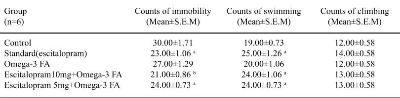
Table 1 shows the results of forced swimming test done at 1 week. One count is
table 1. Effect of escitalopram and omega-3 fatty acid on forced swimming
Group Counts of immobility Counts of swimming Counts of climbing
(n=6) (Mean±S.E.M) (Mean±S.E.M) (Mean±S.E.M)
Control 30.00±1.71 19.00±0.73 12.00±0.58
Standard(escitalopram) 23.00±1.06 a 25.00±1.26 a 14.00±0.58
Omega-3 FA 27.00±1.29 20.00±1.06 12.00±0.58
Escitalopram10mg+Omega-3 FA 21.00±0.86 b 24.00±1.06 a 13.00±0.58
Escitalopram 5mg+Omega-3 FA 24.00±0.73 a 24.00±0.73 a 13.00±0.58
ap<0.05 vscontrol group, bp<0.001 vs control group
duration of 5 sec. The standard group (escitalopram) showed a significant reduction in the counts of immobility (23.00±1.06, p<0.05) along with a significant increase in the counts of swimming (25.00±1.26, p<0.05) compared to the control
3 FA group showed a significant reduction in the counts of immobility (21.00±0.86, p<0.001; 24.00±0.73, p<0.05 respectively) with a significant increase swimming counts in both the groups (24.00±1.06, p<0.05; 24.00±0.73, p<0.05) when compared to the control and escitalopram group. There were no significant increase in climbing counts in any of the groups when compared to control.
table 3. Effect of escitalopram and omega-3 fatty acid on splash test
Group Time in sec.
(n=6) (Mean±S.E.M)
Control 201.00±5.67 Escitalopram 187.00±4.47
Omega-3 FA 201.33±5.50
Escitalaopram.10mg+omega-3 FA 163.00±3.25 b,c
Escitalopram 5mg+omega-3 FA 174.83±3.27 a
a p<0.05 versus group control, bp<0.001 versus control, c- escitalopram 10mg+ omega 3 group compared to escitalopram group;
table 4. Effect of escitalopram and omega-3 fatty acid on MDA and GSH
S.No. Group (n=6) MDA (Mean±S.E.M) GSH (Mean±S.E.M)
1. Control 0.047±0.003 1.141±0.013
2. Escitalopram 0.041±0.001 1.327±0.015a
3. Omega-3 FA 0.045±0.003 1.180±0.007
4. Escitalopram.10mg+omega-3 FA 0.051±0.002 1.525±0.017 a
5. Escitalopram 5mg+omega-3 FA 0.041±0.002 1.345±0.014 a
ap<0.05 versus group control table 2. Effect of escitalopram and omega-3 fatty acid on open field test
Group Number of entries
(n=6) (Mean±S.E.M)
Control 33.50±1.31
Escitalopram 39.50±3.10
Omega -3 FA 29.17±2.73
Escitalopram10mg+omega-3 FA 40.33±2.59
Escitalopram5mg+omega-3 FA 37.33±1.76
Table 2 shows the results of the open field test done at 1 week. The above results does not show any significant difference in the number of entries in any of the groups when compared with control and the standard treatment group (escitalopram).
Table 3 shows the splash test done at 1 week. It indicates that there is a significant difference in the time taken by the escitalopram 10mg+omega-3 FA group and escitalopram 5 mg+ omega 3 group when compared to control (163.00±3.25, p<0.001 and 174.83±3.27, p< 0.05) along with a significant reduction in time taken by the escitalopram 10mg + omega -3 FA group when compared with the standard treatment group (escitalopram) (163.00±3.25, p<0.05).
As shown in table 4 ,there was no significant difference among the control group and the test groups p<0.05 with respect to MDA levels. Levels of glutathione reductase at 1 week was significantly more in the escitalopram group (1.327±0.015, p<0.05 and significantly increase in GSH in test groups, escitalopram 10mg+omega -3 FA (1.525±0.017; p<0.05) and escitalopram 5 mg+omega-3 FA (1.345±0.014; p<0.05) compared to control group
Chronic study
swimming (26.00±1.06, p<0.001) compared to the control group. However, the omega 3 FA group did not show any significant difference in the counts of immobility, counts of swimming and counts of
climbing. The escitalopram 10mg + omega 3 group as well as the escitalopram 5mg + omega-3 FA group showed a significant reduction in the counts of immobility (15.00±0.73, p<0.001; 17.00±0.58,
table 6. Effect of escitalopram and omega-3 fatty acid on open field test
Group Number of entries
(n=6) (Mean±S.E.M)
Control 34.67±1.70
Escitalopram 44.00±1.26 a
Omega-3 FA 41.16±1.90
Escitalopram 10mg+omega-3 FA 47.00±2.67 a
Escitalopram 5mg+omega-3 FA 39.00±2.62
a- P<0.05 versus control group
table 7. Effect of escitalopram and omega-3 fatty acid on splash test
Group Time in secs.
(n=6) (Mean±S.E.M)
Control 210.00±11.94
Escitalopram 182.00±5.46a
Omega-3FA 196.00±4.70
Escitalopram 10mg+omega-3 FA 154.00±2.12b
Escitalopram 5mg+omega-3 FA 168.00±4.03 b
a p<0.05 vs control group;b p<0.001 vs control group
p<0.001 respectively) with a significant increase swimming counts in both the groups (27.33±1.30, p<0.001; 27.00±1.24, p<0.001 respectively) in comparison to the control and escitalopram group. There were no significant increase in climbing counts in any of the groups in comparison to control.
Table 6 .The number of entries increased significantly in the standard group escitalopram (44.00±1.26; p<0.05) when compared with control along with significant increase in the number of
entries in the escitalopram 10mg+omega-3 FA (47.00±2.67; p<0.05) group when compared to control.
As shown in table 7 there was a significant decrease in time in test groups escitalopram (182.00±5.46; p<0.05), escitalopram 10mg+omega-3 FA (154.00±2.12; p<0.001) and escitalopram 5mg+omega-3 FA (168.00±4.03; p<0.001) when compared to control.
As shown in table 8 there was significant increase in MDA in the test group escitalopram table 5. Effect of escitalopram and omega-3 fatty acid on forced swimming test
Group (n=6) Count of immobility Count of swimming Counts of climbing
(Mean±S.E.M) (Mean±S.E.M) (Mean±S.E.M)
Control 29.00±0.96 17.00±0.73 12.00±1.06
Escitalopram 21.00±1.43a 26.00±1.06 a 13.50±0.89
Omega-3 FA 25.00±0.89 23.00±0.86 13.00±1.13
Escitalopram 10mg+omega-3 FA 15.00±0.73 a 27.33±1.30 a 14.33±1.20
Escitalopram 5mg+omega-3 FA 17.00±0.58 a 27.00±1.24 a 13.50±0.96
10mg+omega-3 FA(0.100±0.003;p<0.05) when compared with the control. There was increase in GSH with a significant difference in the standard groups escitalopram (1.410±0.014; p<0.001). The test groups escitalopram 10mg+omega-3 FA (1.625±0.017; p<0.001) and ecitalopram 5mg+omega-3 FA (1.445±0.014; p<0.001) also showed a significant increase in GSH when compared to the control group.
disCussion
This study shows that Omega-3 fatty acids possess significant antidepressant efficacy in their combination with escitalopram. The study was divided into 2 parts acute (1 week) and chronic study (4 weeks).The results showed that omega-3 FA has no effect in short term either alone or in potentiating the escitalopram but in chronic study done over 4 weeks it showed potentiation effect with escitalopram.
The results are consistent with that of previous studies which have reported good efficacy of omega-3 supplementation with antidepressants in uncomplicated MDD.17, 25, 26 The present study
shows the antidepressant effect of omega-3 fatty acids, and its combination with escitalopram will potentiate the antidepressant efficacy than the standard drug escitalopram alone.
The suggestive possible mechanisms are as follows. Inflammatory processes are linked with depression. Omega-3 fatty acids has an effect on brain-derived neurotrophic factor and reduces
inflammatory mediators27. A third mechanism is
omega-3 fatty acids maintain membrane integrity and fluidity28. Role of omega-3 fatty acids in
depression with respect to receptor signaling and neurotransmitter levels have been studied in animal models.29
Proinflammatory cytokines lower precursor neurotransmitter levels, activate hypothalamic-pituitary axis, and alter neurotransmitter metabolism in the central nervous system. Increase in these cytokines are associated with severity of depression30. Different factors such
as psychological stress, infection, trauma, allergies, and toxins increase the cytokines. Omega-3 fatty acids are inhibitors of these proinflammatory cytokines, although the precise mechanism is unclear 31.
There are literature evidences suggesting antidepressants can promote neurogenesis32.
Chronic treatment with omega-3 fatty acids increases brain-derived neurotrophic factor (BDNF), which play a role in the plasticity of adult nervous system 33. This is brought about
by increase in the cyclic AMP (cAMP) signal-transduction which increases of cAMP response element-binding protein (CREB) leading to increase in BDNF . Serum BDNF was found to correlate negatively with the severity of depressive symptoms 34,35. Bourre 36 et al. mentioned that
omega- 3 fatty acids are essential central nervous system membrane components. Any alterations in membrane lipids can alter function due to change in fluidity. Proteins are incorporated into table 8. Effect of escitalopram and omega-3 fatty acid on MDA and GSH
Group (n=6) MDA (Mean±S.E.M) GSH (Mean±S.E.M)
Control 0.055±0.005 1.242±0.013
Escitalopram 0.048±0.004 1.410±0.014 b
Omega-3 FA 0.068±0.008 1.280±0.007
Escitalopram 10mg+omega-3 FA 0.100±0.003a 1.625±0.017 b
Escitalopram 5mg+omega-3 FA 0.058±0.012 1.445±0.014 b
the lipid bilayer and are sensitive to the lipid microenvironment. They act as receptors, enzymes, and transporters37-39 For binding of neurotransmitter
and for signalling within the cell an optimal fluidity is required40.
Haag41 suggested that there are straight
carbon chains in saturated fatty acids. Cis-desaturation of a fatty acid results in more curved carbon chain due to insertion of cis-double bonds in the cell membrane there is curling of the hydrophobic ends of the kinked chains which results in incorporation of more space when it is built into cell membrane phospholipids, thereby increasing the fluidity and functionality of the cell membrane42. Stress alters the phospholipids of
brain membranes thereby decreasing DHA levels
43.
Several studies have shown in mice models which are nutritionally deficient in omega-3 fatty acids are more vulnerable to develop depression44.
Omega-3 fatty acids in neuronal membranes can modulate many of the signal transduction mechanisms Different neurotransmitters such as serotonin, catecholamines and acetylcholine interact with members of a heptahelical transmembrane receptor family45. Murphy et al. and Nicholas et
al46,47 have confirmed that omega-3 fatty acids can
increase adenylyl cyclase activity, increase cAMP, this pathway is used by serotonin, adrenergic and dopamine receptors. It is well known in depression there is decreased serotonergic neurotransmission, Omega-3 fatty acids increase the activity of adenyl cyclase and thus facilitate serotonergic transmission. Fluoxetine increases serotonin synaptic concentration, and in combination with omega-3 fatty acids has potentiating effect. SSRIs have various side effects such as anxiety, insomnia, sexual dysfunction, serotonin syndrome etc., which is not seen with omega-3 fatty acids. So combination can reduce side effects as we can lower the dose of SSRI.As a long-term nutritional supplement, the combination of escitalopram and omega-3 fatty acids can decrease the dose and duration of SSRI administered, it may prove beneficial for
prevention of depression in susceptible population. Also if used in children antidepressant dose could be lowered in combination with omega -3 fatty acids.
limitations
As it is a behavioural study ,sample size of rats should have increased in each group, DHA levels were not measured.
ConClusion
We conclude that omega-3 Fatty acid potentiates antidepressant activity in chronic use but not in acute duration.
aCknowledgeMent
We acknowledge Manipal Academy of Higher Education for their support in conduct of this study.
Conflict of interest
The authors declare no conflicts of interest.
Funding source
Department of Pharmacology, Kasturba Medical College, Manipal
reFerenCes
1. Depression Fact Sheet No. 369.World Health
Organisation (WHO) 2012. Accesed on September 18th Available from :http://www. who.int/mediacentre/factsheets/fs369/en/ 2. Murray CJ, Lopez AD. Alternative projections
of mortality and disability by cause1990– 2020: Global burden of disease study. Lancet;
349:1498–1504 (1997).
3. Nemeroff CB. The burden of severe depression: a review of diagnostic challenges and treatment alternatives. J Psychiatr Res;41:189–206 (2007). 4. Nestler EJ, Barrot M, DiLeone RJ, Eisch AJ, Gold SJ, Monteggia LM. Neurobiology of Neuron;
34:13–25 (2002).
approaches to depression. Nat Rev Neurosci; 2: 343–51 (2001).
6. Schechter LE, Ring RH, Beyer CE, Hughes
ZA, Khawaja X, Malberg JE et al. Innovative approaches for the development of antidepressant drugs: current and future strategies. Neuro Rx; 2: 590–611 (2005).
7. Nemeroff CB, Entsuah R, Benattia I, et
al. Comprehensive analysis of remission (COMPARE) with venlafaxine versus SSRIs.
Biol Psychiatry; 63:424–434 (2008).
8. Shim RS, Baltrus P, Ye J, and Rust G. Prevalence, treatment, and control of depressive symptoms in the United States: results from the National Health and Nutrition Examination Survey (NHANES), 2005–2008. J Am Board Fam Med;
24: 33-38 (2011).
9. Jacka FN, Mykletun A, Berk M, Bjelland I, Tell GS. The association between habitual diet quality and the common mental disorders in community dwelling adults: the Hordaland Health study.
Psychosom Med; 73: 483–490 (2011).
10. Le Port A, Gueguen A, Kesse-Guyot E, Melchior M, Lemogne C, et al. Association between Dietary Patterns and Depressive Symptoms Over Time: A 10-Year Follow-Up Study of the GAZEL Cohort. PLoS ONE;7: e51593 (2012).
11. Kemper KJ and Shannon S. CAM Therapies to Promote Healthy Moods. Pediat ClinNorth Am;
54: 901-926 (2007).
12. Ellis FR, Sanders TA. Long chain polyunsaturated fatty acids in endogenousdepression. J Neurol Neurosurg Psychiatry; 40:168-169 (1977).
13. Fehily AM, Bowey O, Ellis FR, Meade BW.
Plasma anderythrocyte membranelong chain polyunsaturated fatty acids in endogenous depression. Neurochem Int;5: 37-42 (1981). 14. Appleton KM, Rogers PJ and Ness AR. Updated
systematic review and meta-analysis of the effects of n-3 long-chain polyunsaturated fatty acids on depressedmood. Am J Clin Nutr; 91: 757-70 (2010).
15. Lin PY, Su KP. A meta-analytic review of double-blind, placebo-controlled trialsof antidepressant
efficacy of omega-3 fatty acids. J Clin Psychiatry;
68: 1056–1061 (2007).
16. Su KP, Huang SY, Chiu CC, Shen WW. Omega-3 fatty acids in major depressive disorder.A preliminary double-blind, placebo-controlled trial. Eur Neuropsychopharmacol; 13: 267–271 (2003).
17. Nemets B, Stahl Z, Belmaker RH. Addition of omega-3 fatty acid to maintenance medication treatment for recurrent unipolar depressive disorder. Am J Psychiatry; 159: 477–479 (2002). 18. F r a n g o u S , L e w i s M , M c C r o n e P.
Efficacy of ethyl-eicosapentaenoic acid in bipolardepression:randomised double-blind placebo controlled study. Br J Psychiatry;188: 46–50 (2006).
19. Nemets H, Nemets B, Apter A, Bracha Z,
Belmaker RH: Omega-3 treatment of childhood depression: a controlled, double-blind pilot study.
Am J Psychiatry; 163:1098–1100 (2006).
20. Simopoulos AP. Omega-3 fatty acids in
inflammation and autoimmune diseases. JAm
Coll Nutr; 21:495-505 (2002).
21. Young G, Conquer J. Omega-3 fatty acids and neuropsychiatricdisorders. Reprod Nutr Dev; 45: 1-28 (2005).
22. Porsolt RD, Bertin A, Jalfre M. Behavioral despair in mice: a primary screening test for Arch
Int Pharmacodyn Ther; 229: 327–36 (1977).
23. Hall C,Ballachey EL. A study of the rat’s
behavior in a field: A contribution to method in comparative psychology. University of California Publications in Psychology; 6:112 (1932).
24. Isingrini E, Camus V, Le Guisquet AM, Pingaud M, Devers S, Belzung C. Association between repeated unpredictable chronic mild stress (UCMS) procedures with a high fat diet: a model
of fluoxetine resistance in mice. PLoS ONE;5:
e10404 (2010).
25. Gertsik L, Poland RE, Bresee C, Rapaport
Psychopharmacology; 32: 61-64 (2012). 26. Lespérance F, Frasure SN, André E, Turecki G,
Lespérance P, Wisniewski SR. The efficacy of omega-3 supplementation for major depression: a randomized controlled trial. J Clin Psychiatry;
72: 1054-62 (2011).
27. Ikemoto A, Nitta A, Furukawa S, et al. Dietary n-3 fatty acid deficiency decreases nerve growthfactor content in rat hippocampus.
Neurosci Lett;285:99–102 (2000).
28. Yehuda S, Rabinovitz S, Carasso RL, et al. Fatty acids and brain peptides. Peptides; 19:407–419 (1998).
29. Logan AC. Neurobehavioral aspects of omega-3 fatty acids: possible mechanisms and therapeutic value in major depression. Altern Med Rev; 8: 410–425 (2003).
30. Suarez EC. C-reactive protein is associated with psychological risk factors of cardiovascular disease in apparently healthy adults. Psychosom Med ; 66: 684-91 (2004).
31. James MJ, Gibson RA, Cleland LG. Dietary
polyunsaturated fatty acids and inflammatory mediator production. Am J Clin Nutr; 71: 343-348 (2000).
32. Rajkowska G, Miguel-HJJ, Wei J, Dilley G,
Pittman SD, Meltzer HY, Overholser JC et al. Morphometric evidence for neuronal and glial prefrontal cell pathology in major depression.
Biol. Psychiatry; 45: 1085-1098 (1999).
33. Ikemoto A, Nitta A, Furukawa S, Ohishi M,
Nakamura A, Fujii Y et al. Dietaryn-3 fatty acid deficiency decreases nerve growth factor content in rat hippocampus. Neurosci. Lett; 285: 99-102 (2000).
34. Shimizu E, Hashimoto K, Okamura N, Koike
K, Komatsu N, Kumakiri C et al.. Alterations of serum levels of brain-derived neurotrophic factor (BDNF) in depressed patients with or without antidepressant. Biol. Psychiatry; 54: 70-75 (2003).
35. Molteni R, Barnard RJ, Ying Z, Roberts
CK, Gomez-Pinilla F. A high-fat, refined
sugar diet reduces hippocampal brain-derived neurotrophic factor, neuronal plasticity, and learning. Neuroscience;112: 803-814 (2002). 36. Bourre JM, Dumont O, Piciotti M, Clément M,
Chaudière J, Bonneil M, Nalbone G, Lafont H, Pascal G, Durand G. Essentiality of ω3 fatty acids for brain structure and function1. Health Effects of Omega 3 Polyunsaturated Fatty Acids in Seafoods. World Rev Nutr Diet;66:103-117 (1991).
37. Brenner RR. Effect of unsaturated acids on membrane structure and enzyme kinetics. Prog Lipid Res; 23: 69-96 (1984).
38. Spector AA, Yorek MA. Membrane lipid
composition and cellular function. J Lipid Res;
26: 1015-35 (1985).
39. Bourre JM, Bonneil M, Clement M, Dumont O, Durand G, Lafont H et al. Function of dietary polyunsaturated fatty acids in the nervous system. Prostaglandins Leukot Essent Fatty Acids; 48:5-15 (1993).
40. Fernstrom JD. Effects of dietary polyunsaturated fatty acids on neuronal function. Lipids; 34 :161-169 (1999).
41. Haag M. Essential fatty acids and the brain. Can J Psychiatry.; 3: 195-203 (2003).
42. Heron DS, Shinitzky M, Hershkowitz M, Samuel D. Lipid fluidity markedly modulates the binding of serotonin to mouse brain membranes. Proc. Natl. Acad.Sci.USA;77:7463-7467 (1980). 43. Hennebelle M, Balasse L, Latour A,
Champeil-Potokar G, Denis S, Lavialle M, Gisquet-Verrier P, Denis I, Vancasse S.Influence of Omega-3 Fatty Acid Status on the Way Rats Adapt to Chronic Restraint Stress . PloS one; 7 (7): e42142 (2012)
44. Larrieu T and Layé S . Food for Mood:
Relevance of Nutritional Omega-3 Fatty Acids for Depression and Anxiety. Front. Physiol.;9: 1047 (2018)
Wistar rats. Acta Pol Pharm;64: 271-6 (2007).
46. Murphy MG. Membrane fatty acids, lipid
peroxidation and adenylate cyclase activity in cultured neural cells. Biochem . Biophys.Res. Commun;132: 757-63 (1985).