12
Preparation of Metal, Alloy and Semiconductor Nanoparticles
Tran Trong Duc
*, Ngo Gia Long, Hoang Ha My, Nguyen The Binh
Faculty of Physics, VNU University of Science, 334 Nguyen Trai, Hanoi, Vietnam
Received 20 October 2016
Revised 16 November 2016; Accepted 28 December 2016
Abstract: By using Nd: YAG laser, we have successfully prepared noble metal nanoparticles (Au, Ag), semi-conductor nanoparticles (Si), and metalic alloy nanoparticles (Au/Ag) in different clean liquids (such as pure water, ethanol....). Both metal and semi-conductor nanoparticles were synthesized by laser ablation of pure metal or semi-conductor plate in liquids . Au/Ag alloy nanoparticles were prepared by laser induced synthesis from a mixture of colloidal Au and Ag nanoparticles . The influence of the laser power, laser irradiation time and laser wavelength in the synthesis process were studied. The structure and morphology of nanoparticles were characterized by using UV-vis absorption spectroscopy, transmission electron microscopy (TEM), energy-dispersive X-ray spectroscopy (EDX), and X-ray diffraction measurements. The preparation procedures, results and discussion were given in this report.
Keywords:Laser ablation, nanoparticle, laser induced synthesis.
1. Introduction
Metal, semiconductor and metallic alloy NPs are very attractive and interesting area of current research because of their diverse applications in biomedical field. Besides many well known applications of noble metal NPs, many applications of nanoscale semiconductor have been recently studied. It is well known that Si is one of the most important materials for electronic devices. Recently, applications for opto elecronic devices have been examined since efficient visible photoluminescence was observed in Silicon nanocrystallites [1]. Apart from electronic or optical devices, semiconductor nanocrystallites are possible materials for biological applications [2]. Although II-IV semiconductor nanocrystallites such as CdS and CdSe are studied as luminescent markers for biological applications, group V semiconductor nanocrystallites such as Si or Ge have not been studied for biological applications so far. One of the reasons is that the preparation of colloid solutions of silicon is not easy compared with that of II-IV materials. Nanocrystallites embedded in solution are promising for biological applications. The Si-based materials are attractive for biomaterials since they do not include harmful elements such as cadmium or arsenic.
Besides the application of noble metal NPs, Bimetallic Ag-Au nanoparticles (NPs) also have received enormous attention due to their unique electronic, optical and catalytic properties compare to
_______
Corresponding author. Tel.: 84-978880011
those pure monometal NPs, and have various applications such as bio-imaging, sensing [3, 4] and surface-enhanced Raman spectroscopy (SERS) [5, 6].
Up to now, several chemical [7, 8] and physical methods [9, 10] have been developed for the semiconductor and metal NPs synthesis. Among these, Laser ablation technique (LAT) has the most advantages, which is simplicity, versatility, well controlled exposure time, and creation of nanoparticles from colloidal dispersions without any contamination or formation of by-products. In addition, LAT has been used for preparing various NPs such as noble metals [11], alloys [12, 13], oxides [14] and semiconductors [15]. In submerged LAT, a laser beam ablates an immersed solid target at the liquid- solid surface [16]. In doing so, numerous novel possibilities could be reliably realized to create unique experimental conditions. These include laser wavelength, pulse duration, energy per pulse, irradiation time and liquid environments. Such parameters can be used to control the shape and size distribution of the NPs [1]. In this paper we report our results in synthersizeing gold (Au) NPs, silve (Ag) NPs, silicon (Si) NPs by PLAL and and Gold-Silve Alloy NPs by Laser induced synthesis from a mixture of colloidal Au and Ag NPs. The morphological and optical properties of the resultant NPs were respectively characterized by TEM and UV–vis spectroscopy.
2. Experimental
Gold and silver NPs were prepared by laser ablation of corresponding noble metal plate in solution. The noble metal plate (99.9% in purity) was placed in a glass cuvette filled with 10 ml liquid medium. The fundamental wavelength (1064 nm) of the Quanta Ray Pro 230 Nd: YAG pulsed laser, with pulse duration of 8 ns, repetition rate of 10 Hz was focused on the metal plate by a lens having the focal length of 100 mm.
In the experiments of preparation of semiconductor NPs, the target was a single crystal wafer and the solvents were toluene and water. A piece of Si target was immersed in solvent which is put into a quartz cell. The fundamental wavelength (1064 nm) and the second harmonic of a Nd: YAG laser (532 nm) were focused on the target by a 100 mm focal length lens.
To synthersize Au-Ag alloy NPs, a mixtures of Ag and Au NPs colloidal solutions was irradiated by the second harmonic wavelength (532 nm) of the Nd:YAG laser. The experimental scheme is shown in Fig.1
The absorption spectrum of NPs colloidal solutions were recorded by a Shimadzu UV-2450 spectrometer. Chemical composition of alloy NPs was investigated by energy-dispersive X-ray spectroscopy (EDX). The morphology of the NPs was studied by transmission electron microscopy (TEM) in a JEM 1010-JEOL. The size of NPs was determined by ImagieJ 1.37v software of Wayne Rasband (National Institutes of Health, USA). The size distribution was obtained by measuring the diameter of more than 500 particles and using OriginPro 8.5.1 software.
3. Results and discussion
3.1. Preparation of Ag, Au nanoparticles in water
3.1.1. Preparation of Ag nanoparticles
Silver nanoparticles were successfullly synthesized in deionized water by ablating a piece of metal ( 99% purity ) using laser ablation technique. The affection of laser power and laser irradiation time to the absorption spectra of Ag NPs as shown in Fig 2 below:
Fig.2. The absorption spectra of Silver nanoparticles in (a) different irradiation time (b) different laser power.
Using the fundamental wavelength 1064 nm of Nd:YAG laser with average power of 500 mW and irradiation time of 7 minutes we prepared silver NPs in water. The concentration of Ag NPs colloid measured by Atomic absorption spectroscopy (AAS) is 32.04 mg/l.
Fig.3. Absorption spectrum, TEM image and size distribution of silver nanoparticles in deionized water.
The absorption spectrum, TEM image and size distribution of silver NPs in deionized water are presented in Fig.3. The silver NPs have surface plasmon absorption peak at about 395 nm and mean diameter of 17.8nm with size ranging from 3.7 to 67.6 nm.
3.1.2 Preparation of Au nanoparticles
Gold nanoparticles were successfullly synthesized in deionized water by ablating a piece of metal ( 99% purity ) using laser ablation technique. The affection of laser power and laser irradiation time to the absorption spectra of Ag NPs as shown in Fig 4 below:
Fig.4. The absorption spectra of Au NPs in (a) Different irradiation time (b) Different laser power.
Using average power of 400 mW and irradiation time of 15 minutes we prepared gold NPs in deionized water. The AAS measure shows Au NPs colloid concentration of 97.4 mg/l.
The results in Fig.5 show gold NPs are rather spherical in shape with mean diameter of 17.1 nm and size ranging from 2.8 to 47.7 nm. The absorption spectrum of gold NPs exhibits the characteristic peak of the surface plasmon band at about 521 nm.
Fig.5. Absorption spectrum, TEM image and size distribution of gold nanoparticles in water.
3.2. Preparation of Si NPs in water and toluene
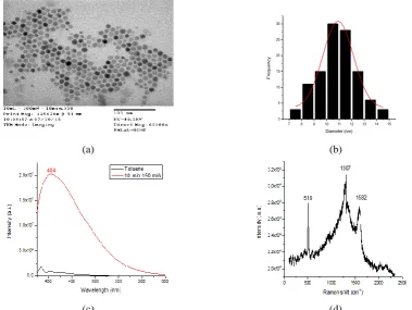
Using the second harmonic of a Nd: YAG laser (532 nm) with average power of 150 mW, irradiation time of 10 minutes we prepared Si NPs in water. The TEM images of Si NPs show that the semiconductor NPs are almost spherical in shape. Analysis from size distribution shows the mean
diameter of Si NPs in water is about 20 nm. TEM image and size distribution histogram is shown in Fig 6 below:
(a) (b)
Fig 6. TEM image of silicon nanoparticle colloidal solution prepared via nanosecond laser ablation in deionized water (average Laser power 150 W, duration of ablation is 10 min). On the right is reported
the corresponding size histogram.
Using the second harmonic of the Nd: YAG laser (532 nm) with average power of 200 mW, irradiation time of 10 minutes we prepared Si NPs in Toluene. The TEM image of Si NPs shows that the Si NPs are almost spherical in shape (see Figure 6 (a)). Analysis from size distribution (Figure 6b) shows the obtained colloidal solution displays an average size of 10 nm and a size distribution varying from 7 nm to 15 nm. The figure indicates that nanoparticles can be obtained by PLA in liquid environments.
7 8 9 10 11 12 13 14 15 0
5 10 15 20 25 30
F
re
q
u
e
n
cy
Diameter (nm)
(a) (b)
(c) (d)
Figure 7 (c) shows the PL spectra of the Si NPs colloidal solutions prepared via PLAL with average power of 150 mW, irradiation time of 10 minutes. No PL emission was observed for the fresh toluene solvent (the small peak around 390 nm originating from Raman emission). The Si NPs colloidal solution displays stable PL emission, characterized by a blue emission centered at 404 nm. The PL spectra look broad ranging from 380 to 500 nm. The PL spectra indicate the emission in the blue region and shows that the Si NPs are blue luminescent in nature.
The Raman scattering of the specimen prepared in toluene was 510 cm-1 (see Figure 7 (d)), which was smaller than that of single crystal Si, 520 cm-1. The Raman scattering peaks 1307 cm-1 and 1582 cm-1 correspond to amide bonds [17] and carbon-carbon bonds [18] respectively, which were formed by reaction of toluene with the environment. These results indicate that the nanoparticles observed by TEM are Si nanocrystallites.
It is known that the photoluminescence peak of Si-NPs redshift as the nanoparticles mean size increases due to quantum confinement effect [19] which occurs when NPs size is comparable to the Bohr radius of the exciton, or larger. Three distinct categories of quantum confinement have been identified from strong regime to weak regime according to the relative value of the bohr radius and nanoparticle size [19]. The mean size of generated Si NPs prepared via nanosecond laser ablation of a silicon target in Toluene is 10 ± 5 nm as reported from the histogram of particle size (Figure 7 (b)). Taking into account the value of 4.9 nm of exciton Bohr radius for silicon, in this case the nanoparticle size is larger than the exciton Bohr radius value and luminescent Si NPs are considered to be in a weak confinement regime. However, there is earlier report which presents the emission color of a very monodisperse size-separated Si NPs with the mean size less than 2nm was in the yellow-orange spectral region [20]. So here we may expect the intense photoluminescence of Si NPs colloidal solution depending on the solvent. This also indicates that surface effect other than the quantum confinement effect should be taken into account [21]. Since PL wavelength is very sensitive to ambient conditions, the PL wave length may be controlled by changing surface condition of the colloid. This character is possible for application to the sensor. Hence further studies with different solvents on the optical emission properties of the Si NPs and their structural studies are planned.
3.3. Preparation of Au-Ag Alloy nanoparticles in water
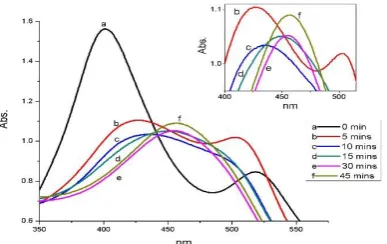
A mixture of the Au and Ag nanoparticle colloids in water was prepared with 1: 1 volume ratio and irradiated by 532 nm wavelength of Nd:YAG laser. The average power of laser was set at 200mW and laser spot diameter is about 6mm. Fig. 8 illustrates the absorption spectra of the Au-Ag NPs colloid mixture in water with different irradiation times.
Before exposure to laser light, two distinct absorbance maxima at 401.4 and 518.7 nm, corresponding to Ag and Au nanoparticle resonance plasmon peaks, respectively, indicate isolated Ag and Au NPs in the colloid mixture. The colloid mixtures exposed to laser radiation at 532 nm give an absorption spectra change as the irradiation time is increased. In the first 5 minutes, the surface plasmon absorption peak of Ag NPs tends to shift to the red (426.5 nm), meanwhile one of Au NPs have trend to move to the blue (503.2 nm). After 10 minutes irradiation, the Au NPs surface plasmon absorption band disappears and a weak shoulder is observed at about 497,7 nm while the Ag NPs surface plasmon absorption peak keeps shifting slightly to the red (435.5 nm). Further irradiation leads to appear a new absorption bands at 450.2 nm, 455.3 nm and 457.2 nm for the sample irradiated during 15 mins, 30 mins and 45 mins respectively. This surface plasmon absorption bands are likely due to the formation of Au-Ag alloys. This result indicates that the irradiated colloidal solutions contained alloy nanoparticles rather than core–shell nanoparticles, or mixtures of monometallic nanoparticles.
Fig.9 shows TEM image (JEM 1010-JEOL) and the size distribution of Au-Ag alloy NPs in the mixed colloidal solution after an exposure time of 45 minutes.
Fig.9. The TEM image and size distribution of Au-Ag alloy nanoparticles in water.
The spherical NPs have mean diameter of 29.4 nm and size ranging from 8.4 to 75.3 nm. EDX microanalysis indicates also that the nanoparticles and the sintered structures are Au-Ag alloys.
4. Conclusion
We have successfully synthesized Gold, Silver and Silicon NPs in different clean liquids by pulsed laser ablation method. Analysis from size distribution shows that in water the mean diameter of Au NPs is 17 nm; mean diameter of Ag NPs is 18 nm, the mean diameter of Si NPs is 20 nm. In Toluene, Si NPs have average size of 10 nm and a size distribution varying from 7 nm to 15 nm.
The creation of NPs depends not only on the laser power and laser irradiation time but also on the liquid environment in laser ablation.
Gold-Silver Alloy NPs were also successfully synthesized by laser induced synthesis method. TEM image analysis shows the shape of Gold-Silver Alloy NPs is rather spherical and size in PVP soluttion ranges from 2 to 10 nm with average diameter of 4 nm and size in water ranges from 8.4 to 75.3 nm with mean diameter of 29.4 nm.
References
[1] Canham L T 1990 Appl. Phys. Lett. 57 1046
[2] Bailey R E, Smith A M and Nie S 2004 Physica E 25 1
[3] R. Jha, A.K. Sharma, J. Opt. A-Pure Appl. Opt. 11, 045502 (2009).
[4] A.K. Sharma, B.D. Gupta, Nanotechnology 17, 124 (2006).
[5] R.A. Alvarez-Puebla, J.P. Bravo-Vasquez, J. Colloid Interface Sci. 333, 237 (2009).
[6] E. Hao, S.Y. Li, R.C. Bailey, S.L. Zou, G.C. Schatz, J.T. Hupp, J. Phys. Chem. B 108, 1224 (2004).
[7] M. Rosso-Vasic, E. Spruijt, Z. Popovic, K. Overgaag, B. Van Lagen, B. Grandidier, D. Vanmaekelbergh, D.
Dominguez-Gutierrez, L. De Cola, and H. Zuilhof, “Amine-terminated silicon nanoparticles: synthesis, optical properties and their use in bioimaging,” J. Mater. Chem. 19(33), 5926–5933 (2009).
[8] X. Zhang, D. Neiner, S. Wang, A. V. Louie, and S. M. Kauzlarich, “A new solution route to hydrogen-terminated
silicon nanoparticles: synthesis, functionalization and water stability,” Nanotechnology 18(9), 095601 (2007).
[9] Y. Khang and J. Lee, “Synthesis of Si nanoparticles with narrow size distribution by pulsed laser ablation,” J.
Nanopart. Res. 12(4), 1349–1354 (2010).
[10] J. Knipping, H. Wiggers, B. Rellinghaus, P. Roth, D. Konjhodzic, and C. Meier, “Synthesis of high purity silicon
nanoparticles in a low pressure microwave reactor,” J. Nanosci. Nanotechnol. 4(8), 1039–1044 (2004).
[11] S. Barcikowski, F. Devesa, and K. Moldenhauer, “Impact and structure of literature on nanoparticle generation by
laser ablation in liquids,” J. Nanopart. Res. 11(8), 1883–1893 (2009).
[12] R. Intartaglia, K. Bagga, F. Brandi, G. Das, A. Genovese, E. Di Fabrizio, and A. Diaspro, “Optical Properties of
Femtosecond Laser-Synthesized Silicon Nanoparticles in Deionized Water,” J. Phys. Chem. C 115(12), 5102– 5107 (2011).
[13] M. Moskovits, Rev. Mod. Phys. 57 (1985) 783.
[14] B. Pettinger, K. Krischer, J. Electron. Spectrosc. Relat. Phenom. 45 (1987) 133.
[15] B. Pettinger, K. Krischer, G. Ertl, Chem. Phys. Lett. 151 (1988) 151.
[16] A.M. Michaelis, J. Jiang, L. Brus, J. Phys, Chem. B, 104 (2000) 11965.
[17] Irvin M. Asher, Kenneth J. Rothschild, Evangelos Anastassakis, H. Eugene Stanley (1976), “Raman
Spectroscopy of Uncomplexed Valinomycin”, Journal of the American Chemical Society, pp. 2026.
[18] S. Reich, C. Thomsen, J. Maultzsch (2008), Carbon Nanotubes: Basic Concepts and Physical Properties, Wiley –
VCH.
[19] P. F. Trwoga, A. J. Kenyon, and C. W. Pitt, “Modeling the contribution of quantum confinement to luminescence
from silicon nanoclusters,” J. Appl. Phys. 83(7), 3789–3794 (1998).
[20] Florian Maier-Flaig, Julia Rinck, Moritz Stephan, Tobias Bocksrocker, Michael Bruns, Christian Kübel, Annie
K. Powell, Geoffrey A. Ozin, and Uli Lemmer, “Multicolor Silicon Light-Emitting Diodes (SiLEDs)” Nano Lett., 2013, 13 (2), pp 475–480
[21] R. Intartaglia, K. Bagga, M. Scotto, A. Diaspro, and F. Brandi “Luminescent silicon nanoparticles prepared by
ultra short pulsed laser ablation in liquid for imaging applications” 1 May 2012 / Vol. 2, No. 5 / Optical Material Express 510