197
Rapid Synthesis of Silver Nanoparticles by a
Marine-derived Fungus Aspergillus Niger and their
Antimicrobial Potentials
A. K. Vala
1*, S. Shah
21- Department of Physics, Bhavnagar University, Sardar Vallabhbhai Patel Campus, Bhavnagar, India
2- Department of Marine and Coastal Studies, Madurai Kamraj University, Madurai, India
(*) Corresponding author: [email protected]
(Received:12 Aug. 2012 and Accepted: 10 Oct. 2012)
Abstract:
Recently, biosynthesis of nanoparticles has received attention due to an increasing need of developing rapid, simple and ecofriendly protocol. Pathogenicity of some of the organisms and lengthy reaction are the drawbacks involved with biosynthesis. We describe a simple protocol for rapid synthesis of silver nanoparticles through
biological route using a marine-derived fungus Aspergillus niger. Silver nanoparticles biosynthesis could be achieved within 3 minutes (which otherwise generally takes about 24h) by altering pH of the reaction mixture. Silver nanoparticles biosynthesized at different pH have been observed to have antimicrobial potentials against four test bacteria (viz. Bacillus megaterium, Proteus vulgaris, Staphylococcus aureus and Shigella sonnei). Further, combined effect of Gentamicin and biosynthesized silver nanoparticles and effect of culture condition (pH) on antimicrobial effect have also been studied. Based on the findings it is concluded that the present study
provides a solution to the drawbacks involved in biosynthesis of silver nanoparticles, an ecofriendly approach.
It is envisaged that the biosynthesized silver nanoparticles alone, or their combination can be potentially used
as effective agents against pathogens.
Keywords: Antibacterial activity, Biosynthesis, Filamentous fungi, Silver nanoparticles, Microbial growth, Environmental preservation.
Int. J. Nanosci. Nanotechnol., Vol. 8, No. 4, Dec. 2012, pp. 197-206
1. INTRODUCTION
Over the years, development of resistance to commercially available antimicrobial agents by
pathogenic microorganisms has been a pressing
problem [1,2]. Consequently, development of new effective bactericides becomes imperative.
Silver salts and elemental silver are widely used as antimicrobial agents. Silver attacks a broad range of targets in microbes, hence, the organisms need to develop a range of mutations simultaneously to protect themselves. Therefore, it’s implausible for microbes to develop resistance against silver [3,4].
However, silver salts and bulk silver face certain limitations for their medical applications, the former may possess quick and uncontrolled silver release while the latter is a sluggish and inefficient releasing system. Silver in the nanoparticle form, not having any such issue, has better prospects for medical applications [5]. This is one of the reasons for a great demand of silver nanoparticles in recent years. However, the physico-chemical protocols generally applied for synthesis of silver nanoparticles suffer
from one or the other limitations like high cost, use
of toxic chemicals, etc.
attention due to an increasing need of developing rapid, simple and ecofriendly protocol. There has been a growing number of reports on harnessing
microorganisms for biosynthesis of nanoparticles
[6-10]. However, compared to their terrestrial counterparts, marine microorganisms are given less attention despite the fact that researches on nanobiotechnology in future may progressively depend more on marine microbes having the capability to grow under extreme conditions [11-13]. Major drawback involved in biological synthesis
of nanoparticles is pathogenicity of some of the
organisms and lengthy reaction as the time required
to complete the reaction generally ranges from
24-120h [9].
In the present investigation, we describe a simple protocol for rapid synthesis of narrowly dispersed silver nanoparticles (AgNPs) by altering the reaction condition. Furthermore, antibacterial activity of the as biosynthesized AgNPs was tested against two Gram negative and two Gram positive bacterial strains. The AgNPs synthesized at pH 10 were found to be the most effective against test bacteria. The present investigation is the first-ever report on effect of physico-chemical conditions on biosynthesis and antimicrobial efficiency of AgNPs by a marine-derived fungus.
Advantages of using Aspergillus niger include (i) easy cultivation (ii) high yield (iii) inexpensive medium requirement iv) it can be obtained as a byproduct from industries and (v) A. niger is
generally regarded to be safe, therefore, can be easily accepted [14,15].
2. MATERIALS AND METHODS
2.1. Organism
A marine-derived fungus Aspergillus niger was selected for the present study. The test isolate was isolated from waters of Bhavnagar coast (Lat. 21º 45’ N and Long. 72º14’ E), Gulf of Khambhat, West Coast of India. The isolate was grown and maintained on potato dextrose agar (PDA) medium [16] and stored at 4ºC until use. The medium was prepared in aged seawater and distilled water at a ratio of 3:1 [17].
One ml inoculum (spore suspension approximately
106/ml) was inoculated in 250 ml Potato Dextrose medium (prepared in 75% ‘aged’ seawater). The inoculated flasks were incubated at room temperature for 4 days. After incubation period, fungal biomass was separated from the medium by filteration and extensively washed with sterile distilled water.
2.2. Biosynthesis silver nanoparticles
Approximately 5g fungal biomass was challenged with 1.0mM silver nitrate at different pH (3-10). The AgNO3 solutions were prepared in sterilized deionized water. The inoculated flasks were incubated at 270C for 72 h under static condition.
Negative and positive controls (without the silver nitrate, only biomass and without biomass only silver nitrate) were also run along with the experimental flasks.
One ml sample from each flask was withdrawn at different time intervals and the spectra were recorded at a resolution of 1 nm using UV– visible spectrophotometer (Elico BL-198). The experiments were carried out in triplicates.
Transmission electron microscopic (TEM) images of biosynthesized silver nanoparticles (mounted on carbon-coated copper grids) were obtained on JEOL (Model GEM 200) transmission electron microscope operated at 200 kV.
2.3. Estimation of total extra cellular protein
After 72h of incubation, the biomass was separated from the flasks by filteration and estimation of total extracellular protein content was carried out using bicinchonic acid (BCA) method [18].
2.4. Antimicrobial activity of the biosynthesized silver nanoparticles
Antimicrobial activity of silver nanoparticles biosynthesized at pH 5, 8, 9, and 10 was examined against two Gram positive (Bacillus megaterium
and Staphylococcusaureus) and two Gram negative
(Proteus vulgaris and Shigella sonnei) bacteria.
199
with antibiotic together with the effect of pH, were also studied.
(A) Disk diffusion method was used to assay biosynthesized nanoparticles for bactericidal activity against test strains on Mueller-Hinton agar
plates.
In this method, Mueller-Hinton agar plates were prepared for each bacterial test strain. Each plate was prepared by aseptically pouring 100μl of bacterial suspension (approaximately 108 CFU/ml)
along with 20ml of sterile molten Mueller-Hinton agar into the sterile petri plates. The plates were allowed to solidify and marked accordingly. Small filter discs approximately of the diameter 6mm were sterilized. Silver nanoparticles synthesized at pH 5, 8, 9 and 10 were used to check their antimicrobial activity.
Each plate was divided into 3 parts and the sterilized discs impregnated with 40μl respective silver nanoparticles were aseptically placed on the
centre of each compartment of the plate. Similar
experiments were carried out with 1mM AgNO3 as
well as Suspension (fungal cell extract) from flask without silver nitrate. After incubation at 370C for
24 hours the zones of inhibition were measured. The assays were performed in triplicate [19].
Synergistic antibacterial activity combined with antibiotic:
Combination of silver nanoparticles with antibiotic Gentamicin, was investigated against test bacteria using the disk diffusion method.
Standard Gentamicin disks (10 µg/disk) were used as positive control, and Gentamicin disks impregnated with silver nanoparticles (10μl) were placed onto the Mueller-Hinton agar medium inoculated with test organisms. Suspension (Fungal cell extract) from flask without silver nitrate was used as the negative control.
These plates were then incubated at 37°C for 24 hours. Similar experiments were carried out with only silver nanoparticles. After incubation, the zones of inhibition were measured. The assays were performed in triplicates and average values are reported here.
The increase in fold area [20] was assessed by
calculating the mean surface area of the inhibition
zone of Gentamicin and Gentamicin+Silver nanoparticles. The fold increase area of different test organisms for Gentamicin and for Gentamicin+Silver nanoparticles was calculated by the equation (B2– A2)/A2, where A and B were zones of inhibition for Gentamicin, Gentamicin+Silver nanoparticles, respectively.
(B) Growth curves of test bacteria exposed to different concentrations of silver nanoparticles
To examine the growth curves of bacterial test strains exposed to biosynthesized silver nanoparticles, Mueller-Hinton broth with different quantity of silver nanoparticles (0, 50, 100, and 150 μl) was used. Respective flasks were inoculated and incubated in a shaking incubator at 370C for
24 h. Growth curves of bacterial cell cultures were obtained through repeated measures of the optical density (O.D.) at 600 nm at regular time interval.
Effect of pH on antimicrobial activity of silver nanoparticles:
Antibacterial testing of the selected silver nanoparticles was also carried out at different pH (pH 6, 6.5, 7.4, 8.0 and 8.5) using disk diffusion
test.
3. RESULTS AND DISCUSSION
3.1. Biosynthesis of silver nanoparticles
Silver nanoparticles are produced by various physico-chemical methods. Recently, biosynthetic route also has been attempted. Generally, biosynthesis involving microbes is time consuming, taking 24-120 h [9]. In our method, to overcome this problem we have developed a simple protocol for rapid synthesis of AgNPs. Biosynthesis of AgNPs was carried out using marine-derived
fungus Aspergillus niger, isolated from waters of Bhavnagar coast, West coast of India, in the pH
range of 3-10 (Plate 1).
When the test fungal biomass was challenged with 1mM AgNO3 at different pH, all the test flasks in the pH range 3-7 showed gradual change in color of reaction mixture from colorless to deep brownish
200
pink within 24h, indicating biosynthesis of AgNPs. A change in color was observed within 1 minute of exposure to fungal biomass to test AgNO3 solution
at pH 10. In case of pH 8 and 9 color change was observed within 2 minutes. Hence, pH in the alkaline range support supported rapid biosynthesis of silver nanoparticles. The negative as well as positive control did not show the characteristic change in color. Biosynthesis boosted with increasing pH reached the maximum at pH 10. The proteins involved in nanoparticles biosynthesis may bind with silver at thiol regions (–SH) forming a –S–Ag bond, a clear indication of which aids the conversion of Ag+ to Ag0. In addition, the alkaline ion (OH-) is
very much required for the reduction of metal ions. At higher pH, efficiency of the enzymes involved in synthesis of silver nanoparticles increases [21]. The present findings help tackling the drawbacks involved with process of biosynthesis of nanoparticles, which could otherwise be reliable and ecofriendly, because as already mentioned, A. niger is generally regarded to be safe and AgNP synthesis by this organism in the present study does not even take 5 minutes, which is a remarkable trait for large scale, commercially viable process.
(*pH 8.9 dilution 1:4; pH 10 dilution 1:8)
Figure 1: UV spectra of biosynthesized silver nanoparticles at different pH*
Characterization of Silver Nanoparticles
Figure 1 shows uv-vis absorbance spectra of Ag-NPs from different pH range at different time intervals. In the spectra, a single surface plasmon resonance (SPR) is observed which originates from elemental silver [22,23].
١ ٢
٣
٤
٥
٦
٧
٨
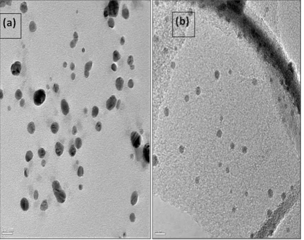
Figure 2: TEM images of silver nanoparticles biosynthesized by marine-derived A. niger at (a) pH 5 and (b) ph 10
201
When transmission electronic microscopic images (Figure 2) were examined it was found that the particles were spherical in all cases. As reported earlier[13], in the previous study using the same test organism, the particles were found to be spherical with size range of 5-26nm, except in the case of samples of pH 8,9 and 10. Here, the particles were spherical in the size range of 1.7-20 nm. More uniformity was observed in size of the particles with
increasing pH.
Smaller size of biosynthesized particles with increase in pH could be due to increased dynamics of the ions and more nucleation regions formed due to the availability of –OH ions. The conversion of Ag+ to Ag0 increases followed by increase in the
kinetics of the deposition of the silver atoms [24].
Possible mechanisms for synthesis of nanoparticles
by fungi have been proposed, however, so far no single generalized mechanism has been identified [25]. Mukherjee et al. (2001) [6] suggested that cell wall and cell wall sugars play important role in reduction of metal ions. A number of studies have suggested role of protein in nanoparticles formation. Bansal et al. (2004) [26]observed that fungus secreted proteins that were capable of extracellularly hydrolyzing compound with zirconium ions and hence, aided in zirconium
nanoparticle formation.
In the succeeding studies with silica and titania nanoparticles it was confirmed [27]. Bharde et al. (2006) [28] suggested role of protein from extracellular extracts of Verticillium sp. in hydrolysia of [Fe(CN)6]3 and [Fe(CN)
6]4. Bhainsa and D’Souza (2006) [29]reported that proteinaceous compound in fungal biomass had a major role in formation of metal nanoparticles. Ingle et al. (2008) [30]reported involvement of NADH-dependent enzyme in reduction of metal ions and demonstrated presence of nitrate reductase in fungal cell filterate. Taking
the importance of fungal proteins in biosynthesis of
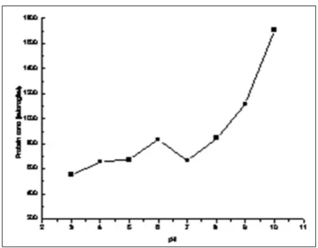
nanoparticles into consideration, total extracellular protein from the fungus was estimated and it was found that extracellular protein content was higher when the fungus was challenged with silver nitrate (Figure 3). These data corroborate with earlier findings that had suggested the role of proteins in
biosynthesis of nanoparticles by fungi.
Figure 3: Extracellular proteins produced by A. niger at different pH
Antimicrobial activity of the biosynthesized silver nanoparticles
The silver nanoparticles biosynthesized by A. niger at pH 5, 8, 9 and 10 were selected for their antibacterial activity against four bacteria: two Gram positive viz. Bacillus megaterium and Staphylococcus aureus and two Gram negative
Proteus vulgaris and Shigella sonnei. Disc diffusion method and bacterial growth kinetic studies were carried out to study the effect in vitro. Effect of pH on antimicrobial activity of Ag-NP also was examined.
(A) Disc diffusion method
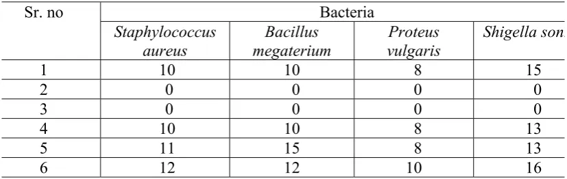
When disc diffusion test was carried out, no inhibition zone was observed when fungal culture filtrate was used, similar observation was recorded in case of silver nitrate also for all test bacteria. Inhibition zones of different diameter (for different bacteria) were observed when biosynthesized silver nanoparticles were used. Hence, biosynthesized silver nanoparticles exerted activity against all test bacteria (Table 1; Plate2).
The present findings about antibacterial activity of silver nanoparticles are similar to the report of Guzmán et al., (2009) [31]. Antibacterial activity of biosynthesized silver nanoparticles in the present study are found to be higher than that reported by
202
Veerasamy et al., (2011)[32]. Antibacterial activity reported by Nithya et al., (2011) [10]was more than observed in the present study. However, strain to strain variation is a logical happening. Bhimba et al., (2011)[33] also reported antibacterial activity of silver nanoparticles synthesized by Hypocrea lixii isolated from mangrove sediment soil.
Interestingly, particles biosynthesized at different pH showed distinct activity at each pH. Invariably, highest anti-bacterial activity was imparted by silver nanoparticles synthesized at pH 10. In general, silver nanoparticles synthesized at pH 5 showed least activity with an exception of S. sonnei. The reason for this could be variation in particle size [34]. Pal et al., (2007) [3] demonstrated that silver nanoparticles undergo a shape-dependent interaction with the Gram-negative organism E. coli. Shukla
et al (2012) [35] reported antibacterial activity of agar-based silver nanoparticles and nanocomposite film against Bacilluspumilis.
Synergistic antibacterial activity combined with antibiotic
Combination of silver nanoparticles with antibiotic Gentamicin, was investigated against test bacteria
using the disk diffusion method. As maximum inhibition zone for all test bacteria was found with silver nanoparticles biosynthesized at pH 10, it was selected for studying its effect with antibiotic Gentamycin. Gentamicin was selected for the study because, as an aminoglycoside antibiotic,
gentamicin is of unquestionable practical interest
and is effective against a large number of Gram negative and Gram positive bacteria. The mode of gentamicin action involves disruption of ribosomal synthesis of protein. Gentamicin is a main antibiotic used to treat severe purulent infection, especially that caused by a resistant Gram-negative bacteria. Being a broad spectrum antibiotic, gentamicin is often used for treatment of patients with mixed infection and also in the case of unidentified infecting agent [36].
When tested for sensitivity against antibiotic Gentamicin, P. vulgaris was the most sensitive followed by Staphylococcus aureus and Bacillus
metagerium. S. sonnei, compared to other test organisms, showed less sensitivity (Table 2; Plate 3). When biosynthesized silver nanoparticles were impregnated on the antibiotic Gentamicin disc, and tested for antibacterial activity, an increase
25
Table 1 :
Zone of Inhibition (mm) in absence and presence of Silver nanoparticles
(40 μl)against
١
test bacteria
٢
٣
٤
٥
٦
٧
٨
٩
١٠
١١
١٢
(1: Ag particles synthesized at pH 5, 2: fungal culture filtrate without AgNO3, 3: AgNO3, 4: Ag particles١٣
synthesized at pH 8, 5: Ag particles synthesized at pH 9, 6: Ag particles synthesized at pH 10)
١٤
١٥
١٦
١٧
١٨
١٩
٢٠
٢١
٢٢
٢٣
٢٤
٢٥
٢٦
Sr. no
Bacteria
Staphylococcus
aureus
megaterium
Bacillus
vulgaris
Proteus
Shigella
sonnei
1
10
10
8
15
2
0
0
0
0
3
0
0
0
0
4
10
10
8
13
5
11
15
8
13
6
12
12
10
16
Table 1: Zone of Inhibition (mm) in absence and presence of Sliver nanoparticles (40ml) against test bacteria
Table 2: Zone of Inhibition (mm) for Gentamicin (10mg/disk) in absence and presence of silver nanoparticles
Table 2 :
Zone of Inhibition (mm) for Gentamicin (10µg/disk)
in absence and presence of silver
١
nanoparticles
٢
٣
٤
٥
٦
٧
٨
(1: Gentamicin, 2: Gentamicin + Ag particles synthesized at pH 10 (10μl), 3 : increase in fold area )
٩
١٠
١١
١٢
١٣
١٤
١٥
١٦
١٧
١٨
١٩
٢٠
٢١
٢٢
٢٣
٢٤
Sr. no
Bacteria
B.
megaterium
S.
aureus
P
.
vulgaris
S.
sonnei
1 25 33 40 20
2 36 40 44 20
3 1.0736 0.4692 0.21
0
203
in fold area was observed in all cases, except S. sonnei. Among all test bacteria, S. sonnei exhibited least sensitivity towards gentamycin, the supplied silver nanoparticles could have been insufficient for exerting additive effect.
Enhancement in antibacterial activity on E .coli and Staphylococcus aureus has been reported by Singh
et al., (2011 ) [37] when Cefazolin is conjugated with silver nanoparticles.
Fayaz et al., (2010) [38] also observed that antibacterial activities of ampicillin, kanamycin, erythromycin, and chloramphenicol were increased in the presence of Ag nanoparticles.
An increase the antifungal effects of drugs
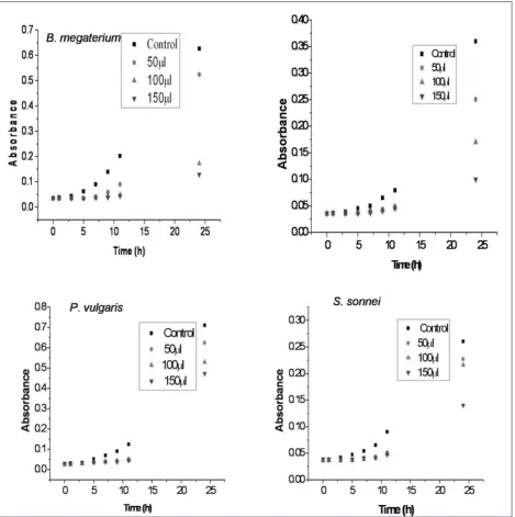
Figure 4: Absorbance indicating bacterial growth in presence of different concentration of
biosynthesized silver nanoparticles
28
١
٢
٣
٤
٥
٦
204
fluconazole and griseofulvin in presence of Ag nanoparticles has been recorded by Noorbakhsh et al., (2011) [39]. Gajbhiye et al., (2009) [8] also
observed enhanced antifungal activity of fluconazole in presence of silver nanoparticles against the test
fungi.
(B) Bacterial growth kinetics in presence of biosynthesized silver nanoparticles
To examine the bacterial growth kinetics in the presence of biosynthesized silver nanoparticles, test bacteria were grown in 100 ml of Mueller Hinton broth supplemented with different doses of silver nanoparticles. It was found that the nanoparticles exerted growth delay at all test concentrations
(Figure 4).
When a comparison is made with disc diffusion test data it was found that the effects were stronger when the test bacteria were grown on agar media than that grown in broth. This could likely be due to changes in exposure dose over the experiments due to nanoparticles’ aggregation and sedimentation in broth, while in agar there is a continued presence of particles on the solid surface.
Cultural condition for bacterial growth also is an important criterion when antimicrobial testing is being carried out. For this obvious reason, antibacterial testing of the selected silver nanoparticles was also carried out at different pH (pH 6, 6.5, 7.4, 8.0 and 8.5).
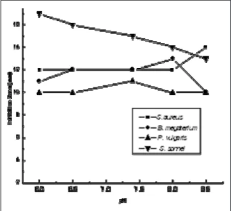
Figure 5 shows the data of inhibition zone for the test microbes when grown at different pH in presence of biosynthesized silver nanoparticles.
Maximum sensitivity of Staphylococcusaureus was observed at pH 8.5, this could be due to an increase in antibiotic uptake and hence killing of the cell [40]. Bacillusmegaterium was found to be the most susceptible at pH 8. Highest antibacterial activity of silver nanoparticles against Proteus vulgaris
was found to be at pH 7.4, at other pH the activity remained the same. Among all test bacteria, S. sonnei was the most sensitive. Highest inhibition zone was observed for S. sonnei when the pH of the medium was 6 followed by pH 6.5, 7.4, 8 and 8.5. Hence, in the acidic condition, the organism was more sensitive towards exposure to the silver
nanoparticles.
4. CONCLUSION
Rapid biosynthesis of silver nanoparticles using marine-derived fungus Aspergillusniger is achieved by altering the pH of the reaction mixture. Alkaline pH range led to rapid biosynthesis. Antibacterial efficiency of the as synthesized silver nanoparticles alone and in combination with antibiotic gentamicin was examined against Gram negative and Gram positive bacteria. The biosynthesized particles are found to be promising antibacterial agents. Effect of pH on antibacterial activity of silver nanoparticles was also studied that indicated more sensitivity of organisms in acidic condition. The present study is the first-ever report on rapid synthesis of silver nanoparticles by marine-derived fungus. These findings are suggestive of possible exploitation of the test fungus for ecofriendly as well as commercially viable biosynthesis of silver nanoparticles.
29
١
٢
٣
Figure 5: In hibition Zone (mm) for silver
nano-particles against test bacteria grown at different pH
ACNOWLEDGEMENTS
:Thanks are due to the Council of Scientific and Industrial Research (CSIR), New Delhi, for financial support sanctioned under Scientists’ Pool Scheme to AKV. Dr. D. Srivastava is gratefully acknowledged for TEM analysis.
205
REFERENCES
1. G. D. Wright, Resisting resistance: new chemical strategies for battling superbugs. Chem. Biol., Vol. 7, (2000), pp. 127-132.
2. G.D. Wright, Bacterial resistance to antibiotics: enzymatic degradation and modification. Adv. Drug Deliv. Rev., Vol. 57, (2005), pp. 1451-1470. 3. S. Pal, Y. K. Tak, J.M. Song, Does the antibacterial
activity of silver nanoparticles depend on the shape of the nanoparticle? A study of the Gram-negative bacterium Escherichia coli. Appl. Env. Microbiol., , Vol. 73, No. 6, (2007), pp. 1712–1720.
4. B. N. Chudasama, A. K. Vala, N. M. Andhariya, R. V. Upadhyay, R. V. Mehta, Highly bacterial resistant silver nanoparticles: Synthesis and antibacterial activities J. Nanopart. Res., Vol. 12, (2010), pp. 1677–1685.
5. G. K. Vertelov, Y. A. Krutyakov, O. V. Efremenkova, A. Y. Olenin, G. V. Lisichkin, A versatile synthesis of highly bactericidal Myramistin_stabilized silver nanoparticles. Nanotechnol., Vol. 19, (2008), pp. 355707 (7 pp).
6. P. Mukherjee, A. Ahmad, D. Mandal, S. Senapati, S. R. Sainkar, M. I. Khan, R. Parischa, P. V. Ajaykumar, M. Alam, R. Kumar, M Sastry, Fungus-mediated synthesis of silver nanoparticles and their immobilization in the mycelial matrix: A novel biological approach to nanoparticle synthesis. Nano Lett., Vol. 1, (2001), pp. 515-519.
7. A. Ingle, M. Rai, A. Gade, M. Bawaskar, Fusarium solani: a novel biological agent for the extracellular synthesis of silver nanoparticles. J. Nanopart. Res., Vol. 11, (2009), pp. 2079–2085.
8. M. Gajbhiye, J. Kesharwani, A. Ingle, A. Gade, M, Rai, Fungus-mediated synthesis of silver nanoparticles and their activity against pathogenic fungi in combination with fluconazole. Nanomed.: Nanotechnol. Biol. Med., Vol. 5, (2009), pp. 382–386. 9. H. Bai, B. Yang, C. Chai, G. Yang, W. Jia, Z. Yi, Green synthesis of silver nanoparticles using Rhodobacter sphaeroides. World J. Microbiol. Biotechnol. Vol. 27, (2011), pp. 2723-2728.
10. G. Nithya, N. Hemashepangam, S. Balaji, Biosynthesis of silver nanoparticle and its antibacterial activity. Arch. Appl. Sci. Res., Vol. 3, No. 2, (2011), pp. 377-380.
11. D. Chandramohan, Marine microbiology: challenges and future directions. In: N. Ramaiah (Ed) Marine Microbiology: Facets & Opportunities, National Institute of Oceanography: Goa, (2004), pp 7-13. 12. K. Kathiresan, S. Manivannan, M. A. Nabeel, B.
Dhivya, Studies on silver nanoparticles synthesized by a marine fungus, Penicillium fellutanum isolated from coastal mangrove sediment. Colloids Surf. B: Biointerfaces., Vol. 71, (2009) 133–137.
13. A. K. Vala, B. Chudasama, R.J. Patel, Green synthesis of silver nanoparticles using marine derived fungus Aspergillus niger. Micro & Nano Lett. Vol. 7, No. 8, (2012), pp. 859-862.
14. E. Schuster, N. Dunn-Coleman, I. C. Frisvad, P. W. M. van Dijd, On the safety of Aspergillus niger - A Review, App.Microbiol. Biotechnol. Vol. 59, (2002), pp. 426-435.
15. A. K. Vala, Aspergillus niger and heavy metal removal: A perception. Res. J. Biotech. Vol. 4, No. 1, (2009), pp. 75-79.
16. Anonymous, Plant Pathologist’s Pocketbook, Commonwealth Mycological Society, KEW, Surrey, England, (1968).
17. C. Schlieper, (Ed.). Research Methods in Marine Biology. Sidgwick and Jackson Ltd, London, (1972). 18. P.K. Smith, R.I. Krohn , G.T. Hermanson, A.K.
Mallia, F.H. Gartner et al. Measurement of protein using bicinchoninic acid. Anal. Biochem. Vol. 150, No. 1, (1985), pp. 76–85.
19. S. Shah, Investigations on antimicrobial activity of silver nanoparticles biosynthesized by a marine-derived fungus Aspergillus niger. Dissertation thesis (2012).
20. S.S. Birla, V.V. Tiwari, A.K. Gade, A.P. Ingle, A.P. Yadav, Raí MK, Fabrication of silver nanoparticles by Phoma glomerata and its combined effect against Escherichia coli, Pseudomonas aeruginosa and Staphylococcus aureus. Lett Appl Microbiol, Vol. 48, (2009), pp. 173-179.
21. R. Sanghi, P. Verma, Biomimetic synthesis and characterisation of protein capped silver nanoparticles. Bioresour Technol, Vol. 100, No. 1, (2009), pp. 501–504.
22. M. Chen, Y. G. Feng, X. Wang, T.C. Li, J. Y. Zhang, D. J. Qian, Silver nanoparticles capped by oleylamine: Formation, growth, and self-organization. Langmuir, Vol. 2, (2007), pp. 5296–5304.
23. P. Gong, H. M. Li, X. X. He, K.M. Wang, J. B. Hu, W. H.Tan, S. C. Zhang, X. H. Yang, Preparation and antibacterial activity of Fe3O4@Ag nanoparticles. Nanotechnol. Vol. 18, (2007), pp. 285604 (7 pp). 24. V. Deepak, K. Kalishwaralal, S. Ram Kumar
Pandian, S. Gurunathan, An insight into the bacterial biogenesis of silver nanoparticles, industrial production and scale-up. In: M. Rai, N. Duran (Eds), Metal Nanoparticles in Microbiology, Springer-Verlag Berlin Heidelberg, (2011), pp 17-35. 25. M. Rai, A. Yadav, P. Bridge, A. Gade,
Myconanotechnology: a new and emerging science. In: M. Rai, P. D. Bridge, Applied Mycology CAB International, (2009) pp. 258-267.
26. V. Bansal, D. Rautray, A. Ahamd, M. Sastry, Biosynthesis of zirconia nanoparticles using the fungus Fusarium oxysporum. J. Mater. Chem. 14 (2004) 3303–3305.
27. V. Bansal, D. Rautray, A. Bharde, K. Ahire, A. Sanyal, A. Ahmad, M. Sastry, Fungus-mediated biosynthesis of silica and titania particles. J. Mater. Chem. Vol. 15, (2005), pp. 2583–2589.
28. A. Bharde, D. Rautray, V. Bansal, A. Ahmad, I. Sarkar, S. M. Yusuf, M. Sanyal, M. Sastry, Extracellular biosynthesis of magnetite using fungi. Small, Vol. 2, No. 1, (2006), pp. 135–141.
29. K. C. Bhainsa, S. F. D’Souza, Extracellular biosynthesis of silver nanoparticles using the fungus Aspergillus fumigatus. Colloids Surf. B: Biointerfaces, Vol. 47, (2006), pp. 160–164.
30. A. Ingle, A. Gade, S. Pierrat, C. Sonnichsen, M. K. Rai, Mycosynthesis of silver nanoparticles using the fungus Fusarium acuminatum and its activity against some human pathogenic bacteria. Curr. Nanosci. Vol. 4, (2008), pp. 141–144.
31. M. G. Guzmán, J. Dille, S. Godet, Synthesis of silver nanoparticles by chemical reduction method and their antibacterial activity. Int. J. Chem. Biol. Eng. Vol. 2, No. 3, (2009), pp. 104–111.
32. R. Veerasamy, T. Z. Xin, S. Gunasagaran, T. Xiang, E. Yang, N. Jeyakumar, S. A. Dhanaraj, Biosynthesis of silver nanoparticles using mangosteen leaf extract and evaluation of their antimicrobial activities. J. Saudi Chem. Soc. Vol. 15, (2011), pp. 113–120.
33. B. V. Bhimba, N. Nath, P. Sinha, Characterization and antibacterial analysis of silver nanoparticles synthesized by the marine fungi Hypocrea lixii MV1 isolated from mangrove sediment soil. J. Pharm. Res. Vol. 4, No. 2, (2011), pp. 477-479.
34. T. J. Brunner, P. Wick, P. Manser, P. Spohn, R. N. Grass, L. K. Limbach, A. Bruinink, W. J. Stark, In vitro cytotoxicity of oxide nanoparticles: comparison to asbestos, silica, and effects of particle solubility. Environ. Sci. Technol. Vol. 40, No. 14, (2006), pp. 4374–4381.
35. M. K. Shukla, R. P. Singh, C. R. K. Reddy, B. Jha, Synthesis and characterization of agar based silver nanoparticles and nanocomposite film with antibacterial applications. Bioresour Technol, Vol. 107, (2012), pp. 295-300.
36. V. G. Maidannik, I. V. Maidannik, Spravochnik sovremennykh lekarstvennykh sredstv (Handbook of Modern Medicines) Izdatel’stvo ‘‘AST’’, Moscow, (2005).
37. P. Singh, R. Kumar, R. Balaji Raja, P.T. Kalaichelvan, Mycobased biosynthesis of silver nanoparticles and studies of its synergistic antibacterial activity combined with cefazolin antibiotic against selected organisms. Australian J. Basic Appl. Sci. Vol. 5, No. 8, (2011), pp. 1412-1427.
38. A. M. Fayaz, K. Balaji, M. Girilal, R. Yadav, P. T. Kalaichelvan, R. Venketesan, Biogenic synthesis of silver nanoparticles and their synergistic effect with antibiotics: a study against positive and gram-negative bacteria. Nanomed. Nanotechnol. Biol. Med. Vol. 6, No. 1, (2010), pp. e103–e109
39. F. Noorbakhsh, S. Rezaie, A. R. Shahverdi, Antifungal effects of silver nanoparticle alone and with combination of antifungal drug on dermatophyte pathogen Trichophyton rubrum, In: Int.Conference on Bioscience, Biochemistry and Bioinformatics IPCBEE, 5: IACSIT Press, Singapore, (2011) pp. 364-367.