INTRODUCTION
Inflammation is the complex process which is frequently associated with pain and involved occurrences such as increased vascular permeability, protein denaturation and membrane alteration. Inflammatory diseases including different types of rheumatic diseases are a major cause of morbidity of the working force throughout world. This has been called the ‘King of Human Miseries’1. Although rheumatism is one of the oldest known diseases of the mankind and affects a large percentage of population of the world, no substantial progress was seen till the synthesis of aspirin in 1899 by the German Company Bayer, the hint of which also was obtained from a plant, the Willow
An Experimental Evaluation of Ageratum conyzoides
on Membrane Stabilization and Protein Denaturation
during Acute Inflammation and Arthritis
ARVIND KUMAR¹, SOURAV GHOSH²* and VAISHALI¹
¹Department of Pharmaceutical Chemistry, ²Department of Pharmacology, S.D. College of Pharmacy & Vocational Studies, Muzaffarnagar (India).
*Corresponding author: E-mail: [email protected]
(Received: September 08, 2011; Accepted: November 03, 2011)
ABSTRACT
Methanolic leaves extract and isolated flavonoides of Ageratum conyzoides was prepared and tested for in-vitro anti-arthritic activity. The in-vitro anti-arthritic activity was studied using the membrane stabilization and protein denaturation model. The activity was expressed as percentage inhibition of heamolysis and protein denaturation. The experiment data was expressed as mean ± SEM, the significance of difference among the various treated groups and control group were analysed by the means of one –way ANNOVA followed by dunnett t-test.
The phytochemical screening revealed that presence of flavonoides in large amount. In membrane stabilizing activity different concentration (100, 250 µg/ml) of extract and isolated compound showed significant inhibitory activity (47.39%,54.79% and 63.69% respectively) and in protein denaturation activity, the same extracts and isolated compound have remarkable inhibitory activity on protein denaturation (45.65%, 56.42% and 76.46% respectively) when compared with the control.
Key words: Ageratum conyzoides, Flavonoides, Heamolysis, Protein denaturation, Anti-arthritic.
to investigate the rationality of their use in modern scientific terms. The scientific studies to work out the actual efficacy and other limitations to these drugs would definitely widen their scope for future use if they come out to be really effective. This is particularly important, firstly due to the gravity of the problem of rheumatism and arthritis and secondly due to the absence of the right type of drug of synthetic origin for its treatment. The presently available drugs provide only symptomatic relief and are not free from side effects. The target should be to discover newer drugs from plant kingdom which may provide therapeutic cure and would be free from undesirable effects as well as economical, which would be accepted by the developing nations like India. [3]
MATERIAL AND METHODS Plant material
The leaves of Ageratum conyzoides Linn was collected from local area of muzaffarnagar, Uttar Pradesh, India in the month of February, 2011 and authenticated by Botanical Survey of India. (Vide no. CNH/I-I/ (292)/2011/Tech.II/333).
Preparation of extracts
Leaf was shade dried at room temperature and powdered to #40 mesh par ticle size. The powder (250 g) of crude plant was defatted with petroleum-ether and subjected to extraction with methanol by using Soxhlet apparatus. The extract was filtered and evaporated at 40°C under vacuum and the residue was freeze-dried.
Phytochemical tests4
Azaretum conyzoides leaf extract was separately tested for the presence of tannins, alkaloids, carbohydrates, steroids, flavonoids.
Test for alkaloids
1. Mayer’s test – 2-3 ml. of solution of extract was added with few drops of Mayer’s reagent.
2. Dragendroff’s test - 2-3 ml. of solution of extract was added with few drops of Dragendroff’s reagent.
3. Hagger’s test - 2-3 ml. of solution of extract was added with few drops of Hagger’s reagent.
Test for tannins
2-3 ml. of solution of extract was added with few drops of 5% FeCl3 solution.
Test for flavonoids
1. 2-3 ml. of solution of extract was added with 95% ethanol and neutral FeCl3 solution. 2. 2-3 ml. of solution of extract was added with
95% ethanol and lead acetate solution.
Test for carbohydrate Molish test
2-3 ml. of solution of extract was added with few drops of alpha napthol solution in alcohole, shaked well and add conc.H2SO4 from sides of the test tube.
Test for steroid Salkowski reaction
2-3 ml. of solution of extract was added with 2 ml. of chloroform and 2 ml. of conc. H2SO4. Isolation of flavonoides
Methanol part of extract (5 gm) was successively extracted with Ethyl acetate, and then the Ethyl acetate extract was concentrated to dryness. The Ethyl acetate extract was subjected to a column of silica gel (60-120 mesh; 3 cm dia, X 60 cm length) being eluted a gradient of Pet ether Ethyl acetate with increasing polarity. 10 main fraction were collected and individual fraction were tested for presence of the active bi-flavonoid compounds. Thus from fraction of ether/ Ethyl acetate (10:90), compound I was separated by using preparative TLC using Hexane/ Ethyl acetate in ratio of 8:2 system as eluent.
There were two different spots on the TLC plate, when illuminated with UV light with Rf value of compound 0.28 respectively from point of origin of sample. The compound with Rf value of 0.28, showed relatively higher concentration of flavonoid tests, (Shinoda and NaOH). Compound labeled as AZ-1 (Rf 0.28). The isolated compound was identified after analyzing spectra obtained from IR Spectra.
Preparation of test sample
Azaretum conyzoides leaf in 0.5 ml water and with phosphate buffer salt solution. According to concentration range from 50 µg/ml, 100 µg/ml and 250 µg/ml and also by dissolving 5 mg of isolated compound of leaf extract in 0.25 ml water and with phosphate buffer salt solution according to concentration 100 µg/ml.
In-vitro anti-arthritic study Membrane stabilizing activity
The test was followed by the method described by shinde et al (1999) [5] with some
modifications. Whole human blood obtains from a healthy human volunteer and transferred to heparinised centrifuge tube. The blood was washed three times with isotonic buffer solution (154 mM NaCl) in 10 mM sodium phosphate buffer solution (pH-7.4) for 10 mins at 3000g. The test sample considered to stock erythrocyte (RBC) suspension(0.5 ml) mixed with 5 ml of hypotonic solution (50 mM NaCl) in 10 mM sodium phosphate buffer solution (pH-7.4) containing the leaf extract and isolated compound solution or indomethacine (0.1 mg/ml). The control sample considered of 0.5 ml of RBC suspension mixed with hypotonic buffer saline solution alone. The mixture was incubated for 10 mins at room temperature and centrifuge for 10 mins at 3000g and the absorbance of supernatant was measured at 540 nm. Each experiment was carried out triplicate and the average was taken. The percentage inhibition of heamolysis or membrane stabilization was calculated by following equation.
% inhibition of heamolysis = 100 X (A1- A2/A1)
Where
A1 = Absorption of hypotonic buffer solution alone. A2 = Absorption of test sample in hypotonic solution. Effect on protein denaturation
Test solution (extract and isolated compound) containing different concentration of plant extract or indomethacine (100µg/ml) was mixed with 1 ml of egg albumin solution (1mM) and incubated at 27±1°C for 15 mins. Denaturation was induced by keeping the reaction mixture at 70°C in a water bath for 10 mins. After cooling the turbidity was measured spectrophotometrically at 660nm6,7. Percentage inhibition of denaturation was calculated from control where no drug was added. Each experiment was carried out in triplicate and the average was taken.
Percentage inhibition = (Abs control – Abs sample) X 100/ Abs control
Statistical analysis
The experiment data was expressed as mean ± SEM, the significance of difference among the various treated groups and control group were analyzed by the means of one –way ANNOVA followed by dunnett t-test.
RESULTS Phytochemical screening
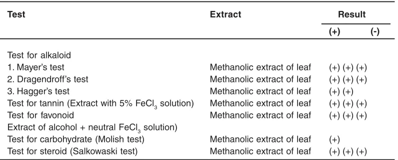
The results of the test for chemical constituents are as follows:
Table 1: Preliminary phytochemical investigation
Test Extract Result
(+) (-)
Test for alkaloid
1. Mayer’s test Methanolic extract of leaf (+) (+) (+)
2. Dragendroff’s test Methanolic extract of leaf (+) (+) (+)
3. Hagger’s test Methanolic extract of leaf (+) (+)
Test for tannin (Extract with 5% FeCl3 solution) Methanolic extract of leaf (+) (+) (+)
Test for favonoid Methanolic extract of leaf (+) (+) (+)
Extract of alcohol + neutral FeCl3 solution)
Table 2: Effect of methanol extract and isolated compound (flavonoides) of Azaretum conyzoides leaves on membrane stabilizing activity
Samples Concentration Absorption at 540 nm % of Inhibition
Hypotonic solution 50mM 0.730 ± 0.06
-Methanolic extracts 50 µg/ml 0.480 ± 0.05 34.24
100 µg/ml 0.384 ± 0.37* 47.39
250 µg/ml 0.330 ± 0.24** 54.79
Isolated compound 100 µg/ml 0.265 ± 0.16** 63.69
Indomethacine 100 µg/ml 0.224 ± 0.29 ** 69.31
Each value represents the mean± SEM; n=3, *p<0.05 and **p<0.01 when compared with blank
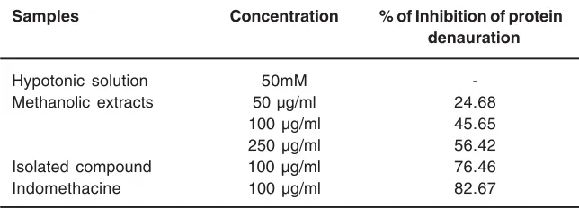
Table 3: Effect of methanol extract and isolated compound (flavonoides) of Azaretum conyzoides leaves on protein denaturation Samples Concentration % of Inhibition of protein denauration
Hypotonic solution 50mM
-Methanolic extracts 50 µg/ml 24.68
100 µg/ml 45.65
250 µg/ml 56.42
Isolated compound 100 µg/ml 76.46
Indomethacine 100 µg/ml 82.67
Preliminary phytochemical investigation reveals the presence of flavonoides. Methanolic extract have showed significant activity in the some of the parameters at higher concentrations (Table 1).
Isolation of flavonoides Compound AZ-1
Brownish yellow crystal, mp. 2250C, R f value 0.28, FT-IR (Jasco-5300) (KBR) V max/cm: 3414 (-OH), 1651 (Unsaturated –C=O), 2928 (C-Me), 1072 cm-1 glycosidic (C-O) groups that are found in flavonoids8-9.
Membrane stabilizing activity
Different concentration (100, 250 µg/ml) of extract showed significant membrane stabilizing activity and isolated compound and indomethacine have significant inhibitory activity (63.69% and 69.31% respectively) which is shown in table 2.
Protein denaturation activity
Different concentrations (100, 250 µg/ml)
of extracts and isolated compound have remarkable inhibitory activity on protein denaturation when compared with the control, which is shown in table 3.
DISCUSSION
Inflammation is the response of living tissue injury. It involves a complex array of enzyme activation, mediator release, extravastation of fluid, cell migration, tissue breakdown and repair10.
expected that compound with membrane stabilizing properties, should offer significant protection of cell membrane against injurious substance13,14,15.
Compounds with membrane stabilizing properties are well known for their ability to interfere with the release of phospholipases that trigger the formation of inflammatory mediators.[16] The extract and
isolated compound has shown significant membrane stabilizing activity, which suggests that its anti arthritic activity observed in this study, may be related to the inhibition of release of phospholipases that trigger the formation of inflammatory mediators.
Denaturation of proteins is well documentated cause of inflammation and rheumatoid arthritis. Several anti-inflammatory drugs have shown dose dependent ability to inhibit thermally induce protein denaturation17. Ability of
Azaretum conyzoides extracts and isolated compound to bring down thermal denaturation of protein is possible contribution factor for its anti-inflammatory activity.
The in-vitro anti-arthritic activity of Azaretum conyzoides found may be due to the presence of flavonoides.
REFERENCES
1. Chatterjee, G.K., and Pal, S.P. “Search for anti-inflammatory agents from Indian Medicinal Plants” - A review. Indian Drugs, 21: 413 (1984)
2. Rainsford, K.D. and Whitehouse, M.W. “Anti-inflammatory/anti-pyretic salicylic acid esters with low gastric ulcerogenic activity.” Agents Action, 10: 451-55 (1980).
3. Huang, K.C. The Pharmacology of Chinese Herbs. CRC Press, London, 199 (1999).
4. Khandelwal, K.R. Techniques and
Experiments, Practical Pharmacognosy, 9th Ed. Nirali Prakashan, 149-59 (2002) 5. Shind, U.A., Phadke, A.S., Nair, A.M.,
Mungantiwar, A.A., Dikshit, V.J.and Saraf, V.O. “Membrane stabilizing activity - a possible mechanism of action of anti-inflammatory activity of cedrus deodara wood oil.” Fitoterapia, 70: 251-57 (1999)
6. Mizushima Y. Screening test for anti -rheumatic drugs. Lancet 2: 443 (1966). 7. Elias, G. and Rao, M.N. “Inhibition of albumin
denaturation and anti-inflammatory activity of dehydrozingerone and its analogs”. Indian. J. Exp. Biol., 26: 540-42 (1988)
8. Kalsi PS. In: Spectroscopy of organic compounds, 5th Ed. Newage publisher 88 (2002).
9. Masbr y, T.J. and Mar kham, K.P. “The systematic Identification of flavonoids”, Springer verlag, 41(1970).
10. Vane, J.R. and Bolting, R.M. “New inside into
the mode of action of anti-inflammatory drugs”. Inflame.Res. 44: 1-10 (1995). 11. Augusto, O., Kunze, K.L. and Montellano, P.R.
“Nphenylprotophorphyrin formation in the haemogolobinphenylhydrazine reaction”. J. Biol. Chem. 257: 6231-41(1982)
12. Ferrali, M., Signorni, C., Ciccoli, L. and Comporti, M. “Iron release and membrane damage in erythrocytes exposed to oxidizing agent, phenylhydrazine, divicine and isouramil.” Biochem. J 285: 295-301 (1992). 13. Maxwell, SRJ. “Prospect for the use of antioxidant therapies”. Drugs 49: 345-61 (1995)
14. Liu, G.T., Zhang, T.M., Wang, B.E. and Wang, Y.W. “Protective action of the seven natural phenolic compounds against peroxidase damage to biomembranes”. Biochem. Pharmacol. 43: 147-152 (1992)
15. Prenez, R.M., Prenez, S., Zavala, M.A. and Salazar, M. “Antiinflammatory activity of the bark of hippocratea excelsa”. J. Ethanopharmacol. 47: 85-90 (1995) 16. Aitadafoun, M., Mounieri, C., Heyman, S.F.,
Binistic, C., Bon, C. and Godhold, J. “4-Alkoxybenzamides as a new potent phospholipase A2 inhibitors.” Biochem. Pharmacol. 51: 737-42 (1996)