EXPERIMENTAL EMPHYSEMA IN THE BLOTCHY MOUSE. A MORPHOMETRIC STUDY USING IMAGE ANALYSIS
BY
ALISON CAROLINE ELLIOTT McCARTNEY M.A., M.B., B.Chir, F.R.C.Path, F.C.Ophth.
Thesis submitted for the degree of DOCTOR OF MEDICINE (M.D.)
FACULTY OF MEDICINE, UNIVERSITY OF LONDON
1993
THE INSTITUTE OF OPHTHALMOLOGY Department of Pathology,
ProQuest Number: U066213
All rights reserved
INFORMATION TO ALL USERS
The quality of this reproduction is dependent upon the quality of the copy submitted.
In the unlikely event that the author did not send a complete manuscript and there are missing pages, these will be noted. Also, if material had to be removed,
a note will indicate the deletion.
uest.
ProQuest U066213
Published by ProQuest LLC(2016). Copyright of the Dissertation is held by the Author.
All rights reserved.
This work is protected against unauthorized copying under Title 17, United States Code. Microform Edition © ProQuest LLC.
ProQuest LLC
789 East Eisenhower Parkway P.O. Box 1346
Abstract.
Smoking remains the major aetiological factor in emphysema but it is paradoxical that most smokers do not appear to develop the disease. Inheritable abnormalities in lung scleroproteins may underlie a propensity to develop emphyse ma.
This hypothesis was tested in two animal models. Experiments were first performed on an animal model, the Wistar rat, which does not exhibit any genetically determined lung disease. Instillation of elastase and saline caused increase in airspace transects in the rat. A sensitive and rapid morphological technique was used to evaluate the light microscopic appearances of the induced lung disease.
The second model was the blotchy mouse, which has an X- linked deficiency of lysyl oxidase, leading to faulty cross linkage of elastin.[ Hemizygous male animals have abnormal lungs, aortic aneurysms and curly whiskers.] This thesis documents the effects on the lungs of increasing age and manipulation of the elastase- antielastase balance in all phenotypes and genotypes of these mice, which were estab
lished as the first outbred colony in the U.K. Scanning electron microscopy was performed on the blotchy mice and their sibling controls.
TABLE OF CONTENTS PAGE NUMBER
FRONTISPIECE
TITLE PAGE
ABSTRACT
TABLE OF CONTENTS
LIST OF FIGURES 12
LIST OF TABLES 15
ACKNOWLEDGEMENTS 16
AIMS 18
CANDIDATES OWN CONTRIBUTION TO RESEARCH 20
TABLE OF CONTENTS
Chapter 1. Introduction 22
1.1 A brief survey of the history of emphysema, 24 its definitions and a survey of theories of pathogenesis 1.1a The history of pulmonary emphysema. 25 Clinical and morphological studies 1721-1948
1.2 Modern morphological studies and measurements. 32 1.2a The age of alpha-1 antitrypsin and the role
of chronic bronchitis. 33
1.2b Smoking and emphysema. 36
1.3 Classification of emphysema. 38 1.4 Relationship of alveolar pores and fenestrae to
emphysema 4 0
1.5 Definitions of emphysema 42
Subtypes of emphysema 45
Centriacinar emphysema Panacinar emphysema Distal acinar emphysema Scar emphysema
1.6 Diagnosis of emphysema 47
1.7 Animal emphysema 49
1.7a Naturally occurring emphysema in animals 49 1.7b Pathogenetic theories and experimentation 51 1.7c The elastase-antielastase balance theory and
its effects on lung research 52
1.7d Cadmium 54
1.7e Hyperoxia and other oxidants 55
1.7f Starvation 57
1.7g Smoking experiments 59
1.7h Enzyme induced emphysema. 60
1.8 Elastases 62
1.8a Porcine pancreatic elastase 64
Effects of elastase on rodent models 65
Short-term effects 65
Medium-term effects 65
Long-term effects 66
Mode of delivery of elastase 66
1.8b Endogenous elastases 69
Human leucocyte elastase 70
Conclusion 71
1.9 The role of the connective tissue matrix and the
development of emphysema 72
1.9a Collagen 73
1.9b Elastin and elastic tissue 76
1.9b.l Elastin 77
1.9b.2 Synthesis 78
1.9b.3 Structure and biochemistry 78 1.9b.4 Desmosine and isodesmosine 81 1.9b.5 Embryological importance of elastin and its
relevance to experimental emphysema 82 1.9b.6 Age and elastic tissue in the lung 84
1.10 Lathyrism 86
1.11 Proteoglycans 87
1.12 Copper, GAGs and connective tissue metabolism 90
1.13 Quantitation of emphysema 91
1.14 Methods of inflation 94
1.15 Measuring emphysema 98
1.15a Examination of lungs 99
Macroscopic assessment 99
1.15b Other problems with measurement 103 Shrinkage phenomena and consequent distortion 103
Compression 105
1.15c Types of microscopic measurement previously
used to assess emphysema 105
1.15d Internal surface area and other indices 106""
1.16 Conclusion 110
Chapter 2 . Experimental emphysema in a rat model instilled with purified porcine pancreatic elastase.
Introduction 111
Effects of elastase on rodent models 112
2.1 The rat model 112
2.2 Rats and elastase in a pilot study 114
2.3 Rat experiment 115
2.3a Experimental protocol 115
Method 116
2.3b Method of preparation of lungs for morphological
analysis 118
Fixation 118
Processing and section cutting 121
Measurement 121
2.3c Method of morphometric analysis 122 2.3d Statistics and choice of method 125 2.4a Results of the effects of instillation of
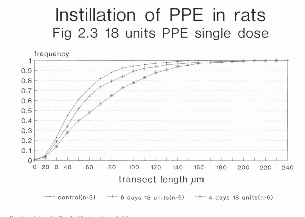
PPE and saline on transect length in rat lungs 128 2.4al Effects of single elastase bolus 128 2.4a2 Effects of a single saline bolus 130 2.4a3 Comparison between the effects of single doses of
saline and elastase at four days (72 hours after
2.4a4 Effects of multiple doses of elastase on
rat lungs 133
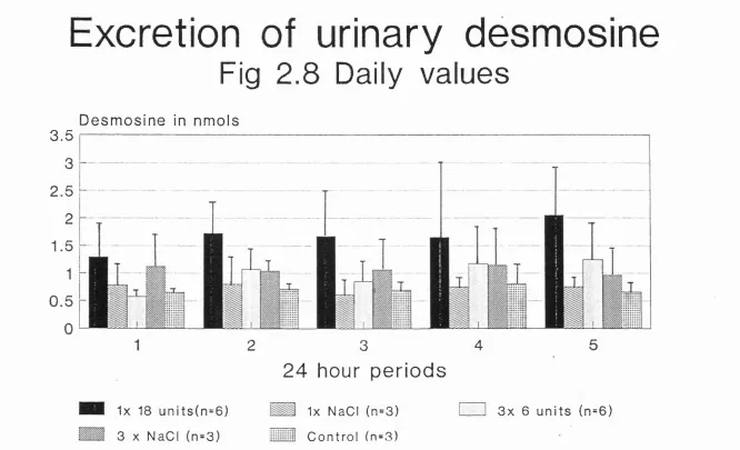
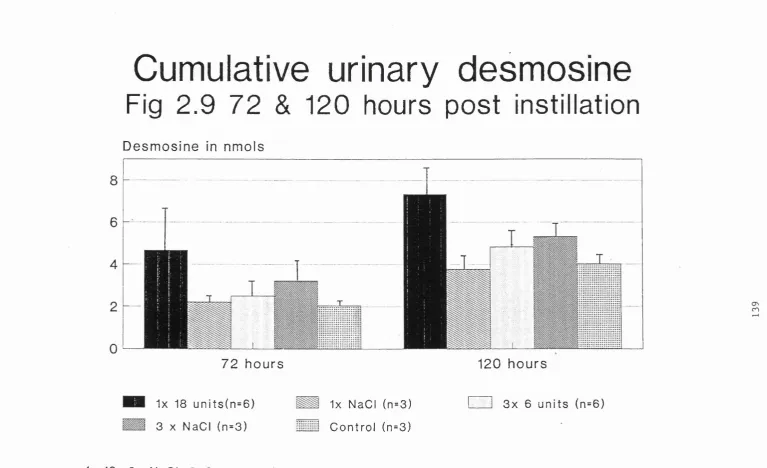
2.4a5 Urinary desmosine 140
2.5 Summary of results 140
2.6 Subjective assessment 143
2.7 Discussion 148
Relevance of the results and is this emphysema ? Type of disease produced in this model 149 2.8 Conclusions from rat experiment 152 Chapter 3. The Blotchy mouse
Chapter 3a. Breeding and anthropometric data on non-instilled mice.
3a.1 Introduction 154
3a.2 Statement 155
3a.2a The Blotchy allele 156
3a.3 Copper and the blotchy mouse 157
3a.4 Connective tissue abnormalities other
than emphysema. 159
3a.4a Cross linking of collagen 159
3a.5 Aortic aneurysms 160
3a.6 Cross linking of elastin 163
3a.7 The blotchy mouse and emphysema 163 3a.8 Blotchy mouse breeding in Charing Cross Hospital 3a.8a Breeding protocols for the blotchy mouse
experiments 164
3a.9 Body weights in blotchy mice 170 3a.10 Lung wet weights in blotchy mice 172 3a.11 Discussion of the biometric data 175 3a.12 Biochemical data from blotchy mice 175
Method Results
3a.12b Measurement of DNA in mouse lungs 178 Method
Results
3a.12c Measurement of hydroxyproline in mouse lungs 178 Method
Results
3a.l2d Measurement of desmosine in mouse lungs 181 Method
Results
3a.13 Measurement of air spaces in blotchy mice 183 3a.13a Results of morphometric analysis 184 3a.14 Results of image analysis using ogive plotting 186
3a.14a Results at three weeks 186
3a.14b Six week data 189
3a.14c Ten week data 189
3a.15 Histology 201
3a.16 Lung volumes in the blotchy mouse model 205
3a.17 Results of lung volumes 208
3a.18 Discussion of biochemical and morphological
measurements in the Blotchy mouse 209 Chapter 3b
Scanning electron microscopy of the Blotchy mouse lung
3b.1 Introduction 215
3b.2 SEM and emphysema 215
Blotchy mouse 233
3b.9 Results 234
3b.10 Conclusion 236
Chapter 4
Effects of instillation of human neutrophil elastase on blotchy mouse lungs.
4.1 Introduction 237
4.2 Experimental protocol 239
4.2a Method 240
4.2b Results 241
4.2b.1 Lung protein and lung DNA 241
4.2b.2 Lung hydroxyproline and desmosine after
instillation of HLE or saline 244
4.2b.4 Lung volumes in instilled mice 244 4.2c Discussion of biochemical results of
instillation experiment 244
4.3 Morphological analysis of instilled animals'
lungs using image analysis 246
4.3a Results of ogive plotting of instillation data
Six week data 252
4.4 Discussion of morphological data from
instillation experiment using HLE and saline 253 Chapter 5
Concluding discussion
5.1 Recap of aims and summary of results 256
5.2 Elastase induced emphysema 259
5.2a The rat model 261
5.3 Discussion of measurement of emphysema in models 263
5.3b Criticism of the method 267
5.4 Pores of Kohn and fenestrae 269
5.5 Instillation in Blotchy mice, a parallel with
smoke induced emphysema in a subset of humans? 271 5.6 HLE and its effects on collagen 271 5.7 Possible fields of investigation suggested
by this current investigation. 273
5.8 Concluding remarks 276
5.9 Originality of research presented in this thesis 281 Appendices
Appendix 1 Biochemical methods and reagents 282 Appendix 1.2 Modified Karnovsky fixative 285 Appendix 2.1 Transect values for rat lungs in 10/xm
intervals 286
Appendix 2.2 Table of critical values of [D] 287 Appendix 3a.1 Point counting technique
Appendix 3a.2. Pooled data from transect length measurements.
Appendix 3a.3 Compliance studies in the blotchy mice Appendix 4. Mean data from transect length
measurements in instillation experiments in Blotchy mice Example of histograms
Examples of values of chi squared for specified comparisons
288
291 295
Appendix 5. Appendix 6.
References
298 300
List of figures
Figure 1.1
Figure 1.2 Figure 2.1
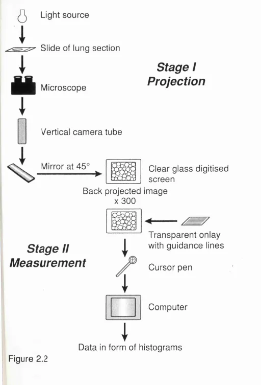
Figure 2.2
Figure 2.3
Figure 2.4
Figure 2.5
Figure 2.6
Figure 2.7
Figure 2.8
Figure 2.9
Figure 2.10
Frontispiece
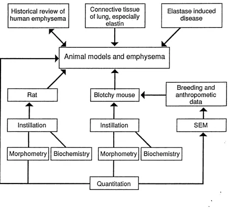
Flow chart showing interrelation of topics discussed in chapter 1 and
experiments in chapters 2-4 26
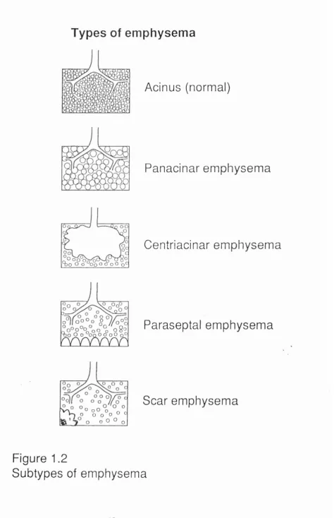
Subtypes of emphysema 45
Constant head apparatus for filling
lungs 119
Diagram illustrating method of projection of image and measurement of image 123 Ogive plot of effects of instillation of PPE on transect length across airspaces in rats: effects of 18 units as a
single dose 129
Ogive plot of effects of instillation of a single dose of saline in rats 131 Ogive plot of effects of instillation measured at 4 days in rats 132 Effects of instillation of PPE in rats: Comparison of effects of triple split dose of 6 units, compared to single dose of 18
units 134
Effects of triple instillation of PPE and saline on transect length across
airspaces 135
on transect length in rats; showing 18 units measured at four days has a greater effect than triple dose measured
at six days. 141
Figure 2.11 Summary ogive plot of effects of
instillation on transect length in rat
lungs 142
Figure 2b.1 Histology from untreated rat x 70 144 Figure 2b.2 Histology from rat treated with 18 units
PPE X 180 144
Figure 2b.3 Histology from rat treated with 18 units
PPE X 450 145
Figure 3a. 1 Colour photograph of Blo/X mouse 167 Figure 3a.2 Colour photograph of Blo/Y mouse 168 Figure 3a. 3 Colour photograph of wild male mouse 169
Figure 3a. 4 Body weight of mice 171
Figure 3a. 5 Lung wet weight of mice 174 Figure 3 . a . 6 Lung protein in mice 177
Figure 3.a.7 Lung DNA in mice 179
Figure 3.a.8 Hydroxyproline in mice 180 Figure 3.a.9 Lung desmosine in Blotchy mice 182 Figure 3.a.10 Ogive plot of transect length of
airspaces in all types of mice at
three weeks 187
Figure 3.a.10a Transect length at three weeks in female mice of each phenotype
Figure 3.a.11 Transect length of airspaces in all types of mice at six weeks
Figure 3.a.11a Transect length of airspaces at six
weeks( female mice of each phenotype) 191 188
Figure 3.a.12 Figure 3.a.13 Figure 3.a.14
Figure 3.a.15
Figure 3.a.16 Figure 3.a.17
Figure 3.a.18
Figure 3.a.19
Figure 3.a.2 0
Figure 3b.1
Figure 3b.2
Figure 3b.3 Figure 3b.4 Figure 3b.5 Figure 3b.6 Figure 3b.7 Figure 3b.8 Figure 3b.9
Figure 4.1
Mean value of transect length in mice 192 Ogive plot of transect length; 10 wks 193 Ogive plot of transect length in adult female mice (older than twelve weeks) 195 Ogive plot of transect length in young adult male mice (12-18 weeks) 196 Transect length of male mice at 32 wks 197 Histology of a) control, b) Blo/X, 202 c) Blo/Y and d) Blo/Blo mice 203 Immersion apparatus for measuring lung
volume by displacement 206
Plot of lung volumes in mice of all
phenotypes 207
Ogive plot of transect lengths in wild male mice to show the effect of aging 211 Composite scanning electron micrograph at low power of all types of mice 223 Wild mouse at ten weeks (SEM, original
magnification x 750) 226
Heterozygous mouse at ten weeks 227 Another heterozygous mouse at 10 wks 228 Hemizygous male mouse at ten weeks 229 Homozygous female mouse at ten weeks 230 Another homozygous mouse at ten weeks 231 Hemizygous male mouse at 180 days 232 Histogram plot to show distribution of
numbers of airspaces in mice of all
phenotypes 235
Figure 4.2
Figure 4.3
against untreated animals at same
age (six weeks) 242
Histogram of lung protein extracted after instillation, compared with controls 243 Histogram of lung hydroxyproline
extracted after instillation compared
to controls 245
Figure 4.4
Figure 4.5
Figure 4.6
Figure 4.7
Figure 4.8
Ogive plot to show transect length after HLE and saline at 3 weeks 247 Ogive plot to show effects of instillation on distribution of airspaces in wild mice
instilled at six weeks 248
Ogive plot to show effects of instillation on six week old Blo/Blo mice 249 Ogive plot to show effects of instllation on six week old heterozygous Blo/X mice 250 Ogive plot showing effect of instillation on all types of female mice at six weeks 251 Appendix A.A.l Standard radioimmunoassay plot 284
List of Tables
ACKNOWLEDGEMENTS.
I should like to thank all of the many people who have been unstinting of their patience, enthusiasm and support of this M.D. thesis but especially Professor John Sloper and Professor Alec Garner for allowing me the facilities and time to carry it out. My sincerest thanks are due to Dr Bernard Fox for his encouragement, advice and criticism of the project since its initiation and for his friendship. Similar thanks are due to Dr Terry Tetley for allowing me to understand the joys and pitfalls of research and for being a constant source of stimulation. I should like to thank Gary Phillips for all the care and attention he lavished on our joint population of mice and Dr Vidya Pai for help with the rat experimental work. Thanks are also due to the staff of the animal unit at Charing Cross Hospital Medical School.
Terry Bull, with his great love of electron microscopy and impeccable standards, has been unfailingly kind and dependable and for technical help, I owe a debt of thanks to Gill Miller, Fathi Gowali, Robin Howes and Stephen Davies amongst many others.
I am also greatly indebted to the secretarial staff of both departments but especially to Mrs Pat Goodwin, who kept track of my references and helped produce the final product. I should like to thank the various librarians who have helped me over the years, but especially the librarians of Charing Cross and the Institute and the Royal Society of Medicine, notably for Tuesday evenings!
Allied Dunbar staff from the Sackville Street branch raised money that enabled the purchase of an image analysis system, a 'tool' with which I was able 'to finish the job'. The machine is for use primarily in eye research but I should like to record their generosity here. The biochemical work was supported by the Chest, Heart and Stroke Association and the Morrison-Davis Trust.
I should also like to thank Dr Heather Morrison and Profes sor Bill Whimster for their helpful advice on completing and refining the thesis.
AIMS
To evaluate two experimental animal models of elastase- induced emphysema: the first in the Wistar rat, which does not have any genetic predisposition to lung disease and the second the Blotchy mouse, an animal not previously bred in this country for investigation of lung disease, but known to have connective tissue abnormalities as a result of lysyl oxidase deficiency.
Aims of experiment 1 on the rat model described in chapter 2
la. To determine whether instillation of relatively small amounts of an exogenous purified porcine pancreatic elastase produces destruction and enlargement of airspaces within a Wistar rat model, using young rats, unlikely to have sponta neous emphysema.
lb. To determine whether this change is produced by single or repeated doses of elastase and whether there is evidence of similar damage when saline, without intrinsic enzyme activity, is used.
Ic. To document change in airspace size by measuring tran sect length across airspaces; the data to be obtained by computerised image analysis of fixed lung sections, plotted using ogives of cumulative frequency and subsequently analy sed using a non parametric method to assess significance.
Aims of experiment 2 on the Blotchy mouse, an animal known to have an X linked deficiency of the cross linking enzyme, lysyl oxidase.(chapter 3.)
2a. To breed a colony of Blotchy mice, using inbreeding and outbreeding to obtain all phenotypes and genotypes of ani mals, including the homozygous Blo\Blo female.
2b. To record whether there were morphometric, including scanning electron microscopic (chapter 3.b), differences in the lungs of these mice by comparing them with their non affected littermates at different ages and to compare this data to the results of biochemical analyses obtained in a parallel study in the younger mice.
Aims of experiment 3, instillation of human leucocyte elas tase in the Blotchy mouse model.(chapter 4)
3a. To determine whether (human) leucocyte elastase, has an effect on transect length across airspaces, in instilled mice of all phenotypes compared to untreated control ani mals .
CANDIDATES OWN CONTRIBUTION
The project was originally suggested to me by Dr Bernard Fox and Professor Abraham Guz. Dr Terry Tetley and her lung biochemistry team, especially Gary Phillips, were responsible for the biochemical analysis and many of the instillations. The animals were after the initial pilot study looked after in the Charing Cross animal house by Dr H.B. Waynforth and his workers, especially Miss Dulcie Gray. Dr Ken MacRae suggested the ogive plotting method and Dr Terry Partridge was responsible for instigating the image analysis. Mr Terry Bull helped me a great deal with the preparation of the SEM specimens and Robin Howes the photog raphy. The Medical Illustration Department of Moorfields Eye Hospital allowed me to use their dry mounting equipment and Jane Fellowes helped with the illustrations.
I am deeply grateful to Mr Phillips and Dr Tetley for allowing me the opportunity to incorporate the biochemical data from our rats and mice to support the morphological data I have obtained.
Chapter 1. Introduction.
Naturally occurring emphysema is rare in animals. The blotchy mouse has an X-linked defect for the enzyme lysyl oxidase, which catalyses the cross linkage of structural proteins. Hemizygous male animals have abnormal lungs and phenotypically differ from their agouti coloured normal siblings and the heterozygous females who have coats flecked with the pale colour giving rise to the name blotchy. My hypothesis was that such animals would be vulnerable to the effects of exogenous events that mimic the effects of smoking.
The reasons for this hypothesis were that, although smoking remains the major aetiological factor associated with the development of emphysema, human emphysema differs from other smoking associated diseases. These diseases, manifested by abnormalities of respiratory and bronchial epithelial cells, are almost ubiquitous, are dose dependent and are to a certain extent reversible on cessation of the activity. Although emphysema is difficult to diagnose clinically, it
elastases and antielastases in the lungs of those who smoke. The elastase-antielastase hypothesis is that in smokers, the antielastase defence system of the lung cannot adequate ly protect the alveolar walls and ducts from damage caused by proteolytic enzymes, particularly neutrophil derived elastase. The 'direct' evidence for this theory was largely accrued from work on the homozygous alAT deficient patients, but the theory is less well supported and the evidence is 'indirect' in alAT nondeficient smokers with supposedly adequate levels of protease inhibitor.
The tissue substrate, primarily elastin, for these elasto- lytic enzymes has been less well investigated. Variation in the pattern of deposition and biochemical or structural anomalies in elastic tissue might also be inheritable and lead to a predisposition to emphysema.
1.1
A brief survey of the history of emphysema, its definitions and a survey of theories of pathogenesis.
Research into emphysema has varied from extensive epide miological surveys to biochemical studies on a molecular scale. Although human studies are the most pertinent, the search for animal models of disease has widened to include genetically determined lung damage such as is seen in the Blotchy mouse . I wished to address questions of morphology in naturally occurring disease and after manipulation of the protease-antiprotease balance, especially with regard to the amount of elastic tissue present and the degree of lung destruction. Quantitative morphology and biochemical analy sis were the two most powerful tools at our disposal coupled with investigation at the ultrastructural level to determine the importance of the fenestrae often observable in emphyse matous lungs. This thesis represents one part of a combined and integrated study.
the biochemical results and the relevance of this type of experimental model to human disease (Figure 1.1).
The historical review will concentrate on the morphology of emphysema and the importance of the underlying connective tissue of the lungs.
1. la
The history of pulmonary emphysema.
Clinical and morphological studies 1721-1948.
Emphysema is derived from the Greek word emphysan, to inflate and was originally used to describe the presence of air within the tissues and as such is still used today in the expression 'surgical (sub cutaneous) emphysema'. Thurlbeck in his review of early work on emphysema (1976) cites the description of Watson (1764), who described a young man who had aspirated vomit. Watson
implies that there was air present within the parenchyma of the lung, which had ' burst through the bronchi and vesicu lar substance,' rather than within the airspaces, with 'air getting loose within the substance of the lungs (which) cannot be parted with on expiration' as well as 'bladders' which 'no pressure on the surface of the lungs could force back' . Rosenblatt (1972) quotes from the earlier work of the Swiss physician Bonet who described in Sepulchretum, in 1679, the correlation of dyspnoea and orthopnoea in cases where there was overdistension of the lungs with air.
Ruysch (1691 and 1721) illustrated enlarged airspaces and these pictures together with the clear illustration by
Morphometry
Biochemistry Biochemistry Morphometry
Blotchy mouse
Instillation
Quantitation Rat
Instillation SEM
Connective tissue of lung, especially
elastin
Breeding and anthropometic
data Elastase induced
disease Historical review of
human emphysema
Animal models and em physem a
Figure 1.1
plate, traditionally thought to illustrate the lung of Samuel Johnson, who died several years after a bout of pneumonia, carries the legend that 'the air cells are much enlarged beyond their normal size and resemble those of amphibians'. In his textbook published eight years later Baillie predates the observations of Laennec by stating that the lungs did not collapse at post mortem. He describes air cells that were large enough to hold a small gooseberry (1799 and 1807). Baillie also recognised the importance of destruction in this disease.
Morgagni (1769) also described two cases of emphysema, likening the distended airspaces to filberts (hazelnuts).
Laennec (1819), working amongst others, on the victims of the cholera epidemic in Paris, is usually credited as the first author to describe pulmonary emphysema correctly and to identify the underlying anatomical basis of production of abnormal air spaces; 'elle consiste dans la simple dilata tion des vésicules ou cellules dont elle compose'. His work, although widely quoted, is difficult to obtain in the origi nal, a manuscript which described the appearances of lungs which in an 'anatomical state of hypertrophy go out of the thoracic cavity to the mediastinum instead of collapsing'.
available in the English language, that most subsequent authors refer. He was aware of the close association of chronic bronchitis with emphysema and emphasised that it was the chief cause of the disease as well as recognising the difference between true emphysema and the enlargement of air spaces associated with increasing senescence. Laennec pre pared his specimens by inflating them and leaving them to dry out, thus recognising the importance of prevention of collapse in fixed specimens. He hypothesised that loss of recoil was an important factor in the genesis of the disease resulting in slower flow and in translation the words 'diminished elasticity of air cells' have been used (Thurl beck 1976).
The association of chronic bronchitis and emphysema was noted by Louis (1835) in his monograph but he denied the importance of this observation, (as did Waters, see below).
Forbes (1824) also gives credit to Avenbrugger, working in Vienna, for the description of right ventricular hypertrophy accompanying lung disease. Rainey in 1848 gives the first description of the microscopy of dilated alveoli and widened meshes of the capillary net, with subsequent pores or gaps in the alveolar walls and the progressive enlargement of these fenestrations, with destruction of the alveolar walls finally leading to bullae. This work was carried out using unstained thick sections.
emphysema; he called obstructive emphysema 'substantive'.
Waters (1862) reviewed the previously held predominating theories about this disease citing the work of Bonet, Morgagni, Floyer (1726), Ruscke (1691) and Baillie (1799). He refers anatomically to air spaces which he describes as air sacs, lobulettes and lobules, painstakingly recording them, (Frontispiece) and documents six to twelve air spaces communicating with the 'dilated extremity of a bronchial tube by a circular opening, smaller than the sac itself, which became polygonal when the lung was properly inflated. He also details the 'point de réunion' of all the air sacs, the common centre of the lobulette and describes the alveoli as 'cyst-like depressions', eight to twenty to an air sac, with circular openings, 'smaller than the cavity to which it
leads'.
Waters predates other observers when he describes the walls of the sacs or alveoli as being composed of 'yellow elastic fibrous tissue' with fibres arranged in bundles and singly encircling the mouths of the air sac and in the wall notes a basement membrane and an epithelium together with
'the capillary vessels of the pulmonary plexus'.
here. Interestingly it has been postulated that fibrosis and emphysema represent divergent responses to a common insult (Niewoehner and Hoidal 1982), and some forms of experimental emphysema are associated with fibrosis (Snider et al 1991). Thirdly he details the emphysematous change extending to the whole lobe or lung.
He was therefore describing dilatation of alveolar ducts and sacs, with fenestration leading to a cribriform appearance and progression to destruction of septa by distension of the existing air sacs , tallying with modern descriptions of the disease.
Strawbridge (1960a) in reviewing the nineteenth century German literature, emphasises the work of Eppinger which detailed the loss of fine elastic fibres, with fraying of the elastic fibres at the edge of recent fenestrations, as well as distinguishing between acute emphysema due to over
inflation and chronic emphysema.
observations. Loeschke (1922) conceded that fenestration led to destruction but did not see pre-existent alterations in elastic fibres, averring that it was the tissue most resistant to the essential atrophy of emphysema. Using corrosion casting he also demonstrated, without recognising its significance, the first illustration of centrilobular emphysema.
1.2
Modern morphological studies and measurements.
The essentially different natures of panlobular and centrilobular disease awaited discovery until 1952 and the work of Gough and the descriptions of McLean (1956, 1957 and
1958). A three dimensional study by Wright in 1961 illustra ted the framework of elastic fibres that support the primary lobule and terminal bronchiole, respiratory bronchiole, alveolar duct and alveoli in the normal lung . These authors also showed focal defects including attenuation of fibres, leading to complete separation and retraction of the unatta ched ends with consequent fenestration of alveolar walls, loss of capillaries and an interesting increase in fibrous tissues. Later quantitative studies were presaged by Har- troft in 1945 who thought that serial section and measure ment were the keys to successful investigation. Gough's work
on the whole lung sections prepared by him and his techni cian Wentworth, allowed pathologists to attempt to quantify the disease and to compare the post-mortem findings with the physiological tests of the clinicians and the appearances of the lungs using X ray where the changes in the lung paren chyma are assessed in relation to the changes in the chest wall, diaphragm, heart and great vessels.
1.2a The age of alpha-1 antitrypsin and the role of chronic bronchitis.
recessive inheritance, with three levels of serum alpha-1 antitrypsin protease inhibitor : normal in the MM phenotype, intermediate in the heterozygote carrier MZ phenotypes and low in the homozygous ZZ phenotypes of whom 50% have obstructive airways disease, usually severe early onset panlobular emphysema. Alpha-1 antitrypsin is a 59 kDa antiprotease providing the major defence against the 'ravages of neutrophil elastase in the lower respiratory tract' (Crystal 1989, Gadek et al 1981). The deficiencies are due to single amino acid substitutions at position 342, close to the carboxy terminal of the 394 amino acid chain, that of the Z variant (PiMZ) , carried by 0.03% of Europeans is of lysine replacing glutamic acid and another polymorphism (PiSM) found in 7%, is associated with substitution also of glutamic acid but by valine at position 264 rather than 342 (Carrell et al 1982) . One of the sur prising elements in patients with this deficiency is the
The roles of proteases and antiproteases and their clinical implications were extensively reviewed in the supplement (1983) to the American Review of Respiratory Disease edited by Cohen.
In cases without antitrypsin deficiency there was gradual awareness that the disease arose as a direct result of smoking cigarettes. This observation came partly as a result of the Clean Air Act in this country and partly as a result of good epidemiological evidence gathered by D.D Reid (1956) and others (Edwards 1961, Lambert and Reid 1970) on the importance of the relationship of chronic bronchitis to the development of emphysema. Clinical and epidemiological studies have similarly shown that 90% of the 62,000 deaths per annum in the U.S.A. occurring in patients with chronic obstructive airway disease (C.O.A.D.) (20,000 per annum in the U.K (Tetley 1992)) are attributable to smoking (Snider, Lucey and Stone 1986) rather than industrial exposure.
The pathology of chronic bronchitis, due to the overactiv ity of the mucous glands, was first described by Florey et al (1932) and consolidated in two works by Lynne Reid in 1954 and 1960 where she accurately defined the extent of the thickness of the mucus-producing glands and their relation ship to the bronchial wall as an index. There were differing diagnostic criteria on either side of the Atlantic leading to confusions as described by Fletcher (1959). The relation ship between the two diseases is not a simplistic cause and effect. Chronic bronchitis was defined in clinical terms by Heard et al (1979) as resulting in expectoration of mucus at some time of the day for at least three months of two conse
emphysema.
The American view of the morphology of 'chronic airway obstruction' was described by Mitchell et al (1976) in a semiquantitative study. This group had been studying this type of diseases since 1956 and the 1976 paper was the culmination of their efforts centred on the lungs of 330 cases.
Continuing epidemiological surveys of patients with these diseases were subjected to multivariate analysis by May and Peto in 1973 to establish the roles of differing pathogens including bacteria. They came to the conclusion that the relationship was due to smoking, the 'final common pathway'
(Thurlbeck 1976).
There may be a lack of sequential evidence to link the two diseases, but in most cases of 'smokers' emphysema, especially in those with incapacitating or severely disabling disease there is evidence of a preexisting respiratory bronchiolitis. This can be seen in young smokers linked to accumulation of brown stained pulmonary macrophages (Lancet editorial 1980) and bronchiolitis accompanies mild centrilobular disease (Leopold and Gough 1957) and has been described as a precursor (Niewhoener et al 1974) . Irregular, less severe forms of emphysema are found in non-smokers and rarely lead to incapacity.
1.2b Smoking and emphysema.
appetite. Unfortunately for those addicted, every puff releases up to two thousand chemicals, apart from nicotine, and a million oxidant molecules, into the respiratory system, as well as substantial amounts of carbon monoxide (10-20mg per cigarette) . The effects of some of these chemicals such as cadmium (Nandi et al 1969) have been the subject of extensive research, (smokers can absorb up to two micrograms per packet) but other compounds are less well
investigated.
Cigarette smoking in those patients not known to have alpha- 1 antitrypsin deficiency, is usually associated with cen- triacinar emphysema, particularly of the upper lobes of the lung. Much of the pathogenetic effect of cigarette smoking has been ascribed to alteration of the elastase- anti- elastase balance (Gadek et al 1979, 1980a) [as well as elastin resynthesis in animal models (Osman et al 1985a and b) ] and techniques such as bronchio-alveolar lavage (BAL)
(Reynolds and Newball 1974) have shown up to a sixfold increase in the numbers of polymorphonuclear leucocytes in the lungs of smokers. Lavage has also shown a metallopro- tease derived from the unstable alveolar macrophages which also secrete a true protease (Bignon et al 1982) probably contributing to extracellular elastolytic activity (Smith et al 1985). The two-pronged attack (Lancet editorial (1980)) results from elastolysis and autodigestion of lung tissue. Suppression of elastolytic inhibition by oxidation of me thionine residues of al-AT has also been suggested but is less well supported.
of terminal bronchioles where they release chemoattractants especially leucotrienes. Even if these macrophages are relatively inefficient at release of these factors,
(Laviolette et al 1986) their increased numbers lead to increased neutrophil adherence through thromboxane release from the arachidonic acid cycle.
The neutrophils are directly affected by smoke and release neutrophil elastase which, in addition to its proteolytic effect, binds to and inactivates alpha-1- proteinase inhibi tor from serum. Although elastase is found in macrophages they can also act as a sink for neutrophil elastase and resecrete it in an activated form (Snider 1984) . Smoke in addition to causing cellular recruitment and consequent damage can liberate active oxygen species including superox ides, free radicals and hydrogen peroxide in addition to carbon monoxide, cadmium, nitrogen oxides and semiquinones in tar residues (Flenley 1986).
1.3
Classification of emphysema.
'Rien ne nuit plus aux progrès d'une science que de dé tourner sans motifs suffisants les noms de leur acception reçue, ou d'en creer de mauvais'(Laennec, quoted by Scadding 1959) .
(Nothing is more harmful to the progress of science than altering accepted names, by creating others which are worse, without adequate motives.)
end-stage of injury and repair in a triphasic organ consisting of gas, liquid and solid matter, whilst the fibrosing diseases, alveolitis or interstitial fibrosis lie at the other (Kilburn 1975).
One of the problems that bedevils research into emphysema is that the disease is usually treated by authors as a * single entity which results in respiratory airspace enlargement with evidence of destruction of airspace walls' (Snider, Ciccolella et al 1991). What becomes increasingly apparent on review is
a) that this 'end-stage', stereotypic disease may arise as a result of several different pathways,
b) that varying patterns of disease emerge at certain preferential sites e.g. centriacinar and centrilobular disease starts in the respiratory bronchioles, is largely confined to the upper parts of the lobes and is associated with smoking, whereas panacinar disease starts at the level of the alveolar walls throughout secondary lobules, usually within the lower parts of the lung and is associated with alAT deficiency. Subpleural or paraseptal disease starts in alveoli next to fibrous septa and paracicatrical or scar emphysema starts, as its name suggests, next to fibrotic scars anywhere within the lung.
c) that these various morphological categories may be due to differing pathogenetic events and that clear thinking may not be aided by trying to find a unifying pathogenetic mechanism to explain them all.
data to suit the fashionable hypothesis.
The problems of pathological and anatomical classification are not confined to the human disease and it can be even more difficult to categorise the lesions produced in experimental emphysema. This problem is addressed later.
Modern classification of emphysema is based on the unit of reference known as the acinus, a structure 8.8mm in length (+/- 1.4mm) (Haelfli-Bleur and Weibel 1988), first described by Rindfleisch in 1878 (quoted by Scadding 1978). The acinus is a three dimensional unit of lung structure de fined as being distal to the terminal bronchiole, an airway without cartilage within its wall. The human lungs are said to contain 28,000 terminal bronchioles and acini, apparently a fixed number of units, acquired shortly after birth and not subject to change (Matsuda and Thurlbeck 1971) although lung growth at the alveolar level occurs for longer, until the adult number of 300 million alveoli is reached.
Although the acinus is the theoretical basis for classifying the disease, practically it is a difficult domain to quantify in both pathological and radiological observations. Many authors with justification have preferred to retain the two dimensional concept of lung lobules which are visible on inspection of fixed and fresh lungs in slices and radiographs.
1.4
discussed them in 1893. These tiny pores are 5-15jum in diameter and are important in both normal lung for rapid equilibration of air pressure 'collateral air drift' whilst at the same time in diseased lung allowing passage of blood, organisms, inflammatory cells and oedema fluid from one alveolus to another without reliance on trans- airway transmission. In emphysema it has been suggested that collateral air drift is an important form of ventilation
(Terry et al 1978).
Pump (1976) suggested that emphysema causes increase in the number or size of the pores of Kohn. He described the progression and evolution of emphysematous airspaces from normal pores of Kohn, which he called Stage 1 fenestrae. These were documented as 2 0 /xm roughly circular openings with a capillary loop border. Stage 2 fenestrae had ruptured or absent capillary loops and were larger and had a more rectangular shape. By the time the third stage was reached almost all of the capillaries in the wall of the alveolus had disappeared and there was heavy elastic tissue deposition around the fenestrae in the form of thick fibres. The fourth and final stage was the formation of emphysematous cysts.
The Pump sequential change and relevance of pores of Kohn to development of emphysema has been questioned (Whimster and Lunkenheimer 1989). These authors concluded from three dimensional reconstruction and image analysis that the pores are normal phenomena, usually covered with surfactant and have a regional distribution dissimilar to that of centri
Another normal type of communicating channel is the canal of Lambert, which runs directly from the pre-terminal bronchiole to the alveoli and is thought to be responsible for the preferential deposition of dust within the peribronchiolar air spaces in coal workers'pneumoconiosis.
1.5
Definitions of emphysema
Before the 1959 guest symposium of British physicians sponsored by Ciba (1959) there had been great confusion between three major lung diseases; asthma, chronic bronchitis and emphysema (Fletcher and Price 1984). This symposium was unsuccessful in defining emphysema in physiological terms and an anatomical compromise was reached, with the evolution of a definition (Scadding 1959). The net result was that diagnosis of emphysema shifted from the clinicians' domain into the pathologists', but even there problems of classification and identification still exist.
The definition advanced by the Ciba symposium was that 'emphysema is a condition of the lung characterised by increase in size of air spaces distal to the terminal bronchiole, arising either from dilatation or destruction of
their walls.• Redefinition was attempted by the World Health
Organisation (1961) and subsequently by the American Thoracic Society (1962).
was also recognised.
A workshop convened in 1985 (Snider, Kleinerman et al) had three goals, the first being to define 'destruction* so that uniform enlargement, due to chromosomal abnormalities such as Down's syndrome (47XX or XY) and the airspace enlargement associated with fibrosing lung disease are not confused with emphysema; secondly to establish criteria for animal models and thirdly to reinvigorate the study of the disease , for which publication rates had diminished.
This 1985 workshop described simple airspace enlargement both congenital or acquired, and emphysema, the latter as a condition of the lung characterised by abnormal permanent enlargement of airspaces distal to the terminal bronchiole, accompanied by the destruction of their walls, and without obvious fibrosis.
This group amplified their definition by defining destruction as, 'nonuniformity in the pattern of respiratory airspace enlargement so that the orderly appearance of the acinus and its components is disturbed and may be lost '. Destruction of lung may be easily recognisable in the moderate and severest forms of the disease but in the less affected lungs, for instance those with mild emphysema, only the fact that the disease is localised may lead to its recognition at post-mortem. It has been suggested however that for emphysema to be macroscopically visible that over 75% of lung respiratory parenchyma may have been lost (Lamb 1990).
Three types of emphysema were described by the workshop in
acinar emphysema. Figure 1.2 (page 45) illustrates these and also shows scar emphysema in diagrammatic fashion. Lately scar emphysema has been reinstated in discussions of the pathogenetic mechanisms involved in the induction of emphysema (Snider et al 1991) , having previously been excluded by virtue of the amount of fibrosis present.
As alluded to above, radiologically it is impossible to relate emphysema to the acinus and one author (Paakko 1981) believes that this is true for pathologists too, whilst relationship to the lobule can be recognised. In the British literature classifications based on the secondary lobule, identifiable by the naked eye remained popular (Heard et al 1979) . Heard et al also draw attention to the practical advantages of defining the disease in terms of destruction rather than dilatation. Their classification was based on a macroscopic evaluation and therefore they preferred the (secondary) lobule, a larger unit, containing four or five acini, and surrounded by connective tissue septae. Some authors remain very confused by this concept of lobular disease, notably Thurlbeck (1976), who illustrates emphysema with reference to the primary (rather than the secondary)
lobule. The primary lobule is a subdivision of the acinus, served by a single respiratory bronchiole.
Subtypes of emphysema Centriacinar emphysema
The emphysema associated with exposure to mineral dusts especially hard coal, with high levels of silica, also
results in centriacinar disease but there is also accumulation of dust in macrophages and the disease is more generally distributed throughout the lung fields. Coal workers' pneumoconiosis, with an associated emphysema is much commoner in those who mine hard coal, such as that found in Wales, in comparison to coal miners in softer rock such as the chalk of Kent (B.Fox personal communication 1992). The disease is worsened by cigarette smoking however. In some cases there may be a combination of patterns with centriacinar and centrilobular disease and areas of paraseptal or scar emphysema.
Panacinar emphysema.
Types of emphysema
Acinus (normal)
5^
Panacinar emphysema
Ooo o
9)0 o o °o q oo oo
Centriacinar emphysema
0^ f o ° ° o 5 7/o°q
%S°o o°°o^o°oo|o
^YYWYY^
Paraseptal emphysema
o o o
^ o ° °o o ° o ° c
O O o ° o
Scar emphysema
Figure 1.2
Subtypes of emphysema
Distal acinar emphysema.
This form of the disease which predominantly involves the alveolar ducts and sacs is associated with the interlobular septa and was previously known as paraseptal emphysema, but is also recognisable where the acinus abuts the pleura or vessels.
Scar emphysema
This form of the disease is associated with any type of scarring within the lung. Previously it was especially associated with post tubercular scars but may follow other causes of consolidation and fibrotic repair and is occasionally seen adjacent to scars generated by neoplasia.
Other terms that are used in description are bleb, which is a collection of air within the layers of the visceral pleura; bulla, which is an emphysematous space with a diameter of more than 1cm in the distended state; cyst, which is a cavity lined by bronchiolar epithelium or fibrous tissue and pneumatocoele which is a large air-filled cyst of at least one third of a lung's volume.
1.6
Diagnosis of emphysema.
Colebatch 1980). Lung functional changes include a decrease in the forced expiratory volume of air exhaled in the first second (FEV^) , regarded as an obstructive phenomenon; an increase in the residual volume (RV) of the lung; a reduced negative intrapleural pressure and an uneven distribution of inspired air (Stead et al 1952). FEV^ is also often expressed as a proportional ratio of the total lung capacity (TLC) and in emphysema the ratio is usually less than 70 percent of that predicted.
In view of the problems apparent in reaching a definition of the disease outlined above, it is not difficult to understand that the disease can be difficult to diagnose both in life and in the inflated post-mortem specimen. There is in addition a lack of correlation between the clinical symptomology, with regard to such factors as wheeze, cough and dyspnoea and objective measurements such as the alveolar surface area calculated from the mean linear intercept obtained from measurement of lungs obtained at post-mortem. The latter is discussed more fully in the section on quantitation. There are signs visible on chest radiographs, such as hyperinflation and reduced vascularity, which are also diagnostic but quantitatively imprecise (Thurlbeck, Henderson et al 1970).
possible to generate a histogram of lung densities from pixels, the picture elements which make up the CT scan. The histogram of lung density is skewed by the presence of large vessels and airways and MacNee and associates have concentrated on the data in the lowest fifth percentile in their in vivo estimations of emphysema using CT densitometry. The measurements are made after the patient has inhaled deeply and has been asked to hold the breath. This technique has meant that this group has been able to correlate their measurements of macroscopic and especially microscopic emphysema in vivo with their published work on the morphometric analysis by measurement of the airspace wall per unit volume (AWUV) (Gould et al 1988) which is discussed below.
In another series, published in abstract, comparison of CT scans from smokers with nonsmokers, showed that there was less than 1% bullous change, attentuation, measured as being equivalent to tracheal lumenal density, and that in smokers the amount of bullous change correlated well with the FEV^\TLC and the residual volume to TLC ratio (Filadiktaki et al 1992) .
1.7
Animal emphysema
gement by proteolytic enzymes. 1.7a
Naturally occurring emphysema in animals.
The description of the lungs of a hawk with *crocke' and of a broken winded horse (Floyer 1698 and 1726; quoted by Rosenblatt 1972, Thurlbeck 1976 and Kilburn 1975) predated the description of emphysema in the human. Budd in 1839 also reportedly described emphysema in the lungs of two horses and attributed at least part of the pathogenesis to the loss of elastic tissue, as well as suggesting the possible role of hereditary factors; something of greater note to the bloodstock breeders of the turf perhaps than the rest of the community, as the role of inheritance in alpha-1 antitrypsin deficiency in humans was not described until 1963.
The equine disease 'heaves'(Foley and Lowell 1966) resulting in onomatopoeic breathing is not only due to emphysema (Alexander 1959) and its consequent expirational difficulty, since Thurlbeck describes similar symptomatology in animals suffering from 'hay fever'(allergic rhinitis) and bronchitis (1976 and 1964). Rats suffering from mycoplasma (mollicute) infections are said by Thurlbeck to have resultant emphysema, and Levame (1980) and Palecek and Holusa (1971) describe emphysema in specified pathogen free (SPF) rats as spontaneous.
Emphysema has also been described as a spontaneous disease in other experimental animals including rabbits (McLaughlin et al 1961 and Strawbridge 1960) and dogs (Hernandez 1966) .
(Greyhounds in particular are said to be affected and represent another form of racing animal in whom form is important.)
acute pulmonary emphysema', occurring ten days after the animals have been allowed to graze on moist green pasture,it is in fact not an acute disease but an exudative disease akin to a usual interstitial pneumonitis and resulting in a chronic interstitial fibrosis and some of the rabbit disease is also similarly fibrotic when observed over prolonged periods of time.(Strawbridge 1960 b, quoted by Karlinsky and Snider 1978).
Our model the blotchy mouse, is described in detail in chapter 3 but in addition to this model of naturally occurring genetically determined disease modulated by the defects in elastin metabolism, there is a model of an elastase-antielastase imbalance in the tight skin (TSK/+) mouse which is an autosomal dominant mutation on chromosome
2 with multiple connective tissue abnormalities. These include increased overgrowth of bone and cartilage, producing an enlarged thoracic cage in which enlarged, extra-compliant lungs are found covered with sub-pleural bullae and cysts of the lung parenchyma are also present. The homozygous animals all die, mostly in utero. There is an increase in the numbers of alveolar macrophages and neutrophils in these animals and some inactivation of the alpha-1 protease inhibitor (Szapiel et al 1981, Rossi et al 1984, Chan and Matulionis 1984).
1.7b
Pathogenetic theories and experimentation
Historically, the concept of obstruction developed from Laennec's description of overdistension, which was thought to arise as a result of overcoming obstruction by 'catarrh'. Many early experimental studies attempted to produce emphysema by obstruction of airways by valves, rings and ligatures or by inducing pneumothorax. These theories and experiments are reviewed by Strawbridge (1960a), who also details experiments on exhaustion, pneumothorax and chest wall deformity and their roles in producing the disease.
1.7c
The elastase-antielastase balance theory and its effects on lung research.
Strawbridge's review in 1960 predates the modern age of emphysema research with its emphasis on the elastase- antielastase theory of the pathogenesis of emphysema. This dates from the discovery of alAT deficiency and its associ ation with 'premature' pulmonary emphysema and the co incidental and 'serendipitous' (Snider 1981) discovery by Gross and co-workers that emphysema was produced in normal and silicotic rats given intratracheal infusions of papain.
(Gross et al 1965).
They noted that four different hypothetical models were in vogue: those that increased the protease burden of the lung usually by the use of elastases; and those in which alpha-1 protease inhibition is modulated and rendered ineffective by intravenous or intratracheal oxidants or by preventing synthesis in the liver. The third group of investigators used models in which there is impaired connective tissue integrity caused by malnutrition or depletion of specific nutrients such as copper or the use of lathyritic agents, and our mouse model is included in this category. The fourth group was and is concerned with increasing the number of lung phagocytes or making them more active by endotoxin: the murine models include the tight skinned mouse.
This workshop defined the animal models in 'non- restrictive fashion' and stated that such models consist of abnormal states of the lungs in which there is enlargement of the airspaces distal to the terminal bronchiole. This airspace enlargement it was thought should be determined qualititatively in appropriate preparations and quantitatively by stereologic methods. It was decided that physiological descriptions should be used if appropriate but recognised that, in view of the great variability of human pathophysiology, it was not possible to establish specific physiologic requirements for animal models of emphysema. The workshop therefore recognised the paramount importance of anatomical description of the disease and of fixing the lungs in inflation so that they can be examined with the dissecting or scanning electron microscope.
classification of human disease. Eiseman et al (1959) remarked that reports are 'biased by the enthusiasm or bitterness of the observer' . Some observers suggest that the pattern of emphysema produced in the papain experiments detailed below is of centriacinar emphysema whilst others believed that in some areas a panacinar picture can be found. Thurlbeck, in discussion thought that the majority of instilled animals showed a prematurely aged lung with widespread overdistension of the acinus and mild panlobular disease, worsening to severe panlobular in the Syrian hamster and 'a poor imitation 'of centrilobular emphysema in other animals' (Kilburn 1976).
Extensive reviews of animal experiments were published by Karlinsky and Snider (1978) and Snider, Lucey and Stone
(1986).
Animal models of emphysema have therefore for the last few years predominantly been of instillation of crude or purified preparations of proteolytic enzymes including elastases, as used in this thesis but a brief review of other modern models and methods is included. These consist of treatment with substances implicated by clinical studies to be important, such as cadmium, involved in mining induced disease and a constituent of cigarette smoke; hyperoxia and other oxidising agents and starvation. Smoke itself has also been used.
1.7d Cadmium
vaporized cadmium were noted to have severe diffuse lung disease thought to be an acute form of centrilobular emphysema (CLE), with fibrosis. A series of experiments using cadmium chloride aerosols or cadmium oxide in rabbits, rats and goats followed, cited by Karlinsky and Snider
1.7e
Hyperoxia and other oxidants
Hypoxia represents another form of experimental disease resulting in airspace enlargement and increased lung volumes but in this case without alteration of the compliance of the lung or the number of alveoli. This air space enlargement occurred concurrently with a 50% increase in the total collagen content of the lung and was prevented by using a proline analogue that inhibited the formation of cross links (Riley et al 1980) . The choice of the rat for the experimental model may not have been ideal as the rat continues to grow throughout its life and it may be that the alteration in the airspace size but not increase in numbers is representative of a process other than emphysema or it may be that the lesion produced is a form of emphysema associated with increased fibrous tissue.
Nitrous oxide, a constituent of smog, is another oxidant that has been used in a variety of animals. Airspace enlargement has been recorded in aerosol experiments on rats (Freeman et al 1968, Glasgow et al 1987, Blank et al 1988) with and without the lathyritic agent BAPN. Enlargement has also been shown in hamsters (Lam et al 1983 and Laufuma et al 1987; all quoted by Snider et al 1991). Mice, rabbits and dogs (Hyde et al 1978) have all been used.
of collagen rose again to control levels by fourteen days even though exposure to NOg continued (Kleinerman and Ip 1979). These experiments were characterised by a bronchioli tis and rapid recruitment of neutrophils to the lung and it is supposed that there was release of the elastase within the neutrophils which were seen migrating through the lung. This model therefore reproduces some of the events known to occur during smoking and as the molecule is a major air pollutant and the levels of atmospheric NO2 may be increas ing, these experiments may be of relevance to induction of disease within nonsmokers. Ozone is another pollutant which is increasing and air space enlargement has been shown in a hamster model exposed to ozone in addition to elastase
(Lucey et al 1990).
The effects of nitrogen oxides are not confined to airspace enlargement and were reviewed by Freeman et al (1968). They have been used alone to produce fatal pulmonary oedema in a concentration of 150 ppm in rats. Smaller doses, 25 ppm in animals lead to an emphysematous-like lesion at forty days accompanied by a chronic bronchitis, for which this may be a better disease model. Increased numbers of alveolar macrophages, up to eight times more than normal, have also been postulated to be a source of elastolytic enzymes.
measure of the alveolar wall or available respiratory epithelial surface of the hamsters (Studies quoted by Snider, Lucey and Stone 1986).
1.7f
Starvation
Starvation studies reviewed by Snider, Lucey and Stone (1986) and Snider et al (1991) show that effects on surface tension and structural integrity, with air space enlargement can be observed in starved rats and hamsters. There are changes in the mean linear intercept (MLI) and internal surface area (ISA), consistent with emphysema but these are reversible on refeeding, in contrast to the definition of permanent destruction . One puzzling observation is that the number of alveoli diminishes (Snider et al 1991) but this may be a reflection of the normal rat's ability to grow throughout life so that cessation of nutrition halts the growth pattern. Sahebjami and Vassallo (1979) noted enlargement of the terminal airspaces in rats starved for ten days showed changes on light and scanning electron microscopy. There was effacement of the interalveolar septa and increased numbers of enlarged pores of Kohn. Papain induced emphysema can also be enhanced by starvation.
enlargement of airspaces does not appear to be due to an imbalance of elastase produced by influx of cells. The production of airspace enlargement may reflect increased lung turnover and depressed growth in these animals rather than true emphysema. Starvation studies do have some clinical relevance. An increased incidence and severity of emphysema was recorded in the Jewish victims of starvation in Warsaw ghettos in wartime Europe. (Sahebjami 1989).
1.7g
Smoking experiments.
Experiments attempting to produce emphysema using cigarette smoke have largely been failures as rodents seem to be resistant to the effects of smoke (Snider et al 1991). This observation is interesting, because rodents, rats especial ly, have a highly effective filtering system with an effi cient mucociliary escalator but do not cough. Gases must however reach the alveoli and alveolar duct wall and the effects of nitrous oxide is detailed above. If rodents fail to succumb to cigarette smoke then it may be deduced
a) that the inciting agent(s) causing emphysema, (or at least airspace enlargement) , in animals such as dogs (see below) should be filterable or else
b) that any possible pathogenetic agent is being removed too rapidly for the damage to occur.