2. Pattan, S.R., Maste, M. and Angadi, J., Indian Drugs, 2002, 39, 429. 3. Javed, S.A., Siddiqui, N. and Drabu, S., Indan J. Heterocycl.
Chem., 2004, 13, 287.
4. Solanki, P.R. and Wadodkar, K.N., Indan J. Heterocycl. Chem., 2003,13, 135.
5. Budzisz, E. , Krajewska, U., Rozalski, M., Szulawska, A., Czyz, M. and Nawrot, B., Eur. J. Pharm., 2004, 502, 59.
6. Gaikawad, M.S., Mane, A.S., Shingare, B.B. and Shingare, M.S.,
Indian J. Heterocycl. Chem., 2004,13, 279.
7. Joshi, H.D. , Upadhyay, P.S. and Baxi, A.J., Indian J. Chem., 2000, 39B, 967.
8. Al-khamees, H.A., Bayomi, S.M., Kandil, H.A. and El-Thahir, K.,
Eur. J. Med. Chem., 1990, 25, 103.
9. Shrama, R.C. and Kumar, D., J. Indian Chem. Soc., 2000, 77, 492.
10. Anonymous, British Pharmacopoeia, University Press, Cambridge, London, 1953, 796.
Accepted 5 June 2006 Revised 18 July 2005 Received 21 March 2005 Indian J. Pharm. Sci., 2006, 68 (3): 377-380
W
W
W
W
Wound Healing Activity of
ound Healing Activity of
ound Healing Activity of
ound Healing Activity of
ound Healing Activity of
Leucas hirta
Leucas hirta
Leucas hirta
Leucas hirta
Leucas hirta
B. K. MANJUNATHA*, S. M. VIDYA1, V. KRISHNA2 AND K. L. MANKANI3
Department of Botany and 1Biotechnology, S. R. N. M. N. College of Applied Sciences, Shimoga-577 201, 2Department of Biotechnology, Kuvempu University, Shankaraghatta-577 451, 3Department of Pharmacology, National College of Pharmacy, Shimoga-577 201, India.
The wound healing efficacy of aqueous and methanolic leaf extracts of Leucas hirta was evaluated in excision, incision and dead space wound models. The parameters studied include rate of wound contraction, period of complete epithelialization, tensile strength of incision wound and granulation tissue, granulation tissue dry weight, hydroxyproline content and histological studies of granulation tissue. Among the two extracts studied, methanol leaf extract was found to possess significant wound healing activity followed by aqueous extract, which was evidenced by decrease in the period of epithelialization, increase in the rate of wound contraction, skin breaking strength, granulation tissue dry weight, hydroxyproline content and breaking strength of granulation tissue. Histopathological study of the granulation tissue evidenced increased collagenation when compared to control group of animals.
The plant Leucas hirta (Roth) Spreng., (Labiateae) is a evaluate the wound healing property of the plant. herb or undershrub, sparsely distributed in Deccan
Peninsula and Western Ghats1. The plant is under threat Leaves of Leucas hirta were collected from the
because of the anthropological activities and Kuduremukha reserve forest of Chikkamagalur District, overexploitation of this herb in and around the forest Karnataka state, during December 2003 and identified by areas by the medical practitioners. The tribal groups of the first author. Taxonomic authenticity was confirmed by the Western Ghats use leaves as antiseptic, wound healer referring to herbarium specimen at Madras Herbarium, in septic wounds, in treating fever, cough, snake bite, Botanical Survey of India, Southern Circle, Coimbatore, and liver disorders2. Review of the literature revealed and a voucher specimen (BKM-234) is deposited in the
that though this plant is known for several departmental herbaria, Department of Biotechnology, pharmacological activities by the tribal groups of the Kuvempu University, Shankaraghatta, as authentic Western Ghat region, it has not been subjected to specimen for future reference. The leaves were shade-scientific evaluation. Hence an attempt has been made to dried for a week, powdered mechanically (sieve no. 10/
44), and stored in airtight containers. About 250 g of the
*For correspondence powdered material was subjected to Soxhlation and
solvent was distilled off at low temperature under reduced pressure using rotary flash evaporator (Buchi, Flawil, Switzerland). The yield was 30.8% w/w. Another 250 g of the powdered material was boiled in distilled water for 30 min, kept for 3 d with intermittent shaking, filtered and concentrated using rotary flash evaporator to get the aqueous extract. The yield was 20.2% w/w. Both the extracts were dried in dessicator and subjected to preliminary phytochemical tests3. Two types of drug
formulations were prepared from each of the extracts. For topical administration, 5% w/w ointment gel was prepared in 2% sodium alginate. For oral administration, suspensions of 35 mg/ml of aqueous and methanol leaf extracts were prepared in 1% gum tragacanth.
Wistar rats of either sex weighing 150-200 g were procured from the National College of Pharmacy, Shimoga, and were maintained at standard housing conditions. The animals were fed with commercial diet (Hindustan Lever Ltd., Bangalore) and water ad libitum during the experiment. The study was permitted by the Institutional Animal Ethical Committee, (Reg. No.144/1999/ CPCSEA/SMG). Acute toxicity study was conducted for both the extracts by staircase method4. The LD
50 of
aqueous and methanol leaf extracts were found to be 350 mg/kg. One-tenth of the dose of the extracts (35 mg/kg) was selected for the evaluation of wound healing activity5. Excision, incision, and dead space wound models
were used to evaluate wound healing activity. The rats were inflicted with excision wounds under light ether anaesthesia6. A circular wound of about 500 mm2 was
made on depilated ethanol-sterilized dorsal thoracic region of rats. The animals were divided into four groups of six each. The group I was considered as the control,
the group II served as reference standard and treated with 1% w/w framycetin sulphate cream (FSC), the group III and IV animals were treated with 50 mg of ointment gel prepared from aqueous and methanol leaf extracts of
Leucas hirta, respectively. The ointment gel was topically
applied once in a day, till the epithelialization was complete, starting from the day of the operation. The parameters studied were wound closure and epithelialization time. The wounds were traced on mm2
graph paper on d 4, 8, 12, 16, and 18 and thereafter on alternate days until healing was complete. Percent wound closure and the period of epithelialization was calculated. In incision wound model, 6 cm long paravertebral incisions were made through full thickness of the skin on either side of the vertebral column of the rat7. The
wounds were closed with interrupted sutures 1 cm apart. The animals were divided into four groups of six animals each. The grouping of experimental animals was similar to that of excision wound model, and the ointment gel containing 5% w/w of aqueous and methanolic leaf extracts in 2% sodium alginate was applied topically once in a day. The sutures were removed on day 8 after inflicting wound. The skin breaking strength of the wounds was measured on day 10 following continuous water flow technique8. In dead space wound model, the
animals were divided into three groups – six rats in each group. The group I served as control, which received 1 ml of 1% gum tragacanth/kg. The animals of group II and III received oral suspensions of aqueous and methanol leaf extracts in the dose of 35 mg/kg p.o., respectively. Under light ether anaesthesia, dead space wounds were created by subcutaneous implantation of sterilized cylindrical grass piths (2.5×0.3 cm), one on either side of the dorsal paravertebral surface of rat9. The granulation
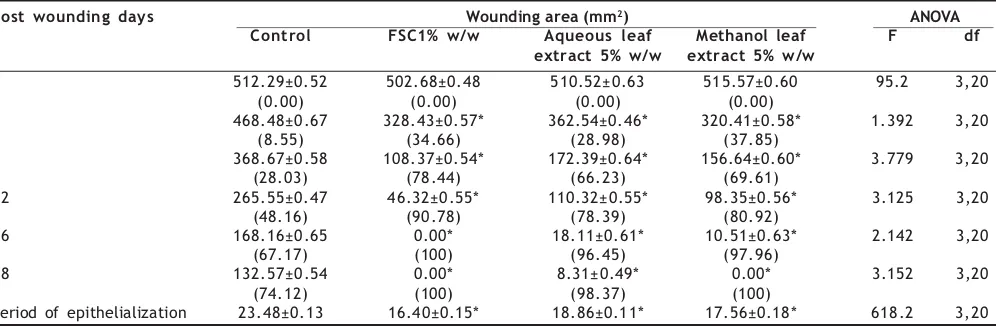
TABLE 1: EFFECT OF TOPICAL APPLICATION OF AQUEOUS AND METHANOL LEAF EXTRACTS OF LEUCAS HIRTA ON EXCISION WOUND MODELS
Post wounding days Wounding area (mm2) ANOVA
Control FSC1% w/w Aqueous leaf Methanol leaf F df extract 5% w/w extract 5% w/w
0 512.29±0.52 502.68±0.48 510.52±0.63 515.57±0.60 95.2 3,20
(0.00) (0.00) (0.00) (0.00)
4 468.48±0.67 328.43±0.57* 362.54±0.46* 320.41±0.58* 1.392 3,20
(8.55) (34.66) (28.98) (37.85)
8 368.67±0.58 108.37±0.54* 172.39±0.64* 156.64±0.60* 3.779 3,20
(28.03) (78.44) (66.23) (69.61)
12 265.55±0.47 46.32±0.55* 110.32±0.55* 98.35±0.56* 3.125 3,20
(48.16) (90.78) (78.39) (80.92)
16 168.16±0.65 0.00* 18.11±0.61* 10.51±0.63* 2.142 3,20
(67.17) (100) (96.45) (97.96)
18 132.57±0.54 0.00* 8.31±0.49* 0.00* 3.152 3,20
(74.12) (100) (98.37) (100)
Period of epithelialization 23.48±0.13 16.40±0.15* 18.86±0.11* 17.56±0.18* 618.2 3,20
tissues formed on the grass piths were excised on day 10 after inflicting wound. The dry weight of the granulation tissue and the breaking strength was measured. Simultaneously, granulation tissue so harvested was subjected to hydroxyproline estimation10 and
histopathological study to evaluate the effect of the extracts on collagen formation. The data were subjected to ANOVA followed by Tukey’s multiple comparison test, and the values of P ≤0.01 were considered statistically significant.
The preliminary phytochemical tests of leaf extracts revealed the presence of flavonoids, alkaloids, tannins, saponins, glycosides, steroids, and triterpenoids. Effect of aqueous and methanol leaf extracts of Leucas hirta on excision wound model is presented in the Table 1. The animals of group I showed complete epithelialization on 23.5±0.13 post wound day, whereas group II animals treated with standard drug FSC showed complete epithelialization on 16.4±0.15 post wound day. Compared to control group, the animal groups treated with aqueous and methanol leaf extract showed decrease in the period of complete epithelialization (18.9±0.11; 17.6±0.18, respectively). Percentage closure of wound area was significantly high in methanol leaf extract treated group followed by aqueous extract treated group of animals. The rate of wound contraction was less in control group of animals, whereas the percentage of wound closure was high in methanol leaf extract treated group followed by
TABLE 2: EFFECT OF AQUEOUS AND METHANOL LEAF EXTRACTS OF LEUCAS HIRTA ON INCISION WOUND MODELS
Group (N) Tissue breaking strength (g)
Control 323.03±2.66
Framycetin sulphate cream 588.79±3.31*
Aqueous leaf extract 483.69±2.80*
Methanol leaf extract 560.67±3.60*
ANOVA
F 1462.0
df 3,20
N=6 animals in each group.*P≤0.01 indicates ‘significant’ when compared to control. Values are expressed as mean±SE
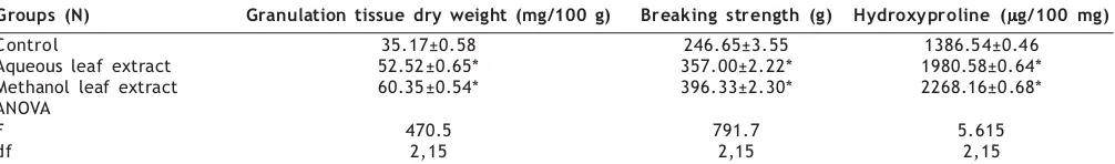
TABLE 3: EFFECT OF AQUEOUS AND METHANOL LEAF EXTRACTS OF LEUCAS HIRTA ON DEAD SPACE WOUND MODELS
Fig. 1: Histology of granulation tissue of control group of animals.
Fig. 2: Histological section of granulation tissue of the aqueous leaf extract treated animal showing moderate collagen deposition.
Fig. 3: Histological section of granulation tissue of the animal treated with methanol leaf extract showing increased collagenation.
The sections were stained with haematoxylin and eosin. They were observed under the magnification of 100x. Arrows in the figures indicate the collagen fibres.
Groups (N) Granulation tissue dry weight (mg/100 g) Breaking strength (g) Hydroxyproline (µµµµµg/100 mg)
Control 35.17±0.58 246.65±3.55 1386.54±0.46
Aqueous leaf extract 52.52±0.65* 357.00±2.22* 1980.58±0.64*
Methanol leaf extract 60.35±0.54* 396.33±2.30* 2268.16±0.68*
ANOVA
F 470.5 791.7 5.615
df 2,15 2,15 2,15
aqueous leaf extract treated animal group, indicating the effect of the plant on promoting healing of excision wound. In incision wound model, significant increase in the skin breaking strength was observed in methanol leaf extract treated groups of animals (561±3.6) followed by aqueous extract treated group of animals (484±2.8), indicating the effect of L. hirta leaf extract in maturation of collagen fibres (Table 2). Dead space wound model was used to study the effect of the extracts on granulation and collagenation of the healing process. Such wound models have been employed for quantitative and qualitative studies of wound healing, such as granuloma breaking strength and hydroxyproline content. Gain in granuloma breaking strength indicates increased collagen maturation by increased cross-linking; hydroxyproline estimation gives the net rate of synthesis and deposition of collagen in healing11. Significant increase in dry weight of
granulation tissue (60.4±0.54; 52.5±0.65), tissue breaking strength (396±2.3; 357±2.2), and hydroxyproline content (2268±0.68; 1981±0.64) was recorded in the animals treated with methanol leaf extract followed by aqueous extract as depicted in Table 3. Histological studies of granulation tissue of the control group showed aggregation of more number of macrophages and less collagen fibres (fig. 1), whereas aqueous extract treated group animals revealed lesser macrophages and moderate collagenation (fig. 2). In methanol leaf extract treated animals, significant increase in collagen deposition with lesser macrophages (fig. 3) was noticed.
Wound healing comprises different phases such as contraction, epithelialization, granulation, and collagenation. Collagen is a major protein of the extracellular matrix and is the component that ultimately contributes to wound strength. Breakdown of collagen liberates free hydroxyproline. Measurement of the hydroxyproline could be used as an index for collagen turnover12. In the present study, significant increase in the
hydroxyproline content of the granulation tissue of the animals treated with methanol leaf extract was recorded followed by aqueous extract when compared to control group. Increase in breaking strength of granulation tissue indicated the enhanced collagen maturation by increased cross-linking. In addition, increase in dry granulation tissue weight indicated the presence of higher protein content13. Increase in the tissue breaking strength,
granulation tissue dry weight, and increased epithelialization could be attributed to the increased hydroxyproline content in the wound tissue14.
Many workers studied the wound healing properties of
several plants such as Merremia tridentata15, Datura alba16 ,
Coronopus didymus17 and Aloe vera18; the wound healing
potency of these medicinal plants may be attributed to the active constituents present in it. Flavonoid reduces lipid peroxidation by preventing or slowing the onset of cell necrosis and by improving vascularity19. Tannins20 and
triterpenoids21 are known to promote the wound healing
process, mainly due to their astringent and antimicrobial property. These active constituents promote the process of wound healing by increasing the viability of collagen fibrils, by increasing the strength of collagen fibres either by increasing the circulation or by preventing the cell damage or by promoting the DNA synthesis22.
The present study revealed that the methanol leaf extract of Leucas hirta possesses better wound healing potency, followed by aqueous extract, which was evident by the increased rate of wound contraction; reduction in the period of epithelialization; increase in collagen deposition, breaking strength, and hydroxyproline in granulation tissue. The potency of the plant in healing the wounds may be attributed to the phytoconstituents like flavonoids, alkaloids, tannins, saponins, glycosides, steroids, and triterpenoids present in it, which may be either due to their individual or additive effect, hastening the process of wound healing. The present investigation offers scientific evidence to the folkloric accounts of the use of leaf extract of Leucas hirta in treating cuts and wounds.
ACKNOWLEDGEMENTS
The authors are grateful to National Education Society for their financial support. The authors express their sincere thanks to Prof. T. S. Ramkumar, Principal; Girimaji N. Raj Gopal, Secretary; S. V. Thimmaiah, Joint Secretary; Prof. Darmanada Rao, Registrar; Prof. Y. N. Manohara; and S. D. Jagadeesh Singh for their valuable guidance and support.
REFERENCES
1. Gamble, J.S., In; Flora of the Presidency of Madras, Botanical Survey of India, Calcutta, 1936, 1153.
2. Manjunatha, B.K., Krishna, V. and Pullaiah, T., In; Flora of Davanagere District, Karnataka, Regency Publication, New Delhi, 2004, 324.
3. Kokate, C.K., Purohith, A.P. and Gokhale, S.B., In; Pharmacognosy, Nirali Prakashan, Pune, 1990, 120.
4. Ghosh, M.N., In; Fundamentals of Experimental Pharmacology, Scientific Book agency, Calcutta, 1984, 153.
5. Jalalpure, S.S., Patil, M.B., Prakash, N.S., Hemalatha, K. and Manvi, F.V., Indian J. Pharm. Sci., 2003, 65, 363.
Spectrophotometric Method for the Determination
Spectrophotometric Method for the Determination
Spectrophotometric Method for the Determination
Spectrophotometric Method for the Determination
Spectrophotometric Method for the Determination
of Cefoperazone Sodium in Pharmaceutical
of Cefoperazone Sodium in Pharmaceutical
of Cefoperazone Sodium in Pharmaceutical
of Cefoperazone Sodium in Pharmaceutical
of Cefoperazone Sodium in Pharmaceutical
Formulations
Formulations
Formulations
Formulations
Formulations
M. SENTHILRAJA* AND P. N. SANJAYPAI1
Department of Pharmaceutical Chemistry, K. M. C. H. College of Pharmacy, Kalapatti Road, Coimbatore-641 035, 1Department of Quality Assurance, Al-Ameen College of Pharmacy, Hosur Main Road, Bangalore-560 027, India.
A new simple and sensitive spectrophotometric method was developed on the basis of a colour reaction of cefoperazone sodium with Folin Ciocalteu’s phenol reagent in presence of sodium carbonate, and it is stable for 20 min. The method is based on the formation of blue coloured chromophore that has an absorption maxima at 668 nm and obeys Beer’s law in the concentration range of 8/40 µg/ml. Results of the analysis were validated statistically and by recovery studies. The method was found to be suitable for routine determination of cefoperazone sodium.
Cefoperazone sodium is a third-generation semisynthetic antibiotic that is used in the treatment of mild to moderate infections caused by susceptible microorganisms1. It is
official in USP2. Chemically, cefoperazone sodium is 7
[R{2-(4-ethyl-2,3-dioxopiperazin-1-yl carboxamide)-2-(4- hydroxylphenyl)acetamide}-3-[1-methyl-1H-tetrazol-5yl-thiomethyl]]-3-cephem-4carboxylate3,4. Reported method of
analysis included colorimetry5. The aim of present
investigation was to develop an improved spectrophotometric method with greater precision and
accuracy. The proposed method is mainly based on the reaction of cefoperazone sodium with Folin Ciocalteu‘s phenol reagent in presence of sodium carbonate, which gives blue colour chromophore that has an absorption maxima at 668 nm. Pure cefoperazone sodium was obtained from Orchid Chemicals and Pharmaceuticals, Chennai.
A Shimadzu-1601 UV/Vis spectrophotometer with 1 cm matched quartz cells was used for all absorbance measurements. All other chemicals used were of Analar grade. Sodium carbonate (20% w/v) and Folin Ciocalteu’s phenol reagent and water (1:2) was prepared. Stock *For correspondence
E-mail: rajdanish2k@rediffmail.com
196, 117.
7. Ehrlich, H.P. and Hunt, T.K., Ann. Surg., 1968, 167, 324. 8. Lee, K.H., J. Pharm. Sci., 1968, 57, 1042.
9. Patil, M.B., Jalalpure, S.S. and Nagoor, V.S., Indian Drugs, 2004, 41, 40.
10. Woessner, J.F., Archiv. Biochem., 1963, 93, 440.
11. Taranalli, A.D., Tipare, S.P., Shivakumar and Torgali, S.S., Indian J.
Pharm. Sci., 2004, 66, 444.
12. Madhura, M.R. and Sushma, A.M., Fitoterapia, 2003, 74, 553. 13. Azad, S., In; Essentials of Surgery, Paras Medical Publishers,
Hyderabad, 2002, 1.
14. Gupta, S. and Gupta, S.K, In; Quarterly Medical Review, Raptakos, Brett and Company Ltd., Mumbai, 1985, 2.
15. Hatapakki, B.C., Hukkeri, V.I., Patil, D.N. and Chavan, M.J., Indian
Drugs, 2004, 41, 532.
16. Priya, K.S., Gnanamani, A., Radhakrishnan, N. and Babu, M.,
J. Ethnopharmacol., 2002, 83, 193.
17. Prabhakar, K.R., Srinivasan, K.K. and Rao, P.G.M., Pharm. Biol., 2002, 40, 490.
18. Udupa, S.L., Udupa, A.L. and Kulkarni, D.R., Fitoterapia, 1994, 65, 141.
19. Tsuchiya, H., Sato, M., Miyazaki, T., Fujiwara, S., Tanigaki, S., Ohyama, M., Tanaka, T. and Iinuma, M., J. Ethnopharmacol., 1996, 50, 27.
20. Ya, C., Gaffney, S.H., Lilley, T.H., Haslam, E., In; Hemingway, R.W. and Karchesy, J.J., Eds., Chemistry and Significance of Condensed Tannins, Plenum Press, New York, 1988, 553.
21. Scortichini, M. and Pia Rossi, M., J. Appl. Bacteriol., 1991, 71, 109.
22. Getie, M., Gebre Mariam, T., Reitz, R. and Neubert, R.H.,
Pharmazie, 2002, 57, 320.