2½-year-old elbow fracture-dislocation
to work after 6 months:
a case report
Lorne J Teperman,
BSc, DC, FCCRS(C), DACRB*
The rehabilitation of elbow fracture and dislocation is not generally considered a mainstream chiropractic concern. The clinician who is able to successfully manage the elbow articulation will rely upon his/her knowledge of functional anatomy, pathobiomechanics, history and examination principles, when selecting the appropriate treatment available. A case is presented of an individual that sustained a radial head fracture and dislocation following a motor vehicle accident. Subsequent to receiving 1½ years of physiotherapy for post-surgical complications (decreased range of motion, pain, stiffness and tingling to the 4th and 5th fingers), the patient was referred to a multidisciplinary clinic for a Work Hardening/Conditioning Program. This article discusses the need for active functional restoration vs. passive therapy, work hardening regimens and outcome measures. After 6 months of rehabilitation and 3 years following his motor vehicle accident, the patient has successfully returned to his previous work environment. A summary of the sequential steps in providing
appropriate management has been provided.
(JCCA 2002; 46(1):22–30)
K E Y W O R D S: fracture, elbow, work hardening,
functional restoration.
La rééducation fonctionnelle à la suite d’une fracture et d’une luxation du coude n’est généralement pas considérée comme un sujet principal en chiropratique. Le clinicien qui peut traiter avec succès l’articulation du coude se fie à sa connaissance de l’anatomie fonctionnelle, de la biomécanique pathologique et des principes d’antécédents et d’examen lors du choix de traitement possible. On a présenté un cas où le patient a subi une fracture de la tête radiale et une luxation lors d’un accident de véhiclue motorisé. Après avoir suivi une physiothérapie d’un an et demi pour des complications post-opératoires (zone motrice réduite, douleurs, raideur et picotements dans les 4e et 5e doigts), le patient a
finalement été référé à une clinique multidisciplinaire pour suivre un programme de réentraînement à l’effort ou de conditionnement au travail. Cet article traite du besoin de rétablissement fonctionnel actif (par opposition à la thérapie passive), de traitements de réentraînement à l’effort et d’analyse des résultats. Six mois de rééducation et trois ans après son accident, le patient a repris son ancien travail. On a également fourni un sommaire des étapes séquentielles d’un traitement approprié.
(JACC 2002; 46(1):22–30)
M O T S C L É S : fracture, coude, réentraînement à l’effort,
rééducation fonctionnelle.
* 78 Quail Run Boulevard, Maple, Ontario L6A 1E9, 416-417-7945. © JCCA 2002.
Case report
A 33-year-old male presented to a multidisciplinary reha-bilitation clinic upon referral from his physiotherapist for injuries sustained in a motor vehicle accident two years and six months earlier. The patient was riding his bicycle
his body. Prior to losing consciousness, the patient recalled that the driver dragged him by both arms as she (i.e. the driver), went into a state of panic.
The patient’s injuries consisted of lacerations to the head, knees and feet. Injury to the left elbow included an undisplaced fracture of the distal medial epicondyle and a fracture-dislocation of the proximal radius. The patient recalled sustaining a fracture to the left elbow 20 years previously.
The patient was hospitalized for 3 days following sur-gery, and was released with a hinged brace specifically designed for his left elbow. Six weeks after the accident, he returned to his duties as a Dangerous Goods Handler. He recalled that while attempting to lift pails of dangerous chemicals, he noted his “left elbow stretching and separat-ing”. Immediately, he experienced pain and numbness in his elbow, 4th and 5th fingers of the left upper extremity.
His family doctor referred him for physiotherapeutic treatment (ultrasound, electrical muscle stimulation, ice and heat) for 8 weeks which he claimed had no effect on his symptoms of persistent elbow and finger discomfort. Following further orthopedic and neurological consulta-tion, additional surgery was suggested. He declined how-ever, and decided to return to physiotherapy where he was treated with ultrasound and EMS (electrical muscle stimu-lation) and seen three times per week for 1½ years.
The patient’s history revealed left elbow stiffness and pain, scar tissue irritation, weakness and 4–5th digits numbness involving the left upper extremity. Joint stiff-ness was intermittent in nature and aggravated by lack of movement from any position. According to the patient re-current dislocation with excessive force to the elbow joint was something he experienced 2–3 times per month.
Examination revealed a pleasant and cooperative 33 year-old-male who appeared not to be in any visual discomfort. At the time of examination, he was wearing his custom-made functional splint, which controlled for excessive varus and valgus forces at the elbow. Scar tissue in the form of moderate keloid formation was evident about the injury site. As well, there was marked atrophy of the left flexor and extensor muscles of the elbow. The Carrying Angle of the elbow, defined as the angle formed by the long axis of the humerus and ulna resulting in an abducted position of the forearm relative to the humerus,1
was measured as 28 degrees and 15 degrees for the left and right elbow respectively.
Range of motion of the right upper extremity was unre-markable. Ranges for the left elbow were reduced during elbow flexion to 125 degrees (N = 145–150 degrees) and supination to 75 degrees (N = 90 degrees). Orthopedic and neurological testing indicated medial laxity involving the ulnar collateral ligament with subsequent hypersensitivity of the ulnar nerve. Manual muscle strength testing for the left extremity was determined to be 5/5, however, there was related weakness present as compared to the right side. Marked guarding upon introduction of varus and valgus stress tests was noted. Tenderness was elicited dur-ing palpation of the left olecranon and medial epicondyle. Orthopedic examination of the left wrist was relatively unremarkable.
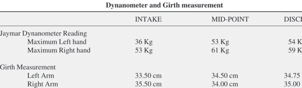
Review of the radiographic report illustrated screw and plate fixation with mild malalignment to the normal carry-ing angle. Dynamometer grip testcarry-ing and muscle girth readings were recorded at initial intake (see Table 1) and followed with additional measurements at the time of final discharge.
In order for the patient to return to work with confi-dence, it was determined he would require a functional restoration program (a term coined by Tom Mayer and Vert Mooney) prior to entering the Work Hardening/Work Simulation program. The primary focus was intended to increase functional ability through techniques used to en-hance strength, endurance, joint mobility and general car-diovascular conditioning.
The patient commenced the program with stretching and strengthening classes which included ice therapy fol-lowed by his choice of aerobic activity (i.e. stationary bike, treadmill, etc.). Isometric exercises preceded resistance training, as the transition was made after the patient ac-quired full range of motion and experienced only minimal pain and tenderness of the left elbow. The hinged brace was worn with any movement that introduced a lateral stress to the elbow joint.
Progressive resistance exercises, as described by Wilk and colleagues (1993),2 were utilized with the intention of
enabling the patient to slowly reach relatively normal strength bilaterally.
com-mitted to entering a Work Hardening program and did not consider surgery as an option. Within appropriate limits, treatment included plyometric exercise (throwing and catching a medicine ball), high speed/high energy strengthening and variations of eccentric muscular con-tractions.2
Aggressive exercise protocols were tolerated well with only minor setbacks. The patient experienced slight pain, joint laxity and occasional tingling in the fingers during vigorous exercises. Such symptoms lasted for 5–10 min-utes, whereas, elbow stiffness remained unless there was
constant movement. There was no report by the patient of elbow dislocation since the onset of treatment. Subse-quently, the patient commenced the Work Hardening Program.
Four months after entering the clinic, the patient partici-pated in a Functional Abilities Evaluation (FAE). A Job Task Analysis was performed with the aid of an occupa-tional therapist, vocaoccupa-tional consultant and employer asses-sor. A detailed job description was essential for the design of the work program. Assessment with the Baltimore Therapeutic Equipment (BTE) Work Simulator was used to determine the patient’s consistency of effort. Scores were compared to L. Matheson protocols.3 Lower and
up-per body strengths were assessed with the use of the Physi-cal Agility Test (PAT) unit.
As a candidate for a Work Hardening regimen, goals (see Table 2) were carefully constructed to compliment the patient’s work environment. As a Dangerous Goods Clerk, he supervised loading and unloading of freight. Occasionally, however, he would lift materials weighing 10 to 50 lbs.
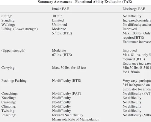
A final Functional Abilities Examination (FAE) was performed at discharge with acceptable results (see Table 3). Overall strength improvement was noted in lifting, car-rying, pushing and pulling activities. There were also im-provements in handling, reaching forward, bending and overhead tasks.
After six weeks of Work Hardening and four months of active rehabilitation, the patient was discharged. It was decided that his job requirements could be met and that he was able to return to regular duties provided that when he
Table 1
Dynanometer and Girth measurement
INTAKE MID-POINT DISCHARGE
Jaymar Dynanometer Reading
Maximum Left hand 36 Kg 53 Kg 54 Kg
Maximum Right hand 53 Kg 61 Kg 59 Kg
Girth Measurement
Left Arm 33.50 cm 34.50 cm 34.75 cm
Right Arm 35.50 cm 34.00 cm 35.00 cm
Left Forearm 31.00 cm 32.50 cm 32.50 cm
Right Forearm 32.00 cm 33.00 cm 33.00 cm
Table 2 Program Goals
Work Hardening Program Goals:
1. Improve strength level in lifting capacity (unload/ load) at different heights including overhead tasks. 2. Improve strength level in carrying capacity
(varia-tion in distance, objects, unilateral and bilateral) 3. Improve functional tolerance in coordination or
manipulation with bilateral activities and in overhead tasks
4. Cardiovascular endurance conditioning
5. Dexterity tasks (Minnesota Turning Test) involv-ing countinvolv-ing, weighinvolv-ing, sortinvolv-ing packaginvolv-ing and unpacking
lifted heavy weights, he would do so with the aid of his co-workers.
He was instructed to wear the hinged brace to prevent excessive loading / fatigue to the elbow joint should his work routine change due to increased demands at the job site.
Five months and two weeks later, having actively par-ticipated in a multidisciplinary rehabilitation program, the patient had returned to work subsequent to a motor vehicle accident, which had taken place three years earlier.
Clinical implications
Extremity cases are by no means rare to the chiropractor. However, the notion that chiropractors devote their atten-tion solely to the back must be dispelled. As musculoskel-etal doctors, chiropractors should not avoid such extremity cases but rather embrace them.
The specialist assessing the elbow joint should have a good working knowledge of the functional anatomy and biomechanics of this structure.
For example, a good knowledge base of elbow
biome-Table 3
Summary Assessment – Functional Ability Evaluation (FAE)
Intake FAE Discharge FAE
Sitting: 30 min. No difficulty
Standing: Limited Increased considerably
Walking: Unlimited No difficulty and unlimited
Lifting: (Lower strength) Moderate Improved
57 lbs. (BTE) Max. 100 lbs. Only 50lbs. is
required(BTE) Endurance increased
(Upper strength) Moderate Improved
67 lbs. (BTE) Max. 81 lbs. only 50 lbs. is
required (BTE) Endurance increased
Carrying: Max. 30 lbs. for 15 feet Min.50 lbs.@ 540 feet
for 1.56min
Pushing/ Pushing: No difficulty (BTE) Very easy -push/pull torque of
315 inch/pound on BTE work Simulator for at least 5 minutes
Crouching: No difficulty (PAT) No difficulty (PAT)
Kneeling: No difficulty No difficulty
Crawling: No difficulty No difficulty
Climbing: No difficulty No difficulty
Twisting: No difficulty No difficulty
Reaching: forward No difficulty No difficulty (MRMT & PP)
Minnesota Rate of Manipulation
Task (MRMT) & Purdue Pegboard (PP)
Bent Minimal difficulty No difficulty
Overhead Moderate difficulty (PAT) No difficulty (PAT)
chanics as in pitching, is recommended when treating to-day’s athlete.4 Many of the concepts for treating athletes
resemble those of everyday patients.
The elbow is a complex joint due to its intricate func-tional anatomy. The ulna, radius and humerus articulate in such a way as to form four distinctive joints. Surrounding the osseous structures are the ulnar collateral ligament complex, the lateral collateral ligament complex and the joint capsule. Four main muscle groups provide move-ment: the elbow flexors and extensors and the flexor-pro-nator and extensor-supiflexor-pro-nator groups.1,5,8
Physical examination of the elbow should include in-spection/observation, palpation, range of motion assess-ment (passive and active), muscle testing, neurological assessment and special tests (Tinel’s sign, test for tennis elbow, and golfer’s elbow, adduction/abduction stress tests, etc.).6,7 Radiographic examination and its variations
such as Magnetic Resonance Imaging (MRI) can be very useful. Depiction of muscles, ligaments and tendons as well as the ability to directly visualize nerves, bone mar-row and hyaline cartilage, are advantages of MRI, relative to conventional imaging techniques. It is suggested that
Figure 1* The classification system for olecranon fractures as developed by DeLee et al.14 *Permission to reproduce granted by Lippincott, Williams and Wilkins.
Figure 2* The classification system for radial head fractures as developed by Mason15 and modified by Johnston.16 *Permission to reproduce granted by Blackwell Publishing, Osney Head, Oxford and the Ulster Medical Journal, Belfast.
III Comminuted
fracture
Radial Head Fracture
I Undisplaced
fracture
II Marginal
fracture
IV Fracture-dislocation
Ulnar Fracture
I Undisplaced
fracture
IIA Avulsion
fracture
IIB Transverse
or oblique fracture
IIC Comminuted
fracture
MRI may be useful when patients have not responded well to conservative therapy and therefore surgical intervention and additional diagnoses would be under consideration.9
Dislocations of the elbow joint are not uncommon and usually result from a fall on the outstretched arm. The literature refers to the most common presentation as being posterolateral.10 Immediate reduction of the joint is
usu-ally performed under regional or general anaesthesia.10,11
When dislocation occurs, reduction is frequently per-formed on site by traction of the forearm as the elbow is flexed to 30 degrees along with counter-traction on the humerus.12 It is suggested that further emergency
proce-dures be conducted to rule out possible complications such as fracture and vascular compromise.10,12 As was
evi-denced in the current report, prior childhood injuries have been known to predispose individuals to recurrent elbow dislocation.13
Fractures most frequently accompany dislocations. DeLee et al. and Mason followed by Johnston (see Figures 1 and 2) have developed a classification system for olecranon and radial head fractures respectively.14,15,16
Other sequelae are nerve complications such as neuritis/ neuropraxia, and compression palsy. Neurovascular com-plications after dislocation occur in up to 5% of cases.17
These nerve palsies usually occur as a result of a traction injury with the ulnar nerve being the most likely nerve susceptible to trauma.18
These complications are said to resolve after a short period however, careful monitoring is necessary to ensure no deterioration of function.17,18
Elbow stiffness occurs from a variety of conditions such as prolonged immobilization, soft-tissue trauma, thermal injury, infection, intra-articular or extra-articular fracture, inflammatory arthritis or degenerative osteoarthritis, and heterotopic bone formation.10,21 Most often emergency
surgical procedures such as open reduction and fixation are necessary treatment for the fractured and / or dislo-cated elbow. Joints that have stiffened and have not gained full mobility after trauma for various reasons may also be considered good candidates for surgery.21,22
Discussion
The rehabilitation specialist, like the surgeon, has a role to play in delivering effective management and treatment. Contemporary chiropractors have positioned themselves as the profession to specialize in the management of
neuromusculoskeletal disorders. Manipulation/ mobiliza-tion is now a widely accepted treatment approach (RAND, Manga, etc.), however, treatment must be delivered in a time-targeted fashion. There tends to be an overemphasis on passive modalities beyond the necessary stages of heal-ing. Excessive use of modalities may even be deleterious to the patient.23 Consensus guidelines and conferences
such as the Agency for Health Care Policy and Research Guidelines, the Mercy Center Conference, the British Standards Advisory Group Guidelines of Low Back Pain are specific in their distinction between active and passive care.23 As well, the Ontario Insurance Commission via the
Quebec Task Force prepared the Commissioner’s Guide-line, on February 15 1996 to help those involved under-stand reasonable therapy and expenses for a person who has sustained a whiplash injury in an automobile accident. The full report is published in the April 15, 1995 edition of Spine and is also available in summary from the Ontario Insurance Commission as “Commissioner’s Guideline No. 1/96”.
Efficiency and effective management of the injured patient is necessary to produce optimal results. Health care providers are hard-pressed to deliver cost-effective care. Today, many patients and third party payers are demanding the best possible care for their money. The shift in health care from case management to cost-con-tained outcome management, has propelled the study and use of valid and reliable outcome tools. Outcome assess-ment which has many benefits (see Table 4) may be de-fined as measuring the symptom and/or function of a patient’s clinical status.25 Implementing these
measure-ments begins at the initial intake in order to establish a baseline. Patient directed questionnaires are completed at different periods during various phases of care. Ques-tionnaires which assess areas of perceived pain, psycho-social, lifestyle/disability and job dissatisfaction are available throughout the literature and can be examined in greater detail.24,25,26,27
patient’s overall feeling of improvement towards good health is depicted in his self-perceived reports. At the com-mencement of the work hardening program, our patient scored 149/200 on the P.A.C.T. Spinal Function Sort. The purpose of this survey is to indicate a person’s level of function with respect to 50 Activities of Daily Living. Upon discharge, the patient scored a perfect 200/200 dem-onstrating his ability to perform all 50 activities illustrated without difficulty.
It is not suggested that all chiropractors need to become rehabilitation specialists, however, chiropractors should attempt to follow the rehabilitation paradigm in their prac-tices. Understanding the guidelines and reports (AHCPR, Mercy, RAND, WAD, Manga, etc.) compels practitioners to follow treatment protocols which promote active therapy as opposed to passive therapy. Practitioners that choose to provide traditional chiropractic care are essential to health care, but must understand that active rehabilita-tion protocols are needed from the beginning. Reaching the subacute stage (between 1 and 4 weeks) requires some form of active therapy and treating chiropractors who are unable to provide active therapy, should continue to pro-vide the appropriate care but be willing to refer patients to a treatment center which provides active functional resto-ration. Mercy guidelines emphasize the need “to proceed
to the rehabilitation phase as soon as possible, to minimize dependency on passive forms of treatment/care.”24
Our patient presented to the clinic after receiving 1½ year of physiotherapy which included, but was not limited to, ultrasound, interferential current, diathermy, heat and cold therapy. He had been off work for 2½ years with minimal progress. Despite the chronicity and exist-ent complications, he did not appear to fall into the chronic pain patient model. The exercise treatment re-gime was tolerated well and he approached the program with enthusiasm and determination. There were no psy-chosocial signs of fear avoidance, job dissatisfaction, anxiety, depression, etc. The fact that our patient had been off work for almost three years, did not deter his remarkable recovery.
Our objective for management was to return this patient to his pre-accident status and position of employment. Work Hardening/Simulation, which involves supervision, prepares the individual with a gradual build-up of activi-ties resembling the usual work demands.
Patients must be suitable to enter this type of program and may require additional passive and active treatment prior to commencement. Our patient required preliminary active therapy prior to entering the work hardening pro-gram. Low-tech rehabilitation therapies are numerous and readily available to thedetermined practitioner.2,27–32
The transition to a Work Hardening Program is indi-vidually based and should be started as soon as strength and functional stability have been reached.
Evaluation is assessed with a functional ability/capacity examination. Functional Capacity Examination (FCE) is a comprehensive, objective test of a person’s ability to per-form work-related tasks. Issues of safety, reliability, valid-ity, practicality and usefulness are key components to a successful examination.33 There are widespread reports
of successful work hardening programs, however, more carefully documented, randomized and controlled studies are needed to determine which programms are of optimal benefit.34–41
Summary
Effectiveness and management are the key principles in delivering appropriate health care. The following list was incorporated in the management of this case and is offered as a guideline for interested practitioners:
1 Take a detailed history and examination; Table 4
Benefits of Outcome measurement24
OUTCOME ASSESSMENT BENEFIT
1. Documentation of improvement to the patient, clinician, and third parties.
2. Justification for the type, duration, and frequency of care.
3. Indication of the point of maximum therapeutic improvement.
4. Suggestions to modify the goals of treatment when necessary
5. Help in uncovering problems in care, including patient non-compliance
6. A database for clinical research and effectiveness of care over time.
2 Utilize outcome assessment tools from the initial visit
and thereafter on a regular basis;
3 Develop a specific diagnosis and attainable goals for
the patient;
4 Rule out other possible influences (cancer, infection,
fracture, cauda equina syndrome, etc.);
5 If necessary, refer to other specialists and/or other
di-agnostic testing;
6 Prepare Individual written rehabilitation report with
clearly delineated diagnosis, objectives and goals, treatment and therapy to be administered, treatment duration, barriers to recovery, and prognosis, etc.;
7 Discuss goals, objectives and costs with patient, third
party payer, case manager, vocational consultant, and employer, etc.;
8 Appropriately manage with time-targeted passive
modalities, manipulation and/or adjunct therapies;
9 Identify complicating factors and those patients, which
have the potential to develop Abnormal Illness Behaviors, or the tendencies of becoming become Chronic Pain patients;
10 Quick transition to an Active Functional Restoration
program which incorporates Iow or high- technical re-habilitation principles and addressing key areas such as stretching, strengthening, proprioception, stability cardiovascular conditioning, stress management and educational principles;
11 Approved candidates to enter a Work Hardening/
Simulation program to include Functional Capacity Evaluation, job evaluation (vocational and case man-agement), job description and ergonomic analysis;
12 Discharge when Functional Capacity Evaluation has
been performed to optimal levels;
13 Return the rehabilitated worker to original or new
work position in full or graduated return-to-work tran-sition;
14 Follow up to determine patient status;
15 Analyze Outcome Assessments to develop objective
principles and protocols for future patients.
The above is presented as a guideline only. It is not to be assumed that rehabilitation protocols must follow these 15 steps. Many practitioners may accomplish their objec-tives with fewer or more steps.
Conclusion
A case of fracture and dislocation to the elbow following a motor vehicle accident has been presented. Key issues per-taining to the elbow joint and particularly with respect to the case at hand, have been presented. Rehabilitation protocols and principles involving work hardening/simu-lation and active functional restoration have been dis-cussed. The need for outcome measures and sequential objectives is highly recommended in establishing reputa-ble multidisciplinary rehabilitation centers. Understanding when and how to implement rehabilitation principles are determinants with respect to satisfactory treatment and recovery. In this case, the patient returned to work after 3 years and has had no complications to this author’s knowledge. Possibly, had he presented to our clinic imme-diately following his accident, he may have been able to return to work much sooner and as a result, would have saved the employer, insurance payer and the tax payer considerable amounts of money. More research is needed to objectively make such claims, however, multidis-ciplinary rehabilitation centers are definitely part of our future. Such centers in future will require the incorporation of cardiac and neurological programs. As well, all patients should have the choice of being treated in an environment, which includes basic rehabilitation principles.
Approaching rehabilitation need not be a tedious effort. Many difficult and challenging cases present themselves to our clinics and whether we do or do not adopt rehabilita-tion protocols it is our obligarehabilita-tion as primary health care professionals, to provide the best resources available for our patients.
References
1 Stroyan M, Wilk KE. The functional anatomy of the elbow complex. JOSPT 1993; 17(6):279–288.
2 Wilk KE, Arrigo C, Andrews JR. Rehabilitation of the elbow in the throwing athlete. JOSPT 1993; 17(6):305–316.
3 Industrial Rehabilitation Quarterly. 1989; 2(1): . 4 Werner SL, Fleisig GS, Dillman CJ, Andrews JR.
Biomechanics of the elbow during baseball pitching. JOSPT 1993; 17(6):274–278.
5 Peterson-Kendall F, KendalI-McCreary E. Muscles: Testing and Function. 3rd. ed. Williams & Wilkins, 1983: 77–98.
7 Cipriano JJ. Photographic Manual of Regional
Orthopaedic and Neurological Tests. 2nd. ed. Williams & Wilkins, 1991: 101–150.
8 Platzer W. Color Atlas and Textbook of Human Anatomy: Volume 1, Locomotor System. 3rd. r. ed. Georg Thieme Verlag, 1986:116–121.
9 Fritz RC, Steinbach LS. Magnetic resonance imaging of the musculoskeletal system. Clin Orthop 1996; 324:321–337.
10 Rettig AC. Elbow, forearm and wrist injuries in the athlete. Sports Med 1998; 25(2):115–130.
11 Soon JCC, Kumar VP, Satkunanartham K. Elbow dislocation with ipsilateral radial shaft fracture. Clin Orthop 1996; 329:212–215.
12 Andrews JR, Whiteside JA. Common elbow problems in the athlete. JOSPT 1993; 17(6):289–295.
13 Abe M, Ishizu T, Nagaoka T, Onomura T. Recurrent posterior dislocation of thehead of the radius in post-traumatic cubitus varus. J Bone Joint Surg [Br] 1995; 77–B: 582–585.
14 DeLee JC, Green DP, Wilkins KB. Fractures and dislocations of the elbow. Rockwood CA, and Green DP. (eds.): Fractures. Philadelphia JB. Lippincott, 1984. 15 Mason ML. Some observations on fractures of the head of
the radius with review of one hundred cases. Br J Surg 1954; 42:123.
16 Johnston GW. A follow-up of one hundred cases of fracture of the head of the radius with review of the literature. Ulster Med J 1962; 31:51.
17 Limb D, Hodkinson SL, Brown RF. Median nerve palsy after posterolateral elbow dislocation. J Bone Joint Srug [Br] 1994; 76–B:987–988.
18 Grobler GP. Unusual cause of ulnar nerve palsy. Clin Orrhop 1996; 323:192–193.
19 Noonan K J, Blair WF. Chronic median-nerve entrapment after posterior fracture-dislocation of the elbow. J Bone Joint Surg 1995; 77–A(10):1572–1574.
20 Sojbjerg JO. The stiff elbow: How I do it. Acta Orthop Scand 1996; 67(6):626–631.
21 Modabber MR, Jupiter JB. Reconstruction for post-traumatic conditions of the elbow joint. J Bone Joint Surg 1995; 77–A(9):1431–1441.
22 Teasdali R, Savoie FH, Hughes JL. Comminuted fractures of the proximal radius and ulna. Clin Orthop 1993; 292:37–47.
23 Liebenson C. Commentary: Rehabilitation and chiropractic practice. JMPT 1996; 19(2):134–140.
24 Chapman-Smith D. Measuring results: the new importance of patient questionnaires. Chdo Rep 1992; 7:1–6.
25 Yeomans SG, Liebenson C. Applying outcomes
management to clinical practice. JNMS 1997; 5(1):1–14. 26 Liebenson C. Rehabilitation of the Spine: A Practitioner’s
Manual. Williams & Wilkins, 1996: 73–95.
27 Bonutti PM, Windau JE, Ables BA, Miller BG. Static progressive stretch to reestablish elbow range of motion. Clin Orthop 1994; 303:128–134.
28 Lephart SM, Pincivero DM, Giraldo JL, Fu FH. The role of proprioception in the management and rehabilitation of athletic injuries. Am J Sports Med 1997; 25(1):130–137. 29 Keith RA. Treatment strength in rehabilitation. Arch Phys
Med Rehabil 1997; 78:1298–1304.
30 Jette AM, Delitto A. Physical therapy treatment choices for musculoskeletal impairments. Phys Ther 1997; 77(2):145–154.
31 Irrgang JJ, Delitto A, Hagen B, et al. Rehabilitation of the injured athlete. Sports Med 1995; 26(3):561–577. 32 Baumert -Jr PW. Acute inflammation after injury: quick
control speeds rehabilitation. Postgrad Med 1995; 97(2):35–48.
33 Hart DL, Isernhagen SJ, Matheson LN. Guidelines for functional capacity evaluation of people with medical conditions. JOSPT 1993; 18(6):682–86.
34 Lechner DE. Work hardening and work conditioning interventions: do they affect disability? Phys Ther 1994; 74(5):471–493.
35 Jacobs MD. Rehabilitation, conditioning and work hardening: New concepts in the evaluation of industrial/ chiropractic interface. ACA J Chiro 1989; ergonomics: 43–45.
36 Niemeyer LO, Jacobs K, et al. Work hardening: Past, present, and future-The work programs special interest section National Work-Hardening Outcome Study. Am J Occup Ther 1994; 48(4):327–335.
37 King P. Outcome analysis of work-hardening programs. Am J Occup Ther 1993; 47(7):595–603.
38 Werneke MW, Harris DE, Lichter RL. Clinical effectiveness of behavioral signs for screening chronic iow-back pain patients in a work-oriented physical rehabilitation program. Spine 1993; 18(16): 2412–2418. 39 Beissner KL, Saunders RL, McManis BG. Factors related
to successful work hardening outcomes. Phys Ther 1996; 76(11):1188–1200.
40 Greenberg SN, Bello RP. The work hardening program and subsequent return to work of a client with low back pain. JOSPT 1996; 24(1):37–45.
41 Schmidt SH, Oort-Marburger D, Meijman TF. Employment after rehabilitation for musculoskeletal impairments: The impact of vocational rehabilitation and working on a trial basis. Arch Phys Med Rehabil 1995; 76:950–954.
Help Support Chiropractic Research
Become a member of the