S T A N D A R D A R T I C L E
Lymphatic endothelial cell immunohistochemical markers for
evaluation of the intestinal lymphatic vasculature in dogs with
chronic inflammatory enteropathy
Sara A. Wennogle
1| Simon L. Priestnall
2| Alejandro Suárez-Bonnet
2|
Sirikul Soontararak
1| Craig B. Webb
11
Department of Clinical Sciences, College of Veterinary Medicine, Colorado State University, Fort Collins, Colorado 2
Department of Pathobiology and Population Sciences, Royal Veterinary College, Hatfield, United Kingdom
Correspondence
Sara A. Wennogle, Department of Clinical Sciences, College of Veterinary Medicine, Colorado State University Veterinary Teaching Hospital, 300 West Drake Road, Fort Collins, CO 80525.
Email: sara.wennogle@colostate.edu
Funding information
Naniboujou Legacy Fund; Rocky's Research Fund; Royal Canin
Abstract
Background:
Lymphatic endothelial cell (LEC) immunohistochemical markers have
identified intestinal lymphatic vasculature abnormalities in humans with inflammatory
bowel disease, but have not been used to evaluate intestinal lymphatic vasculature in
a group of dogs with chronic inflammatory enteropathy (CIE).
Objectives:
To utilize LEC markers to identify and measure intestinal lymphatic
vas-culature in endoscopic biopsy samples of CIE dogs. To evaluate whether measured
lymphatic vasculature variables correlate with serum albumin concentrations.
Animals:
Twenty-four dogs with CIE; n = 13, serum albumin concentration <2.5 g/dL
(CIE-protein-losing enteropathy [PLE]), n = 11, serum albumin concentration
≥
2.5 g/dL
(CIE-N).
Methods:
Prospective study. Lymphatic endothelial cell immunolabeling with Prox-1
and LYVE-1 performed on endoscopic biopsy samples from 24 dogs with CIE.
Duo-denal and ileal villous lacteal width (VLW) and proprial mucosal lacteal width (MLW)
were determined for each case and analyzed for correlation with serum albumin
con-centration. Lacteal dilatation scores using routine H&E histopathology were assessed
for correlation with immunohistochemistry (IHC)-calculated VLW and MLW.
Results:
Lower serum albumin concentrations were correlated with increased VLW
(rho =
−
.4644;
P
= .02) and MLW (rho =
−
.6514;
P
< .001) in the ileum. Lymphatic
endothelial cell IHC identified presumptive proprial mucosal lymphangiectasia in
some dogs that was not recognized with routine H&E staining. Lacteal dilatation
scores were correlated with VLW in duodenum (rho = .4634;
P
= .02) and ileum
(rho = .5292;
P
= .008), but did not correlate with MLW.
Abbreviations:CCECAI, canine chronic enteropathy clinical activity index; CD, Crohn's disease; CIE, chronic inflammatory enteropathy; CIE-N, chronic inflammatory enteropathy with serum albumin concentrations≥2.5 g/dL; CIE-PLE, chronic inflammatory enteropathy with protein-losing enteropathy; IHC, immunohistochemistry; IL, intestinal lymphangiectasia; LD, lacteal dilatation; LEC, lymphatic endothelial cell; LYVE-1, lymphatic vascular endothelial hyaluronic acid receptor; MLW, mucosal lacteal width; PLE, protein-losing enteropathy; Prox-1, human prospero homeobox; VLW, villus lacteal width.
DOI: 10.1111/jvim.15545
This is an open access article under the terms of the Creative Commons Attribution-NonCommercial License, which permits use, distribution and reproduction in any medium, provided the original work is properly cited and is not used for commercial purposes.
© 2019 The Authors.Journal of Veterinary Internal Medicinepublished by Wiley Periodicals, Inc. on behalf of the American College of Veterinary Internal Medicine.
Conclusions and Clinical Importance:
Lymphatic endothelial cell immunolabeling
identified presumptive proprial mucosal lymphangiectasia in CIE dogs, particularly in
the ileum of hypoalbuminemic dogs. Routine evaluation of villous lacteals likely
underestimates abnormalities of the lymphatic vasculature in dogs with CIE.
K E Y W O R D S
canine, enteropathy, immunolabeling, lacteals
1
|
I N T R O D U C T I O N
Chronic inflammatory enteropathy (CIE) refers to conditions of the
intes-tinal tract that are characterized by the presence of gastrointesintes-tinal signs
of at least 3 weeks' duration, histologic evidence of intestinal
inflamma-tion, and the exclusion of neoplastic, infectious, and extra-gastrointestinal
causes of gastrointestinal signs.1-3Intestinal lymphangiectasia (IL) is a
disorder of dilated lymphatic vasculature at any level of the intestinal
lymphatic system. Although it can be a primary condition, it also can
occur secondary to a variety of other intestinal disorders, including CIE.
When it occurs secondary to CIE, it is presumably a result of increased
lymphatic pressure associated with inflammatory infiltrates in the
intes-tine, and may result in hypoalbuminemia, or protein-losing enteropathy
(PLE), because of direct loss of protein-rich lymph, lymphatic
dysfunc-tion, or both.4-6
Several recent studies have found a relationship between serum
albumin concentrations and IL in cases of idiopathic CIE in the dog,
however lymphatic abnormalities have been inconsistently reported.7-9
Lymphangiectasia can be underappreciated on routine histopathologic
examination of the intestine, because it can have a segmental
distribu-tion and, in some cases, be confined to deeper layers of the intestine
that may not be sampled endoscopically.5,6 Lymphatic abnormalities
can be similarly confined to the deeper layers of the intestine in cases
of Crohn's disease (CD),10-12a type of inflammatory bowel disease in
humans characterized by idiopathic inflammation that can be localized
to the ileum or found diffusely throughout the small intestine.13 In
some cases of CD, dilated lymphatics are not identified in the
superfi-cial mucosa, but rather found in the deeper mucosa, submucosa, and
muscularis layers of the intestine, as well as in the mesentery.14,15In
humans with CD, immunolabeling with lymphatic endothelial cell
(LEC)-specific markers is superior to standard microscopy for identifying
abnormalities of the lymphatic vasculature, including lymphangiectasia,
and obstructed lymphatics.15
Lymphatic endothelial cells are derived from venous progenitor
cells and express various antigens that distinguish them from blood
ves-sel endothelial cells.16Lymphatic endothelial cell markers are numerous
and include human prospero homeobox (Prox-1), a nuclear transcription
factor,17 and lymphatic vascular endothelial hyaluronic acid receptor
(LYVE-1).18 Prox-1 and LYVE-1 immunolabeling previously has been
used to differentiate types of angiosarcomas in the dog,19but to our
knowledge has not been used to evaluate the intestinal lymphatic
vas-culature in dogs with CIE.
Our objective was to utilize the LEC markers Prox-1 and LYVE-1 to
identify and evaluate the intestinal lymphatic vasculature in endoscopic
biopsy samples of dogs with CIE, and to determine whether
abnormali-ties associated with the lymphatic vasculature were related to serum
albumin concentrations in dogs with idiopathic CIE.
2
|
M A T E R I A L S A N D M E T H O D S
2.1
|
Study population
Client-owned dogs presented to the Colorado State University
Veteri-nary Teaching Hospital for evaluation of chronic gastrointestinal signs
(eg, decreased appetite, vomiting, diarrhea, and weight loss) of >3 weeks'
duration were recruited for participation in the study. To be eligible
for inclusion, dogs were required to have had routine fecal screening
with no parasites detected, no evidence of clinically relevant
non-gastrointestinal illness, as assessed by routine hematology and serum
biochemical profile, and a histopathologic diagnosis of inflammatory
enteritis for which no distinct cause could be identified. Dogs had to
have had duodenal and ileal biopsies performed. Dogs with
histopath-ologic evidence of intestinal neoplasia were excluded. To be eligible
for the study, exclusion of exocrine pancreatic insufficiency as a cause
of clinical signs with a fasted serum canine trypsin-like
immunoreactiv-ity concentration >5.0 ng/mL was required. Hypoalbuminemic dogs
(serum albumin concentration <2.5 g/dL) also were required to have
no clinically relevant proteinuria (negative urine dipstick test result or
urine protein:creatinine ratio <0.5) and no evidence of clinically
rele-vant hepatic disease based on normal fasted and postprandial bile acid
concentrations or normal synthetic liver function and enzyme activity
on routine serum biochemistry profile.
Recorded data included age, breed, sex, weight, duration of illness,
clinicopathological data, and results of any diagnostic imaging
per-formed. Additionally, at the time of enrollment, owners were asked to
score appetite, activity level, vomiting, fecal consistency, fecal
fre-quency, weight loss, and pruritus for each dog. After the results of the
serum biochemical profile (serum albumin concentration) and
abdomi-nal ultrasound examination (peritoneal effusion), and using the owner's
score, a canine chronic enteropathy clinical activity index (CCECAI)1
was calculated for each dog. The Clinical Review Board at Colorado
State University approved all procedures and written consent was
2.2
|
Histopathologic evaluation
Twelve duodenal and 5 ileal biopsy samples were obtained from each
dog for histopathologic evaluation. Histopathologic evaluation of
endo-scopically obtained intestinal tissue from CIE dogs was performed by a
board-certified veterinary pathologist (S.L.P.) and pathologist-in-training
(A.S.-B.) blinded to clinical data and clinicopathologic information. Biopsy
samples were assessed as adequate for evaluation, and both
patholo-gists evaluated duodenal and ileal tissues and reached a consensus for
the presence and severity of morphologic criteria (villous stunting,
epi-thelial injury, crypt distension, lacteal dilatation [LD], and mucosal
fibro-sis) and inflammatory criteria (intraepithelial lymphocytes, lamina propria
eosinophils, lamina propria lymphocytes or plasma cells, and lamina
propria neutrophils) based on World Small Animal Veterinary
Associa-tion (WSAVA) guidelines.20For the severity of each change, the
follow-ing scores were applied based on established criteria: 0 = normal,
1 = mild, 2 = moderate, and 3 = marked. Scores for LD were based on
the most severely affected villus in each case. If the lacteal occupied
0%-25% of the villus width, a score of 0 was given, 25%-50% was a
score of 1, 50%-75% was a score of 2, and >75% of the width of the
vil-lus resulted in a score of 3.
2.3
|
Immunohistochemistry
All immunohistochemical (IHC) labeling was performed using a Leica
Bond III immunostainer (Leica Biosystems Inc, California, Illinois).
Formalin-fixed paraffin-embedded tissues were sectioned at 5μm and
mounted on positively charged slides for IHC. Dewaxing and epitope
retrieval were performed using the Bond III instrument. Epitope
retrieval was performed using the ER1 solution (Leica Biosystems Inc),
a pH 6.0 citrate buffer. Antibodies against Prox-1 (rabbit polyclonal;
Angiobio [Delmar, California]) and LYVE-1 (rabbit polyclonal; Abcam
[Cambridge, Massachusetts]) were diluted 1:100 in PowerVision IHC
Super Block (Leica) and incubated on tissue sections for 20 minutes.
All wash steps were performed in triplicate using Leica Bond wash
buffer. Tissue sections then were incubated for 20 minutes with goat
secondary antibody at 1:200 dilution and alkaline phosphatase
poly-mer. The Leica Fast Red chromogenic substrate for alkaline
phospha-tase was used to detect specific immunoreactivity of each antibody.
Upon completion of immunolabeling, samples were counterstained
with hematoxylin. Isotype-matched irrelevant primary antibodies were
used as negative controls.
2.4
|
Immunohistochemical evaluation
The Prox-1 and LYVE-1 IHC slides as well as negative control slides were
digitally scanned using Philips Ultra Fast Scanner slide (Philips IntelliSite
Pathology Solution, Philips Electronics, Amsterdam, the Netherlands) and
analyzed with the use of Philips Image Management System viewer
(ver-sion 2.4.1.2; Phillips IntelliSite Pathology Solution, Phillips Electronics) by
a single evaluator (S.W.), who was blinded to the case data. To be
coun-ted or measured as a lymphatic vessel, a visible lumen was required in
addition to immunolabeling with the LEC markers. Lymphatic vessels
were measured using LYVE-1 immunolabeling. The Prox-1 slides
subse-quently were evaluated to verify the structures as lymphatic vessels.
On 4×, 10 well-oriented villi associated with cryptal tissue and
dis-persed throughout the slide were selected for measurement of villous
lymphatic vessel width (VLW). Lacteal width (μm) was taken as the
dis-tance from 1 side of the lacteal to the other, measured perpendicular to
the midline of the lacteal. Next, well-oriented areas of the propria
mucosa were evaluated at 4×for immunolabeling. In 10 distinct 20×
fields in the propria mucosa, lymphatic vessels were identified and
lac-teal width (μm) was measured perpendicular to the midline of the
lac-teal (mucosal lymphatic vessel width [MLW]). If no laclac-teals were
identified in the field, the next field was examined. No more than 2
lac-teals were measured per 20×field. If >2 lacteals were identified in the
field, all visible lacteals were measured, and the widest and most
nar-row lacteal of the group were used in the analysis. In addition, the
viewing trail feature was used to ensure that all areas of the slide were
assessed. For both VLW and MLW, the mean of the 10 measurements
was recorded for each tissue in each case. In 2/24 cases, only 5
defini-tively measurable mucosal lymphatics could be identified, and therefore
those cases had MLW scores calculated as the mean of the 5
measure-ments, rather than 10.
2.5
|
Statistical analysis
Descriptive statistics were calculated for age, sex, weight, duration of
illness, CCECAI scores, and histopathological scores. The distribution
of data for statistical analysis was assessed by the Shapiro-Wilk test.
Data were not normally distributed. Spearman (rank-based)
correla-tion was used to evaluate relacorrela-tionships between the lymphatic
vari-ables and serum albumin concentration in each section of intestine.
Spearman (rank-based) correlation also was performed to assess the
relationship between the routine LD score as assessed by the blinded
pathologists based on WSAVA guidelines and the VLW and MLW in
each section of the intestine. For Spearman, a statistically significant
correlation score of (+/−) 0.3-0.5 was considered a weak correlation,
(+/−) 0.5-0.7 a moderate correlation, and (+/−) 0.7-1.0 a strong
corre-lation.21All statistical analysis was performed using GraphPad Prism
scientific statistic software (Graph Pad Prism, GraphPad Software, Inc,
San Diego, California). Statistical significance for all statistical
compari-sons was set atP< .05.
3
|
R E S U L T S
Thirty dogs were screened for inclusion in the study. All dogs were
fasted for a minimum of 24 hours before endoscopic examination and
biopsies. After routine histopathologic evaluation, 1 dog was diagnosed
with intestinal lymphoma and therefore was excluded from the study.
Ileal biopsy samples were not obtained in 2 dogs, which prompted
exclusion from the study. In 3 additional cases, the quality of the
endo-scopic biopsy samples was considered inadequate for IHC evaluation of
LEC markers because of either the presence of only villus tips, or
orientation. Therefore, 24 clinical cases were included in the final data
analysis. Twenty-two dogs had a basal serum cortisol concentration
>2μg/mL or normal response to ACTH, ruling out hypoadrenocorticism
as a cause of their clinical signs. All dogs had routine abdominal
ultraso-nography performed by or under the supervision of a board-certified
veterinary radiologist to evaluate for intestinal disease or
extra-luminal intestinal masses before endoscopic examination.
Of the 24 cases, 13 dogs had serum albumin concentration
<2.5 g/dL, which was defined as chronic inflammatory enteropathy
with PLE (CIE-PLE), and 11 dogs had serum albumin concentration ≥2.5 g/dL (CIE-N). Breeds with CIE-PLE included Bernese Mountain Dog (2), Labrador Retriever (2), mixed breed dog (2), and 1 each of the
following: Australian Shepherd, English Bulldog, Great Pyrenees,
Pem-broke Welsh Corgi, Pug, Rottweiler, and Yorkshire Terrier. Breeds
with CIE-N included mixed breed dog (3), and 1 each of Australian
Terrier, Bernese Mountain Dog, Brittany Spaniel, Cavalier King
Charles Spaniel, German Shepherd dog, German Shorthaired Pointer,
Labrador Retriever, and Siberian Husky. Dogs with CIE-PLE
con-sisted of 7 neutered males and 5 spayed females, and dogs with
CIE-N consisted of 8 neutered males and 3 spayed females.
Addi-tional descriptive statistics of interest are presented in Table 1. Age
and sex were not different between dogs with CIE-PLE and dogs
with CIE-N.
The Prox-1 and LYVE-1 immunolabeling of lymphatics from a
non-study dog with a histopathologic diagnosis of marked lymphangiectasia
in the duodenum and ileum was used as a positive control. Human
prospero homeobox has been reported to be variably expressed in
enteroendocrine epithelial cells in the crypts.22In our cases, Prox-1
labeling was visible in some individual crypt epithelial cells but was
eas-ily differentiated from lymphatic vessel endothelial labeling. Although
LYVE-1 is expressed by some macrophages in the intestine,23 this
staining was easily distinguishable from lymphatic vessel endothelial
labeling. Lymphatic endothelial cell labeling was not observed on any of
the negative control slides. Examples of immunolabeled villus
lym-phatics are shown in Figure 1. Examples of immunolabeled proprial
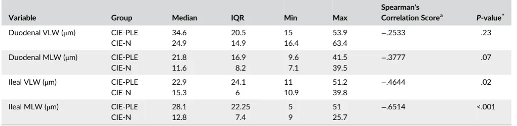
mucosal lymphatics are shown in Figure 2. Villus lacteal width in the
ileum was weakly negatively (rho =−.4644) correlated with serum
albu-min concentration (P= .02). Proprial MLW in the ileum was moderately
negatively (rho =−.6514) correlated with serum albumin concentration
(P< .001).21Summary statistics and correlation data for lymphatic
vari-ables with serum albumin concentration are presented in Table 2.
Routine H&E LD scores as determined by blinded board-certified
pathologist and pathologist-in-training were assessed for correlation
to the width of the villus and proprial mucosal lymphatics (VLW and
MLW, respectively) as determined by IHC for each tissue section.
Duodenal LD was weakly positively correlated with duodenal VLW
(rho = .4634; P = .02) but not correlated with duodenal MLW
(rho = .2767;P= .19). Ileal LD was moderately positively correlated
with ileal VLW (rho = 5292;P= .008) but not correlated with ileal
MLW (rho = .3889;P= .06).
4
|
D I S C U S S I O N
We utilized LEC-specific markers to evaluate the intestinal lymphatic
vasculature in dogs with CIE with and without PLE, as defined by serum
albumin concentration <2.5 g/dL. Lymphatic endothelial cell IHC was
successful in labeling the lymphatic vasculature in dogs with CIE, and
identified apparently dilated lymphatics in the proprial mucosa that
were not identified as lymphatics by routine H&E assessment for
lymphangiectasia. In addition, in our 24 dogs with CIE, serum albumin
concentrations were negatively correlated with villus and MLW in the
ileum. For both the duodenum and the ileum, average VLW as
deter-mined by use of LEC IHC was correlated with LD scores as traditionally
assessed using routine H&E staining. Proprial MLW as determined by
the use of IHC was not correlated with LD scores as traditionally
assessed by routine H&E staining in either section of intestine.
The most striking finding of our study was the presence of apparent
proprial mucosal lymphangiectasia in the intestine of dogs with CIE, in
particular in the ileum of several dogs with CE and serum albumin
con-centrations <2.5 g/dL. In some of these dogs with apparently dilated
proprial mucosal lymphatics, villus lymphatics were not concurrently
dilated and these dogs had not been diagnosed with lymphangiectasia
on routine histopathologic examination. The appearance of the
dis-tended proprial mucosal lymphatics in the ileum of these dogs may be
similar to what has been identified in humans with CD,15an idiopathic
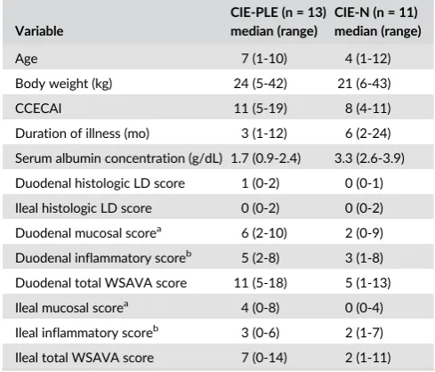
T A B L E 1 Selected descriptive statistics for dogs with chronic inflammatory enteropathy with (CIE-PLE) and without (CIE-N) protein-losing enteropathy
Variable
CIE-PLE (n = 13) median (range)
CIE-N (n = 11) median (range)
Age 7 (1-10) 4 (1-12)
Body weight (kg) 24 (5-42) 21 (6-43)
CCECAI 11 (5-19) 8 (4-11)
Duration of illness (mo) 3 (1-12) 6 (2-24)
Serum albumin concentration (g/dL) 1.7 (0.9-2.4) 3.3 (2.6-3.9)
Duodenal histologic LD score 1 (0-2) 0 (0-1)
Ileal histologic LD score 0 (0-2) 0 (0-2)
Duodenal mucosal scorea 6 (2-10) 2 (0-9)
Duodenal inflammatory scoreb 5 (2-8) 3 (1-8)
Duodenal total WSAVA score 11 (5-18) 5 (1-13)
Ileal mucosal scorea 4 (0-8) 0 (0-4)
Ileal inflammatory scoreb 3 (0-6) 2 (1-7)
Ileal total WSAVA score 7 (0-14) 2 (1-11)
Notes: LD, mucosal, and inflammatory histologic scores reported in this table were obtained by blinded evaluation of H&E samples.
Abbreviations: CIE-N, chronic inflammatory enteropathy with serum albumin concentration≥2.5 g/dL; CIE-PLE, chronic inflammatory enteropathy with protein-losing enteropathy (serum albumin <2.5 g/dL); LD: lacteal dilatation; WSAVA: World Small Animal Veterinary Association. a
Total score for villus stunting, epithelial injury, crypt distension, lacteal dilatation, and mucosal fibrosis.
bTotal score for intraepithelial lymphocytes, lamina propria
F I G U R E 1 Immunolabeled villous lymphatics of dogs with chronic inflammatory enteropathy (CIE). A, Duodenal villi from dog with CE and serum albumin concentration of 1.7 g/dL and central villous lymphatic dilation (arrow; lacteal dilation score = 2); human prospero homeobox immunohistochemistry (IHC). Severe lamina propria lymphoplasmacytic inflammation is also visible. B, Ileal villi from dog with CIE, serum albumin concentration 1.9 g/dL, and central villous lymphatic dilation (arrow; lacteal dilation score = 2). Cytoplasmic immunoreactivity of lymphatic endothelial cells shown with lymphatic vascular endothelial hyaluronic acid receptor IHC. Apparent proprial mucosal lymphangiectasia can also be seen (arrows)
chronic inflammatory intestinal disease most commonly found in the
ileum.13In humans with CD, lymphangiectasia also has been identified
in the submucosa, muscularis propria and subserosa.14,15,24Therefore,
it is possible that full thickness intestinal biopsy samples with LEC
immunolabeling may identify more abnormalities in the lymphatic
vas-culature of dogs with CIE than are currently seen in endoscopic biopsy
samples.
Presumptive proprial mucosal lymphangiectasia also was identified
in the duodenum of some dogs with CIE, including in 2 dogs with
serum albumin concentrations≥2.5 g/dL. Proprial MLW in the
duode-num was not significantly correlated with serum albumin
concentra-tion. It is possible that with an increased sample size, a statistical
difference may have been detected. A previous study comparing
his-tologic findings in the duodenum versus ileum in a group of dogs with
chronic small intestinal enteropathies found hypoalbuminemia to be
correlated with ileal LD, but not with duodenal LD.25The albumin
cor-relation results of our study also suggest that lymphatic abnormalities
can differ among sections of the intestine, lending additional support
to the recommendation to always obtain ileal samples in the
diagnos-tic evaluation of dogs with CIE.
In many cases, it is unknown whether lymphatic abnormalities are
a cause or consequence of intestinal disease. Regardless, they likely
represent an important component of the disease process. In addition
to their role in the transport of intestinal immune and inflammatory
cells, the lymphatic vasculature is responsible for regulation of the
pressure of interstitial fluid in tissues, and transport of excess fluid
back to the circulation. Furthermore, lymphatic vessels are the main
route of absorption of fat, cholesterol, fat-soluble vitamins, and
gas-trointestinal hormones.24,26 Obstruction or dysfunction of the
lym-phatic vasculature or both should have important consequences, and
the recognition of lymphatic abnormalities is likely important to the
management of dogs with CIE. Although response to treatment and
outcomes were not evaluated in our study, follow-up information was
available for several dogs. Six dogs in the study had an average ileal
mucosal lymphatic width >30μm, all of which had serum albumin
con-centrations <2.5 g/dL. Blinded ileal LD scores were 0 in 4/6 of these
dogs. Of these dogs, 3 were euthanized as a consequence of their
dis-ease, 1 was euthanized because of unrelated disdis-ease, and 2 were alive
at the time of publication. Of the 3 dogs that were euthanized because
of their disease, 2 received traditional treatments including commercial
gastrointestinal diets and glucocorticoids for >2 weeks after their
diag-nosis with no clinically relevant improvement noted. The third dog that
was euthanized initially was glucocorticoid-responsive but then relapsed
when glucocorticoids were tapered and the owner chose not to pursue
further treatment. The other 3 dogs had persistent clinical signs despite
treatment with glucocorticoids, immunosuppressive drugs, vitamin
sup-plementation, supportive care, and commercially available hydrolyzed
and low-fat diets. All 3 of these dogs ultimately had an apparent clinical
response once switched to a veterinary nutritionist-formulated
home-cooked diet, formulated to be lower in fat (10%-15% by metabolizable
energy) than the commercially available diets. Two of those dogs were
alive at the time of publication; the third was euthanized because of
splenic hemangiosarcoma 12 months after response to the
home-cooked diet. Although anecdotal, and only a small number of cases,
this population of dogs may represent a subset of dogs with CIE
that have important abnormalities of their lymphatic vasculature
that require the administration of a diet that is lower in fat than
what is commercially available.
Our study had some limitations. First, biopsy samples from healthy
control dogs were not available, and we cannot accurately determine
normal lymphatic width in the propria mucosa of the intestine. Further
studies of the lymphatic vasculature in dogs with CIE ideally should
include evaluation of samples from healthy control dogs. In addition,
although effort was made to standardize evaluation of the intestinal
lym-phatic vasculature, the sectioning of intestinal tissue, in particular
endo-scopically obtained biopsy samples, cannot be entirely uniform. It is
possible that in some cases the lymphatics were less or more visible
because of the angle of sectioning, and this may have affected the
results. In 2 cases (1 ileal sample from a hypoalbuminemic dog and 1
duo-denal sample from a normoalbuminemic dog), only 5 measurable mucosal
lymphatics could be definitively identified, compared to 10 in all other
cases, which may have affected the results. Despite the use of a team of
T A B L E 2 Intestinal villous and mucosal lymphatic scores for dogs with chronic inflammatory enteropathy with (CIE-PLE) and without (CIE-N) protein-losing enteropathy
Variable Group Median IQR Min Max
Spearman's
Correlation Scorea P-value*
Duodenal VLW (μm) CIE-PLE CIE-N 34.6 24.9 20.5 14.9 15 16.4 53.9 63.4
−.2533 .23
Duodenal MLW (μm) CIE-PLE CIE-N 21.8 11.6 16.9 8.2 9.6 7.1 41.5 39.5
−.3777 .07
Ileal VLW (μm) CIE-PLE CIE-N 22.9 15.3 24.1 6 11 10.9 51.2 39.8
−.4644 .02
Ileal MLW (μm) CIE-PLE CIE-N 28.1 12.8 22.25 7.4 5 9 51 25.7
−.6514 <.001
Abbreviations: CIE-N, chronic inflammatory enteropathy with serum albumin concentration≥2.5 g/dL; CIE-PLE, chronic inflammatory enteropathy with protein-losing enteropathy (serum albumin <2.5 g/dL); MLW, mucosal lacteal width; IQR, interquartile range; VLW, villus lacteal width.
aSpearman correlation performed between lacteal variables and serum albumin.
a blinded veterinary pathologist and veterinary pathologist-in-training to
score histopathologic lesions in the intestine using established
guide-lines, histopathologic evaluation of the intestine in dogs is known
to be subjective with significant interobserver variation.27,28A final
limitation is that 2 dogs in the study did not have hypoadrenocorticism
definitively excluded before their enrollment in the study. Both dogs
had previously been treated with glucocorticoid treatment without
clin-ical improvement and hypoadrenocorticism therefore was considered
unlikely. Ideally, it would have been excluded definitively.
In conclusion, the use of LEC IHC allowed for identification of
both villus and apparent proprial mucosal IL in dogs with CIE. Several
abnormalities of the intestinal lymphatic vasculature were correlated
with lower serum albumin concentrations. The most notable finding
was the discovery of apparently distended proprial mucosal intestinal
lymphatics using LEC IHC, the most striking of which was seen in the
ileum of dogs with CIE and concurrent PLE. This apparent proprial
mucosal lymphangiectasia had not been recognized on routine H&E
evaluation of the lymphatics. Routine LD scoring using H&E did not
correlate with the changes in the proprial mucosal lymphatics, which
suggests that evaluation of the villus lacteals alone can underestimate
abnormalities to the lymphatic vasculature in dogs with CIE. This
find-ing should be assessed in a larger group of dogs with CIE with and
without concurrent hypoalbuminemia because the identification of
lymphangiectasia deeper in intestinal biopsy samples will impact the
therapeutic management of these cases.
A C K N O W L E D G M E N T S
The authors acknowledge Brendan Podell and CSU Veterinary
Diag-nostic Lab's Biopsy and Histopathology service and Ethos DiagDiag-nostic
Science/STAT Veterinary Laboratory for their technical support. This
work was performed with the support of Royal Canin as well as the
Naniboujou Research Legacy.
C O N F L I C T O F I N T E R E S T D E C L A R A T I O N
Authors declare no conflict of interest.
O F F - L A B E L A N T I M I C R O B I A L D E C L A R A T I O N
Authors declare no off-label use of antimicrobials.
I N S T I T U T I O N A L A N I M A L C A R E A N D U S E C O M M I T T E E
( I A C U C ) O R O T H E R A P P R O V A L D E C L A R A T I O N
The Clinical Review Board at Colorado State University approved all
procedures.
H U M A N E T H I C S A P P R O V A L D E C L A R A T I O N
Authors declare human ethics approval was not needed for this study.
O R C I D
Sara A. Wennogle https://orcid.org/0000-0002-6486-3644
Alejandro Suárez-Bonnet https://orcid.org/0000-0003-0296-5896
R E F E R E N C E S
1. Allenspach K, Wieland B, Grone A, et al. Chronic enteropathies in dogs: evaluation of risk factors for negative outcome.J Vet Intern Med. 2007;21:700-708.
2. Dandrieux JR. Inflammatory bowel disease versus chronic enteropa-thy in dogs: are they one and the same?J Small Anim Pract. 2016;57: 589-599.
3. German AJ, Hall EJ, Day MJ. Chronic intestinal inflammation and intestinal disease in dogs.J Vet Intern Med. 2003;17:8-20.
4. Dossin O, Lavoué R. Protein-losing enteropathies in dogs.Vet Clin North Am Small Anim Pract. 2011;41:399-418.
5. Okanishi H, Yoshioka R, Kagawa Y, Watari T. The clinical efficacy of die-tary fat restriction in treatment of dogs with intestinal lymphangiectasia. J Vet Intern Med. 2014;28:809-817.
6. Larson RN, Ginn JA, Bell CM, Davis MJ, Foy DS. Duodenal endoscopic findings and histopathologic confirmation of intestinal lymphangiectasia in dogs.J Vet Intern Med. 2012;26:1087-1092.
7. Rossi G, Cerquetella M, Antonelli E, et al. The importance of histologic parameters of lacteal involvement in cases of canine lymphoplasmacytic enteritis.Gastroenterol Hepatol Bed Bench. 2015;8:33.
8. Wennogle SA, Priestnall SL, Webb CB. Histopathologic characteristics of intestinal biopsy samples from dogs with chronic inflammatory enteropathy with and without hypoalbuminemia.J Vet Intern Med. 2017;31:371-376.
9. Moser K, Mitze S, Teske E, et al. Correlation of clinical, diagnostic and his-topathological parameters in dogs with chronic lymphocytic-plasmacytic enteropathy.Tierarztl Prax Ausg K Kleintiere HeimtiereTierarztl Prax Ausg K Kleintiere Heimtiere. 2018;46:15-20.
10. Van Kruiningen HJ, Colombel JF. The forgotten role of lymphangitis in Crohn's disease.Gut. 2008;57:1-4.
11. Alexander JS, Chaitanya GV, Grisham MB, Boktor M. Emerging roles of lymphatics in inflammatory bowel disease.Ann N Y Acad Sci. 2010; 1207:E75-E85.
12. von der Weid PY, Rehal S, Ferraz JG. Role of the lymphatic system in the pathogenesis of Crohn's disease.Curr Opin Gastroenterol. 2011; 27:335-341.
13. Cerquetella M, Spaterna A, Laus F, et al. Inflammatory bowel disease in the dog: differences and similarities with humans.World J Gastroenterol. 2010;16:1050-1056.
14. Van Kruiningen HJ, Hayes AW, Colombel JF. Granulomas obstruct lymphatics in all layers of the intestine in Crohn's disease.APMIS. 2014;122:1125-1129.
15. Sura R, Colombel JF, Van Kruiningen HJ. Lymphatics, tertiary lym-phoid organs and the granulomas of Crohn's disease: an immunohis-tochemical study.Aliment Pharmacol Ther. 2011;33:930-939. 16. Pedica F, Ligorio C, Tonelli P, Bartolini S, Baccarini P. Lymphangiogenesis
in Crohn's disease: an immunohistochemical study using monoclonal antibody D2-40.Virchows Arch. 2008;452:57-63.
17. Oliver G, Sosa-Pineda B, Geisendorf S, et al. Prox 1, a prospero-related homeobox gene expressed during mouse development.Mech Dev. 1993;44:3-16.
18. Banerji S, Ni J, Wang SX, et al. LYVE-1, a new homologue of the CD44 glycoprotein, is a lymph-specific receptor for hyaluronan.J Cell Bio. 1999;144:789-801.
20. Washabau RJ, Day MJ, Willard MD, et al. Endoscopic, biopsy, and his-topathologic guidelines for the evaluation of gastrointestinal inflam-mation in companion animals.J Vet Intern Med. 2010;24:10-26. 21. Mukaka MM. A guide to appropriate use of correlation coefficient in
medical research.Mal Med J. 2012;24:69-71.
22. Petrova TV, Nykänen A, Norrmén C, et al. Transcription factor PROX1 induces colon cancer progression by promoting the transition from benign to highly dysplastic phenotype.Cancer Cell. 2008;13:407-419. 23. Kim KE, Sung HK, Koh GY. Lymphatic development in mouse small
intestine.Dev Dyn. 2007;236:2020-2025.
24. Alexander JS, Ganta VC, Jordan PA, Witte MH. Gastrointestinal lym-phatics in health and disease.Pathophysiology. 2010;17:315-335. 25. Procoli F, Mõtsküla PF, Keyte SV, Priestnall S, Allenspach K.
Compari-son of histopathologic findings in duodenal and ileal endoscopic biop-sies in dogs with chronic small intestinal enteropathies.J Vet Intern Med. 2013;27:268-274.
26. Miller MJ, McDole JR, Newberry RD. Microanatomy of the intestinal lymphatic system.Ann N Y Acad Sci. 2010;1207:21-28.
27. Jergens AE, Evans RB, Ackermann M, et al. Design of a simplified his-topathologic model for gastrointestinal inflammation in dogs.Vet Pat-hol. 2014;51:946-950.
28. Willard MD, Jergens AE, Duncan RB, et al. Interobserver variation among histopathologic evaluations of intestinal tissues from dogs and cats.J Am Vet Med Assoc. 2002;220:1177-1182.
How to cite this article: Wennogle SA, Priestnall SL,
Suárez-Bonnet A, Soontararak S, Webb CB. Lymphatic endothelial cell
immunohistochemical markers for evaluation of the intestinal
lymphatic vasculature in dogs with chronic inflammatory
enteropathy.J Vet Intern Med. 2019;1–8.https://doi.org/10.