RVC OPEN ACCESS REPOSITORY – COPYRIGHT NOTICE
This is the peer-reviewed, manuscript version of the following article:
Mehl, N. S., Srisuwatanasagul, S., Swangchan-Uthai, T., Sirivaidyapong, S. and Khalid, M. 'GnRH-agonist implants suppress reproductive function and affects ovarian LHR and FSHR expression in prepubertal female cats', Theriogenology.
The final version is available online: http://dx.doi.org/10.1016/j.theriogenology.2016.09.003.
© 2016. This manuscript version is made available under the CC-BY-NC-ND 4.0 license http://creativecommons.org/licenses/by-nc-nd/4.0/.
The full details of the published version of the article are as follows:
TITLE: GnRH-agonist implants suppress reproductive function and affects ovarian LHR and FSHR expression in prepubertal female cats
AUTHORS: N.S. Mehl, M. Khalid, S. Srisuwatanasagul, T. Swangchan-uthai, S. Sirivaidyapong
JOURNAL TITLE: Theriogenology PUBLISHER: Elsevier
GnRH-agonist implants suppress reproductive function and affects ovarian LHR and FSHR
expression in prepubertal female cats
N.S. Mehla, M. Khalidb*, S. Srisuwatanasagulc, T. Swangchan-uthaia, S. Sirivaidyaponga
aDepartment of Obstetrics, Gynaecology and Reproduction, Faculty of Veterinary Science, Chulalongkorn University, Bangkok,
Thailand.
b Department of Production and Population Health, The Royal Veterinary College, Hertfordshire, The United Kingdom.
c Department of Anatomy, Faculty of Veterinary Science, Chulalongkorn University, Bangkok, Thailand.
Abstract
Effect of a GnRH-agonist (deslorelin) was studied on reproductive function and ovarian luteinizing hormone
receptor (LHR) and follicle stimulating hormone receptor (FSHR) expression in prepubertal female cats that
were either implanted with 4.7-mg deslorelin (implanted: n = 6) or not (controls: n = 18) or
ovariohysterectomized at prepubertal age (prepubertal OVH: n = 6). Body weights, fecal estradiol, and
sexual behavior of implanted and control cats were monitored for 48 weeks followed by collection of
ovaries and uteri. Ovaries and uteri were collected from control cats at follicular, luteal, and inactive stage (n
= 6/group) and from prepubertal OVH cats at prepubertal age. Ovaries and uteri were analyzed for
anatomical/histological characteristics. Ovaries were also analyzed for LHR and FSHR expression.
Statistical analysis showed higher (P ≤ 0.05) body weight in control than implanted cats only during 22nd to
26th weeks of the study. Estrus was observed in control cats only. Deslorelin reduced (P ≤ 0.05) ovarian
weight and number of antral follicles but did not affect endometrial thickness and gland diameter. However,
myometrial thickness of implanted cats was significantly lower than control cats at follicular and luteal
stage. Ovarian LHR mRNA expression was lower (P ≤ 0.05) in implanted cats than control cats at follicular
stage. FSHR mRNA and LHR protein expression did not differ among the three groups. FSHR protein
expression was lower (P ≤ 0.05) in prepubertal OVH cats and was not affected by deslorelin. In conclusion,
deslorelin suppresses reproductive function in prepubertal female cats for at least 48 weeks possibly through
a change in the ovarian mRNA expression of LHR.
Introduction
Overpopulation of cats has become a serious problem in big cities across the world. Surgical
contraception is one of the first choices as a tool for population control. However, it is an expensive and
invasive method, which requires anesthesia and proper postsurgical care [1]. Previous studies have
suggested that nonsurgical contraception could be an alternative method of population control especially in
those animals which are at surgical and/or anesthesiological risks. In this regard, GnRH agonists have been
used to suppress the pituitary gland and/or the release of gonadotropins. Nonsurgical contraception in the
female cat is thought to be potentially a challenge, considering the unique pattern of estrous cycle in this
species [2]. However, GnRH-agonists can be used for the purpose of contraception in mammals at a wide
range of age and at different stages of estrous cycles [1], [3], [4], [5], [6] and [7]. Although the use of
GnRH-agonist for the control of cat population may still be too expensive, cat breeders might consider it as
an option to control estrus in breeding queens.
Previous studies in female cats have shown the ability of GnRH-agonists to suppress their reproductive
tract and/or function, but an upregulation/flare-up effect was reported during the early stages of the treatment
that resulted into exhibition of estrus symptoms which is undesirable [1] and [3]. However, such an
upregulation effect was not observed with the implantation of GnRH-agonist in the prepubertal female dogs
[4]. Although the absence of upregulation effect in these dogs has been attributed to their prepubertal status,
there are no reports regarding the use of GnRH-agonist treatment in prepubertal female cats. Recent studies
on postnatal female cats using 1.6-mg GnRH-agonist (deslorelin) found that puberty in the treated cats was
delayed for 16 months and the number of primordial, primary, secondary, and antral follicles in the treated
cats were significantly lower than the controls [7] and [8]. However, there is not enough information regarding
how long GnRH-agonist treatment could delay puberty in prepubertal female cats. Moreover, the underlying
mechanism by which GnRH-agonist suppresses the reproductive system is not known. The objectives of this
study were to investigate (1) the effect of GnRH-agonist implantation on the reproductive function, and the
structures and characteristics of uteri and ovaries in prepubertal female cats, (2) a possible link of these effects
with the ovarian luteinizing hormone receptor (LHR) and follicle stimulating hormone receptor (FSHR)
expression, and (3) to see how closely GnRH-agonist implantation maintains the prepubertal status of these
2. Materials and methods
2.1Experiment design and animals
Three groups of 3-month-old prepubertal female cats were used in this study. Cats in group 1
(implanted; n = 6) were implanted with 4.7-mg deslorelin GnRH-agonist (Suprelorin 4.7 mg, Virbac Animal
Health, France) in the interscapular area, cats in groups 2 (control; n = 18) and 3 (prepubertal
ovariohysterectomized [OVH]; n = 6) were left without implants. To collect the ovaries and uteri, cats in group
3 (prepubertal OVH) were ovariohysterectomized while they were still 3 month old, i.e., at their prepubertal
status, whereas the implanted and control cats were housed in separate cages in an open air room with natural
daylight in the Department of Obstetrics, Gynecology and Reproduction, Faculty of Veterinary Science,
Chulalongkorn University, Thailand. During the study period of 48 weeks, they were fed with a commercial
diet twice daily with water always available ad libitum. The study was performed under the license of
Chulalongkorn University Laboratory Animal Center number 13310056.
Implanted animals were monitored for any potential adverse effects like tissue reaction and/or infection
at the site of implantation for a period of 1 week. Any rashes, edema, erythema of the implantation area, and
other possible lesions were recorded. Body temperature was measured daily for 1 week after the implantation
to monitor any infection.
Body weight of all the implanted and control cats was also recorded fortnightly throughout the
experimental period. Estrus behavior (rubbing and rolling, lordosis, tail deflection, treading of the hind limbs,
vocalization, and the acceptance of mating with intact males) of all the cats was monitored as described
previously [2] and [3] throughout the experimental period of 48 weeks. Estrus detection was confirmed by
vaginal cytological pattern consisting of a clear background with more than 80% of superficial cells [9] and/or
serum estradiol concentrations more than 20 pg/mL [2] and [6] or 42.2 to 157.8 pmol/L [10]. Estrous behavior
was taken as an indication of ovarian follicular activity. Four cats from each of the groups 1 and 2 were housed
singly, and their feces were collected at two-day intervals for a period of 4 weeks (28th–32nd week of the
study period). Fecal samples were analyzed for estradiol concentrations, which were used to confirm the
ovarian activity/cyclicity. At the end of study period, all implanted and control cats were
were at their follicular (n = 6), luteal (n = 6), and inactive (n = 6) stage of the estrous cycle. Blood samples
were collected only once for serum estradiol and progesterone just before ovariohysterectomy at the end of
the study period. Stage of the estrous cycle was determined in these cats mainly by their estrous behavior,
vaginal cytology, and the structures on the ovary but was confirmed by serum estradiol and progesterone
concentrations. Cats with serum estradiol concentrations of more than 20 pg/mL were considered to be at their
follicular stage of the cycle, and cats with serum progesterone concentrations of 1.5 to 20 ng/mL were
considered to be at their luteal stage of the estrous cycle. Cats with serum estradiol and progesterone
concentrations of less than 20 pg/mL and less than 1.5 ng/mL, respectively, were considered to be either in
interestrus or anestrus, which for the purpose of this article was defined as inactive stage of the estrous cycle
[2]. Structures present on the ovaries (follicles and corpora lutea) obtained after ovariohysterectomy were also
used to confirm the stage of the estrous cycle.
2.2Morphology of female reproductive organs
After ovariohysterectomy, ovarian weight, structures present on the ovary, and their morphological
appearance were recorded. The ovaries and uteri were divided into two parts; one part was fixed in 4% (wt/vol)
paraformaldehyde for 48 to 72 hours and then stored in 70% ethanol until processing, whereas the other part
was snap frozen with liquid nitrogen and stored at −80 °C until RNA extraction. Fixed uterine and ovarian
tissues were embedded in paraffin wax and cut into 5-μm sections by a rotor microtome, applied to
gelatin-coated slides, and left to dry in an incubator at 37 °C, then stained with hematoxylin and eosin staining.
Histological investigation of the uterus was performed under light microscope, five sections per uterine
horn and five fields per section at × 40 magnification were captured for the measurement of the thickness of
endometrium and myometrium, and five fields per section at × 200 magnification for the measurement of the
uterine gland diameter. Different types of follicles (primordial, primary, secondary, and antral) and corpora
lutea were counted from five sections per ovary and five fields per section under light microscope at × 100
magnification. The number of different types of follicles was recorded per mm2 of the ovarian cortex area.
Blood samples collected just before ovariohysterectomy at the end of the study period were sent to
Bangkok RIA group clinical laboratory to measure serum estradiol and progesterone by radio immunoassay
(RIA) and CMIA (Chemiluminescent Microparticle Immunoassay), using “ARCHITECT Progesterone” and
“ARCHITECT Estradiol” kits (Abbot Ireland, Longford, Ireland), respectively as described earlier [10] and
[11].
After collection, fecal samples were stored in a freezer at −20 °C. Fecal estradiol concentrations were
measured by the Khao Kheow open zoo laboratory in Bangkok, Thailand, using enzyme immune assay as
described previously [12]. Fecal estradiol was used to analyze the ovarian function of both implanted and
control cats.
2.4Luteinizing hormone receptor and FSHR expression
Ovarian sections were deparaffinized with Xylene (J.T. Baker, PA, USA) and rehydrated through
ascending concentrations of alcohol (50%, 70%, 90%, 99.7%, and 100%). The immune-histochemical staining
was performed as described previously [13]. Briefly, to de-mask epitopes, tissue sections/slides were placed
in boiling 0.01-M sodium citrate solution, then, cooled down to room temperature for 35 minutes. Slides were
then rinsed three times in phosphate buffered saline (PBS). Endogenous peroxidase activity was inactivated
by immersing slides in 1% (v/v) hydrogen peroxide in methanol for 10 minutes, then rinsed again three times
in PBS. Sections were subsequently blocked for 60 minutes in a humidified chamber using a blocking solution,
comprising 1% normal horse serum (Vector Laboratories, CA, USA) diluted in PBS and 20% (v/v) avidin
solution (Avidin/Biotin blocking kit; Vector Laboratories, CA, USA). After washing three times in PBS, the
slides/sections were incubated overnight at 4 °C in a humidified chamber with LHR (H–50) polyclonal
antibody (Santa Cruz biotechnology, Inc., USA) at a dilution of 1:50 or FSHR (N–20) polyclonal antibody
(Santa Cruz biotechnology, Inc., USA) at a dilution of 1:50. The primary antibodies were diluted in PBS to
which 20% (v/v) biotin solution (Avidin/Biotin blocking kit; Vector Laboratories, CA, USA) was added. The
negative control sections were treated in the same manner with PBS and biotin mixture in the absence of
primary antibodies. After incubation, sections were washed with PBS three times (3 × 10 minutes). Then,
secondary antibody (Biotinylated anti-mouse anti-rabbit IgG, Vector Laboratories, Inc., USA for LHR
applied to the sections and incubated for 30 minutes. Sections were washed again three times in PBS and
incubated at room temperature with 20% (v/v) avidin-biotin complex solution (VECTASTAIN Vector
Laboratories, Inc., USA) for 30 minutes. Tissue sections were then incubated with DAB peroxidase substrate
(Vector Laboratories, Inc., USA) until color development. All slides were counterstained with Mayer's
hematoxylin. Brown staining was observed on tissue sections with positive staining for both LHR and FSHR
and no staining was observed for negative controls for either receptor.
At least 2 sections for both positive antibody staining and negative controls were examined from each
animal.
2.5Quantification of the immunohistochemistry staining
The pattern and intensity of protein staining for LHR and FSHR were determined semi-quantitatively
using a histochemical score method. Ten fields per section of each tissue sample were assessed blindly by one
investigator using a light microscope at × 200 magnification. The intensity of staining was classified by one
assessor on a scale of 1 to 3, where 1 = weak staining, 2 = moderate staining, and 3 = strong staining [13] and
[14]. Percentage of cells stained at each level of staining (weak, moderate, or strong) in a certain area of the
section was assessed using the Image-pro plus 7.0 program (Media Cybernetics, Inc., MD, USA). An
expression index was calculated for each tissue sample based on the percentage of positively stained cells and
the intensity of staining using the following formula:
EI = %total stained cells x [(1 x %weak) + (2 x %medium) + (3 x %strong)]/100
A mean expression index was calculated to represent the protein expression of LHR or FSHR in each
ovarian tissue section from an individual animal [14], [15] and [16].
Quantitative real-time polymerase chain reaction (qPCR) for the LHR and FSHR mRNA in the ovarian
tissue.
2.6Extraction and reverse transcription of mRNA
Total RNA was extracted from the whole frozen ovaries after grinding with a homogenizer (at
10,000–20,000 RPM for 10–20 seconds), using the RNeasy mini kit (QIAGEN, Alameda, CA, USA)
spectrophotometer (ND-2000, NanoDrop, Wilmington, DE, USA). The RNA samples were stored at −80 °C
before qPCR analysis.
2.7 Quantitative real-time PCR
Conventional PCR was performed for the preparation of standards and analyzing the optimal melting
and annealing temperature for each gene (LHR, FSHR, and glyceraldehyde-phosphate-dehydrogenase
(GAPDH) as reference gene). The thermal cycler (G-Storm Thermal Cycler, Somerset, United Kingdom) was
set at the condition of 15 minutes at 95 °C to activate Taq DNA polymerase, 30 cycles of 30 seconds at 94 °C
for denaturing, 90 seconds at 57 °C for annealing, 30 seconds at 72 °C for extension, and 10 minutes at 72 °C
for the final extension. Previously published [17] and [18] forward and reverse primers for feline LHR and
FSHR and GAPDH were used as shown in Table 1. Each reaction was contained with the Qiagen Multiplex
PCR Kit (QIAGEN, Alameda, CA, USA). Amplified products were run on 1.2% agarose gel (Sigma-Aldrich,
St. Louis, MO, USA) and visualized under UV gel document and analysis (Syngene Cambridge, United
Kingdom) to confirm the presence of single products without dimers. Purification of the amplified products
was performed with the QIAquik PCR purification kit (QIAGEN, Alameda, CA, USA). Purified products
were quantified by spectrophotometer (ND-2000, NanoDrop, Wilmington, DE, USA) and used to prepare
standards for qPCR.
2.7Statistical analysis
Real-time qPCR amplification was performed using the CFX96 Thermal cycler (Bio-Rad Laboratories,
Inc., Hercules, CA, USA) with the Bio-Rad CFX manager 3.1 software (Bio-Rad Laboratories, Inc.). Each
reaction (20 μL) was contained with 10 μL of 2× qPCR BIO SyGreen Mix Lo-ROX (PCR Biosystems Ltd.,
London, United Kingdom), 0.8 μL of each forward and reverse primer, 5 μL of a DNA template (5 ng/μL),
and RNase free water was added up to make up to the volume of 20 μL. RNase free water was added instead
of cDNA template in the Non-template control. Thermocycler was set for 38 cycles of denaturing at 95 °C for
5 seconds following with the optimum annealing temperature of 61.4 °C, 61.4 °C, and 61.4 °C for 30 seconds
and melting temperature of 82 °C, 80 °C, and 76 °C for 10 seconds for GAPDH, FSHR, and LHR, respectively
microgram (fg/μg) of total RNA. Standards of each gene were used to determine the absolute value of mRNA
in each reaction.
2.8 Statistical analysis
The statistical analysis was performed using SAS (SAS Institute INC., 2002). Body weight was
compared between implanted and control cats using independent t test. Simple presence of estrogen peaks in
a cat was taken an indication of the ovarian follicular activity.
The ovarian weight, endometrial gland diameter, thickness of endometrium and myometrium, number
of primordial, primary, secondary, and antral follicles, and mRNA and protein expression of LHR and FSHR
were tested for normality using univariate procedure and were compared among the three groups. i.e.,
implanted, control cats at follicular, luteal and anestrus stages of estrous cycle and prepubertal OVH cats using
general linear model procedure. Least-squared means were obtained for each group and compared by using
least significant difference test. The number of CL was compared among groups using Wilcoxon rank sum
test. The differences in the mean values were considered significant at P ≤ 0.05.
3. Results
Following GnRH implantation, no adverse effects or infection were observed in any of the implanted
cats. Control cats had significantly higher (P ≤ 0.05) body weight compared with the implanted cats but only
during the 22nd to 26th weeks of the study period. However, for rest of the treatment period, no difference
was observed in the body weight of cats between these two groups (Fig. 1).
No estrus was observed in the implanted cats throughout the experimental period, whereas estrus was
observed in all the control cats during the experimental period. Among control cats, two cats started to show
signs of estrus from the 10th week after the start of the treatment, whereas rest of the other control cats showed
signs of estrus from the 12th week after the start of the study in June 2012.
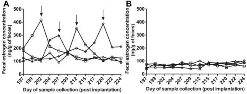
Figure 2 shows the fecal estradiol concentrations from the 28th to 32nd week of the treatment period
in implanted and control cats. Although control cats showed estradiol peaks, no estradiol peak was observed
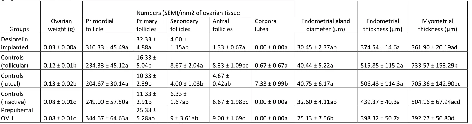
The ovarian weight of the implanted cats was significantly lower (P ≤ 0.05) than that of the prepubertal
OVH and the control cats at all stages of their estrus cycle. Significantly lower (P ≤ 0.05) ovarian weight was
recorded in the prepubertal OVH cats compared with the control cats at their luteal and estrus stage of the
estrous cycle. However, no difference was observed in the ovarian weight between prepubertal OVH cats and
control cats at the inactive stage of their estrous cycle (Table 2). The number of primordial follicles did not
differ among the different groups. Number of primary follicles was significantly higher (P ≤ 0.05) in implanted
cats compared with control cats at all the stages of the estrous cycle. However, it did not differ from that in
prepubertal OVH cats (Table 2). The number of secondary follicles did not differ among the three groups of
cats. The number of antral follicles was, however, significantly lower (P ≤ 0.05) in implanted cats compared
with other groups except the luteal stage control cats (Table 2). The number of CL in the control cats at luteal
stage was significantly higher (P ≤ 0.05) than that in the implanted, prepubertal OVH, and the control cats at
the follicular and inactive stages of the estrous cycle (Table 2).
Protein expression of LHR was observed in the cytoplasm of theca cells, interstitium of the ovarian
tissue and granulosa cells of large antral follicles only, whereas the FSHR protein expression was found in the
granulosa cells of antral follicles only. No difference was observed in the protein expression of the LHR
among different experimental groups of cats studied (Fig. 4A). Prepubertal OVH cats had significantly lower
(P ≤ 0.05) protein expression of FSHR compared to the implanted and the control cats at the luteal stage of
the estrous cycle. However, there was no difference in the protein expression of FSHR among the implanted
and control cats at the follicular and inactive stages of the estrous cycle (Fig. 4A).
Ovarian LHR mRNA expression was significantly lower (P ≤ 0.05) in the implanted cats than the
control cats at the follicular stage of the estrous cycle. However, there was no difference in the mRNA
expression of LHR among the implanted, prepubertal OVH, and the control cats at their luteal or inactive
stages of the estrous cycle (Fig. 4B). Moreover, no difference was observed in the mRNA expression of FSHR
between the implanted, prepubertal OVH. and the control cats (Fig. 4B).
4. Discussion
This study was designed to investigate the effects of the treatment of prepubertal female
LHR and FSHR. Moreover, to see how closely GnRH-agonist implantation maintains the prepubertal status
of the parameters studied, tissues were also obtained and analyzed from the prepubertal animals.
The prepubertal female cats at the age of 3 months tolerated the deslorelin implantation very well
without the need of any local or general anesthesia; no adverse effects were observed in the implanted animals.
This supports the previous studies in which tolerance of deslorelin implantation has been reported in
prepubertal cats [7] and [19]. General health of the implanted and the control animals was also monitored by
recording the weekly body weights of the cats throughout the study period. The body weight of implanted and
control cats did not differ during most of the study period of 48 weeks except during the 20th to the 26th week
when body weight of the implanted cats was found to be significantly lower than that of the controls. As this
difference in the body weight was observed only 22 weeks after the treatment and only for a brief period of
time, it seems highly unlikely that it might have been due to deslorelin implantation, rather it might have
resulted from the individual variation among the cats used in the study.
Fecal estradiol in felid species is considered to be an indication of ovarian activity [20]. The presence
of fecal estradiol peaks in the control cats and the absence of such peaks in the implanted cats simply prove
that deslorelin inhibits the ovarian activity in prepubertal cats. Moreover, unlike the previous studies [20], we
did not notice any upregulation or flare-up effect because of the deslorelin implantation. In addition to fecal
estradiol, ovarian activity was also monitored in these animals at the end of the study period by recording
ovarian weight and number of ovarian structures including follicles and CLs. No differences were observed
in the number of primordial follicles between the experimental groups, which may indicate that all the
experimental groups had the same potential of follicle development. In the implanted group, however, follicle
growth seemed to be arrested at the primary follicle stage with little growth to the secondary stage and almost
negligible growth to the antral stage of follicle development. This was also evidenced by the significantly
lower ovarian weight recorded in this group. In the control cats, follicle growth basically reflected the stage
of the estrous cycle. Understandably although, the highest number of antral follicles was observed in the
prepubertal OVH cats that may be due to an absence of ovulation in this group. As expected, the number of
CLs was the highest in the luteal group; however, CLs were also found in follicular phase queens but in only
few instances. However, no evidence of ovulation or CL was found in the prepubertal OVH or implanted
spontaneous ovulation may have occurred in our experimental animals as reported previously [21] and [22].
Gonadotropins (LH and FSH) play an important role to control the overall activity of female reproductive
tract including follicle growth [23]. Gonadotropins act by the binding to their receptors on the ovarian cells,
so a change in either the gonadotropins' concentrations or the number of their receptors may modulate the
effects of the gonadotropins on follicular development, ovulation and/or luteinization. Luteinizing hormone
receptor in the interstitium is also found to be responsible in modulating the production of steroid hormones,
which are necessary for the functions of the reproductive tract [24].
The mRNA expression of LHR in the implanted cats was significantly lower than that in the control
animals at their follicular phase while there were no differences in the LHR mRNA expression between the
implanted cats and the control animals during the luteal and inactive stages. This may suggest that receptors
for LH are activated in the control cats during their follicular stage of the estrous cycle in concomitantly with
an increase in the LH release itself [22]. It is worth noting that mRNA expression of LHR was not different
during the other stages (luteal and inactive) of the estrous cycle when LH release remains basal with mild
fluctuations [22]. Although LH release was not monitored in this study, it is well documented that GnRH
administration does cause suppression of LH in almost all the species including dogs [25] and [26]. It is,
therefore, a possibility that the decreased levels of estradiol observed in the implanted cats may be the result
of suppressed gonadotropins in these animals [3]. In this study, LH suppression by deslorelin implantation
seems to be quite effective and consistent, and might have resulted in the lower expression of LHR mRNA
observed in the implanted cats. However, the observed lack of difference in the LHR mRNA expression
between the prepubertal OVH and control cats suggests that LHR synthesis does occur during the prepubertal
period but it remains suppressed in the implanted cats. In spite of the differences observed in deslorelin-treated
cats regarding the LHR mRNA, no difference was observed in the protein expression of the LHR between the
implanted and the control cats, even at the follicular stage of their estrous cycle. This suggests that LH plays
a role in the translation of LHR mRNA into LHR protein. The action of LH, however, normally occurs after
its release induced by copulation. However, the control cats in this study were not exposed to copulation yet
some of these cats ovulated and were in luteal phase as evidenced by progesterone concentrations. As none of
these queens could have possibly been bred, there is a possibility that they might have experienced a
laboratory-maintained animals has been reported to depend on individual conditions and housing of cats [21]. However,
in our case, it is difficult to identify the housing conditions that could have caused spontaneous ovulation as
our study was not designed for this purpose, unlike other studies [27] and [28]. Interestingly, we did not record
any spontaneous ovulation in the implanted cats. Considering the possible roles of both the LHR and the LH
itself in the observed suppression of estradiol production in the implanted cats, one may conclude that LH
itself might have played a major role rather than any contribution by a change in the LHR expression.
FSHR is normally detected in the granulosa cells of antral follicles and is considered to play a role in
follicular development. The fact that the number of antral follicles was significantly higher in the prepubertal
OVH than the implanted cats in spite of significantly lower expression of FSHR protein in the prepubertal
OVH females, simply points out the fact that the ligand itself plays the critical role required for the follicle
development rather than changes in its receptor expression. However, a lack of difference in the FSH mRNA
between these two groups suggests that translation was compromised in the prepubertal animals.
The endometrial glands are responsible for uterine secretions that nourish the embryos during early
pregnancy [29], and they are also active during certain stages of the estrous cycle. The significantly lower
endometrial gland diameter in the prepubertal OVH cats suggests their lower activity in these animals
compared with the control cats during their follicular and luteal phase of the estrous cycle. Although the
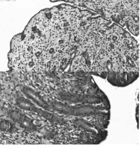
endometrial gland diameter of implanted cats was not lower than that of the control cats, the epithelium of the
endometrial glands of the implanted cats consisted of a single layer of cuboidal cells while that of control cats
during their luteal and follicular stages of the estrous cycle had a single layer columnar epithelial cells with
greater height and abundant secretion in the glands. This simply suggests a negative effect of deslorelin on the
structure/morphology and function/activity of the endometrial glands. No differences were observed in
endometrial thickness between implanted and control cats during any stage of the estrous cycle, but
myometrial thickness of the implanted cats was significantly lower than that of control cats during their luteal
and follicular phases of the estrous cycle. Moreover, myometrial thickness of prepubertal OVH cats was also
significantly lower than that of the control cats during their follicular and luteal phases of the estrous cycle.
The lower thickness of myometrium in implanted cats may be a result of suppressed estrogen concentrations
Nevertheless, results of this study suggest that the endometrial glands and the myometrium of the implanted
cats does not seem to be fully functional compared to the control cats with active ovaries. These data therefore
support the notion that both the myometrium and the endometrial glands are not quite functional due to the
suppression of ovarian function induced by the GnRH-agonist implantation.
In conclusion, the results of this study have demonstrated that in prepubertal queens, a subcutaneous
implant of 4.7-mg deslorelin suppresses ovarian weight, follicle development, estrogen production, and
myometrial thickness for a period of at least 48 weeks without any adverse effects. Associated to the
suppression of estrogen production and other ovarian functions, sexual behavior in deslorelin-implanted cats
was also suppressed. And this suppression of reproductive function due to deslorelin implantation seems to
have partly been achieved through changes in the ovarian mRNA expression of LHR. These studies could be
extended to investigate both the actual duration of suppression of reproductive function beyond the 48 weeks
and other possible mechanism(s) by which GnRH-agonist treatment could act apart from the ovarian LHR and
FSHR expression.
5. Acknowledgement
This study was supported by the Royal Golden Jubilee Ph.D. program (PHD/0161/2553), the 90th
Anniversary of Chulalongkorn University (Ratchadaphiseksomphot Endowment Fund) and the Research
Unit of Obstetric and Reproduction in animals of Chulalongkorn University, Bangkok, Thailand. The
authors acknowledge Dr. Zhangrui Cheng from the Royal Veterinary College, University of London, for
laboratory assistance and Dr. Em-on Olanratmanee from the Faculty of Veterinary Medicine, Rajamangala
University of Technology Tawan-Ok, Thailand for help in the statistical analysis of the data.
References
[1] Goericke-Pesch S, Georgiev P, Atanasov A, Albouy M, Navarro C, Wehrend A. Treatment of queens in estrus and after estrus with a GnRH-agonist implant containing 4.7 mg deslorelin; hormonal response, duration of efficacy, and reversibility. Theriogenology 2013;79:640–6.
[2] Johnston SD, Root Kustritz MV, Olson PNS. The feline estrous cycle. Canine and feline theriogenology. First edition. Philadelphia, PA 19106: Saunders Elsevier; 2001. p. 396–405.
[3] Toydemir TS, Kilicarslan MR, Olgac V. Effects of the GnRH analogue deslorelin implants on reproduction in female domestic cats. Theriogenology 2012;77:662–74.
[5] Pelican KM, Wildt DE, Howard JG. GnRH agonist Lupron (leuprolide acetate) pre-treatments prevent ovulation in response to gonadotropin stimulation in the clouded leopard (Neofelis nebulosa). Theriogenology 2006;66:1768–77.
[6] Munson L, Bauman JE, Asa CS, Jochle W, Trigg TE. Efficacy of the GnRH analogue deslorelin for suppression of oestrous cycles in cats. J Reprod Fertil Suppl 2001;57:269–73.
[7] Carranza A, Faya M, Merlo ML, Batista P, Gobello C. Effect of GnRH analogs in postnatal domestic cats. Theriogenology 2014; 82:138–43.
[8] Carranza A, Faya M, Merlo ML, Batista P, Gobello C. Histologic effect of a postnatal slow-release GnRH agonist on feline gonads. Theriogenology 2015;83:1368–72.
[9] Faya M, Carranza A, Miotti R, Ponchon T, Furlan P, Gobello C. Fecal estradiol-17beta and testosterone in prepubertal domestic cats. Theriogenology 2013;80:584–6.
[10] Axner E, Gustavsson T, Strom Holst B. Estradiol measurement after GnRH-stimulation as a method to diagnose the presence of ovaries in the female domestic cat. Theriogenology 2008;70:186–91.
[11] Goodrowe KL, Howard JG, Wildt DE. Comparison of embryo recovery, embryo quality, oestradiol-17 beta and progesterone profiles in domestic cats (Felis catus) at natural or induced oestrus. J Reprod Fertil 1988;82:553–61.
[12] Kinoshita K, Ohazama M, Ishida R, Kusunoki H. Daily fecal sex steroid hormonal changes and mating success in captive female cheetahs (Acinonyx jubatus) in Japan. Anim Reprod Sci 2011;125: 204–10.
[13] Ponglowhapan S, Church DB, Scaramuzzi RJ, Khalid M. Luteinizing hormone and follicle-stimulating hormone receptors and their transcribed genes (mRNA) are present in the lower urinary tract of intact male and female dogs. Theriogenology 2007;67:353–66.
[14] Chowdhury MWH, Scaramuzzi RJ, Wheeler-Jones CPD, Khalid M. The expression of angiogenic growth factors and their receptors in ovarian follicle throughout the estrous cycle in the ewe. Theriogenology 2010;73:856–72.
[15] Ishibashi H, Suzuki T, Suzuki S, Moriya T, Kaneko C, Takizawa T, et al. Sex steroid hormone receptors in human thymoma. J Clin Endocrinol Metab 2003;88:2309–17.
[16] Ponglowhapan S, Church DB, Khalid M. Differences in the expression of luteinizing hormone and follicle-stimulating hormone receptors in the lower urinary tract between intact and gonadectomised male and female dogs. Domest Anim Endocrinol 2008;34:339–51.
[17] Tharasanit T, Thongkittidilok C, Sananmuang T, Techakumphu M. Recombinant human follicle stimulating hormone and growth factors improve the meiotic and developmental competence of cat oocyte. Thai J Vet Med 2014;44:107–15.
[18] Sano J, Nagafuchi S, Yamazaki J, Oguma K, Kano R, Hasegawa A. Effect of antineoplastic drugs on the expression of Bcl-2 and Bcl-xL genes in the feline T-cell leukemia cell line. Res Vet Sci 2005;79: 197–201. [19] Risso A, Corrada Y, Barbeito C, Diaz JD, Gobello C. Long-term-release GnRH agonists postpone puberty in domestic cats. Reprod Domest Anim 2012;47:936–8.
[20] Brown JL, Wasser SK, Wildt DE, Graham LH. Comparative aspects of steroid hormone metabolism and ovarian activity in felids, measured noninvasively in feces. Biol Reprod 1994;51:776–86.
[21] Brown JL. Comparative endocrinology of domestic and nondomestic felids. Theriogenology 2006;66:25– 36.
[22] Bristol-Gould S, Woodruff TK. Folliculogenesis in the domestic cat (Felis catus). Theriogenology 2006;66:5–13.
[23] Orosz SE, Morris PJ, Doody MC, Niemeyer GP, Cortelyou Lee J, Eaton NL, et al. Stimulation of folliculogenesis in domestic cats with human FSH and LH. Theriogenology 1992;37:993–1004.
[24] Saint-Dizier M, Malandain E, Thoumire S, Remy B, ChastantMaillard S. Expression of follicle stimulating hormone and luteinizing hormone receptors during follicular growth in the domestic cat ovary. Mol Reprod Dev 2007;74:989–96.
[25] Wright PJ, Verstegen JP, Onclin K, Jochle W, Armour AF, Martin GB, et al. Suppression of the oestrous responses of bitches to the GnRH analogue deslorelin by progestin. J Reprod Fertil Suppl 2001;57:263–8. [26] Ponglowhapan S. Clinical applications of GnRH agonist deslorelin in dogs and cats. Thai J Vet Med Suppl 2011;41:59–63.
[28] Gudermuth DF, Newton L, Daels P, Concannon P. Incidence of spontaneous ovulation in young, group-housed cats based on serum and faecal concentrations of progesterone. J Reprod Fertil Suppl 1997;51:177– 84.
[29] Chatdarong K, Rungsipipat A, Axner E, Linde Forsberg C. Hysterographic appearance and uterine histology at different stages of the reproductive cycle and after progestagen treatment in the domestic cat. Theriogenology 2005;64:12–29.
Table 1: Description of forward and reverse primers for GAPDH as housekeeping gene and feline LHR and FSHR as target genes.
Gene Primer sequence (5′-3′)
Length (bp)
Annealing
temperature (°C) Reference
GAPDH
F: GGAGAAAGCTGCCAAATATG 20
61.4
Tharasanit et al. 2014 [17], Sano et al. 2005 [18] R: AGGAAATGAGCTTGACAAAGTGG 23
LHR
F: CTAATGCCTTTGACAACCTAATA 23
61.4 Tharasanit et al. 2014 [17] R: CCCATTGAATGCATGACTTTGTA 23
FSHR
F: CATGCTGCTAGGCTGGATCTT 21
61.4 Tharasanit et al. 2014 [17] R: CTTGGCGATCTTGGTGTCACT 21
Abbreviations: F, forward; FSHR, follicle stimulating hormone receptor; GAPDH, glyceraldehyde-phosphate-dehydrogenase; LHR, luteinizing hormone receptor; R, reverse.
Table 2: Mean ± standard error of the mean (SEM) ovarian weight (g), numbers of structures (follicles and corpora lutea) per mm2 of ovarian tissue, uterine gland diameter (μm), endometrial and myometrial thickness (μm) in deslorelin-implanted (n = 6), control (follicular, luteal, and inactive) (n = 6/phase), and prepubertal OVH (n = 6) female cats.
Groups
Ovarian weight (g)
Numbers (SEM)/mm2 of ovarian tissue
Endometrial gland diameter (μm) Endometrial thickness (μm) Myometrial thickness (μm) Primordial follicle Primary follicles Secondary follicles Antral follicles Corpora lutea Deslorelin
implanted 0.03 ± 0.00a 310.33 ± 45.49a
32.33 ± 4.88a
4.00 ±
1.15ab 1.33 ± 0.67a 0.00 ± 0.00a 30.45 ± 2.37ab 374.54 ± 14.6a 361.90 ± 20.19ad Controls
(follicular) 0.12 ± 0.01b 234.33 ± 45.12a
16.33 ±
5.04b 8.67 ± 2.04a 8.33 ± 1.09bc 0.67 ± 0.67a 40.44 ± 5.22a 515.85 ± 115.2a 733.57 ± 153.29b Controls
(luteal) 0.13 ± 0.02b 204.67 ± 30.14a
10.33 ±
2.39b 4.00 ± 1.03b
4.67 ±
0.42ab 7.33 ± 0.99b 40.75 ± 6.17a 506.43 ± 114.3a 705.36 ± 142.90bc Controls
(inactive) 0.08 ± 0.01c 249.00 ± 57.50a
11.33 ± 2.91b
6.33 ±
1.67ab 6.67 ± 1.98bc 0.00 ± 0.00a 32.60 ± 4.11ab 439.37 ± 40.3a 504.16 ± 67.94acd Prepubertal
OVH 0.08 ± 0.01c 344.67 ± 64.63a
25.33 ±
5.28ab 9 ± 3.61ab 9.00 ± 1.69c 0.00 ± 0.00a 25.13 ± 7.56b 398.32 ± 50.7a 392.27 ± 56.80d
Figure legends
Figure 1: The mean (± SEM) body weight (kg) in cats (n=6/group) with or without deslorelin implantation. Cats were implanted with deslorelin at the age of 3 months for a period of 48 weeks. Black arrow indicates the weeks (22nd to 26th) during which the control group had significantly higher (P ≤ 0.05) body weight than the deslorelin-implanted group.
Figure 3. Histological sections of uterus showing epithelia of endometrial glands (G) in implanted cats having a single layer of cuboidal cells (upper panel), and control luteal and follicular (lower panel) cats having a single layer columnar epithelial cells of greater height and abundant secretion in the glands. Black arrows identifies the epithelial cell of the endometrial glands.
Figure 4. The mean (±standard error of the mean) level of protein (left) and mRNA (fg/μg of RNA) (right) expression for the ovarian LHR and FSHR in cats that were either prepubertal OVH (n = 6) or implanted with deslorelin (n = 6) at the age of 3 months for 48 weeks or nonimplanted controls at the inactive,