Subtle radiographic presentation of a pleural
effusion secondary to a cancer of unknown
primary: a case study
Marc-André Blanchette, DC, MSc
1Julie-Marthe Grenier, DC, DACBR/FCCR(C)
21 Public Health PhD Program
School of Public Health, University of Montreal, Montreal, QC
2 Associate Professor
Department of Chiropractic, Université du Québec à Trois-Rivières, Trois-Rivières, QC Corresponding author: Marc-André Blanchette, DC, MSc
Sources of Financial Support: Dr. Marc-André Blanchette is funded by a PhD Fellowship from the CIHR. Dr. Julie-Marthe Grenier receives no funding.
©JCCA 2014
Carcinoma of unknown primary sites is a clinical syndrome that represents many types of cancer. The mortality rate associate to this type of cancer is elevated and a rapid medical referral is required for patients presenting this condition. Pleural effusion may be the only visible sign. We report a case of pleural effusion secondary to a cancer of unknown primary site in a 60-year-old man that sought chiropractic care for radiating low back pain. The radiographic studies revealed a pleural effusion as one of the only significant finding. This article will address the clinical presentation, radiographic studies and a discussion on the radiographic detection of pleural effusion.
(JCCA 2014;58(3):273-279)
k e y w o r d s : cancer of unknown primary, metastasis, pleural effusion
Les cancers de sites primaires inconnus représentent un syndrome clinique englobant de nombreux types de néoplasie. Le taux de mortalité associé à ce type de cancer est élevé et une consultation médicale rapide est nécessaire chez les patients présentant cette affection. Un épanchement pleural peut être le seul signe radiographique visible. Nous rapportons un cas d’épanchement pleural secondaire à un cancer de sites primaires inconnus chez un homme de 60 ans qui consultait en chiropratique pour une lombalgie irradiante. Les études radiographiques ont révélé un épanchement pleural comme une trouvaille fortuite. Nous avons inclus la présentation clinique, les examens radiographiques et une discussion sur la détection d’un épanchement pleural.
JACC 2014;58(3) :273-279)
Introduction
Cancer of unknown primary origin (CUP), or occult pri-mary malignancy, is not a single entity, but rather a com-plex clinical syndrome that represents many types of can-cer. Patients with CUP presents with histologically con-firmed metastatic cancer for which the primary site cannot be identified, even following a sophisticated work-up.1
CUP is not rare, it is the seventh to eighth most frequent cancer in the world1,2 and represents 2% of all
malignan-cies diagnosed in the United-States3. CUP represents the
fourth leading cause of cancer deaths in both sexes.4 The
overall age-standardised incidence per 100,000 people per year is 7-12 cases in the USA, 18-19 in Australia, 5-7 in the Netherlands, and 4-6 in Switzerland.1 The median
age at presentation is 59-66 years.4,5 CUP is slightly more
common in men than in women and predominantly affects adults (less than 1% of patient with non-hematologic CUP are children).4 Patients may demonstrate a wide variety
of clinical presentations such as palpable masses, pain or dyspnea, as well as abnormal radiographic findings such as multiple lung nodules and destructive bone lesions.6
The natural history of patients with CUP differs con-siderably from patients with known primary tumours. CUP shows several fundamental characteristics: short history with symptoms and signs associated with meta-static sites, early dissemination in the absence of primary tumour, aggressive clinical course, and occasionally un-predictable metastatic pattern (frequency and location of metastases different from those of known primary tu-mours).7 Early dissemination is responsible for the lack
of primary tumour-related clinical signs.4 Additionally,
more than 50% of CUP patients have metastatic lesions in more than one location at the time of diagnosis.4 Optimal
therapy for patients with CUP is still under debate and may depend on the histological type of the lesions found: adenomatous, squamous, neuroendocrine or poorly dif-ferentiated cell types.8 The prognosis is poor with an
ap-proximate median survival following the diagnosis of 6-9 months.4 Post-mortem studies have been able to establish
that the primary tumours in 73% of patients, the most common primary sites includes lungs (27%), pancreas (24%), liver or bile duct (8%), colo-rectal (7%), genital system (7%) and stomach (6%).9
CUP exceeds the scope of practice of chiropractors, however, it is important for clinicians to be mindful that this condition as well as many other cancers initially
present with non-specific and vague symptoms. Recogni-tion and quick referrals are key to an appropriate manage-ment. This case illustrates this situation well. A review of the history eliciting important details such as dyspnea, loss of appetite; an appropriate examination revealing among other findings, hepatomegaly and a careful obser-vation of important radiographic findings of pleural effu-sion allowed for a quick referral. Even if the findings of pleural effusion are not specific to CUP, they are serious enough to refer the patient for rapid medical care in order to identify and treat the underlying condition.
Case presentation
A 60 year-old man sought chiropractic care after suffering from episodes of low back pain extending down the left leg for approximately 6 weeks. He described the pain as stiffness that prevented him from walking comfortably or crossing his legs while in a seated position. Initially, the pain was sporadic, but for the last week, it had been rather constant, prompting him to seek care.
Upon further questioning, the patient reported a loss of appetite resulting in a loss of approximately 20 pounds in the last 6 months. He also suffered from insomnia, fa-tigue and dyspnea. The patient was a long-time smoker. His past medical history included hyperlipidemia, chronic renal failure, myocardial infarction (at age 46). He had been hospitalised for pleural effusion in the past few months. The patient was treated with medication for high blood pressure, anemia, hyperlipidemia, gastric ulcer and prevention of angina.
The orthopaedic and neurological assessment find-ings were consistent with left sacroiliac joint dysfunction. Nearly all the ranges of motion for the lumbar, hip and knee joints were decreased but none were painful. The vascular examination of the lower limbs was unremark-able.
During part of the examination, the patient was placed in a prone position and signs of facial plethora were no-ticed. The discoloration faded while the patient was in an upright position and returned each time with a recumbent position. Abdominal and pulmonary examinations were then performed. The liver was found to be enlarged but not tender and the pulmonary examination was within normal limits.
anorexia combined with the presence of dyspnea, insom-nia, malaise and facial plethora.
Radiological findings
Bone density appeared decreased, especially considering the age and body habitus of the patient. Atherosclerotic plaque was visible in the abdominal aorta and the com-mon iliac arteries. Mild degenerative disc disease was present throughout the lumbar spine, but especially in-volving L5-S1, where a posterior osteophyte was present. Hepatomegaly was also observed, in concordance with the clinical examination. Hepatomegaly is not easily de-termined on radiographs. Generally, it can be suspected if the liver measures more than 16 cm in length at the mid-clavicular line on the AP lumbar radiograph and extends
below the level of the iliac crest.10,11 (Figure 1) The liver
could not be measured precisely on the radiographs since the superior border could not be seen but its measure-ment was more than 20 cm. The medial border of the liver as outlined by the air-filled colon also extended past the level of the right kidney, another sign of hepatomegaly.12
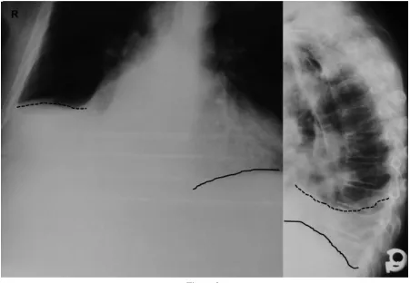
The chest radiographs (with incomplete field of view due to technical processing damage) demonstrated im-portant blunting of the costophrenic sulci on the right. This is seen on the lateral view with rounding of the costovertebral angle as well as on the frontal view with the disappearance of the right costophrenic angle. The right hemidiaphragm appeared elevated partly because of the accumulation of fluid in the pleural space. This could also be accentuated by the hepatomegaly. (Figure 2) Mild Figure 1:
AP and lateral lumbar radiographs showing degenerative disc disease with posterior osteophytosis at L5 (solid arrow) Hepatomegaly was also observed, especially on the frontal radiograph. The liver measured more than 16 cm at the
blunting of the costophrenic sulci (posterior and lateral) is also noted on the left. The cardiac silhouette also ap-peared enlarged, when grossly assessed with the cardio-thoracic ratio.13
The combination of hepatomegaly, mild cardiomegaly and recurrent pleural effusion, along with the past history raised serious concerns about this patient’s health status. The patient was referred to the hospital for more assess-ment and testing.
The patient was hospitalized and underwent a full work-up. The diagnosis of carcinoma with unknown pri-mary source was communicated by the patient’s wife to one of the authors during a phone conversation, approxi-mately two weeks after the initial visit. Metastatic lesions had been found in the lungs, brain and bones. The patient
was given a grim prognosis: less than one year to live. He passed away two months later.
Discussion
This case highlights the importance of adequately cor-relating examination with radiological findings. Although the clinical presentation raised serious «red flags» for the presence of disease, the radiological signs were subtle and could have been easily missed, especially if the clinician had omitted to assess the soft tissues. Although the as-sessment of hepatomegaly on radiograph is imprecise at best, the findings of pleural effusions are relatively easy to visualize if one knows where to look. Chiropractors do not have the necessary resources to diagnose a CUP, but they can recognise a pleural effusion. Pleural effusion is Figure 2:
PA and lateral chest radiographs with incomplete field of view due to technical processing damage. The right diaphragm (dashed line) appeared elevated on both views.
not uncommon in patients with CUP, it might be the only visible sign in many patients (as in this case study).4
Pathophysiology
The leading aetiologies of pleural effusion are cancer (27%), hearth failure (20%), pneumonia (18%), tuber-culosis (9%), pericardial diseases (3.5%) and cirrhosis (3%).14(table1) The normal pleural space contains a small
amount of liquids, which allows the lungs to expand and deflate with minimal friction during respiratory move-ments.15,16 Pleural fluid is normally produced by the
sys-temic capillaries of the parietal pleural surface and ab-sorbed into pulmonary capillaries at the visceral pleural surface.17 Lymphatic vessels also play an important role
in removing pleural liquids.14,15,17-19 There is pleural fluid
accumulation whenever the rate of pleural fluid forma-tion exceeds that of its reabsorpforma-tion. Pleural effusion as-sociated with bacterial pneumonia, bronchiectasis or lung abscess is called parapneumonic effusion, while the pres-ence of pus in the pleural space is named empyema.17
According to the composition of the pleural fluid, pleural effusions are classically divided in two type: tran-sudates and exudates.20 Transudates (low level of protein)
occur when there is:
– Increased hydrostatic pressure (e.g.: congestive heart problems),
– Decrease oncotic forces (e.g.: hypoproteinemia), – Increase negative intrapleural pressure (e.g.:
atelectasis),
– Movement of ascitic fluid through the dia-phragm (e.g.: hepatic hydrothorax).14,18,19
Exudates (high amount of protein) results of:
– Increase in the permeability of the capillarysec-ondary to infection or neoplastic process and/or – Reduction of lymphatic drainage resulting from
obstruction of the latter caused by proliferative (e.g.: malignancy) or inflammatory (e.g.: parap-neumonic effusions) process.14,18,19
Clinical approach to pleural effusion
The patient history may be very helpful to recognize the signs of pleural effusion and guide the investigation of its potential causes. For example, a typical viral prodrome (low-grade fever, sore throat, upper respiratory symp-toms) might indicate a viral pleuritis. A history of con-gestive heart failure, liver disease, uremia, or malignancy will direct the etiologic investigation of the effusion. Symptoms are often caused by an underling disease and not the effusion itself.17 Small pleural effusion can be
en-tirely asymptomatic.14,17 Large effusions will cause
dysp-nea, trepopdysp-nea, with or without chest pain (shooting, dull aching) or dry cough. The chest pain is usually exacer-bated by deep inspiration or coughing and may refer to the abdomen or the ipsilateral shoulder.14,17,21 Trepopnea is
a positional dyspnea where the patient has less symptoms when lying of the affected side.14
Classic signs during physical examination are: dimin-ished breath sounds, dullness to the percussion, decrease tactile fremitus, and localized pleural friction rub.16,17
Auscultatory percussion (method of Guarino) might also Table 1:
Causes of pleural effusion14,17,19 Transudates Congestive heart failure
Cirrhosis with ascites Nephrotic syndrome Hypoalbuminemia Myxedema
Peritoneal dialysis Glomerulonephritis
Superior vena cava obstruction Pulmonary embolism
Exudates Malignant
Primary lung Mesothelioma
Pulmonary/pleural metastases Lymphoma
Infections
Bacterial pneumonia Bronchiectasis
Lung abscess Tuberculosis
Viral illness
Connective Tissue Disease Rheumatoid arthritis
Systemic lupus erythematosus Abdominal/Gastrointestinal Disorders Pancreatitis
Subphrenic abscess Esophageal rupture Abdominal surgery Miscellaneous Pulmonary infarction Uremia
have some value for detecting small effusion.22 Many of
the patient’s medical conditions might have been respon-sible of the previous hospitalisation for pleural effusion. Among the conditions listed in table 1, the patient had heart and renal failure.
The presence of facial plethora, as demonstrated by this patient is not a classic sign of pleural effusion. It may be a manifestation of retrosternal goiter but may also occur with lung carcinoma, lymphoma, thymoma, or aortic aneurysms.23 By having the patient in a prone
position with both arm elevated on the arm rest, we may accidentally have reproduce elements of the Pemberton’s manoeuvre.24 During this classic manoeuvre, the patient
raises both arms above his head as high as possible for one minute. The manoeuvre is positive if the patient ex-perience facial plethora (Pemberton’sign). Pemberton sign occurs when the thoracic inlet becomes obstructed during positional changes, resulting in compression of the jugular veins.
Radiography
Radiographs are the easiest and least expensive way to confirm a clinical suspicion of pleural effusion.25 Chest
radiography may also reveal pleural effusion as an in-cidental finding. Blunting of a costophrenic angle is the classic sign for pleural effusion. It is important to note that minor blunting may be caused by scarring or chronic atel-ectasis. Effusions first become apparent on lateral upright radiographs with blunting of the posterior costophrenic angle.17,19 An accumulation of 200 ml of fluid is necessary
for the effusion to affect the lateral angles of frontal stand-ing radiographs.19,25 Lateral decubitus radiograph with the
affected side down is the more sensible view to identify an effusion of 5 to 15 ml.26 It is possible that effusion
un-noticed if the radiograph is taken in the supine position. The fluid then layers superiorly and posteriorly. In this case, an effusion should be considered when there is an opacification of the apical portion of the lung.19 Other
im-aging techniques such as ultrasound, CT and MRI may Table 2:
Differential diagnosis aids with pleural effusion14
Radiological characteristics Potential Diagnoses
Massive pleural effusion Malignancy, parapneumonic/empyema, tuberculosis, hepatic hydrothorax Massive effusion without
contralateral mediastinal deviation Lung cancer, mesothelioma
Heart failure, malignancy, lupus pleuritis and other systemic Bilateral pleural effusion inflammatory conditions
Parapneumonic/empyema, tuberculosis, hemothorax, Located effusion malignancy, pleurodesis, plumonary embolism, heart failure Air-fluid level in the pleural Bronchopleural fistula, gas-forming pleuropulmonary infection,
space spontaneous pneuymothorax, trauma, oesophageal rupture
Foca1 consolidation Pneumonia, lung contusion, Lung cancer Apical Infiltrate Tuberculosis or loculated fluid
Heart failure, viral pneumonia, lymphangitic carinomatosis, Interstitial infiltrates rheumatoid arthritis
Lung nodules or masses Malignancy, multifocal infection, rheumatoid arthritis Tuberculous, empyema, asbestos-related pleural disease,
Pleural calcification trauma
Pericardial calcification Constrictive pericarditis
Rib fissure or fracture Trauma
be helpful in localising effusions and distinguishing tran-sudate from exudates. Table 2 summarizes useful radio-graphic signs that can guide the investigation for potential diagnosis.
Even if radiography is an effective way to identify a pleural effusion, advance imaging, pleural fluid analysis and when applicable pleural biopsy are key elements to uncover the aetiology of the underlying disease. The role of the chiropractor is to detect the effusion and quickly refer the patient for further investigation. Patients with identified pleural effusion should be referred for medic-al investigations and treatment since the majority of the underlying condition requires rapid medical attention.
Conclusion
The confluence of findings including pleural effusion led to appropriate referral and diagnosis of CUP. Pleural ef-fusion may be caused by a variety of serious underlying conditions and should be considered in the differential diagnosis of patients presenting with dyspnea, dry cough or trepopnea, with or without chest pain. Pleural effusion might also be an incidental finding on thoracic or lumbar radiography. Chiropractors should look for an asymmetry of the hemi diaphragm, or a blunting of the costophrenic angle on every film where the diaphragm can be visual-ised. In order to identify and treat the underlying condi-tion, patients should be referred for rapid medical care.
References
1. Pavlidis N, Fizazi K. Carcinoma of unknown primary (CUP). Crit Rev Oncol Hematol. Mar 2009;69(3):271-278. 2. Fizazi K, Greco FA, Pavlidis N, Pentheroudakis G.
Cancers of unknown primary site: ESMO Clinical Practice Guidelines for diagnosis, treatment and follow-up. Ann Oncol. Sep 2011;22 Suppl 6:vi64-68.
3. Ettinger DS, Agulnik M, Cates JM, et al. Occult primary. J Natl Compr Canc Netw. Dec 2011;9(12):1358-1395. 4. Pavlidis N, Briasoulis E, Hainsworth J, Greco FA.
Diagnostic and therapeutic management of cancer of an unknown primary. Eur J Cancer. Sep 2003;39(14):1990-2005.
5. Tan. WW, Amar. S, Shahab. N, Perry. M. Metastatic Cancer With Unknown Primary Site. 2012; http://
emedicine.medscape.com/article/280505-overview#a0199. Accessed August 28th, 2013, 2013.
6. Taylor MB, Bromham NR, Arnold SE. Carcinoma of unknown primary: key radiological issues from the recent
National Institute for Health and Clinical Excellence guidelines. Br J Radiol. Jun 2012;85(1014):661-671. 7. Pavlidis N, Pentheroudakis G. Cancer of unknown primary
site. Lancet. Apr 14 2012;379(9824):1428-1435.
8. Balaker AE, Abemayor E, Elashoff D, St John MA. Cancer of unknown primary: does treatment modality make a difference? Laryngoscope. Jun 2012;122(6):1279-1282. 9. Pentheroudakis G, Golfinopoulos V, Pavlidis N. Switching
benchmarks in cancer of unknown primary: from autopsy to microarray. Eur J Cancer. Sep 2007;43(14):2026-2036. 10. Brant WE, Helms CA. Fundamentals of diagnostic
radiology. 3rd ed: Lippincott, Williams & Wilkins; 2007: 758.
11. Unal B, Bilgili Y, Kocacikli E, Bagcier S, Huvaj S, Kara S. Simple evaluation of liver size on erect abdominal plain radiography. Clinical Radiology. 2004;59(12):1132-1135. 12. Baker SR. The abdominal plain film. East Norwalk,
Conneticut1990: 49.
13. Novelline RA. Squire’s fundamentals of radiology. Harvard University Press; 2004: 179.
14. Porcel JM, Light RW. Pleural effusions. Dis Mon. Feb 2013;59(2):29-57.
15. Netter F. Atlas d’anatomie humaine (5 éd.). elsevier-masson; 2011: 184-199.
16. Bates B, Bickley LS, Hoekelman RA. Guide de l’examen clinique. 4th ed. Reuil-Malmaison cedex: Arnette, 2001:239-246.
17. Marx JA, Hockberger RS, Walls RM, Adams JG. Rosen’s emergency medicine: concepts and clinical practice. Vol 1: Mosby Incorporated; 2010.
18. Gallardo X, Castaner E, Mata JM. Benign pleural diseases. Eur J Radiol. May 2000;34(2):87-97.
19. Ferrer J, Roldan J. Clinical management of the patient with pleural effusion. Eur J Radiol. May 2000;34(2):76-86. 20. Light RW, Macgregor MI, Luchsinger PC, Ball WC, Jr.
Pleural effusions: the diagnostic separation of transudates and exudates. Ann Intern Med. Oct 1972;77(4):507-513. 21. Brims FJ, Davies HE, Lee YC. Respiratory chest pain:
diagnosis and treatment. Med Clin North Am. Mar 2010;94(2):217-232.
22. Guarino JR, Guarino JC. Auscultatory percussion: a simple method to detect pleural effusion. J Gen Intern Med. Feb 1994;9(2):71-74.
23. Antonarakis ES. Pemberton sign. Mayo Clin Proc. Jul 2007;82(7):859.
24. Pemberton H. Sign of submerged goitre. The Lancet. 1946;248(6423):509.
25. Mocelin HT, Fischer GB. Epidemiology, presentation and treatment of pleural effusion. Paediatr Respir Rev. Dec 2002;3(4):292-297.