Abstract
Background:To describe an esthetic orthodontic treatment using aligners in an adult patient with class II subdivision associated with crowding and dental crossbite. An 18-year-old hyperdivergent male patient with skeletal class II from mandibular retrusion presented for an orthodontic treatment. Occlusally, the patient presents class II subdivision, crossbite at tooth 4.4, an upper midline deviated towards the left with respect to the lower and facial midlines, and slight crowding in both arches. The patient refused conventional fixed multibracket treatment in favor of aligners. Pre- and post-treatment records as well as 1-year follow-up records are presented.
Findings:Treatment objectives were achieved in 12 months, and the patient was satisfied with the functional and esthetic outcomes, which were stable at 1 year.
Conclusion:Combining aligners with appropriate auxiliaries is an efficacious means of resolving orthodontic issues such as class II, dental crossbite, and crowding in a time-frame comparable to that of conventional fixed
orthodontics. Furthermore, this system is associated with optimal oral hygiene and excellent esthetics.
Background
Nowadays, there is a growing demand for esthetic treatment among both adolescents [1] and adults [2]. Indeed, a recent study estimated that 45% of adults are unhappy with their smile and that 20% of these have considered undergoing orthodontic treatment to improve their appearance [3].
Hence, aligner systems must now be able to treat various types of malocclusion, and over recent years, many studies have shown their great efficacy in correcting crowding, misalignment and diastems,
and even complex cases featuring extraction,
open-bite, and poor occlusal relationships [4–8].
Case report
This case report describes an adult male patient with class II subdivision malocclusion, dental crossbite, and crowding treated successfully with aligners.
Diagnosis and etiology
An 18-year-old hyperdivergent male patient presented for treatment. Extraoral photos (Fig. 1) and frontal examination revealed good incisor exposure; however,
buccal corridors and upper midline deviation towards the left with respect to the facial midline were present. The profile had a convex aspect characterized by man-dibular retrusion and increased lower facial height.
Clinical examination revealed class II subdivision with lower midline deviation towards the right of the upper midline, dental crossbite, slight crowding in both arches, and small alteration of the upper right lateral incisor morphology.
Periodontal biotype and oral hygiene were good (Fig. 2).
Panoramic radiography revealed full dentition, a lack of bone defects, no infection and no temporomandibular joint abnormalities (Fig.3).
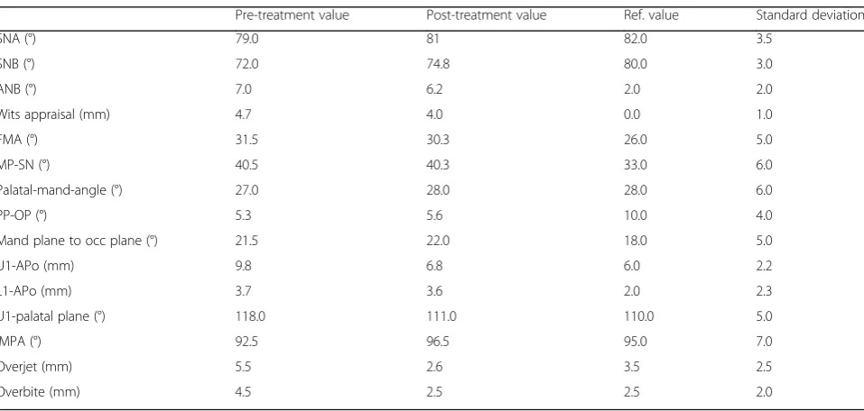
Latero-lateral teleradiograpy (Fig. 4) showed skeletal class II from mandibular retrusion, and a hyperdivergent facial type; maxillary incisor were proclined and man-dibular incisors had a correct inclination. Overbite and overjet were increased as reported in Table1.
Clinical examination showed no sign of bad habits.
Treatment objectives
The primary objective was to achieve a molar and ca-nine class I and centering the upper midline with the
* Correspondence:[email protected]
Postgraduate School of Orthodontics, University of Ferrara, Ferrara, Italy
lower and facial midlines. Additional objectives were to correct the crowding and dental crossbite, obtain ideal overjet and overbite (Fig. 5), improve facial es-thetics, and reduce black buccal corridors during smile.
Treatment alternatives
As there were no major skeletal discrepancies, a com-bined orthodontic/surgical approach was ruled out. Fixed multibracket treatment with extraction of four premolars was considered, but also excluded due to potential worsening of the profile. The patient was therefore offered a treatment plan involving unilateral distalization by fixed multibracket appliances in order to center the upper midline with the lower and facial midlines. However, the patient refused this option due to the unsightliness of the device, and we therefore agreed upon a non-extractive treatment with F22 aligners (Sweden & Martina, Due Carrare, Italy) for unilateral distalization and mesialisation of the lower arch in order to correct the class II relationship.
Treatment progress
The virtual set-up dictated 20 treatment steps for each arch. To achieve upper midline correction, the plan in-volved distorotation of teeth 1.6 (22°) and 1.7 (13°) in as-sociation with distalization. The use of class II elastics had a double function: the anchorage, used to obtain simultaneous distalization of the elements of the quad-rant I and support the correction of the lower midline.
In the lower arch, the plan involved mesorota-tion of teeth 4.6 and 4.7 associated with mesial tipping. The plan also involved alignment of the arches and retroclination of the upper incisors.
In order to achieve correct alignment and valid intercuspidation, vestibular grip points on teeth 1.6, 1.7, 4.5, 4.4, and 4.3 were planned, alongside 0.2 mm of stripping at each interproximal point in the lower right sector, from the mesial surface of tooth 4.6 to the distal surface of 4.2.
In order to promote achievement of class I, 6 oz. intermaxillary elastics were to be hooked directly on the apposite notches in the aligners at the upper canines and lower first molars from
Fig. 1Pre-treatment extraoral photos
step 1 onwards (Fig. 6). The patient was instructed to wear each aligner for 22 h per day and to move on to the next one in the series after 14 days.
After 10 months of treatment, the treatment objectives had been successfully fulfilled, although it was necessary to plan another three steps per arch for detailed finish-ing of the case and complete class correction. Specific-ally, the derotation of teeth 4.5, 1.6, and 1.5 was improved.
Treatment results
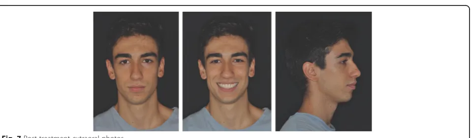
Post-treatment records demonstrate satisfactory final re-sults with all objectives achieved. Extraoral photos show a good profile, correct incisor exposure during smile and the absence of buccal corridors (Fig.7). Intraoral exam-ination reveals the achievement of all planned objectives, namely class I, centered midlines, and crowding correc-tion (Fig. 8). Post-treatment panoramic radiography (Fig. 9) showed good root parallelism, no sign of crestal bone height reduction, and no evidence of apical root
resorption. Cephalometric indices and post-treatment latero-lateral teleradiograpy show good vertical control and proclination of the lower incisors (Table 1). Superimpos-ition of pre- and post-treatment cephalometric tracings (Figs. 4, 10, 11, 12, and 13), carried out according to the methodology described in the images captions as developed by Professor Arne Björk [9,10], highlight the distal tipping
Table 1Pre- and post-treatment cephalometric values
Pre-treatment value Post-treatment value Ref. value Standard deviation
SNA (°) 79.0 81 82.0 3.5
SNB (°) 72.0 74.8 80.0 3.0
ANB (°) 7.0 6.2 2.0 2.0
Wits appraisal (mm) 4.7 4.0 0.0 1.0
FMA (°) 31.5 30.3 26.0 5.0
MP-SN (°) 40.5 40.3 33.0 6.0
Palatal-mand-angle (°) 27.0 28.0 28.0 6.0
PP-OP (°) 5.3 5.6 10.0 4.0
Mand plane to occ plane (°) 21.5 22.0 18.0 5.0
U1-APo (mm) 9.8 6.8 6.0 2.2
L1-APo (mm) 3.7 3.6 2.0 2.3
U1-palatal plane (°) 118.0 111.0 110.0 5.0
IMPA (°) 92.5 96.5 95.0 7.0
Overjet (mm) 5.5 2.6 3.5 2.5
Overbite (mm) 4.5 2.5 2.5 2.0
Fig. 6Combine use of aligners and class II intermaxillary elastics
Fig. 5Set-up viewer: the initial occlusion is shown in white and the post-treatment objectives in green
movement of the right upper sector, the retroclination achieved at the upper incisors, and the proclination of the upper incisors with respect to the basal bone—an accept-able outcome due to the morphology and conformation of the patient’s symphysis.
Check-up at 1 year demonstrates the excellent stability of results (Figs.14and15).
The last pair of aligners was used for retention due to the elastic propriety of the thermoplastic material [11].
Restoration of the 1.2 was performed in order to im-prove its morphology.
Discussion
Aligners associated with intermaxillary elastics en-abled resolution of the malocclusion within a
treat-ment time comparable with that required for
conventional fixed orthodontics, providing the patient with a comfortable, practical, and esthetic appliance. This case report is very similar to those presented in 2010 by Schupp et al. [12]. In our case, in order to prevent unwanted extrusion and/or rota-tion and further enhance the esthetics, we used
notches in the aligners at the upper canines and lower first molars rather than buttons bonded dir-ectly onto the teeth for attaching the intermaxillary elastics. Unfortunately, a direct comparison of the cephalometric indices, especially pertaining to the lower incisor proclination, was not possible, as these were not provided in the report.
From a biomechanical perspective, the dental movements occurred as planned, thanks to the fact that they were planned within the correct range of predictability [13] and the excellent properties of the material that the F22 aligners are made of [11].
In accordance with findings from Janson et al. [14], and due to the age of the patient, the movements brought about by the use of intermaxillary elastics were predominantly dentoalveolar in nature and led to a slight reduction in the SNA angle, a slight increase in the
Fig. 8Post-treatment intraoral photos
IMPA, and retroclination of the upper incisal sector. Their main effect was to provide anchorage for the upper arch, thereby promoting distal tipping and retro-clination of the upper incisors.
The treatment plan selected proved to be a winning solution not only in terms of biomechanics, but also as regards esthetics and periodontal health. Indeed, previ-ous studies [15] have shown that fixed multibracket ap-pliances, whether labial or lingual, are associated with an increase in plaque retention, which in turn may cause an increase inS. mutansconcentration and gingival inflam-mation. Furthermore, the use of such devices can in-crease the chromium and nickel concentration in a patient’s mucosa, potentially resulting in damage to DNA [16], whereas aligners have not been linked to any type of cytotoxicity [17].
Moreover, Abbate et al. [18] revealed the microbio-logical and periodontal changes that may occur during orthodontic treatment; comparing aligners and fixed ap-pliances, they found that aligners were associated with greater compliance, better oral hygiene, less accumulation of plaque, and less gingival inflammation than fixed appli-ances. These findings are in line with those reported in a previous study by Mietheke et al. [19], and in another that showed an increase in periodontopathic bacteria associ-ated with a worsening of periodontal health in fixed multi-bracket orthodontics with respect to aligners [20].
Summary and conclusions
Combined use of aligners and auxiliaries is an efficacious means of resolving orthodontic issues such as class II, dental cross-bite, and crowding within a time-frame
Fig. 12Superimposition of the maxilla. The only stable structure in the maxilla is the anterior contour of the zygomatic process [9]
Fig. 13Superimposition of the mandible. The stable anatomical structures of the mandible are:•The anterior contour of the chin. •The inner cortical structure at the inferior border of the symphysis. •Trabecular structures related to the mandibular canal [10]
comparable to conventional fixed orthodontics, but with excellent esthetics and oral hygiene.
Funding
The authors declare that they have not received funding.
Availability of data and materials
The authors declare that the materials are available.
Authors’contributions
LL is responsible for the treatment planning decision and clinical patient treatment. CA did the article test production. CA had a hand in the digital elaboration set-up and planning. OT led the clinical treatment of the patient. SG contributed in the treatment planning decision and clinical patient treatment. All authors read and approved the final manuscript.
Ethics approval and consent to participate
The study was performed in accordance with the Declaration of Helsinki. It is a case report, and the treatment plan was approved by the Chairman of Postgraduate School of Orthodontics, University of Ferrara.
Consent for publication
Written informed consent was obtained from the patient for publication of this short report and any accompanying images.
Competing interests
The authors declare that they have no competing interests.
Publisher’s Note
Springer Nature remains neutral with regard to jurisdictional claims in published maps and institutional affiliations.
Received: 26 February 2018 Accepted: 14 June 2018
References
1. Walton DK, Fields HW, Johnston WM, Rosenstiel SF, Firestone AR, Chirstensen JC. Orthodontic appliance preferences of children and adolescents. Am J Orthod Dentofac Orthop. 2010;138(6):698.e1–12. 2. Feu D, Catharino F, Duplat CB, Capelli Junior J. Esthetic perception and
economic value of orthodontic appliances by lay Brazilian adults. Dental Press J Orthod. 2012;17(5):102–14.
3. British Orthodontic Society. News release,www.bos.org.uk/news/ NOWYouGovSurvey. Accessed 9 Apr 2013.
4. Boyd RL, Miller RJ, Vlaskalic V. The Invisalign system in adult orthodontics: mild crowding and space clousure cases. J. Clin. Orthod. 2000;34(4):04–203. 5. Womack WR. Four-premolar extraction treatment with Invisalign. J Clin
Orthod. 2006;40:493–500.
6. Hönn M, Göz G. A premolar extraction case using the Invisalign system. J Orofac Orthop. 2006;67:385–94.
7. Boyd RL. Complex orthodontic treatment using a new protocol for the Invisalign appliance. J Clin Orthod. 2007;41:525–47.
8. Boyd RL. Esthetic orthodontic treatment using the Invisalign appliance for moderate to complex malocclusions. J. Dent. Ed. 2008;72:948–67. 9. Björk A, Skieller V. Growth of the maxilla in three dimensions as revealed
radiographically by the implant method. Br J Orthod. 1977;4:53–64.
Fig. 14Extraoral photos at 1 year
14. Janson G, Sathler R, Fernandes TM, Branco NC, Freitas MR. Correction of class II malocclusion with class II elastics: a systematic review. Am J Orthod Dentofac Orthop. 2013;143(3):383–92.
15. Lombardo L, Ortan YÖ, Gorgun Ö, Panza C, Scuzzo G, Siciliani G. Changes in the oral environment after placement of lingual and labial orthodontic appliances. Prog Orthod. 2013;11(14):28.
16. Hafez HS, Selim EM, Kamel Eid FH, Tawfik WA, Al-Ashkar EA, Mostafa YA. Cytotoxicity, genotoxicity and metal release in patients with fixed orthodontic appliances: a longitudinal in-vivo study. Am J Orthod Dentofac Orthop. 2011;140(3):298–308.
17. Eliades T, Pratsinis H, Athanasiou AE, Eliades G, Kletsas D. Cytotoxicity and estrogenicity of Invisalign appliances. Am J Orthod Dentofac Orthop. 2009; 136(1):100–3.
18. Abbate GM, Caria MP, Montanari P, Mannu C, Orrù G, Caprioglio A, Levrini L. Periodontal health in teenagers treated with removable aligners and fixed orthodontic appliances. J Orofac Orthop. 2015;76(3):240–50.
19. Miethke RR, Vogt S. A comparison of the periodontal health of patients during treatment with the Invisalign system and with fixed orthodontic appliances. J Orofac Orthop. 2005;66(3):219–29.


![Fig. 13 Superimposition of the mandible. The stable anatomicalstructures of the mandible are: • The anterior contour of the chin.• The inner cortical structure at the inferior border of the symphysis.• Trabecular structures related to the mandibular canal [10]](https://thumb-us.123doks.com/thumbv2/123dok_us/827295.1580268/6.595.56.291.459.702/superimposition-anatomicalstructures-cortical-structure-symphysis-trabecular-structures-mandibular.webp)
