CASE REPORT PEER REVIEWED | OPEN ACCESS
Primary angiosarcoma of the small bowel: A case report
Maria Olim Sousa, Ricardo Cabrita Viveiros, Diana Fernandes,
Rómulo Ribeiro, Lídia Ferreira, Ana Filipa Capelinha
ABSTRACT
Introduction: Small bowel angiosarcomas are exceedingly rare neoplasms with unspecific symptomatology which may lead to a delay in the diagnosis and consequently a worst prognosis. Case Report: A 73-year-old male patient presented with nausea, vomiting and abdominal pain. The blood test showed a mild anemia. Computed tomography (CT) scan revealed an ileal tumor. The patient was submitted to an exploratory laparotomy and segmental enterectomy. Pathology findings described an angiosarcoma. The patient had disease progression and died after two months. Conclusion: Angiosarcomas are high grade rapidly progressive neoplasms and have a very poor prognosis with a high mortality rate. The average life expectancy is 2–6 months after diagnosis.
Keywords: Small bowel, Cancer, Primary angio-sarcoma, Obstruction
Maria Olim Sousa1, Ricardo Cabrita Viveiros1, Diana
Fer-nandes1, Rómulo Ribeiro2, Lídia Ferreira2, Ana Filipa
Cape-linha2
Affiliations: 1MD, Hospital Dr. Nélio Mendonça – Resident,
General Surgery, Funchal, Madeira, Portugal; 2MD, Hospital
Dr. Nélio Mendonça – Consultant, General Surgery, Fun-chal, Madeira, Portugal.
Corresponding Author: Maria Olim Sousa, Avenida Luís de Camões Nº 57, Funchal, Madeira, Portugal, 9004-514; Email: [email protected]
Received: 22 February 2017 Accepted: 21 April 2017 Published: 16 May 2017
How to cite this article
Sousa MO, Viveiros RC, Fernandes D, Ribeiro R, Ferreira L, Capelinha AF. Primary angiosarcoma of the small bowel: A case report. Case Rep Int 2017;6:21–26.
Article ID: 100037CRINTMS2017
********* doi:10.5348/crint-2017-37-CR-6
INTRODUCTION
Angiosarcomas account for 1–2% of soft tissue sarcomas and are preferentially localized to skin and superficial soft tissue. Rare cases are described in heart, liver, spleen and adrenal glands, being the digestive tract localization an exceptionally rare occurrence [1–3].
Malignant tumors of the small intestine make up only 1– 1.6% of all gastrointestinal tract tumors.
The most widely recognized predisposing factors for angiosarcomas of skin and soft tissue are radiation and chronic lymphedema (Stewart-Treves syndrome). There is also a strong association with contact to some chemical agents such as thorium dioxide, arsenic, vinyl chloride or to foreign material introduced iatrogenically like vascular graft material or by trauma such as foreign bodies [1, 4].
Clinically, like other tumors of the small bowel, angiosarcomas have nonspecific symptoms including recurrent gastrointestinal bleeding, abdominal pain and nausea [2, 5–7]. The few cases described in the literature report rapid dissemination and very reserved prognosis with average survival from two to six months after diagnosis [3, 6].
CASE REPORT
A 73-year-old male, with irrelevant personal history and no known exposure to chemical toxins, surgery, chemotherapy or radiation was admitted to the emergency department with abdominal pain in the right lower quadrant and vomiting for three days. Physical examination revealed a palpable mass in the mesogastric region of the abdomen. Laboratory testing detected a mild anemia (Hb 11.3 mg/dL) with no other analytic abnormalities. The computed tomography demonstrated an abdominal tumor measuring approximately 12.3x11.6x7 cm in the proximal ileum, with regional lymph nodes and a moderate amount of intra-abdominal free fluid (Figure 1).
Laparotomy showed a small bowel obstructing tumor and a minimal amount of bloody ascites. No other lesions were observed. A segmental enterectomy was performed (Figure 2). Pathological analysis revealed an angiosarcoma and immunohistochemistry shows positivity for factor VIII, CD 34, CD31 and negativity for CD 117, S100 and MITF (Figure 3). The tumor was 7 cm in larger axis and had metastasis in 4 of 16 nodes removed.
Due to the rarity of randomized trials and prospective studies, the management guidelines for other soft tissue sarcomas tend to be utilized when dealing with angiosarcoma. According to 7th edition of AJCC Soft-Tissue Sarcoma Staging System, this case was classified as pT2bN1M0. The presence of positive nodes (N1) in M0 tumors is considered stage III.
There were no complications during the postoperative period. Adjuvant chemotherapy was decided by multidisciplinary team discussion.
One month after surgery was readmitted with abdominal pain, vomiting, ascitic abdominal distension and worsening anemia (Hb 9.2 mg/dL). Paracentesis was performed and 1000 mL of serous-hematic fluid was drained. The ascitic fluid was positive for malignant cells. CT scan showed extensive peritoneal carcinomatosis (Figure 4). Patient experienced a progressive abdominal distention, worsening of pain and anemia despite transfusions; loss of weight with severe malnutrition and rapid deterioration with multiorgan failure; he died within tw0 months after the initial diagnosis.
DISCUSSION
The primary angiosarcoma of the small bowel like other neoplasms on this location present with nonspecific symptomatology that may include abdominal pain, nausea, vomiting, intestinal obstruction, gastrointestinal bleeding and anemia [2, 4].
The patient, in our case report, presented with nonspecific complaints of pain and vomiting, and had no identifiable risk factors described in the medical literature [1]. This may have been the reason for delayed diagnosis and consequent poor prognosis.
Magnetic resonance imaging (MRI) scan, computed tomography (CT) scan, abdominal X-rays and ultrasound can be used for diagnosis, but all of them have limited diagnostic utility [5, 6].
Immunohistochemistry is the only method to confirm the diagnosis [7]. Immunohistologically, intestinal angiosarcomas are positive for endothelial markers as CD31, CD34, Von Willebrand factor and vascular endothelial growth factor and negative for epithelial,
Figure 1: Computed tomography scan showing an abdominal tumor.
Figure 2: Laparotomy findings: small bowel obstructive tumor. A segmental enterectomy was performed.
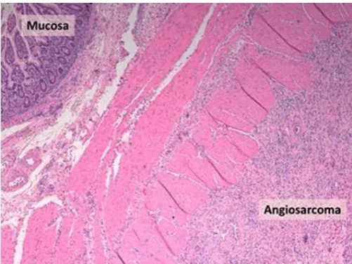
Figure 3: Histological sections of intestinal tissue observed through an optical microscope (hematoxylin eosin). Specimen showed an angiosarcoma. Immunohistologically: Factor VIII+, CD34+, CD31+, CD117-, S100- and MITF-.
neuronal and melanocytic markers as Keratins, S-100 and HMBE-45 [1, 4, 7].
Despite its aggressive behavior, angiosarcoma of the small bowel is an extremely rare entity [3]. In a literature review only 22 cases of truly primary angiosarcoma of small intestine cases were described (Table 1) [8].
There is no established standard treatment, and treatment itself becomes difficult owing to late detection due to inaccessible localization and the nonspecific symptoms.
The attempt to treat these patients requires a multidisciplinary team that may include radiologists,
Table 1: Cases of confirmed primary angiosarcoma involving the small intestine reported in English literature
Authors Sex/age
(years) Site Immuno-histochemical staining
History of prior
radiation or other
predisposing factor
Presentation Treatment Follow-up
Siderits
et al. M/79 Small bowel Strongly positive forCD31 None Obstruction Resection Unknown Taxy and
Battifora M/64 Small bowel Positive for Factor VIII, collagen type IV and vimentin
Not available Gastrointestinal
bleeding Resection Died 1 year after the initial diagnosis Taxy and
Battifora F/57 Small bowel Positive for Factor VIII, collagen type IV
Not available Not available Resection Died shortly after surgery Chami et al. M/59 Small
bowel Weakly positive for factor VIII-related antigen, Ulex
europaeus I antigen and
cytokeratin
None Gastrointestinal bleeding, bowel obstruction, anorexia and weight loss
Resection and
transfusions Died on the 11th day after surgery
Ordonez
et al. M/80 Small bowel Positive immunoreaction for FVIII-RAG
None Anemia, undue tiredness and weakness
Resection Died on the 20th
postoperative day
Hwang
et al. F/60 Small bowel Positive for Ulexeuropaeus agglutinin 1 History of radiotherapy Diffuse abdominal pain Resection Died 2 months after discharge Mohammed
et al. F/25 Small bowel Not available None Intermittent abdominal pain, weight loss, abdominal distension, hematemesis and malaena
Resection Died on the 11th day after surgery
Fraiman
et al. M/85 Small bowel Strong positivity forvimentin and CD31; focal positivity for factor
VIII and CD34
None Weight loss, anemia, weakness and abdominal pain
Resection and
thalidomide Not available
Selk et al. M/57 Small
bowel Not available History of radiation therapy
Progressive abdominal distention and shortness of breath
Resection Died 4 months after surgery
Berry et al. M/51 Small
bowel Positive for Ulexeuropaeus and vimentin
History of 3-year irradiation
Peritonitis Resection, adriamycin and dacarbazine
Khalil et al. M/68 Small
bowel Strongly positive forCD31, CD34 and vimentin
30 year history of heavy occupational exposure to radiation and polyvinyl chloride
Gastrointestinal bleeding and melaena
Resection Died 6 months after initial presentation
Suzuki et al. F/61 Ileum Positive for factor VIII-related antigen and Ulex europaeus agglutinin 1
20 year history of
radiotherapy
Abdominal pain Resection and intraabdominal cisplatin
Died 1 year after initial presentation Delvaux
et al. M/67 Small bowel Positive for CD 31,CD 34, factor VIII-related
antigen and keratin
Not available Weight loss, intermittent severe abdominal pain
and melaena
Resection Died 3 months after diagnosis Policarpio-Nicolas et al. F/51 Small
bowel Positive for CD 31, CD 34 and factor VIII-related antigen
History of
irradiation Abdominal pain Resection Died 10 months after laparotomy Hansen
et al. F/76 Small bowel Positive for factor VIII and vimentin
History of
irradiation Watery diarrhea, vomiting, weight loss and abdominal pain
Resection Died 5 months after operation
Aitola et al. F/50 Small
bowel Positive for CD 31, CD 34 and factor VIII-related antigen
≥10 year history of
radiotherapy
Intestinal
obstruction Resection followed by combination chemotherapy with
doxorubicin
1 year and 9 months after diagnosis, she was alive
Ogawa et al. M/36 Small
bowel Positive for factorVIII-related antigen Not available Abdominal pain and nausea
Resection Not available
Liu et al. F/39 Terminal
ileum Positive for CD31and CD34 None Increasing right iliac fossa pain, abdominal bloating
and vomiting
Resection and
chemotherapy Not available
Kelemen
et al. M/76 Small bowel Positive for CD31 None Abdominal pain and fatigue
Resection Died of cardiac arrest on the 9th
day after surgery Fohrding
et al. M/84 Small bowel Positive for CD31,cytokeratin and vimentin;
slightly weaker for CD34;
Focally positive for factor VIII
Not available Gastrointestinal
bleeding Resection, adjuvant chemotherapy with paclitaxel and transfusion Not available Grewal
et al. M/73 Small bowel Positive for CD31 None Gastrointestinal bleeding, weakness and melaena
Resection Died within 4 months of the diagnosis
Qingquianq
Ni et al. M/33 Small bowel Liver metastasis
Positive for
CD31 and vimentin None Abdominal pain, vomiting, weight loss, fatigue, fever
Resection, adjuvant chemotherapy
pathologists, surgical oncologists, medical oncologists and radiation oncologists. Although the cornerstone of the treatment is surgical complete resection when possible to achieve maximal locoregional control, treatment often includes palliative resection of the bleeding or obstructing lesions, chemotherapy, radiotherapy and best supportive care which may include massive blood transfusions [3].
In 1999, Aitola et al. reported a case of a small bowel tumor resection followed by combination chemotherapy with doxorubicin which survived one year and nine months after diagnosis [9]. The patient described had 14 years previously undergone total hysterectomy and salpingo-oophorectomy for a stage I adenocarcinoma of the uterine corpus and received 55.6 Gy external radiation therapy to the lower pelvis. In May 1997, at the age of 50 years, she was again admitted to hospital due to repeated symptoms of intestinal obstruction. Complementary study demonstrated a constant 5-cm-long stricture at the terminal ileum. Laparotomy revealed a 20 cm long segment of thickened terminal ileum, an extended ileocecal resection was performed. The patient received 6 adjuvant doses of doxorubicin (110 mg). Relaparotomy was undertaken one year and nine months after diagnosis of the angiosarcoma from the operative specimen, and this showed wide intra-abdominal spread and retroperitoneal recurrence. This case is the one with the highest survival described in literature in patients with this disease. Most patients die within a few months after diagnosis with an average survival from two to six months after diagnosis secondary to refractory bleeding and disease progression [2, 3, 6, 10–13].
CONCLUSION
Angiosarcoma of the small bowel is an exceptionally aggressive and rare entity with very poor prognosis. Early diagnosis is a challenge due to the nonspecific symptoms. Imaging studies can be extremely important in timely finding these lesions, but high clinical suspicion based on clinical history is necessary for diagnosis. There are no defined guidelines or demonstrated efficacy of adjuvant treatment due to the low incidence of this pathology, thus, the multidisciplinary approach of these patients is of utmost importance.
*********
Acknowledgements
We are thankful to Emanuele Parodi, Hospital Dr. Nélio Mendonça, Consultant, General Surgery, Funchal, Madeira, Portugal, and Fernando Jasmins, Hospital Dr. Nélio Mendonça, Consultant, General Surgery, Funchal, Madeira, Portugal for their help and support in preparing the manuscript.
Author Contributions
Maria Olim Sousa – Substantial contributions to conception and design, Acquisition of data, Analysis
and interpretation of data, Drafting the article, Revising it critically for important intellectual content, Final approval of the version to be published
Ricardo Cabrita Viveiros – Substantial contributions to conception and design, Acquisition of data, Analysis and interpretation of data, Drafting the article, Revising it critically for important intellectual content, Final approval of the version to be published
Diana Fernandes – Substantial contributions to conception and design, Acquisition of data, Analysis and interpretation of data, Drafting the article, Revising it critically for important intellectual content, Final approval of the version to be published
Rómulo Ribeiro – Substantial contributions to conception and design, Acquisition of data, Analysis and interpretation of data, Drafting the article, Revising it critically for important intellectual content, Final approval of the version to be published
Lídia Ferreira – Substantial contributions to conception and design, Acquisition of data, Analysis and interpretation of data, Drafting the article, Revising it critically for important intellectual content, Final approval of the version to be published
Ana Filipa Capelinha –Substantial contributions to conception and design, Acquisition of data, Analysis and interpretation of data, Drafting the article, Revising it critically for important intellectual content, Final approval of the version to be published
Guarantor
The corresponding author is the guarantor of submission.
Conflict of Interest
Authors declare no conflict of interest.
Copyright
© 2017 Maria Olim Sousa et al. This article is distributed under the terms of Creative Commons Attribution License which permits unrestricted use, distribution and reproduction in any medium provided the original author(s) and original publisher are properly credited. Please see the copyright policy on the journal website for more information.
REFERENCES
1. Weiss SW, Goldblum JR, Folpe AL. Enzinger & Weiss’s Soft Tissue Tumors. 6ed. Philadelphia Saunders; 2013. p. 703–32.
2. Lopes RH, Resende FAM, Fraga JBP, et al. Angiosarcoma of small intestine: Case report and literature review. J Bras Patol Med Lab 2016;52(5):345–8.
4. Zemheri E, Engin P, Ozkanli S, Ozemir IA. Primary angiosarcoma of small intestine presenting with intestinal perforation: A case report. J Med Cases 2014;5(2):113–5.
5. Mohammed A, Aliyu HO, Liman AA, Abdullahi K, Abubakar N. Angiosarcoma of the small intestine. Ann Afr Med 2011 Jul–Sep;10(3):246–8.
6. Young RJ, Brown NJ, Reed MW, Hughes D, Woll PJ. Angiosarcoma. Lancet Oncol 2010 Oct;11(10):983– 91.
7. Aziz MT, Tabrez MO. Angiosarcoma of small intestine presenting with intestinal obstruction. Int Surg J 2016;3(2):956–8.
8. Ni Q, Shang D, Peng H, Roy M, Liang G, Bi W, Gao X. Primary angiosarcoma of the small intestine with metastasis to the liver: A case report and review of the literature. World J Surg Oncol 2013 Sep 25;11:242. 9. Aitola P, Poutiainen A, Nordback I. Small-bowel
angiosarcoma after pelvic irradiation: A report of two cases. Int J Colorectal Dis 1999 Dec;14(6):308–10.
10. Zacarias Föhrding L, Macher A, Braunstein S, Knoefel WT, Topp SA. Small intestine bleeding due to multifocal angiosarcoma. World J Gastroenterol 2012 Nov 28;18(44):6494–500.
11. Allison KH, Yoder BJ, Bronner MP, Goldblum JR, Rubin BP. Angiosarcoma involving the gastrointestinal tract: A series of primary and metastatic cases. Am J Surg Pathol 2004 Mar;28(3):298–307.
12. Kelemen K, Yu QQ, Howard L. Small intestinal angiosarcoma leading to perforation and acute abdomen: A case report and review of the literature. Arch Pathol Lab Med 2004 Jan;128(1):95–8.
13. Taxy JB, Battifora H. Angiosarcoma of the gastrointestinal tract a report of three cases. Cancer 1988 Jul 1;62(1):210–6.
Access full text article on