Research Article
CODEN: IJPNL6
PHYTOCHEMICAL SCREENING, ANTI OXIDANT POTENTIAL AND ANTI
INFLAMMATORY ACTIVITY OF
LEUCAS DIFFUSA
PLANT EXTRACT
Ramachandran Somasundaram
1*, Priyanka Vaddadi
1, Dhanaraju Magharla Dasaratha
21
Department of Pharmacology, GIET School of Pharmacy, Rajahmundry, Andhra
Pradesh-533296, India
2
Research Lab, GIET School of Pharmacy, Rajahmundry, Andhra Pradesh-533296, India
*Corresponding author e-mail:
[email protected]
ABSTRACT
The plants of the genus ‘Leucas’ have been found to be useful in various diseases. Leucas diffusa (LD) widely distributed throughout India as a weed in cultivated fields, wastelands & roadsides. There is no scientific report published indicating utility of this plant. The present study was performed to evaluate the antioxidant and anti-inflammatory activities by using hydroxyl radical, reducing power & hydrogen peroxide scavenging abilities and through acetic acid induced vascular permeability model in mice & acetic acid induced colitis in rats significantly. LD (1000 mg/kg, p.o.) presented a significant anti-inflammatory activity towards acetic acid induced vascular permeability model in mice in comparison to Diclofenac sodium(10 mg/kg, s.c.) and acetic acid induced colitis in rats in comparison to 5-ASA. Our findings suggest that, LD contains potential antioxidant and anti-inflammatory compounds which will aid us to conduct bioactivity guided isolation & characterization of leading compounds in due course.
Keywords: Leucas diffusa, Antioxidant, Flavanoid, vascular permeability, Colitis, anti-inflammatory.
INTRODUCTION
Natural antioxidants obtained from plants scavenge harmful free radicals from our body. Free radical reactions have been implicated in the pathology of numerous diseases. [1] It is possible to reduce the risk of chronic diseases & prevent disease progression by either enhancing the body’s natural antioxidant defences or by supplementing with proven dietary antioxidants. [2] Synthetic antioxidants like ascorbic acid, butylhydroxyanisole (BHA) and butyl hydroxyl toulene (BHT) are commonly used in foods have their potential health risks & toxicity. Thus the need for alternative sources of antioxidant is paramount & the search for natural antioxidant, especially of plant origin has received much attention.[3] Plant polyphenolic compounds, such as flavanoids are described as scavengers of ROS. [4] Most sources of natural antioxidant originate from plant material. It has been also reported that reactive oxygen species (ROS) participate in the process of inflammation in
various tissues. [5] In addition to their role in acute inflammation, several factors are recognized to contribute to the pathogenesis of inflammatory bowel disease (IBD) including an over generation of reactive oxygen species (ROS). Therefore, compounds that have scavenging activities toward these radicals may be expected to have therapeutic potentials for several inflammatory diseases. [6, 7] Medicinal plants are believed to be an important source of new chemical substances with potential therapeutic effects. Plants of genus Leucas are widely used in traditional medicine to cure many diseases such as cough, cold, diarrhoea and inflammatory skin disorder. The genus Leucas comprises of about 2,500 species. The highest species diversity has been found in East Africa. In India 43 species are available. [8]
Leucas diffusa (LD) (family-lamiaceae) is an annual herb found throughout India as a weed in cultivated fields, wastelands and roadsides. Leaves are opposite,
International Journal of Pharmacy
oval shaped with tapered end. Whitish hairs are generally present on the outer surface of upper lip of the corolla. There is no scientific report published indicating utility of this plant material.
The main aim of the present work is to evaluate the antioxidant potential of the extract using tests such as hydroxyl radical scavenging activity, reducing power activity & hydrogen peroxide scavenging activity and total flavanoid content to support the pharmacological and phytochemical investigation of this plant.
MATERIALS AND METHODS
Chemicals used: The following were purchased from SIGMA- ALDRICH CHEMICALS COMPANY (BANGALORE): apigenin, reagents for estimation of MPO and SOD, ascorbic acid, Evans blue dye, Aluminium chloride, EDTA, ferric chloride
Plant material and extract preparation: LD (lamiaceae), a branched herb collected from the Tirupathi region, A.P. India, in January 2013. It was authenticated and certified by Dr. K. Madhava chetty, Assistant professor, Department of Botany, Sri Venkateswara University, Tirupathi. Accession number 1123.
The whole plant was dried in shade, separated and made to dry powder. A weighed quantity (500gm) of the powder was subjected to continuous hot extraction in Soxhlet Apparatus & extracted successively with n-hexane, ethyl acetate and methanol solvents. The methanolic extract yielded a brown sticky mass weighing 19.2g (6.14%). [9]
Preliminary chemical characterization of the extract Leucas diffusa: The extracts prepared were tested for the type of chemical constituents present by known qualitative tests.
Determination of Total Flavanoid content: Total flavanoid contents were estimated based on the formation of a complex flavanoid-aluminium. Apigenin was used to make the calibration curve [25, 50, 100, 150, 200 µg/ml in 99.9% methanol (v/v)]. The standard solutions or extracts (0.5 ml) were mixed with 1.5 ml 95% methanol (v/v), 0.1 ml 10% AlCl3, 0.1 ml of 1 mol/l sodium acetate and 2.8 ml H2O2. Blank contains 10% AlCl3 and the same volume of distilled water. After 30 min, the absorbance of the reaction mixture was measured at 415 nm using UV-Visible spectrophotometer. All determinations were done in triplicate and values were calculated from calibration curve obtained from
apigenin and total flavonoid content is expressed as μg of Apigenin equivalents (AE)/mg of extract. [10]
Evaluation of in vitro antioxidant activity
Hydroxyl radical-scavenging activity: 1ml of the final reaction solution consisted of aliquots (500 μl) of various concentrations of the extract, 1 mM FeCl3, 1 mM EDTA, 20 mM H2O2, 1 mM L-ascorbic acid, and 30 mM deoxyribose in potassium phosphate buffer (pH 7.4). The reaction mixture was incubated for 1 h at 37 °C, and further heated in a boiling water bath for 15 min after addition of 1 ml of 2.8% (w/v) TCA and 1 ml of 1% (w/w) 2-TBA. The color development was measured of 532 nm against a blank containing phosphate buffer. [11] The ability of the plant extract to scavenge hydroxy radical was calculated by the equation:
Hydroxy radical scavenging activity = {(Abs 1– Abs 2)/ (Abs0)} × 100
Where; Abs1 is the absorbance of extract; Abs 2 is the absorbance of ascorbic acid; Abs0 is the absorbance of control
Reducing power activity: Different concentrations of extracts were mixed with 2.5 ml of phosphate buffer (200 mM, pH 6.6) and 2.5 ml of 1%potassium ferricyanide. The mixtures were incubated for 20 min at 50°C. After incubation, 2.5 ml of 10% trichloroacetic acid were added to the mixtures, followed by centrifugation at 3000rpm for 10 min. The upper layer (5 ml) was mixed with 5 ml of distilled water and 1 ml of 0.1% ferric chloride and the absorbance of the resultant solution were measured at 700 nm. Increased absorbance of the reaction mixture indicated higher reducing power of the plant extract. [12]
Hydrogen peroxide-scavenging activity : The extract was dissolved in 3.4 ml of 0.1M phosphate buffer (pH 7.4) and mixed with 600 μl of 43 mM solution of hydrogen peroxide. The absorbance value (at 230 nm) of the reaction mixture was recorded at 10 min intervals between zero and 40 min. for each concentration, a separate blank sample was used for background substraction. [13] The amount of hydrogen peroxide radical inhibited by the extract was calculated using the following equation:
H2O2 radical scavenging activity = {(Abs control – Abs sample)/ (Abs control)} × 100
Where; Abs control - absorbance of H2O2 radical + methanol; Abssample - absorbance of H2O2 radical +sample extract or standard.
GIET School of Pharmacy animal house & were maintained in a controlled environment at 22±2 ◦C and 55±10% humidity with12 h light–dark cycle and fed with standard pellet food and water ad libitum. The experimental design & research plan along with animals handling and disposal procedure were placed before the institutional ethics committee. The committee granted approval after carefully evaluating research project during their meeting held in January
2012. Animal house registration
no1069/PO/ac/07/CPCSEA and IHEC project approval no- GSP/IAEC/2013/04/09.
Acute toxicity: Acute oral toxicity test for the methanolic extract of Leucas diffusa (MELD) plant was carried out as per Organization for Economic Cooperation and Development (OECD) Guidelines 423. The animals were divided into four groups containing six animals each. MELD was dissolved in 1%CMC and administered orally as a single dose to mice at different dose levels viz.300, 500, 1000 and 2000 mg/kg of body weight (b.w.). The Mice were observed periodically for symptoms of toxicity and death within 24 h and then daily for next 14 days.
Anti-inflammatory activity
Acetic acid induced vascular permeability in mice:
Four groups (n=6) of swiss mice were fasted for 18h before the experiment. One group received low dose of MELD1 (500mg/kg) and other group received high dose of MELD 2 (1000mg/kg), third group received Diclofenac sodium (1000mg/kg). Control group received distilled water. Each animal were injected with freshly prepared 0.6% acetic acid in Nacl 0.9% solution. The volume injected should be 1ml/100g through intraperitoneal route. Then, 10mg/kg of 10% (w/v) evan’s blue was injected intravenously through the tail vein immediately after administration of acetic acid. After 30min of evans blue injection, the animals were hold by a flap of abdominal wall & the viscera irrigated with distilled water over a petri dish. The dye leaking out into the peritoneal cavity was measured using UV spectra at 610nm. [14]
Acetic acid induced colitis in Albino rats: Healthy albino rats, weighing 150–200 g, were used in the study and were divided into five groups (n=6) as follows: Group A (normal control) received 1% CMC 10 ml/kg/day p.o., Group B (test) received MELD 1(500mg/kg/day p.o), Group C (test) - MELD 2 (1000mg/kg/day p.o), Group D (standard) –5-aminosalicylic acid (5-ASA) 100 mg/kg/day p.o, Group E (Toxic control) – no treatment. All the animals were pre-treated with the respective drugs (volume of drug was kept constant at 5 ml/kg) for 5
days. On the fifth day, animals were fasted overnight and colitis was induced the next morning in Groups B, C and D, E by administration of 1 ml of 4% acetic acid solution. The administration should be done rectally using paediatric catheter under low dose of ether anaesthesia. [15]
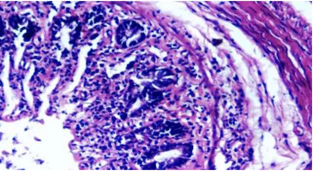
The rats were kept for 30 seconds in head-down position to prevent leakage. All the animals were sacrificed after 48 hours of colitis induction, by ether overdose. Abdomens were opened and colons were exposed. Distal 10 cm of colon was excised. Colon was opened by a longitudinal incision. Wash the mucosa with saline solution and mucosal injury was assessed macroscopically [16] – no damage (score 0); localised hyperaemia but no ulceration (score 1); linear ulcer without significant inflammation (score 2); linear ulcer with significant inflammation at one site (score 3); two or more sites of ulceration and inflammation (score 4) and two or more sites of ulceration and inflammation or one major site of inflammation and ulcer extending >1 cm along the length of colon (score 5). Disease activity indexes (DAI), degree of tissue oedema were also measured. [17] Histological analysis was done by excising a 6–8 mm sample block of the inflamed colonic tissue from a region of grossly visible damage. Formalin-fixed tissue samples were embedded in paraffin and stained with haematoxyllin–eosin (HE). Colonic tissues were scored for histological damage [18] 0=intact tissue with no apparent damage; 1=damage limited to surface epithelium; 2=focal ulceration limited to mucosa; 3=focal, transmural inflammation and ulceration; 4=extensive transmural ulceration and inflammation bordered by normal mucosa; 5=extensive transmural ulceration and with inflammation in entire area.
Biochemical Assessments
Preparation of the sample: The proximal 5 cm of the dissected colon specimen was collected. The colonic samples were minced and homogenised using a Polytron homogenizer. Then, centrifuge the samples at 3000rpm for 20min. The supernatant was used for biochemical analysis of MPO, CAT & SOD.
460 nm. One unit of MPO activity is defined as the change in absorbance per minute at room temperature, in the final reaction. [19]
MPO activity (U/g) = X/weight of the piece of tissue taken,
Where X=10×change in absorbance per minute/volume of supernatant taken in the final reaction.
Malondialdehyde (MDA) level: 0.75ml of rat serum was taken. To this, 3ml of the reagent (75mg of Thiobarbituric acid (TBA) was dissolved in 15% TCA, to this 2.08ml of 0.2N HCL was added, the volume was made up to 100 ml using 15% TCA) was added. The test tubes were kept in a boiling water bath for 15 minutes. They were cooled and centrifuged for 10 minutes at 10000rpm. Absorbance of the supernatant was read against the blank at 535nm.The results were expressed in nmol/ml of serum. [20]
Evaluation of antioxidant status in colonic tissue CAT estimation: Phosphate buffer (2.5 ml, pH 7.8) was added to the supernatant and incubated at 25°C for 30 minutes. Absorbance was measured at 240 nm spectrophotometrically. Hydrogen peroxide (650 μl) was added and change in absorbance was measured for 3 minutes. Values were expressed as μmol/min/mg of proteins. [21]
SOD estimation: The colonic samples were mixed with 3.0ml of potassium phosphate buffer, centrifuged at 2000 rpm for 10 minutes and the supernatants were collected. To that add 0.2ml of NADH to the mixture and incubated at 30°C for 90 seconds and finally the reaction was arrested by the addition of 1.0ml of glacial acetic acid. Then add 4ml of n-butanol to the reaction mixture, allowed to stand for 10 minutes and centrifuged. The absorbance was measured at 560nm in a spectrophotometer. [22]
Statistical analysis: Results were expressed as mean±SD. Statistical analysis was performed by one-way analysis of variance (ANOVA) followed by the tukey’s test for multiple comparisons. Differences were considered significant at P < 0.05.
RESULTS
Preliminary phytochemical tests: The preliminary phytochemical tests revealed the presence of flavanoids, terpenoids in methanol, ethylacetate and hexane extracts. Steroids, tannins, glycosides were found in methanolic and ethyl acetate extracts. Saponins, alkaloids, carbohydrates were found only in methanolic extract (Table 1)
Total flavanoid content: Total flavonoids compound in methanolic & ethanolic plant extract of LD was found to be 125 μg/g of methanolic & 37.5 μg/g of ethanolic extract calculated as Apigenin equivalent. (r2=0.985). (Fig 1)
Antioxidant activity
Hydroxyl radical scavenging activity: MELD showed hydroxyl radical scavenging activity with about 95.80% and 98.88% at concentration of 5μg/ml, 500μg/ml. Because the MELD was high in its flavonoid content, it may act as antioxidant and scavenge hydroxyl radical generated from the Fenton reagent (Table 2).
Reducing power activity: At concentration 5μg/ml
and 500μg/ml Ascorbic Acid (Reference) and MELD showed absorbance with about 0.382-0.509 and 0.019-0.995 respectively (Table 3). Thus MELD exhibited reducing activity & might be due to hydrogen donating ability.
Hydrogen peroxide-scavenging activity: Removal of hydroxyl free radical is necessary to protect life. The hydroxyl free radical in the cells can easily cross cell membranes and react with most biomolecules causes tissue damage, cancer and cell death. The Scavenging affect of various fractions of Ascorbic Acid (reference compound) (5μg to 500μg) and MELD (5μg and 500μg) was found to be 74.75%-81.04% and 29.45%-78.86% (Table 4).
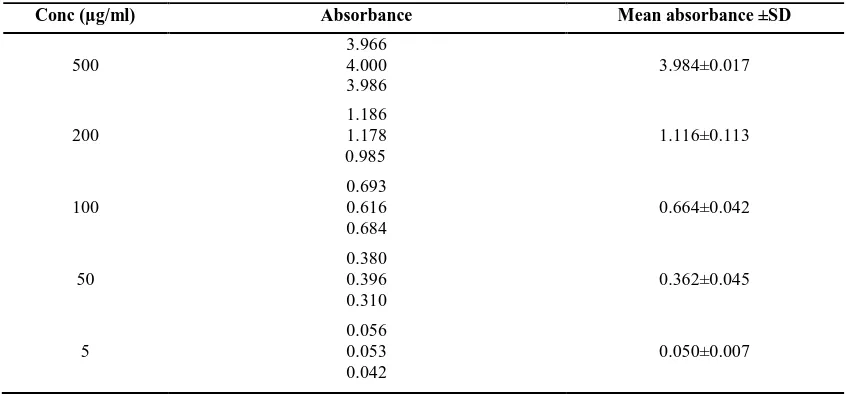
Calibration curve of Ascorbic acid (Table 5, Figure 2): Based on the results obtained MELD showed free radical scavenging activity not remarkably different than reference compound Ascorbic Acid and major antioxidative component seems to contain flavonoids. Therefore, it can be concluded that the MELD could be considered for prevention and treatment of human diseases and its complications as potent antioxidant.
Anti-inflammatory activities
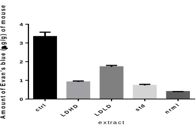
Acetic acid induced vascular permeability: As shown in Figure 3, the amount of dye passed across vessel in control group was 3.339±0.09 μg/g b.w. The extract (1000 mg/kg) reduced significantly (p<0.05; versus control group). The amount of dye retrieved in peritoneal fluid was 1.721±0.03 (25.91%) and 0.913±0.02 (60.66%) μg/g of mouse, respectively. For comparison, the vessel leakage of the dye in the diclofenac treated group was 0.726±0.02 μg/g of body weight (68.52%) (Table 6, Figure 3).
in rat colon along with significant mucosal injury microscopically (P<0.05), when compared to the normal control group. Also, there was significant change in biochemical parameters including tissue levels of MPO, MDA, CAT, SOD and serum MDA (P<0.05), indicating oxidative stress due to colon damage and colonic inflammation. MELD has shown significant activity against experimentally induced IBD when compared to that of the experimental control (P<0.05) animals, with an improved picture of colon architecture both macroscopically as well as microscopically (Table 7). There is reduction of oxidative stress with significant improvement in tissue levels of CAT, SOD (P<0.05), showing its antioxidant potential. There is also significant improvement in the levels of MPO, showing its potential anti-inflammatory activity (P<0.05) (Table 8, Figure 5, 7, 8). As for the standard drug 5-ASA, its activity against IBD was significantly better than that of Leucas difffusa extract with regard to all the parameters (P<0.05). The 5-ASA showed near normalisation of DAI and macroscopic and microscopic score as compared to normal control (Table 7, figure 6).
DISCUSSION
Some plants possess pharmacologically active constituents. Isolation of pure, pharmacologically active constituents from plants remains a long process. Chemical screening involves isolation of new or useful constituents with potential activities. This procedure recognises known metabolites in extracts, at the earliest stages of separation and is thus economically very important. Flavonoids, a large group of naturally occurring plant polyphenolic compounds, also known as nature’s tender drugs, possess numerous biological and/or pharmacological activities. Flavonoids exert beneficial effects on some diseases involving lipid peroxidation. They interact with protein phosphorylation, antioxidant and free radical scavenging activity. [23 to 28] Antioxidants play an important role in cardiovascular disease, cancer, and inflammatory disorders, scavenge free radicals from the body. [29] The extract was screened for its potential antioxidant activities. The in-vitro antioxidant assay showed Leucas diffusa methanolic extract posses potent antioxidant activity when compared with reference compound ascorbic acid. Leucas diffusa could be useful for preparation of neutraceuticals as potent antioxidant to treat various human diseases and its complications. The crude methanolic extract of Leucas diffusa was also tested for acute & chronic anti-inflammatory activity and it showed significant anti-inflammatory activity at a dose of 1000mg/kg. Vascular permeability change
participates in pathophysiology of inflammation with leakage of vascular contents to interstitial tissue. That was assessed by the amount of Evans blue dye which extravasated to peritoneal fluid in acetic acid induced peritonitis in the mice. The extract of Leucas diffusa reduced significantly vascular permeability. The extract contains chemical components which could interfere with the metabolism of released vascular active mediators .Acetic acid induced colitis affects the distal colon portion and causes massive epithelial damage. The inflammatory response initiated by acetic acid includes activation of cyclooxygenase and lipooxygenase pathways. The results showed that methanolic extract of Leucas diffusa has got a significant protective activity against experimental colitis in rats. MPO is a good marker of inflammation, an enzyme mainly found in azurophilic granules of neutrophils. Pretreatment with Leucas diffusa exhibits significant reduction in MPO activity. Oxidative damage may represent crucial pathogenic factor in IBD because intestinal inflammation is accompanied by increased production of reactive oxygen and nitrogen species. MDA is as an important indicator of lipid peroxidation [30], which is found to be increased in rats treated with acetic acid. This might be due to lipid peroxidation. Rat pretreatment with Leucas diffusa showed protection against lipid peroxidation characterised by significant decrease in MDA level. Oxidative stress plays a key role in the pathogenesis of IBD-related intestinal damage. Increased free radical production and a low concentration of endogenus antioxidant defence can be seen in IBD related damage. The antioxidant enzymes, mainly SOD, CAT are first line defensive enzymes against free radicals. [31] In the present study it was observed that the methanolic extract of Leucas diffusa significantly increase antioxidant parameters (CAT and SOD) in colitis induced rats.
CONCLUSION
The present study indicates that the MELD have got profound antioxidant effect and showed significant anti-inflammatory activity at higher dose. This may be due to presence of flavanoids in the methanolic extract. This novel finding will aid us to conduct bioactivity guided isolation and characterization of leading compounds in due course.
ACKNOWLEDGEMENT
Table 1: Qualitative chemical test for phytoconstituents
S. No Name of the test Hexane extract
Ethyl acetate extract
Methanolic extract
1 Terpenoids + + +
2 Steroids - + +
3 Saponins - - +
4 Alkaloids - - +
5 Carbohydrates - - +
6 Flavanoids + + +
7 Tannins - + +
8 Glycosides - + +
Table 2: Hydroxyl radical scavenging activity of MELD
Conc (µg/ml)
DMSO inhibition% MELD%
5 96.43 95.80
500 99.63 98.88
Table 3: Reducing power activity of MELD
Conc (µg/ml) Ascorbic acid
(absorbance)
MELD (absorbance)
5 0.382 0.019
500 0.509 0.995
All the values are means of three independent determinations, n=3, analyzed in triplicate.
Table 4: Shows hydrogen peroxide scavenging activity of MELD
Conc(µg/ml) Ascorbic acid inhibition % MELD%
5 74.75% 62.06%
500 81.04% 73.12%
All the values are means of three independent determinations, n=3, analyzed in triplicate
Table 5: Calibration curve of Ascorbic acid
Conc (µg/ml) Absorbance Mean absorbance ±SD
500
3.966 4.000 3.986
3.984±0.017
200
1.186 1.178 0.985
1.116±0.113
100
0.693 0.616 0.684
0.664±0.042
50
0.380 0.396 0.310
0.362±0.045
5
0.056 0.053 0.042
Table 6: Effect of MELD on acetic acid induced vascular permeability
Table 7: Effect of MELD on Acetic acid Induced Colitis
Groups Macroscopic score DAI Microscopic score
Normal 0.0±0.0 3±0.51 0±0
Colitis control 4.21±0.06* 7.16±1.01* 4.33±0.21*
Standard drug 1.11±0.03** 1.66±0.42** 1.33±0.21**
MELD -1 2.31±0.07** 3.16±1.66** 2.66±0.21**
MELD -2 2.05±0.02** 1.83±0.33** 2.16±0.16**
Values expressed as mean±SEM (n=6). *P<0.05 when compared to normal control; **P<0.05 when to experimental control; ANOVA followed by Tukey’s multiple comparison test; DAI – Disease activity index.
Table 8: Effect of MELD on Colitis
Groups Tissue MPO
(U/g)
MPO inhibition %
Serum MDA (nmol/ml)
Tissue CAT (μmol/min/mg)
Tissue SOD (U/mg of proteins)
Normal 62.42±1.23 -- 3.28±0.04 1.066±0.010 7.59±0.042
Colitis control 196.5±1.21* -- 5.58±0.11* 0.467±0.007* 2.94±0.020*
Standard 78.16±0.04** 60.19 3.06±0.01** 0.970±0.007** 5.15±0.014**
MELD -1 96.03±1.27** 51.12 2.41±0.02** 0.670±0.006** 5.21±0.002**
MELD -2 88.15±0.33** 55.12 2.27±0.01** 0.631±0.038** 5.36±0.016** Values expressed as mean±SEM (n=6). *P<0.05 when compared to normal control; **P<0.05 when to experimental control; ANOVA followed by Tukey’s multiple comparison test
Figure 1 Calibration curve of Apigenin
Group Dose (mg/kg,
po)
Amount of Evans blue dye (µg/g)
% inhibition
Normal --- 0.396±0.00 --
Control -- 3.339±0.09 --
Diclofenac 10 0.726±0.02 68.52
MELD -1 500 1.721±0.03 25.91
Figure 2 Calibration curve of Ascorbic acid
c tr l
L D HD
L DL D s t
d
n rm l 0
1 2 3 4
e x t r a c t
A
m
ou
nt
o
f E
va
n’
s
bl
ue
(
g
/g
)
o
f
m
o
u
s
e
Figure 3Effect of MELD on vascular permeability in mice
LDHD-Leucas diffusa high dose; LDLD- Leucas diffusa low dose
Figure 4 (Normal control) Normal mucosal architecture
Figure 6 (Standard) Near normalisation of architecture with mucosal infiltration only
Figure 7 (MELD-1) Focal ulceration limited to mucosa
REFERENCES
1. Madhavi DL, Deshpande SS, Sulunkhe D. New York Marcel dekker; 1996.
2. Stanner SA, Hughes J, Kelly CN, Buttriss JA. Public Health Nutrition, 2000; 7: 401-22. 3. Branen AL. J of American oil chemist’s society, 1975; 52: 59-63.
4. Chen S, Hwang J, Deng PSK. Archives of biochemistry and biophysics, 1993; 303: 72-77. 5. Rice-Evans C, Miller NJ. Methods in enzymology, 2002; 234: 279-93.
6. Grisham MB. Lancet, 1994; 344:859–61.
7. Keshavarzian A, Banan A, Farhadi A, Komanduri S, Mutlu E ,Zhang Y, Fields JZ. Gut, 2003; 52:720–28. 8. Aruna kumari D, Sushmitha K, Ratna madhuri T, Spandana M. IJPWR, 2012; 3(2): 1-20.
9. Das BK, Das B, Arpita FK, Morshed MA, Uddin A, Bhattacherjee A, Hannan JMA. IJPSR, 2011; 2(7):1746-52.
10. Tushrendra Singh.Int. J. of Pharm. & Life Sci, 2011; 2(3): 613-16.
11. Halliwell B, Guttridge JMC, Aruoma OI. Analytical biochemistry, 1987; 165: 215-19. 12. Yen Duh PD.Journal of the American oil Chemistry Society, 1993; 70: 383-86. 13. Ruch RT, Cheng SJ, Klaunig JE. Methods in enzymology, 1984; 105: 198-209. 14. Whittle BA. British J. Pharmacol, 1964; 2: 246-53.
15. Murat Z, Mustafa K, Erhan A, Ozgur F, Murat A, Gokhan İ. Turk J Gastroenterol, 2004; 15: 243-9.
16. Morris GP, Beck PL, Herrigge MS, Depew WT, Szewcdzuk MR, Wallace JL. Gastroenterology, 1989; 96: 795-803.
17. Ko JK, Lam FY, Cheung AP. World J Gastroenterol, 2005; 11:5787-94. 18. Wallace JL, Keenan CM. Am J Physiol, 1990; 258: 527-34.
19. Krawisz JE, Sharon P, Stenson WF. Gastroenterology, 1984; 87: 1344-50. 20. Satoh K. Clin Chim Acta, 1978; 90(1): 37-43.
21. Beers RF Jr, Sizer IW. J Biol Chem, 1952; 195:133-40.
22. Kakkar P, Das B, Viswanathan PN. Indian J Biochem Biophys, 1984; 21(2):130-32.
23. Saija A, Scalese M, Lanza M, Marzullo D, Bonina F. Free Rad Biol Med, 1995; 19(4):481-86. 24. Moon YJ, Wang X, Morris ME. Toxicology, 2006; 20:187-210.
25. Veitch NC. Nat. Prod. Rep, 2007; 24:417-64.
26. Jiang H, Zhan WQ, Liu X, Jiang SX. Nat. Prod. Res, 2008; 22(18):1650-56. 27. Kim HP, Son KH, Chang HW, Kang SS. J. Phamacol. Sci, 2004; 96:229-45.
28. Van Acker SA, van den Berg DJ, Tromp MN, et al. Free Rad Biol Med, 1996; 20:331-42.
29. Cioffi G, D’Auria M, Braca A, Mendez J, Castillo A, Morelli I, De Simone F, De Tommasi N. J. Nat. Prod, 2002; 65:1526-29.
30. Zama D , Meraihi Z, Tebibel S , Benayssa W, Benayache F, Benayache S et al. Indian J Pharmacol, 2007; 39(3):145-50.