R E S E A R C H
Open Access
Modeling of interaction between cytochrome c
and the WD domains of Apaf-1: bifurcated salt
bridges underlying apoptosome assembly
Daria N. Shalaeva
1,2, Daria V. Dibrova
3, Michael Y. Galperin
4and Armen Y. Mulkidjanian
1,2,3*Abstract
Background:Binding of cytochromec, released from the damaged mitochondria, to the apoptotic protease activating factor 1 (Apaf-1) is a key event in the apoptotic signaling cascade. The binding triggers a major domain rearrangement in Apaf-1, which leads to oligomerization of Apaf-1/cytochromeccomplexes into an apoptosome. Despite the availability of crystal structures of cytochromecand Apaf-1 and cryo-electron microscopy models of the entire apoptosome, the binding mode of cytochromecto Apaf-1, as well as the nature of the amino acid residues of Apaf-1 involved remain obscure.
Results:We investigated the interaction between cytochromecand Apaf-1 by combining several modeling approaches. We have applied protein-protein docking and energy minimization, evaluated the resulting models of the Apaf-1/ cytochromeccomplex, and carried out a further analysis by means of molecular dynamics simulations. We ended up with a single model structure where all the lysine residues of cytochromecthat are known as functionally-relevant were involved in forming salt bridges with acidic residues of Apaf-1. This model has revealed three distinctive bifurcated salt bridges, each involving a single lysine residue of cytochromecand two neighboring acidic resides of Apaf-1. Salt bridge-forming amino acids of Apaf-1 showed a clear evolutionary pattern withinMetazoa, with pairs of acidic residues of Apaf-1, involved in bifurcated salt bridges, reaching their highest numbers in the sequences of vertebrates, in which the cytochromec-mediated mechanism of apoptosome formation seems to be typical.
Conclusions:The reported model of an Apaf-1/cytochromeccomplex provides insights in the nature of protein-protein interactions which are hard to observe in crystallographic or electron microscopy studies. Bifurcated salt bridges can be expected to be stronger than simple salt bridges, and their formation might promote the conformational change of Apaf-1, leading to the formation of an apoptosome. Combination of structural and sequence analyses provides hints on the evolution of the cytochromec-mediated apoptosis.
Reviewers:This article was reviewed by Andrei L. Osterman, Narayanaswamy Srinivasan, Igor N. Berezovsky, and Gerrit Vriend (nominated by Martijn Huynen).
Keywords:Apoptosis, WD40 domains, Hydrogen bond, Salt bridge, Apoptosis, Protein-protein interactions, Caspase, Molecular dynamics simulations, Sequence analysis, Evolution
* Correspondence:[email protected] 1
School of Physics, Osnabrück University, 49069 Osnabrück, Germany 2School of Bioengineering and Bioinformatics, Lomonosov Moscow State
University, Moscow 117999, Russia
Full list of author information is available at the end of the article
© 2015 Shalaeva et al.; licensee BioMed Central. This is an Open Access article distributed under the terms of the Creative Commons Attribution License (http://creativecommons.org/licenses/by/4.0), which permits unrestricted use, distribution, and reproduction in any medium, provided the original work is properly credited. The Creative Commons Public Domain Dedication waiver (http://creativecommons.org/publicdomain/zero/1.0/) applies to the data made available in this article, unless otherwise stated.
Background
Apoptosis is a mechanism of programmed cell death that is involved in numerous processes in humans, including organism development, immune system response and aging. The intrinsic apoptotic pathway is believed to be triggered by an increased production of reactive oxygen species (ROS) in the electron-transfer chain of mito-chondria, see [1–5] for reviews. One of the key subse-quent events in mitochondria-mediated apoptosis is permeabilization of the inner and outer mitochondrial membranes by direct damage or by transition pore for-mation, followed by swelling of mitochondria [3, 6–8]. Formation of these pores, as well as rupture of the outer mitochondrial membrane, allows proteins resid-ing in the intermembrane space to escape into the cyto-plasm [9, 10].
A comparison of the intrinsic apoptotic pathways in different multicellular organisms shows that they have some common properties but also some differences [10–12]. In vertebrates, the apoptotic cascade in the cytosol is triggered by the release of cytochromecfrom mitochondria [1, 13]. Within mitochondria, cytochrome c resides in the intermembrane space and transfers electrons from the ubiquinol:cytochromec oxidoreduc-tase (cytochrome bc1 complex, or respiratory Complex
III) to the cytochrome c oxidase (respiratory Complex IV) whereby cytochrome c docks to acidic patches at the surface of the cytochrome bc1 complex or
cyto-chrome c oxidase by using a set of positively charged lysine residues [14]. After getting into the cytoplasm, cytochrome c binds between the two tryptophan (W) and aspartate (D)-rich WD domains of the apoptotic protease activating factor (Apaf-1) [3, 9, 15, 16].
WD domains (also known as WD40-repeat domains) are among the top 10 most abundant domains in eukaryotic genomes and are also widespread in bacteria [17, 18]. The common function of WD domains is to serve as scaffolds for protein-protein interactions and to coordinate downstream events, such as ubiquitina-tion or histone methylaubiquitina-tion [19]. Each WD repeat com-prises a four-stranded antiparallel β-sheet secured by hydrogen bond network between the conserved resi-dues [20]; a single WD domain is aβ-propeller that can contain from 4 to 8 WD repeats as blades [21]. More generally, proteins of the β-propeller fold are widely used in nature as structural scaffolds for ligand binding, protein-protein interactions and enzymatic activity. Despite the diversity of β-propellers, their blades fre-quently show sequence similarity indicative of a com-mon ancestry and are thought to be a result of independent amplification of an ancient blade-sized fragment [22, 23]. Specifically, in case of Apaf-1, cyto-chrome c binds between its 8-bladed C-terminal WD domain (hereafter WD-8 domain) and the 7-bladed
WD domain (hereafter WD-7 domain) that is separated from the 8-bladed domain by a flexible loop.
Upon cytochrome cbinding to Apaf-1, the WD-7 do-main rotates to accommodate the cytochrome cglobule between the two WD domains [24–26]. This cytochrome c-induced movement of WD-domains is thought to fa-cilitate the nucleotide exchange within the nucleotide binding domain (NBD) [25, 26]. Replacement of ADP (or dADP) nucleotide within the NBD by ATP (or dATP) molecule is associated with large rotational movement of the NBD and the neighboring helix do-main 1 (HD1) [24, 25], as well as with the release of the N-terminal caspase activation and recruitment domain (CARD). These events lead to the “open”, cytochrome c- and dATP/ATP-bound conformation of Apaf-1 pro-teins which then oligomerize into a heptameric platform of apoptosome [24, 27]. The CARD domains of oligomer-ized Apaf-1 monomers form a disc-like structure that binds the CARD domains of procaspase-9 to create asym-metric holo-apoptosome ready to activate the downstream caspases in the apoptotic cascade [25, 26, 28].
Functional studies that measured the ability of diverse cytochrome c variants/mutants to activate caspase-9 in the presence of Apaf-1 identified several residues of cytochrome c that were likely to be involved in the cytochromec/Apaf-1 interaction [29–35], see also [10, 16] for comprehensive reviews. The most crucial role appeared to be played by Lys72 (hereafter, the numbering matches the mature horse [PDB:1HRC] and human [PDB:1J3S] cytochrome c sequences without the N-ter-minal methionine). Replacement of Lys72 by Arg, Trp, Gly, Leu or Ala in horse cytochrome c (expressed in Escherichia coli) led to the strongly diminished activity as compared to the wild-type [29–33]. When the metazoan cytochrome c was expressed in yeast cells, it got N-trimethylated in the Lys72 position and lost its ability to trigger the assembly of apoptosome [36]. Interestingly, the yeast cytochrome cexpressed in E. coli was not methyl-ated and showed certain pro-apoptotic activity, albeit well below that of the wild-type horse cytochrome c [29]. In addition to Lys72, mutations of residues Lys7, Lys8, Lys13, Lys25, Lys27, Lys39, Lys86, Lys87, and Lys88 were found to reduce pro-apoptotic activity of cytochrome c [29–35]. In some cases, the impact of mutations was shown to be additive. Specifically, Lys7Glu/Lys8Glu and Lys25Pro/Lys39His double mutants showed a ~10-fold re-duction in caspase activation [29]. The only non-lysine residue mutations (of the total of 13 tested) that affected the activation of caspase were the Glu62Asn replacement in the horse cytochrome c and the muta-tions of the neighboring residues 63–65 [29].
surprising. However, the behavior of the cytochrome c from Drosophila with a set of functionally important ly-sine residues was more complicated. This cytochrome c could activate horse Apaf-1 protein and trigger the apop-tosome formation [28]. Surprisingly, the same fly cyto-chromecfailed to induce caspase activation inDrosophila cell lysate that contained a fly homolog of Apaf-1 [9, 37, 38] capable of oligomerization into an apoptosome, which, however, contains no cytochromes c [39]. Apparently, while promoting the formation of an apoptosome, the lysine residues of cytochromecinteract with a particular set of the Apaf-1 residues, absent from the fly homolog of Apaf-1. However, as long as no sufficiently well resolved crystal structure of the cytochrome c/Apaf-1 complex is available, the nature of these key residues of Apaf-1 re-mains obscure.
A single-particle electron density map of human apop-tosome at ~9.5 Å resolution was obtained by Yuan and co-workers in 2010 [24]. Later, the same authors have improved the structure [25] by combining their single-particle electron density map [24] with the available struc-tures of the full-length mouse Apaf-1 [PDB:3SFZ] [26], a truncated human Apaf-1 [PDB:1Z6T] [40], and the oxi-dized bovine cytochrome c [PDB:2B4Z] [41], see Fig. 1a and b. While providing powerful insight into the structure of an active apoptosome and the conformational changes in the domains of Apaf-1, this model, because of its low resolution, did not provide sufficient information either on the exact orientation of cytochromecin the lobe between
the two WD domains of Apaf-1 or on the residues of Apaf-1 that are involved in binding of cytochromec.
In this work, we have combined several molecular mod-eling approaches to scrutinize the interaction between the human cytochromecand the WD domains of Apaf-1. We were encouraged by recent results of Kokhan, Wraight and Tajkhorshid [42] who have studied the interaction between the yeast cytochrome cand the cytochrome bc1
complex using molecular dynamics (MD) simulations. Kokhan and colleagues have found that many dynamic hydrogen bonds and salt bridges, transiently showing up in their MD simulations [42], were absent from the avail-able high-resolution crystal structures [43, 44]. Specifically, many salt bridges between the patch of lysine residues of cytochrome c(such as Lys79, Lys86, and Lys87) and the polar residues of the cytochrome bc1 complex (such as Asn169, Gln170, Asp232, Glu235, and Glu99) were shown to have a dynamic nature and were not detectable in the crystal structure [42]. The authors concluded that “the static nature of x-ray structures obscures the quantitative significance of nonbonded interactions between highly mobile residues, and that short-range electrostatic in-teractions are substantially involved in cyt c binding” [42]. These results support the earlier observations that all potential hydrogen bonds are not necessarily simul-taneously present in the protein and vary depending on relevant physiological conditions [45]. The observation that even the availability of highly resolved structures does not guarantee the identification of all physiologically relevant
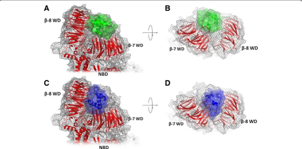
Fig. 1Structural models of the Apaf-1/cytochromeccomplexes.a, b- the cryo-EM based model of Yuanet al.[PDB:3J2T] [25], top and side views;c, d -the Patchdock’model (this work), top and side views. The cryo-EM map is shown as gray mesh, proteins are shown in cartoon and surface representation, Apaf-1 is red, cytochromecin the cryo-EM based model [PDB:3J2T] [24] is green, the structure of cytochromecin the PatchDock’model is shown in blue
interactions between proteins served as an additional justi-fication for our study.
Following the approach of Kokhan and coworkers [42], we analyzed the interaction between cytochrome c and the WD domains of Apaf-1 by MD simulations. The surfaces of the WD domains carry a significant number of aspartate and glutamate residues, so it could be antic-ipated that the interaction with cytochrome c might be mediated by salt bridges similar to those described by Kokhan and coworkers for the interaction of cytochrome cwith the cytochromebc1complex [42]. Indeed, by
com-bining molecular modeling and MD simulations we have found a specific arrangement of cytochromecbetween the two WD domains of Apaf-1 where cytochromecwas em-bedded in an extended network of salt bridges; these brid-ges involved all the lysine residues of cytochromecknown to be functionally important for apoptosome formation. Sequence analysis revealed a clear evolutionary pattern for the acidic residues of Apaf-1 that interacted with ly-sine residues of cytochrome c in the model structure, which may support the functional relevance of the found position of cytochrome c between the two WD domains of Apaf-1.
Here we scrutinized the interaction between human cytochrome cand Apaf-1 by combining several molecu-lar modeling approaches with molecumolecu-lar dynamics simu-lations. The resulting model structure of the Apaf-1/ cytochromeccomplex rationalizes the literature data on functional importance of particular residues of cyto-chromec. The identification of particular salt bridges in-volved in the interaction allowed us to identify the residues of Apaf-1 that might be involved in binding of cytochrome c and to investigate the co-evolution of the interacting residues in cytochromecand Apaf-1.
Results
Structure analysis
The most recent model of the human apoptosome [PDB:3J2T] [25], as shown in Fig. 1a and b, contains struc-tures of Apaf-1 in complex with cytochromecthat are fit into an electron density map, obtained earlier at ~9.5 Å resolution [24, 25]. The electron density map provides only the overall information about the relative location of cytochrome c in the cleft between the WD domains of Apaf-1. Since the Apaf-1 surface is enriched with nega-tively charged residues and cytochromechas a plethora of lysine residues, almost any orientation of cytochromecin the cleft between WD-domains of Apaf-1 would provide several salt bridges between the proteins. However, experi-mental data clearly indicate that this interaction is specific and requires not just a positively charged patch on the sur-face of cytochromec,which is involved in the interaction with the cytochromebc1complex and cytochromec
oxi-dase, but a whole set of lysine residues located on the
opposite sides of the protein globule [29–35]. This specifi-city of interaction implies a single functionally relevant binding mode of cytochrome c, which we have searched for usingin silicoapproaches.
To position the cytochrome c molecule between the two WD domains of Apaf-1 we have started from mo-lecular modeling. We treated the binding of cytochrome c to Apaf-1 as a docking problem and therefore started from using the available programs for rigid protein-protein docking and manually editing of the results ob-tained (see Methods). Using this approach, we obob-tained four predicted model structures of the Apaf-1/cytochrome ccomplex: one model by ClusPro software, one model by PatchDock software, and two models by ZDOCK software. These model structures were manually adjusted to resolve possible clashes between proteins and provide as many lysine-aspartate/glutamate pairs as possible. For the Patch-Dock model, the manual adjustment yielded an additional, alternative conformation (hereafter PatchDock’structure) with cytochromecthat was slightly tilted respective to the original PatchDock structure. The model of the apopto-some complex obtained from the electron density map at ~9.5 Å resolution [PDB:3J2T] [25] was treated as one more model structure under investigation.
The residues 785–805 of Apaf-1 form a loop that is completely exposed to the solution and is expected to be flexible. Therefore, during manual editing, we adjusted the position of this loop in all model structures to provide salt bridge partners for the nearby lysine residues of cyto-chromec.
All of the resulting six models placed cytochromec in the lobe between two WD domains of Apaf-1 in agree-ment with the cryo-EM data and in each of these models the lysine residues of cytochrome c formed several salt bridges with Apaf-1 (Table 1).
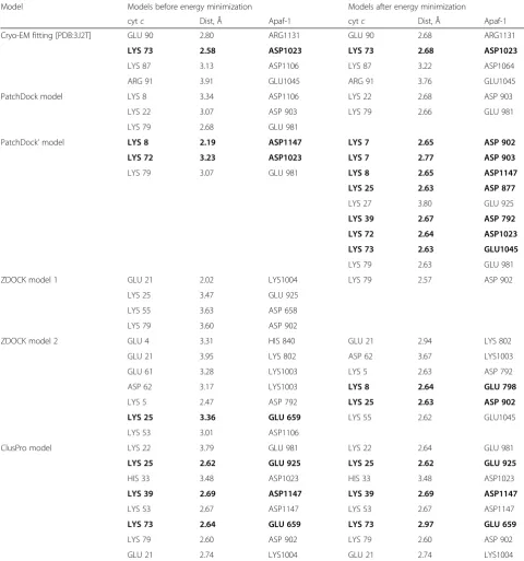
We performed energy minimization for all 6 structures and checked for salt bridges between cytochrome cand Apaf-1 before and after the energy minimization proced-ure (Table 1). After the energy minimization treatment, the models with the highest number of salt bridges in-volving conserved, functionally relevant lysine resides were the ClusPro server prediction and the PatchDock’ model (Table 1). Notably, the ClusPro model changed in-significantly after energy minimization, while the manually edited PatchDock’ model gained 6 new salt bridges after the energy minimization procedure (Table 1).
Thus, MD simulations revealed one model (the PatchDock’ model, Fig. 1c, d and 2) that retained the proper domain architecture and intact geometry dur-ing the MD simulation (Additional file 1: Figure S1). The same model had the largest number of stable salt bridges involving all important conserved residues of cytochrome c known to be involved in the interaction with Apaf-1 (Table 1, Fig. 2). These contacts involve
residues at the opposite sides of cytochrome c globule and are evenly distributed between domains WD-7 and WD-8 of Apaf-1 (Fig. 2, Table 3). Some of these brid-ges are so-called complex salt bridbrid-ges, involving more than two residues. In three cases, bifurcated (as de-fined in [46] in relation to the crystal structure of gly-cine [47], see also [48]), three-partite salt bridges involve a lysine amino group of cytochrome c that Table 1Salt bridges in putative models of Apaf-1/cytochromeccomplex before and after energy minimization
Model Models before energy minimization Models after energy minimization
cytc Dist, Å Apaf-1 cytc Dist, Å Apaf-1
Cryo-EM fitting [PDB:3J2T] GLU 90 2.80 ARG1131 GLU 90 2.68 ARG1131
LYS 73 2.58 ASP1023 LYS 73 2.68 ASP1023
LYS 87 3.13 ASP1106 LYS 87 3.22 ASP1064
ARG 91 3.91 GLU1045 ARG 91 3.76 GLU1045
PatchDock model LYS 8 3.34 ASP1106 LYS 22 2.68 ASP 903
LYS 22 3.07 ASP 903 LYS 79 2.66 GLU 981
LYS 79 2.68 GLU 981
PatchDock’model LYS 8 2.19 ASP1147 LYS 7 2.65 ASP 902
LYS 72 3.23 ASP1023 LYS 7 2.77 ASP 903
LYS 79 3.07 GLU 981 LYS 8 2.65 ASP1147
LYS 25 2.63 ASP 877
LYS 27 3.80 GLU 925
LYS 39 2.67 ASP 792
LYS 72 2.64 ASP1023
LYS 73 2.63 GLU1045
LYS 79 2.63 GLU 981
ZDOCK model 1 GLU 21 2.02 LYS1004 LYS 79 2.57 ASP 902
LYS 25 3.47 GLU 925
LYS 55 3.63 ASP 658
LYS 79 3.60 ASP 902
ZDOCK model 2 GLU 4 3.31 HIS 840 GLU 21 2.94 LYS 802
GLU 21 3.95 LYS 802 ASP 62 3.67 LYS1003
GLU 61 3.28 LYS1003 LYS 5 2.63 ASP 792
ASP 62 3.17 LYS1003 LYS 8 2.64 GLU 798
LYS 5 2.47 ASP 792 LYS 25 2.63 ASP 902
LYS 25 3.36 GLU 659 LYS 55 2.62 GLU1045
LYS 53 3.01 ASP1106
ClusPro model LYS 22 3.79 GLU 981 LYS 22 2.64 GLU 981
LYS 25 2.62 GLU 925 LYS 25 2.62 GLU 925
HIS 33 3.48 ASP1023 HIS 33 3.48 ASP1023
LYS 39 2.69 ASP1147 LYS 39 2.69 ASP1147
LYS 53 2.67 ASP1147 LYS 53 2.67 ASP1147
LYS 73 2.64 GLU 659 LYS 73 2.97 GLU 659
LYS 79 2.60 ASP 902 LYS 79 2.60 ASP 902
GLU 21 2.74 LYS1004 GLU 21 2.74 LYS1004
Bonds, as formed by conserved cytochromecresidues known to be involved in activation of the apoptosome, areshown in bold font
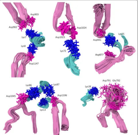
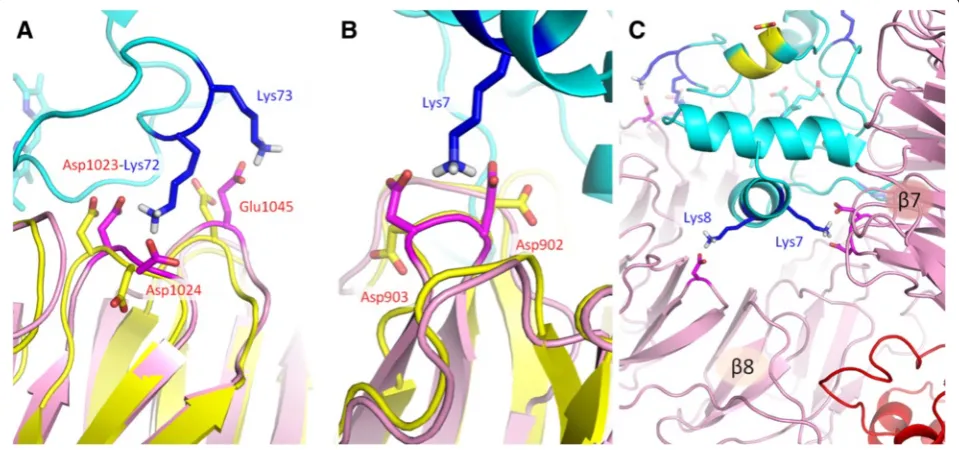
interacts with two neighboring acidic resides of Apaf-1. Namely, Lys72 interacts with residues Asp1023 and Asp1024 of Apaf-1 (Figs. 2 and 3a), Lys7 forms a salt bridge with the Asp902-Asp903 pair of Apaf-1 (Figs. 2 and 3b), and Lys39 forms salt bridges with the Glu791-Asp792 pair of Apaf-1 (Fig. 2). A pair of neighboring
lysine residues Lys7/Lys8 provides a connection between the two WD domains, creating a relatively rigid link at the bottom of the cytochrome c binding pocket (Figs. 2 and 3c).
We also calculated the electrostatic properties of the human cytochromecand Apaf-1 (see Fig. 4). While the
Fig. 2The PatchDock’model of the Apaf-1/cytochromeccomplex after energy minimization (see text). Contacts between cytochromecand Apaf-1 are shown in blue (lysine residues) and magenta (aspartate and glutamate residies). The negatively charged patch of conserved residues 62–65 of cytochromecis shown in green. The cytochromecbackbone and the heme are shown in cyan, the WD domains are shown in pink, and the rest of Apaf-1 monomer is colored red. Amino acid numbering is as in [PDB:3J2T]
surface of the cleft between the two WD domains of Apaf-1 is negatively charged, the surface of cytochrome c is mostly positively charged but has a distinct nega-tively charged patch that corresponds to Asp62 and neighboring residues. The Glu62Asn replacement at this position and mutations of the neighboring residues 63–65 are the only non-lysine mutations that are known to affect the activation of Apaf-1 [29] (the horse cyto-chromecsequence, used in these experiments, contains a glutamate residue in the 62nd position, while the hu-man cytochrome c has an aspartate). In the PatchDock’ model, this negatively charged area on cytochromec sur-face is facing outside from the WD domains cleft (Fig. 4).
The PatchDock’structure showed a good fit to the experimental electron density map with correlation coefficient of 0.9463 as compared to 0.9558 for the model structure that had been obtained earlier from cryo-EM data by Yuan et al. [PDB:3J2T] [24, 25], see Fig. 1. However, the cytochrome cposition appears to
be different in the two models. In the PatchDock’ structure, the cytochrome c globule sits deeper in the lobe between the two WD domains (Fig. 1c and d), while in cryo-EM-based structure of Yuanet al.[PDB:3J2T] [25] cytochrome c is not reaching the bottom of the lobe between two WD domains, which leaves some un-occupied electron density (Fig. 1a and b). In contrast, the deeper position of cytochromecin the PatchDock’ model results in an unoccupied density in the cryo-EM map close to the surface of the WD domains (Fig. 1c and d).
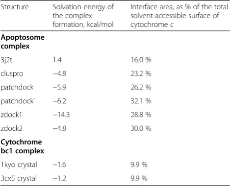
For all model structures that were obtained after energy minimization, as well as for the cryo-EM-based structure by Yuanet al. [25], we have estimated the change in the solvation energy upon the binding of cytochrome c to Apaf-1 (ΔGs), see Table 2. All model structures that were obtained via docking programs and the subsequent editing procedures showed negative values ofΔGs(Table 2). Most beneficial interaction interfaces were provided by the
Fig. 4Electrostatic properties of the interacting surfaces of Apaf-1 and cytochromecas calculated with the APBS (Adaptive Poisson-Boltzmann Solver [77]) and PDB2PQR [75, 76] software packages. The linear color scale was set from−3 (red) to 3 (blue) kcal/mol.a, WD domains of Apaf-1 are shown in a surface representation colored according to electric charge (red, negative; blue, positive), other domains of Apaf-1 are not colored, cytochrome c is not shown to reveal the negative charge of the binding interface;b, Surfaces of cytochromecand WD domains of Apaf-1 are shown simultaneously, the negatively charged spot (colored red) on the cytochromecsurface is facing the outside;c, cytochromecis shown in a cartoon representation with lysine residues shown as sticks (conservative residues shown in blue) and conserved residues 62–65 matching the negatively charged spot shown in green; d, the cytochrome c/Apaf-1 complex is shown in a“back view”, rotated by 180° as compared to panelsa–c. Apaf-1 is shown in a cartoon representation, the acidic surface residues of WD domains potentially accessible to cytochromecare shown as red sticks, the conservative acidic residues that are remote from the cytochrome c binding interface of the WD domains are shown as black sticks
model structures ZDOCK1 and PatchDock’. The cryo-EM-based structure of Yuan and co-workers [25] was characterized by a positive value of ΔGs (Table 2). In addition, by using the same approach, we calculated, for comparison, the values of ΔGs for the available high-resolution structures of the complexes of cytochromec with the cytochromebc1complex (see Table 2).
For all structures listed in Table 2, we have also calcu-lated the fraction of cytochrome c surface involved in the interactions with the domains of Apaf-1 and with the surface of the cytochrome bc1complex, respectively
(see Table 2).
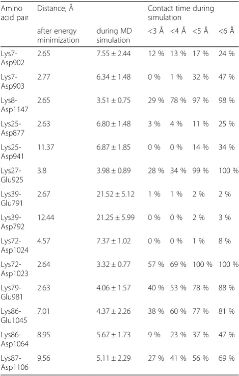
Molecular dynamics simulations provided an ensemble of conformations which allowed further evaluation of sta-bility of the protein contacts and of their impact on the structure of the PatchDock’complex (Figs. 5, 6 and 7). Salt bridges, including the H-bonded interactions, are gener-ally considered within a 4 Å cutoff [42, 49–54]. However, interactions over 4Åshould not be disregarded. Lee et al. described the distance dependence of electrostatic interac-tions between charged amino acids by a coulombic func-tion that captured the experimental and theoretical data [55]. Pairwise coulombic interactions among surface charges were significantly weakened at distances of inter-action greater than 5Å. Under conditions of“physiological” ionic strength (0.1 M), the energy of interaction between charges separated by 10 Å was approximately 0.1 kcal/ mol, but increased up to 0.5–1.0 kcal/mol already when the charges were separated by 5 Å [55]. At the binding interface, we observed both short and long-range inter-actions between charged residues (Table 3). Each of the salt bridges in Table 3 became shorter than the
classical 4 Å cutoff [42, 43, 49–53], at least transiently, during the MD simulation (Fig. 5).
All the interactions listed in Table 3 stayed throughout the 45-ns free MD simulation, with the exception of Lys39 contacts with residues Glu791 and Asp792, which got broken (Fig. 5, the top trace, and Fig. 7). The resi-dues Glu791–Asp792 of Apaf-1 are part of a loop that comprises residues 785–805; this highly mobile loop floated as a whole away from cytochromecsurface during the MD simulation (see also Additional file 1: Figure S1). Generally, the dynamic behavior of said bonds was mainly due to the side chain fluctuations and was not notably influenced by protein backbone mobility, with the exception of contacts formed by Lys39 (Fig. 7). However, neither of the observed contacts was long-living. Instead, each particular contact was lost and then regained at picoseconds. The only exceptions were the salt bridges between residues Lys25 and Asp941 as well as Lys8 and Asp1147, which could be maintained for up to 10 ns (Fig. 5).
Figure 2 reveals multiple bifurcated salt bridges that involve a single lysine residue of cytochromecas a proton donor and carboxyl groups of two aspartate or glutamate residues of Apaf-1 as proton acceptors. In addition to the three aforementioned bridges where the lysine residues of cytochromecinteract with pairs of neighboring acidic res-idues of Apaf-1, there are also interactions of Lys25 with Asp877 and Asp941, and Lys86 with Asp1064 and Glu1045 (see Table 3). In some of these bifurcated bonds the hydrogen bonds are not equivalent, so that the strong (“major”) and weak (“minor”) components can be identi-fied. To describe the components of bifurcated salt brid-ges, we have plotted the distances from each proton donor group to the two available acceptors against each other (Fig. 6).
The interaction of Lys7 with Asp902 and Asp903 (Fig. 6a) shows two distinct states, characterized by a ly-sine residue shifted to either one or the other aspartate residue, respectively. However, the population of these states is low (13 % for the conformations with Lys7 shifted to Asp902, and 26 % for the conformations with Lys7 shifted to Asp903); in all the other conformations the amino group of Lys7 is“scattered”between the two carb-oxyl groups. In contrast, the interactions of Lys25 residue with Asp877 and Asp941 (Fig. 6b) are not characterized by distinct states. The interactions of Lys72 with Asp1023 and Asp1024 (Fig. 6c) are shifted in favor of forming a salt bridge between Lys72 and Asp1023, which can be consid-ered a major state in this case. The interactions of Lys86 with Asp1064 and Glu1045 are biased in favor of a salt bridge between Lys86 and Glu1045 (Fig. 6d).
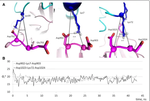
An important geometrical feature of bifurcated, com-plex salt bridges is the angleΘbetween the Cαatoms of interacting amino acids [53]. We measured theΘangles in Table 2Properties of structural models of the cytochrome c/
Apaf-1 complex as compared to the crystallographic data on the interactions of cytochromecwith the cytochromebc1
complex
Structure Solvation energy of the complex formation, kcal/mol
Interface area, as % of the total solvent-accessible surface of cytochromec
Apoptosome complex
3j2t 1.4 16.0 %
cluspro −4.8 23.2 %
patchdock −5.9 26.2 %
patchdock’ −6.2 32.1 %
zdock1 −14.3 28.8 %
zdock2 −4.8 30.0 %
Cytochrome bc1 complex
1kyo crystal −1.6 9.9 %
Fig. 5Distances between the charged groups involved in ionic bonds between cytochromecand Apaf-1, as measured during the free MD simulation. Distances were measured between the nitrogen atoms of the amino groups of lysine side chains and the closest oxygen atoms of the side chains of aspartate and glutamate residues of Apaf-1
the PatchDock’model structure after energy minimization and during the MD simulations to establish whether the bifurcated salt bridges in the model were coopera-tive or not. The small values of theΘangles (Fig. 8) in-dicate high cooperativity of the salt bridges, see also the Discussion section.
Sequence analysis
To substantiate the deduced structure of the complex between cytochromecand Apaf-1, we performed a comparative ana-lysis of the cytochromecand Apaf-1 sequences in differ-ent organisms.
Upon PSI-BLAST search of cytochromecsequences in the RefSeq database, 168 proteobacterial, 56 fungal, and 209 metazoan sequences were retrieved after the third iteration. Multiple alignments of these three groups were
used to obtain the logo diagrams (Fig. 9). As already noted, an interesting feature of the cytochrome c se-quences is the presence of a set of positively charged ly-sine residues which interact with the negatively charged
“docking”patches at the surface of its functional partners [14]. We have checked how this pivotal set has evolved. As shown in Fig. 9 by arrows, the number of lysine resi-dues has increased in the course of evolution from proteo-bacteria to Metazoa. Apparently, the higher number of lysine residues facilitated the binding of cytochromec to its functional targets.
We also performed a comparative sequence analysis of the Apaf-1 proteins (Fig. 10 and Additional file 1: Figure S2). Using our model of the cytochromec /Apaf-1 complex, we have traced the evolution of acidic resi-dues of Apaf-1 that were involved in formation of the
salt bridges in the PatchDoc’structure and checked for correlation with the evolution of the functionally import-ant cytochrome c lysine residues. The Apaf-1 residues involved in cytochrome c binding in the PatchDock’ model are conserved among the vertebrates, in agreement with the common apoptosome assembly pathway and conserved cytochrome cresidues (red arrows in Fig. 10). The Apaf-1 sequences of planarian flatworm Schmidtea
mediterranea and sea urchinStrongylocentrotus purpura-tus (phylum Echinodermata), for which the cytochrome-dependent apoptosome formation has been shown [12], contain some of these acidic residues, but not all of them (see in the Additional file 1: Figure S2). Noteworthy, the Asp1023-Asp1024 pair that forms a salt bridge with Lys72 in the PatchDock’structure is one of the most conserved, with at least one aspartate residue being present generally
Fig. 7Mobility of complex salt bridges between cytochromecand Apaf-1 in the course of MD simulations. Conformations of particular complex salt bridges observed in MD simulation were superimposed individually for each group of contacts. Protein backbone fragments are shown in cartoon representations: cytochromecin cyan, Apaf-1 in magenta. Interacting residues are shown in stick representation: lysine residues in blue, aspartate and glutamate residues in red
in all Metazoa, which mirrors the conservation of Lys72 residue through all Metazoa as well (Fig. 9). A peculiar replacement of one aspartate in this pair to histidine is observed in Aves (birds), although apoptotic pathways have only been well studied in chicken cells, and the chicken Apaf-1 has aspartates or glutamates in all posi-tions proposed to be important for apoptosome assembly. For the 791–792 and 902–903 pairs of acidic residues there is a clear evolutionary trend of their prevalence within chordates. A comparison of Figs. 9 and 10 shows that while proteins with the Apaf-1 domain architecture are already seen inNematostella vectensisandTrichoplax adhaerens, the set of potent ligands of cytochrome c de-scribed in this work has fully evolved at the level ofС hor-dates, probably after their branching from Echinoderms and Hemichordates.
Discussion
In this work, we present a model of the Apaf-1/cyto-chrome c complex which could serve as a basis for de-tailed investigation of specific interactions that underlie the apoptosome assembly. For the lysine residues that are known to be crucial for the ability of cytochromecto in-duce apoptosis, we have identified acidic counterparts in Apaf-1. In three cases, acidic “duplets” (pairs of adjacent aspartate and/or glutamate residues) were involved in complex salt bridges with lysine residues of cytochromec.
We estimated the changes in the solvation energy due to the interface formation (ΔGs), as well as fractions of the cytochrome c surface involved in the interaction with Apaf-1 for all the model structures including the one that had been obtained earlier from cryo-EM data by Yuan and co-workers [25], see Table 2. For all our model struc-tures the calculated values of solvation energiesΔGswere distinctly negative, unlike the cryo-EM-based structure of Yuan and co-workers [25] for which the ΔGs value was positive (Table 2). This positive value correlated with the smallest fraction of cytochromec surface involved in the interactions with the domains of Apaf-1 in this structure as compared with the model structures that were obtained by using docking programs (see Table 2). It is noteworthy that the cryo-EM-based model structure of Yuan and co-workers was obtained by maximizing the correlation with electron density as experimentally measured in [24], while our model structures were obtained by docking methods that generally search for maximal energy gains and the lar-gest interaction interfaces for the docking partners. The PatchDock’ model structure showed the largest inter-action surface.
The smaller, albeit negative values ofΔGs, as calculated for the high-resolution complexes of cytochrome c with the cytochromebc1complexes (Table 2) can be explained
by smaller interactions surfaces: while in the cytochrome c/Apaf-1 complex both sides of cytochromecinteract with the domains of Apaf-1, only one side of cytochromec in-teracts with the cytochromebc1complex.
The role of the conserved negatively charged patch of residues 62–65 in the PatchDock’structure might be in providing orientation of cytochromecin its binding cleft between the two negatively-charged surfaces of the Apaf-1 domains. Noteworthy, this region faces away from the contact interface, as it also does in the complexes of cyto-chromecwith the cytochromebc1complex [43].
All of the initial six models placed cytochromecin the lobe between two WD domains of Apaf-1, in agreement with the cryo-EM data, and in each of these models lysine residues of cytochromecformed salt bridges with Apaf-1. However, only some of these models invoked the func-tionally important lysine residues and only the PatchDock’ model included a salt bridge formed by Lys72 from the very beginning (Table 1).
Table 3Salt bridges between Apaf-1 and cytochromecin the PatchDock’model
Amino acid pair
Distance, Å Contact time during
simulation
after energy minimization
during MD simulation
<3 Å <4 Å <5 Å <6 Å
Lys7-Asp902
2.65 7.55 ± 2.44 12 % 13 % 17 % 24 %
Lys7-Asp903
2.77 6.34 ± 1.48 0 % 1 % 32 % 47 %
Lys8-Asp1147
2.65 3.51 ± 0.75 29 % 78 % 97 % 98 %
Lys25-Asp877
2.63 6.80 ± 1.48 3 % 4 % 11 % 25 %
Lys25-Asp941
11.37 6.87 ± 1.85 0 % 0 % 14 % 34 %
Lys27-Glu925
3.8 3.98 ± 0.89 28 % 34 % 99 % 100 %
Lys39-Glu791
2.67 21.52 ± 5.12 1 % 1 % 2 % 2 %
Lys39-Asp792
12.44 21.25 ± 5.99 0 % 0 % 2 % 3 %
Lys72-Asp1024
4.57 7.37 ± 1.02 0 % 0 % 1 % 8 %
Lys72-Asp1023
2.64 3.32 ± 0.77 57 % 69 % 100 % 100 %
Lys79-Glu981
2.63 4.06 ± 1.57 40 % 53 % 78 % 88 %
Lys86-Glu1045
7.01 4.37 ± 2.26 38 % 60 % 77 % 81 %
Lys86-Asp1064
8.95 5.67 ± 1.73 9 % 23 % 37 % 47 %
Lys87-Asp1106
9.56 5.11 ± 2.29 27 % 41 % 56 % 69 %
Specifically, the position of the functionally important Lys72 residue in the PatchDock’structure indicates the possibility of a complex salt bridge formation with the Apaf-1 residues Asp1024 and Asp1023 (Fig. 3a), although in the latter case the 4.6 Å distance between the charged moieties after energy minimization is larger than usually expected for salt bridges (see the discussion of the cut-off distances below). In contrast, in the model of Yuan and colleagues [PDB:3J2T] [25], it is the neighboring residue Lys73 that is forming the salt bridge with Asp1023, while Lys72 of cytochromecand Asp1024 of Apaf-1 are facing away from interaction interface. It is tempting to speculate that binding of Lys72 might play a guiding role in docking of cytochromecto Apaf-1.
Interactions involving more than two charged residues are commonly referred to as “complex” or “networked” salt bridges. Complex salt bridges have been investigated for their role in stabilizing protein structure and protein-protein interactions [52, 56–60]. While playing an import-ant role in connecting elements of the secondary structure and securing inter-domain interactions in proteins, com-plex salt bridges are commonly formed by partners that
are separated by 3–4 uninvolved residues in the protein chain. Repetitive cases within the same protein domain with neighboring residues of the same charge being in-volved in bifurcated interactions, three of which are pre-dicted in the PatchDock’structure, to the best knowledge of the authors, have not been reported until now. This is not surprising, since the repulsion between two negatively charged residues could hardly contribute to the protein stability [61]. Still, in the case of Apaf-1, there is a clear pattern of emergence and evolutionary fixation of several Asp-Asp motifs (Fig. 10) that, as the modeling suggests, might be involved in binding the lysine residues of cyto-chromec.
The geometry of the interactions between acidic and basic residues is similar in simple and complex salt brid-ges. Adding a residue to a simple interaction represents only a minor change in the geometry but yields a more complex interaction, a phenomenon that may explain the cooperative effect of salt bridges in proteins. Energetic properties of complex salt bridges vary depending on the protein environment around the salt bridges and the geometry of interacting residues. Detailed analyses of the
Fig. 8Geometry of bifurcated salt bridges.a, Values of the angle between Cαatoms for complex salt bridges in the PatchDock’model structure after energy minimization.b, Values of the angle between Cαatoms for the same structure during the MD simulation. Values for the Asp792-Lys39-Glu793 salt bridge are not shown due to the high mobility of the respective loop of Apaf-1 (residues 785–805)
net energetics of complex salt bridge formation using double- and triple-mutants gave conflicting results. In two cases, complex salt bridge formation appeared to be co-operative, i.e., the net strength of the complex salt bridge was more than the sum of the energies of individual pairs [62, 63]. In one case, formation of a complex salt bridge was reported to be anti-cooperative [64].
Statistical analysis of complex salt bridge geometries performed on a representative set of structures from the PDB revealed that over 87 % of all complex salt bridges formed by a basic (Arg or Lys) residue and two acidic partners have a geometry such that the angle formed by their Cαatoms,Θ, is < 90° [53]. Same preferred geometry was observed in the two aforementioned instances when the energetics of complex salt bridge formation was co-operative [62, 63], while in the reported anti-coco-operative complex salt bridge [64] the value of Θ was close to 160°. The anti-cooperativity of complex salt bridges with Θ= 150° was also established by measuring the stability of model proteins [53].
It is noteworthy that complex salt bridges can be also found at the interfaces of cytochrome c with other pro-teins; due to dynamic nature of such interactions they are not always reflected in crystallographic structures. Crystal
structures are available for cytochrome c bound to the cytochromebc1complex [43, 44], the cytochromec
perox-idase [65], the photosynthetic reaction center [66], along with a theoretical model of the complex with cytochrome c oxidase [67]. Most of interactions described for cyto-chromeclysine residues can be classified as long-distance electrostatic interactions with distances between charged groups in the 4 to 9 Å range [43, 44, 65–67]. Still, some of these interactions involve pairs of negatively charged resi-dues, and in few cases – even pairs of neighboring resi-dues [44].
The geometry of bifurcated salt bridges in the Patch Dock’model of the Apaf-1/cytochromec complex shows surprising resemblances to the known cytochromec inter-actions with other partners.For example, on the interface between cytochrome c (chain W in [PDB:3CXH]) and cytochromec1of the yeast cytochromebc1complex (chain
O in [PDB:3CXH]) the bifurcated salt bridge between Lys96 (Lys87 in human) of cytochromecand the duplet of aspartate residues of cytochromec1(Asp231 and Asp232)
showsΘ= 22.8°. This value indicates cooperativity between the bonds involved in these interactions. The bifurcated salt bridges in the PatchDock’cytochrome c/Apaf-1 com-plex, described above, show pretty small values for theΘ
angle, around 15–20° (Fig. 8). According to Gvritishvili et al. [53], such small angles would indicate high coopera-tivity for these bonds. However, an important destabilizing factor in this interaction might be the conformational ten-sion in the protein backbone. The bifurcated salt bridges reported here include acidic residues located next to each other on relatively loose loops between the β-strands of WD domains, so the energetic gain upon insertion of a positive charge between two negatively charges moieties can be accompanied by a loss in protein backbone mobil-ity. In addition, with the introduction of a positively charged lysine residue, the carboxyl groups of two Asp residues are being forced to come closer together (Fig. 3a
and b), which might create tension in the protein back-bone structure and trigger certain conformational changes in the Apaf-1 protein.
Within Apaf-1, the signal about the binding of cyto-chromecto the WD domains should be mechanistically transmitted towards the nucleotide-binding domain. Formation of bifurcated salt bridges may be involved in this signaling, since such interactions: (i) are specific to the apoptotic pathway; (ii) should cause conformational changes in those loops that carry the neighboring pairs of acidic residues (Fig. 3a and b); and (iii) might be ener-getically favorable to an extent sufficient to initiate a con-formational rearrangement of the whole Apaf-1 structure
Fig. 10Conservation of negatively charged residues in the sequences of Apaf-1 homologs. The numeration of residues corresponds to the human Apaf-1. Sequence logos were generated with WebLogo [89] from multiple alignments of 22 sequences from group I, which included Chordates (Vertebrates and Cephalochordates), and 15 sequences from group II (Hemichordates, Echinoderms, Platyhelminthes, Cnidaria, Arthropods, and Placozoa). Each position in the logo corresponds to a position in the alignment while the size of letters in the position represents the relative frequency of corresponding amino acid in this position
enabling transmission of a signal to the partner from the other side of the WD domain.
We would like to emphasize that our structure, as shown in Figs. 1c, d, 2, and 4 is just a theoretical predic-tion; the ultimate structural solution of the Apaf-1/cyto-chrome c complex would come, hopefully, in the near future, along with a well-resolved crystal and/or cryo-EM structure of the complex. Although we hope that this structure would match our prediction, there is obvi-ously no guarantee. Taking into account the large num-ber of lysine residues that are spread all over the surface of cytochromec, one could not exclude some alternative arrangement of cytochromec between the two WD do-mains, which also would satisfy the existing functional constrains. It also seems plausible that binding of cyto-chrome c between the two WD domains, as well as its release from a mature holo-apoptosome, might both be multistep processes, so that the structure in Fig. 1 might correspond to only one of the structural intermediates. Our goal was, however, to identify the residues of Apaf-1 that are involved in binding of cytochromec. Accordingly, we believe that the acidic“duplets”, which are particularly abundant in the Apaf-1 sequences of vertebrates, would withstand the scrutiny of further experimental studies as the key players in promoting the apoptosome formation.
Replacement of key lysine residues of cytochrome c has been shown to decrease its ability to cause caspase activation [29–35]. Accordingly, the appearance of these lysine residues at the surface of cytochrome c in the course of evolution (Fig. 9) should have increased the ability of cytochromecto promote apoptosis - provided that new acidic counterparts for these lysine residues emerged concurrently on the interacting surfaces of the WD domains, which seems to be the case,cfFig. 9 with Fig. 10 and Additional file 1: Figure S2. Bifurcated salt bridges, which should be stronger than the simple ones, could further contribute to the ability of cytochrome c to promote apoptosome formation.
This scenario, as well as our model, lead to an experi-mentally testable prediction that replacement of the acidic residues of Apaf-1, identified in this work, would decrease the ability of cytochrome c to promote apop-tosis. Such experimental validation might be useful also for other WD domains (tryptophane and aspartate-rich) as salt bridges formed by these acidic residues might ac-count for the ability of these domains to mediate protein-protein interactions also in other cell systems.
While the number of acidic residues of Apaf-1 in the regions facing cytochrome c is increased in vertebrates as compared to other taxa, there are also conserved as-partate residues on the sides of WD domains that are opposite to the cytochrome c-interacting sides (black boxes in Fig. 10). As these residues cannot be involved in the binding of cytochrome c, their conservation,
perhaps, indicates their involvement in the interaction of Apaf-1 with some other partner (s). Several proteins, be-sides cytochromec, can bind to Apaf-1 and affect its ac-tivation, see [68] for a review. For example, specific binding to the WD domains of Apaf-1 was demonstrated for the anti-apoptotic Bcl-2 family member Boo [69]. Particularly interesting are the positions 754 and 755 of Apaf-1 (Figs. 4 and 10) where a clear evolutionary trend of emergence of an aspartate duplet can be seen. These aspartate residues are very likely to bind one of the Apaf-1-modulating proteins.
WD-40 repeat-containing proteins are abundant among the conserved clusters of orthologous groups of eukaryotic proteins [70]. These proteins are subunits of major, eukaryote-specific protein complexes, such as the rRNA processosome [71], and the presence of numerous paralogs indicates that architecture of these complexes, with the unique functions of individual sub-units, almost entirely evolved at a very early stage of eukaryotic evolution via multiple duplications of genes for superstructure-forming proteins [72]. Thus, all nu-merous paralogous proteins containing WD-40 repeats are expected to function as structural components of multisubunit complexes [72], with WD domains medi-ating interactions between protein domains [25, 73, 74], the function that we have addressed here on the ex-ample of Apaf-1. It is tempting to speculate that WD domains, generally, mediate interactions between pro-teins by changing their conformation in response to various impacts that affect the acidic residues of the loops that connect the rigidβ-blades.
Conclusions
Methods Structures used
We used coordinates of the full-length human Apaf-1 pro-tein in cytochromec-bound state [PDB:3J2T] [25] and the NMR solution structure of reduced human cytochromec [PDB:1J3S] (Jeng WY, Shiu JH, Tsai YH, Chuang WJ. 2009. Solution structure of reduced recombinant human cytochromec, unpublished).
Electrostatic calculations
We used the APBS (Adaptive Poisson-Boltzmann Solver) and PDB2PQR software packages designed for analysis of the solvation properties of small and macro-molecules such as proteins, nucleic acids, and other complex sys-tems. We used PDB2PQR [75, 76] to prepare the setup (structures and parameters) for calculations, and APBS [77] for modeling properties of protein surfaces by solving the Poisson-Boltzmann equation. We used the versions implemented as web servers hosted by the National Biomedical Computation Resource (http://nbcr-222.ucsd. edu/pdb2pqr_2.0.0/). Protonation states of residues were assigned with the PROPKA software [78], separately for the Apaf-1 and cytochromecstructures.
Modeling of the cytochromecbinding to Apaf-1
To predict the orientation of cytochromecin its binding cleft we used several rigid protein-protein docking soft-ware packages that are based on different approaches, namely PatchDock [79], ZDOCK [80], and ClusPro [81], and combined them with manual editing and evaluation of the obtained models.
The PatchDock algorithm is inspired by object recog-nition and an image segmentation technique used in computer vision and applies geometric hashing and pose-clustering matching to match convex and concave patches of interacting surfaces [79]. The web server is located at http://bioinfo3d.cs.tau.ac.il/PatchDock/.
ZDOCK is a fast Fourier transform (FFT)-based pro-tein docking program which searches all possible bind-ing modes in the translational and rotational space between the two proteins and evaluates each pose using an energy-based scoring function [80]. The web server is at http://zdock.umassmed.edu/.
ClusProalso uses the FFT-based rigid docking with an addition of low energy results clustering under the as-sumption that a native binding site will have a wide free-energy attractor with the largest number of results [81]. The web server is at http://cluspro.bu.edu/.
Additionally, the orientation of cytochrome c in the cryo-EM fitted structure of apoptosome [PDB: 3J2T] [25] was also treated as a model under investigation.
The software that we used for calculating the protein-protein docking operates with rigid bodies (ZDOCK and PatchDock servers) or incorporates only side-chain
flexibility (ClusPro). Thus, we used manual editing, en-ergy minimization procedure, and, at the final stage, free molecular dynamics simulations to refine the model structures and examine the flexible interacting interfaces.
Structure editing and evaluation were done manually using PyMOL [82]. During the analysis of the obtained structural models we were mainly considering the num-ber of salt bridges and hydrogen bonds between the interacting proteins. At each stage of modeling we used the PISA service at the European Bioinformatics Insti-tute (http://www.ebi.ac.uk/pdbe/pisa/) [83] to list salt bridges and hydrogen bonds between the proteins in the complex (Table 1). PISA was also used for estimating the change of the solvation energy of the cytochrome c structure due to the interface formation (ΔGs) (Table 2), as well as the fraction of cytochromec surface involved in the interactions with Apaf-1 and the cytochrome bc1
complex, respectively (Table 2).
We have used the UCSF Chimera package [84] to fit the model structures into the experimental cryo-EM data [24] and to calculate the correlation coefficients.
Molecular dynamics (MD) simulations
For the MD simulations we used the Gromacs v.4.5.5 soft-ware with MPI implementation at the supercomputer SKIF “Chebyshev” (the Computational Center of the Lomonosov Moscow State University). The protein mole-cules were modeled with the CHARMM36 force field. The system for simulation consisted of an Apaf-1/cyto-chromeccomplex placed in the simulation box that was big enough to provide at least 12 Å distance from protein atoms to periodic cell walls. Each model was placed in a water box with addition of Na+ and Cl− ions to balance the total charge of the system and create 0.2 M total salt concentration.
Energy minimization
Energy minimization for each structure was performed by using the steepest descent algorithm with an initial step size 0.02 nm. Minimization converged when the max-imum force became smaller than 1 kJ mol−1nm−1. Free MD simulation
Prior to the free MD simulation, we performed a pres-sure equilibration in constant temperature and volume (NVT) ensemble with positional restraints applied to all non-hydrogen protein atoms. Subsequent free MD was set in the NPT ensemble (with constant pressure and temperature). The reference temperature of 298 K was maintained by using a Nose-Hoover extended ensemble with the time constant of the temperature fluctuations at equilibrium of 0.4 ps. The pressure was maintained at 1 atm by the Parrinello-Rahman extended-ensemble
pressure coupling where the box vectors are subject to an equation of motion, with isotropic pressure coupling with the time constant of 1 ps. Non-bonded interactions were computed by using particle mesh Ewald method with 10 Å real space cut-off for electrostatic interactions and the switching functions between 10 and 12 Å for the van der Waals interactions. The multiple time-step method was employed for the electrostatic forces; the non-bonded interaction list was constructed using a cut-off of 14 Å, updated every 20 steps. The covalent bonds involving hydrogen atoms were constrained using the SHAKE algorithm (with the MD integration step size, 2 fs). Trajectory coordinates were written down every 0.2 ns of simulation.
The resultant trajectories were visualized and analyzed by means of VMD (Visual Molecular Dynamics) software [85]. Structures of all models under investigation after energy minimization are available as Additional files 2, 3, 4, 5, 6 and 7.
Sequence analysis
The initial sequence search in the RefSeq database of fully sequenced genomes [86] was performed with PSI-BLAST [87] using the horse cytochromecand the human Apaf-1 sequences as queries. Multiple alignments were con-structed with Muscle [88]. The logo diagrams were cre-ated and visualized with WebLogo [89].
Reviewers’comments
We thank the reviewers for their valuable comments and helpful suggestions that helped us improve the manuscript.
Reviewer’s report 1: Prof. Andrei L. Osterman, Sanford-Burnham Medical Research Institute, La Jolla, California 92037, USA
Reviewer 1: The manuscript by D. Shalaeva et al.“ Model-ing of interaction between cytochrome c and Apaf-1: bi-furcated salt bridges underlying apoptosome assembly”is addressing an intriguing and fundamentally important problem. How the two seemingly unrelated proteins with distinct evolutionary history, molecular functions and compartmentalization recognize each other and form a unique molecular machine of cellular self-distraction? The importance of this recognition and assembly is quite obvi-ous considering devastating potential consequences of im-precision, the untimely cell death or even more dangerous immortality (as in malignant transformation). Remarkably, while numerous experimental studies in this subject area provided rich and diverse data, none of them proposed a sufficiently detailed mechanistic model. In addition to ap-parent experimental difficulties, this is also due to the gen-eral tendency of such (or any other) studies to focus on one particular technology, which often falls short of pro-viding sufficient resolution to effectively address such a
complex task. An integrative approach combining dy-namic structural modeling with advanced evolutionary analysis allowed the authors of this study to produce plausible and potentially testable hypotheses about atomic-level interactions, a unique electrostatic bar-code driving apoptosome assembly. The choice of both princi-pal technological components of this analysis is perfectly justified by the dynamic nature of the two underlying (al-beit very distinct) processes, heterooligomerization of the apoptosome components and their co-evolution. While, the latter aspect is fascinating by itself, the applied co-evo-lutionary trajectory approach was also particularly instru-mental in elucidating the interacting amino acid residues. This was especially helpful for supporting one of the key hypotheses about rather unusual (but not unprecedented) dual electrostatic interactions between lysine residues emer-ging in eukaryotic cytochromes with adjacent pairs of dicar-boxylic amino acid residues in Apaf-1, as well as about their special role in the apoptosome assembly process. Overall, this elegant study provides us with a remarkable example of insightful structural bioinformatic analysis in the postgenomic era. Despite the unavoidably speculative nature of its conclusions, the overall picture is quite com-pelling and will likely withstand the scrutiny of further ex-perimental studies. Triggering and guiding the follow-up verification experiments, even if potentially refuting some of the conjectures in the work of Shalaeva et al., may be considered one of the key impacts of this publication
that would lead to the activation of Apaf-1. Further selection would just have increased the specificity of interaction between cytochrome c and Apaf-1.
In the revised manuscript, we discuss in more detail the evolutionary implications from our study, as well as the potential verification experiments.
Reviewer’s report 2: Prof. Narayanaswamy Srinivasan, Molecular Biophysics Unit, Indian Institute of Science, Bangalore 560 012, India
Reviewer 2: Authors of this manuscript are proposing a three-dimensional model for the complex between cyto-chrome c and Apaf-1 which contains WD domain. The basis of generation of this model is a strategic integration of extensive sequence, structural and evolutionary analyses with molecular dynamics simulations. Among the multiple models initially arrived at, authors favor one of the models which is consistent with known interaction properties, mutations, conservation of important residues etc. Interestingly the proposed model is radically different from a previously derived model which was deter-mined on the basis of a low-resolution cryoEM map; but, the proposed model too fits quite well in the cryoEM density map as reflected by a good correlation coefficient.
Authors’response: We thank the reviewer for the comments. We would not say that our model structure radically differs from the cryo-EM-based model of Yuan and co-workers[24, 25]. In fact, we built upon their model, which revealed the location of cytochrome c within the lobe between the two WD domains. Our modeling procedures aimed at refining the
orientation of cytochrome c within this lobe.
Reviewer 2: The approach of the authors is quite ef-fective and the final model seems to fit-in not only in the cryoEM density map, but, also is quite consistent with current understanding of molecular processes in apoptosome. I wish this article is published as it pro-vides an opportunity to those working in this area of apoptosome to consider an alternate effective struc-tural model. However authors may want to consider following points before the possible publication of this work:
Question 1. It is not clear if the flexibilities associated with the tertiary structures of cytochromecand Apaf-1 have been used when authors performed protein-protein docking using various methods. I thought, at some stage in the docking (perhaps at least in the final stages after the interaction patches are recognized), it is appropriate to allow some flexibility in the struc-tures of the two associating interfaces.
Authors’response: The software that we used for calculating the protein-protein docking operated with rigid bodies (ZDOCK and PatchDock servers) or incorporated only side-chain flexibility (ClusPro). Therefore, to refine model structures and to examine the flexible interfaces, we have used manual editing and energy minimization procedures and, at the final stage, free molecular dynamics simulations. We have added the respective clarification to the Methods.
Question 2. When authors fitted their model in the cryoEM density map, have they used flexible fitting? Use of flexible fitting in the density map is likely to re-sult in a better fitting. When flexible fitting is done, are the structural interaction features proposed by the authors remaining undisturbed?
Authors’response: We have used a rigid fitting procedure as implemented in Chimera software. It could not be excluded that, if applying flexible fitting, we would end up with a model structure similar to the structure of Yuan and co-workers as shown in Fig. 1a and b and described in [25]; these authors, upon producing their model structure, have used a
sophisticated flexible fitting routine complemented by a manual evaluation. Our more modest fitting routine has been applied just to demonstrate that our model structure is compatible with the cryo-EM data.
Question 3. After authors fitted their model in the cryoEM density map, are there any densities within the zone of cytochrome c and Apaf-1 complex in the map that is unoccupied by any part of the proposed model?
Authors’response: The arrangement of the WD domains of Apaf-1 in our model structure matched perfectly the arrangement of these domains in the cryo-EM-based model of Yuan et al. [25]. However, cytochrome c“sits”more deeply in the PatchDock’ model than in the cryo-EM-based model of Yuan et al. [25]. In the latter case, cytochrome c is less deeply buried in the cavity between the two WD domains of Apaf-1,“peeking”slightly out of the estimated electron density (Fig. 1a and b) and, consequently, leaving a part of the electron density map
underneath cytochrome c unoccupied. In contrast, the deeper position of cytochrome c in the
PatchDock’model results in an unoccupied density in the cryoEM map close to the surface of the WD domains (Fig. 1c and d). In the revised version of the manuscript, we have updated the respective figure by showing the structural models in two projections (see Fig. 1) to make the difference between the fits of the crystal structures into the electron density map, as
obtained in [24], for the PatchDock’model and the cryo-EM based structure [PDB:3J2T] [25],
respectively, more clear. We also described the differences between the fits in more detail.
Question 4. What are the calculated energies of interaction between the two proteins in the proposed model and in the model proposed previously?
Authors’response: In the revised manuscript, we provide estimates of the changes in solvation energy of the cytochrome c upon its binding to Apaf-1 (ΔGs
) for all model structures that were obtained after energy minimization, as well as for the model structure by Yuan et al. [25]; the results are presented in the new Table 2 and discussed.
Reviewer’s report 3: Dr. Igor N. Berezovsky, Bioinformatics Institute, Agency for Science, Technology and Research (A*STAR), Singapore 138671, and Department of Biological Sciences, National University of Singapore, Singapore, 117597, Singapore
Reviewer 3: The contribution of bifurcated salt bridge to the assembly of apoptosome is hypothesized and ex-plored in this work. Specifically, interactions between cytochrome C and Apaf-1 protein were studied by means of protein-protein docking followed by molecular dynam-ics simulations. Sequence analysis was used for checking the evolutionary conservation of pairs of acidic residues in Apaf-1 involved in formation of bifurcated salt bridges. The novelty of this research is in unraveling potential role of bifurcated salt bridges in stabilization of the protein-protein interface.
The salt bridge is typically provided by electrostatic in-teractions and/or hydrogen bonds, depending on the ionization state of relevant residues. The term ‘ bifur-cated hydrogen bonds’ was first introduced almost 50 years ago [93], the omnipresence of these bonds in proteins was later shown, and geometric characteristics were analyzed in detail [94]. Coincidentally, this reviewer worked on the analysis of hydrogen bonding in protein [58], which revealed substantial role of bifurcated (one acceptor of the proton interacts with two donors) and double (one donor interacts with two acceptors) hydro-gen bonds in forming native structures of proteins [59]. Specifically, it appears that about two-thirds of all hydro-gen bond in the protein are involved into bifurcated or double bonds (or both). In addition to archetypal back-bone hydrogen bonds i-(i + 4) inα-helices, there are also i-(i + 3) hydrogen bonds in about 85 % cases. Overall majority (89 %) of hydrogen bonds in α-helices partici-pate in bifurcated or double bonds. Noteworthy, rigorous geometric criteria used in our analysis [45] delineates all potential hydrogen bonds, which are not necessarily
simultaneously present in the protein and vary depending on relevant physiological conditions. MD simulations used by authors allow one to detect dynamic interactions – temporal bonds that can be absent in the crystal structure. While thorough quantitative analysis of the contribu-tion from bifurcated bonds to protein stability remains to be performed, this work unravels another important aspect of these bonds relevant to protein-protein inter-actions. Pending experimental verification, role of bi-furcated bonds in stability of interfaces is a valuable addition to our understanding of the protein-protein interactions and the mechanisms of their formation and stability.
Authors’response: We are grateful to the Reviewer for these comments and for providing useful references to the earlier studies of the complex salt bridges/ hydrogen bonds in proteins. We have incorporated these references into the revised manuscript.
We also appreciate the notion that, according to the current terminology for hydrogen bonding“our” complex salt bridges, where one donor interacts with two acceptors, should be called“double salt bridges” instead of“bifurcated salt bridges”. And still we have retained the designation“bifurcated salt bridges”in the revised manuscript because of the following reasons.
First, the term“double salt bridge”has become ambiguous; it is also used to describe a combination of two pairs of residues forming two“parallel”, simple salt bridges next to each other, usually on the interface of two proteins or domains [95]. Second, Google Scholar retrieved the first usage of the term“bifurcated bond”as early as in 1941 and in relation to the bonds within a glycine crystal where an amino group of one molecule made a bifurcated bond with carboxyl groups of two neighboring glycine molecules [46,47]. Apparently, this arrangement is exactly the one that we have described for the bifurcated salt bridges in the Apaf-1/cytochrome c complex. In addition, the general theory of hydrogen bonding in solids calls the bonds “bifurcated”,“trifurcated”and“multifurcated”depending on number of proton acceptors interacting with a single donor [48].
Thus, we decided to keep to the term“bifurcated”as it clearly reflects the main steric feature of the described interactions: a residue of one protein interacts with two residues of the other protein.
in the paper what kind of interactions are detected in this case, to compare characteristics of obtained bonds with those typical for ion pairs and hydrogen bonds.
Authors’response: For protonation state assignment we have used the PROPKA [78] software that is based on empirical approach and not on electrostatics
calculations. The desolvation effects, hydrogen bonds and interaction between charges are described by a set of empirical rules, with function formulas and
numerical values were“ultimately chosen based on trial and error”[78]. Based on an available protein structure and said empirical relationships, this method, from our experience, enables fast and reliable, as compared to experimental data, predictions of pKa values within a couple of seconds. For the Apaf-1 and cytochrome c, PROPKA predicted the lysine residues to be protonated (positively charged) whereas residues of aspartate and glutamate to be deprotonated
(negatively charged). Of course, this is not always the case in proteins, and for buried, functionally relevant amino acid residues deviations from this rule were described [96]. However, as long as the residues that were implied in the formation of salt bridges between cytochrome c and Apaf-1 were exclusively surface located, these trivial assumptions on their protonation states seem to be reasonable. The pairs of neighboring acidic residues on the surface of Apaf-1 could, in principle, share a proton even in spite of their surface location. However, in the presence of a positively charged lysine residue (see Figs. 2 and 3) even partial protonation of these carboxyl groups is extremely unlikely because of straightforward electrostatic reasons.
Question 2. Referring to“dynamic nature”of interac-tions that can be observed in MD simulainterac-tions, it would be interesting to analyze Fig. 5 in terms of major states (long-living interactions) existing between correspond-ing residues.
Authors’response: We thank the reviewer for this comment. Indeed, the key feature of the interactions described is their dynamic nature; none of the contacts observed was long-living. Instead, each particular contact was lost and then regained at picoseconds. The only exceptions were salt bridges between residues Lys25 and Asp941 as well as Lys8 and Asp1147, which could be maintained for up to 10 ns, see Fig. 5. In the revised manuscript, we have updated Fig. 5 to include the graph for distance between Lys86 and Asp1064, and have rescaled the Y axis (distances) to better illustrate the mobility of residues. To provide further information about the dynamic properties of
the salt bridges, we have added a new Table 3 into the revised manuscript. In addition, we plotted the distances between proton donor and acceptor atoms of interacting residues against each other for each of the three stable bifurcated bridges (see the new Fig. 6).
Question 3. The binding of cytochrome C to WD domains of the apoptotic activating factor Apaf-1 is generalized/hypothesized in the discussion onto the potential role of WD domains in“transmitting mechan-ical signals rather than their purely structural role”. This idea should be explained and formulated in more clear way.
Authors’ response: We have expanded the respective section of the Discussion.
Reviewer’s report 4: Prof. Gerrit Vriend, Centre for Molecular and Biomolecular Informatics, Radboud University Medical Centre, Nijmegen, The Netherlands Reviewer 4: I am not familiar with cytochrome c at all and poorly read-in on apoptosis, which, I guess, dis-qualifies me a bit as a referee. But I will do my best.
1) As a bioinformatician, I generally get worried when I read that protein structures got‘improved’by molecular dynamics. MD is a nice technique, but our YASARA experiences [85] made clear that MD normally drives structure models away from the true minimum.
Authors’response: We fully agree with the notion
that MD simulations might drive structures away from the true energy minima. Therefore, in our article, we first obtained energy minimized model structures and only then used MD simulations to tackle the dynamics of some of them. In the revised
version we have replaced‘improved’with a more
appropriate wording.
2) Along the same lines as point 1, I am worried about salt-bridges by lysines, especially bifurcated ones. Lysine is the residue with the most flexible side chain, so one would expect it to move around liberally enjoying its entropy, while passing by its negatively charged friends frequently. The chance that all of them actually form a salt bridge at the same time seems rather small.
Authors’response: We fully agree with this comment.
Lysine-involving salt bridges, particularly at the interfaces, are known to be highly mobile and could
be present only temporarily ([42] and references
therein). The mobility of lysine residues follows also from our MD simulations. While the original
manuscript already contained a figure (Fig.7in the
![Fig. 4 Electrostatic properties of the interacting surfaces of Apaf-1 and cytochromethe negatively charged spot (colored red) on the cytochrome c as calculated with the APBS (Adaptive Poisson-Boltzmann Solver[77]) and PDB2PQR [75, 76] software packages](https://thumb-us.123doks.com/thumbv2/123dok_us/365776.1529157/7.595.60.538.87.397/electrostatic-properties-interacting-cytochromethe-negatively-cytochrome-calculated-boltzmann.webp)