R E S E A R C H A R T I C L E
Open Access
Cosmeceutical activities of ethanol extract
and its ethyl acetate fraction from coffee
silverskin
Song Hua Xuan, Keon Soo Lee, Hyo Jin Jeong, Young Min Park, Ji Hoon Ha and Soo Nam Park
*Abstract
Background:Coffee silverskin is a thin film that covers the raw coffee bean. In general, coffee silverskin, which detaches during the coffee roasting process, is disposed as firelighters or dispatched to landfills and can cause serious environmental pollution. The aim of this study was to investigate the feasibility of using coffee silverskin as a functional material in cosmetics by evaluating its bioactive ingredients, antioxidative activity, cytoprotective effect, matrix metalloproteinase-1 (MMP-1)-inhibiting effect, and anti-melanogenesis effect.
Results:To this end, a 50% ethanol (EtOH) extract and its ethyl acetate (EtOAc) fraction were prepared from coffee silverskin; caffeine was found to be the major compound in the extract. Both the 50% EtOH extract and its EtOAc fraction exhibited antioxidant activities. However, the EtOAc fraction showed a greater radical-scavenging activity and reducing power than that shown by the 50% EtOH extract. Furthermore, the EtOAc fraction increased cell viability in a UVB-irradiated human keratinocyte injury model and significantly suppressed UVB-induced MMP-1 expression andα-melanocyte-stimulating hormone (α-MSH)-stimulated melanin production in HaCaT keratinocytes and B16F1 melanocytes, respectively. Interestingly, caffeine, the major component of the EtOAc fraction, did not show an inhibitory effect. Thus, the antioxidant capacity of the coffee silverskin extract may be attributable to some compounds that exhibit a high antioxidant capacity even at low concentrations or the total antioxidant capacity of various constituent phenolic compounds.
Conclusion:Our findings indicate that coffee silverskin has the potential for application as a natural functional material in multifunctional cosmetics.
Keywords:Coffee silverskin, Reactive oxygen species, Antioxidant activity, Cytoprotective effect, Metalloproteinase-1, Melanogenesis
Background
The demand for functional cosmetics is on the rise be-cause of population aging and an increasing interest in health and beauty. Furthermore, it has been long known that Asians, particularly the Korean, Chinese, and Japanese people, are interested in functional cosmetics, especially whitening products. In recent years, there has been an increasing trend of developing multifunctional cosmetics with benefits such as anti-aging, whitening, moisturizing, and ultraviolet (UV) radiation-protective efficacy. Sunlight exposure due to outdoor activities
leads to ultraviolet light directly acting on the skin, the largest organ of the human body and the first protective
shield against the external environment [1]. UV
irradi-ation not only induces DNA damage by causing the for-mation of cyclobutane–pyrimidine dimers (CPDs) and pyrimidine-pyrimidone (6–4) photoproducts in cells but also causes the generation of a variety of reactive oxygen
species (ROS) such as singlet oxygen (1O2), superoxide
anion radical (O2•−), hydrogen peroxide (H2O2),
hy-droxyl radical (•OH), alkoxyl radical (•OR), and peroxyl
radical (• OOR) [2]. UV-induced ROS accelerate skin
aging by initiating protein oxidation and lipid peroxida-tion and promoting the expression of matrix metallopro-teinases (MMPs) to breakdown collagen and elastin fibers, which are the matrix components of the dermis * Correspondence:snpark@seoultech.ac.kr
Department of Fine Chemistry, Cosmetic R&D center, Cosmetic Industry Coupled Collaboration Center, Seoul National University of Science and Technology, 232, Gongneung-ro, Nowon-gu, Seoul 01811, Korea
© The Author(s). 2019Open AccessThis article is distributed under the terms of the Creative Commons Attribution 4.0 International License (http://creativecommons.org/licenses/by/4.0/), which permits unrestricted use, distribution, and reproduction in any medium, provided you give appropriate credit to the original author(s) and the source, provide a link to the Creative Commons license, and indicate if changes were made. The Creative Commons Public Domain Dedication waiver (http://creativecommons.org/publicdomain/zero/1.0/) applies to the data made available in this article, unless otherwise stated. Xuanet al. Biomaterials Research (2019) 23:2
layer [3]. In addition to photoaging, facial spots and freckles are caused by the accumulation of oxidative
stress through excessive melanin production [4, 5].
Therefore, it is essential to develop a multifunctional cosmetic with whitening and anti-photoaging effects, along with human skin cell-protective activity character-ized by an effective removal of excess ROS due to UV
radiation or absorption of UV radiation [6].
Coffee is the world’s most widely consumed beverage,
with its consumption increasing every year [7]. As
re-ported by the International Coffee Organization, coffee consumption increased with a compound annual growth rate of 1.9% from 148 million of 60 kg bags in 2013 to 157
million in 2017 [8]. Large amounts of residues are
gener-ated in the process of brewing coffee, which can cause ser-ious environmental pollution. The thin film that covers
the raw coffee bean is called coffee silverskin [9], which
detaches during the coffee roasting process. Roasted coffee beans are ground and brewed with near-boiling water to prepare coffee; most of the silverskin is disposed as fire-lighters or for landfills [10]. Recently, however, coffee sil-verskin has been reported to have several biological
activities such as prebiotic characteristics [11] and
hyal-uronidase inhibitory activity [12]. Because of its
applica-tion value, interest in coffee silverskin has gradually increased. However, there is still not much evidence of its
effect on total antioxidant activity in Fe3+-EDTA / H2O2
system, cytoprotective effect against UV-induced human keratinocyte damage, and anti-photoaging and whitening activity. Therefore, the present study was conducted to in-vestigate the feasibility of using coffee silverskin as a func-tional cosmetic by evaluating its bioactive ingredients, antioxidative activity, cytoprotective effect, and inhibition of MMP-1 and melanin production activities.
Methods
Reagents and chemicals
Folin-Ciocalteu’s phenol reagent, the 2,2-diphenyl-1-pi-crylhydrazyl (DPPH) radical, luminol,
ethylenediaminetet-raacetic acid (EDTA), H2O2,
3-(4,5-dimethythiazol-2-yl)-2,5-di-phenyltetrazolium bromide (MTT), FeCl3· 6H2O, and L-ascorbic acid were purchased from Sigma-Aldrich (St. Louis, USA). Various solvents such as ethanol (EtOH) and ethyl acetate (EtOAc) were of analyt-ical grade. In addition, Dulbecco’s modified Eagle’s
medium (DMEM), fetal bovine serum,
penicillin-streptomycin, and trypsin used for cell culture were
pur-chased from Capricorn Scientific (Ebsdorfergrund,
Germany).
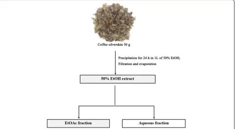
Preparation of the EtOH extract and its EtOAc fraction from coffee silverskin
The coffee silverskin used in this study was obtained by
roasting coffee beans (Coffea arabica) at the Roasting
Barn Co. (Seoul, Korea). The process of preparing the 50% EtOH extract and its EtOAc fraction from coffee
silverskin was as follows (Fig. 1). Dried coffee silverskin
powder (50 g) was extracted with 1 L of 50% EtOH for 24 h at room temperature. The extracts were then fil-tered using a Buchner funnel and concentrated with a rotary evaporator to obtain a 50% EtOH extraction pow-der. The 50% EtOH extract was fractionated with EtOAc and concentrated with a rotary evaporator. Then, the
dried samples were stored at −80 °C in the refrigerator
(Cosmetic R&D Center, Seoul National University of Sci-ence and Technology, Seoul, Korea) until use.
Determination of total phenolic content
The total phenolic contents of the 50% EtOH extract of coffee silverskin and its EtOAc fraction were determined by slightly modifying the method described by Alves et al
[13]. Briefly, 80μL of the samples was added to 20μL of
Folin-Ciocalteu reagent (50% v/v in H2O), mixed
thor-oughly, and incubated at room temperature for 5 min.
Then, 200μL of sodium carbonate solution (2% w/v in
H2O) was added, and the mixture was incubated at room temperature for 30 min. Absorbance was measured at 760 nm by using an ELISA reader. The total phenolic contents were determined by preparing a standard curve of con-centration (0–50 mg/L) by using chlorogenic acid as a ref-erence material (y = 0.0078 x + 0.0226,R2= 0.9968).
Characterization of coffee silverskin extracts by using HPLC and LC/ESI-MS
A component analysis of the silverskin extract and deter-mination of caffeine content were performed using a Shimadzu LC-20A high-performance liquid chromatog-raphy (HPLC) system with a UVD 170 s DIONEX de-tector and Shim-pack VP-ODS C18 column (L: 250 mm,
LD: 4.6 mm, 5μm) (Shimadzu, Japan). The mobile phase
was solvent A (2% acetic acid in H2O) and B (0.5% acetic acid in 50% acetonitrile aqueous solution), and the
fol-lowing linear scheme was used: 0–5 min, 0–10% (v/v) of
B; 5–20 min, 10–100% (v/v) of B; 20–25 min, 100–100% (v/v) of B; 25–30 min, 100–10% (v/v) of B; and 30–35 min, 10–10% (v/v) of B. The flow rate was 1.0 mL/min, and the sample was confirmed at a wavelength of 254– 400 nm. Quantitation of caffeine content was performed
using a commercial standard (50–500 mg/L; R2= 0.9955)
Free radical-scavenging activity
A free radical with an unpaired electron is unstable and very reactive. The high reactivity of free radicals dam-ages not only the skin but also disturbs the body com-position. To evaluate the free radical-scavenging activity of the EtOH extract of coffee silverskin and its EtOAc fraction, the reducing power of the sample was mea-sured based on its electron-donating ability against DPPH, which is a relatively stable radical. An equal vol-ume of EtOH and the sample (0.4 mL) was added to 0.4 mL of 0.2 mM DPPH solution dissolved in methanol. The samples were then mixed and allowed to stand at room temperature for 10 min. Absorbance was measured
at 517 nm using a UV/Vis spectrophotometer [14].
(+)-α-Tocopherol was used as a positive control.
ROS-scavenging activity using luminol-dependent chemiluminescence in the Fe3+-EDTA/H2O2system
The Fe3+-EDTA/H2O2system was used to determine the
ROS-scavenging activity as described previously [14].
Various concentrations of the sample (50μL) were added
to 1.78 mL of distilled water and then 40μL of EDTA
(2.5 mM), 10μL of FeCl3·6H2O (5 mM), and 80μL of
luminol (35 mM) were added. Then the tube was placed in the cell holder of the chemiluminescent device and
incubated for 5 min. After incubation, 40μL of H2O2
(150 mM) was added to the mixture, and chemilumines-cence was measured for 25 min. In the control group, the solvent was added instead of the sample. The blank
was the same as that used in the experiment but distilled
water was added instead of H2O2and FeCl3·6H2O. The
ROS-scavenging activity (active oxygen-scavenging activ-ity [OSC50]) was defined as the concentration of the sample required for 50% scavenging of ROS:
ROS Scavengingð Þ ¼% cpmcontrol−cpmexperiment cpmcontrol−cpmblank l00
Measurement of intracellular ROS generation
Intracellular ROS generation was assessed using the fluorescence dye 2′,7′-dichlorodihydrofluorescein diace-tate (H2DCF-DA, Sigma-Aldrich, USA) as our previous reports [3].
Determination of the cytoprotective effect
HaCaT cells (CLS Cell Line Service, Germany) were cul-tured up to 70–80% confluence in a cell culture medium
and then irradiated with 400 mJ/cm2UVB (280–320 nm)
in Dulbecco’s phosphate-buffered saline (DPBS) contain-ing various doses of the samples by uscontain-ing a CL-1000 Ultraviolet Cross linker (UVP Co., USA) device. Our
preliminary study showed that 400 mJ/cm2 of UVB
ir-radiation reduced cell viability by about 70% compared with the unirradiated control (data not shown). After washing twice with DPBS, the cells were cultured for 24 h in serum-free DMEM culture conditions. The cytoprotective effect of the 50% EtOH extract of coffee
Fig. 1Fractionation scheme for the 50% EtOH extract of coffee silverskin and its EtOAc fraction
silverskin and its EtOAc fraction against UVB-induced cell damage was measured by confirming cell viability by using the 3-(4,5-dimethyl-2-thiazolyl)-2,5-diphenyl-tetrazolium bromide (MTT; Sigma-Aldrich) assay as previously described [15].
Quantitative analysis of MMP-1 expression
HaCaT cells were seeded into 60-mm plates and cultured
up to 70–80% confluence in a cell incubator maintained
at 37 °C. After treatment with different concentrations of
the samples, the cells were exposed to 80 mJ/cm2UVB,
which does not affect cell survival, and then cultured further for 48 h in fresh medium. Immunoreactivity for MMP-1 within the culture medium was measured using an ELISA kit (R&D Systems, USA) according to the manufacturer’s instruction.
Western blot analysis
Western blot analysis was performed as previously
de-scribed [3] with specific antibodies against caspase-3
(Signalway Antibody, USA), MMP-1 (Abnova, Taiwan), andβ-actin (Cell Signaling Technology, USA).
Measurement of melanin content
B16F1 melanoma cells, which obtained from Kyung Hee University Skin Biotechnology Center (Seoul, Korea), were seeded in 6-well plates and cultured in a cell
incu-bator until 70–80% confluence was achieved. Then the
cells were treated with α-melanocyte-stimulating
hor-mone (α-MSH) and various concentrations of the
sam-ples in a fresh medium and further incubated for 72 h. After washing with DPBS, the cells were collected and
100μL of 1 N sodium hydroxide solution containing
10% DMSO was added to the pellet. Absorbance was measured at 410 nm using the ELISA microplate reader.
Statistical analysis
The results were repeated at least three times and expressed as the mean ± standard deviation (S.D.) values. Statistical significance was determined by one-way ANOVA with SPSS 17.0 (SPSS Inc. Chicago, IL). Differ-ences were considered statistically significant atp <0.05.
Results
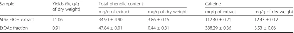
Yield and total phenolic compound content
As shown in Table1, the yields of the 50% EtOH extract
of coffee silverskin and its EtOAc fraction were 11.06%
(w/w) and 0.91% (w/w), respectively, and were
deter-mined based on dried coffee silverskin (50 g). Phenolic compounds are natural antioxidants and known to be very important factors in determining the antioxidant
activity of natural products [16]. In this study, the
phen-olic content was expressed as the amount contained in 1 g of the 50% EtOH extract or its EtOAc fraction or the amount contained in 1 g of dried coffee silverskin weight. The contents of phenolic compounds in the 50% EtOH extract of coffee silverskin and its EtOAc fraction were converted to the amount of chlorogenic acid, and the results show that the content in the EtOAc fraction (47.84 mg/g of coffee silverskin extract, 0.44 mg/g of cof-fee silverskin) was higher than that in the 50% EtOH ex-tract (34.90 mg/g of coffee silverskin exex-tract and 3.86 mg/g of coffee silverskin) (Table1).
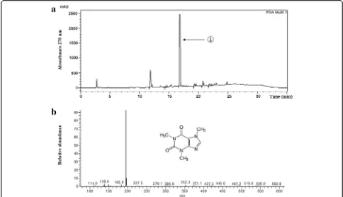
HPLC and LC/ESI-MS analyses and determination of the caffeine content in coffee silverskin
HPLC and LC/ESI-MS were used to analyze the compo-nents of coffee silverskin. The HPLC analysis of the 50% EtOH extract of coffee silverskin and its EtOAc fraction showed that the retention time of the major components was 16.8 min and the retention times were consistent with
those of caffeine standards (Fig.2a). For a more accurate
analysis, the [M-H]+ ion was detected at m/z 195 in the
positive ion mode of LC/ESI-MS. Thus, the major compo-nent of the silverskin extract was identified as caffeine (Fig.
2b). Determination of the caffeine content using a
stand-ard calibration curve revealed the caffeine content in the 50% EtOH extract of coffee silverskin to be 112.40 mg/g
and that in its EtOAc fraction to be 388.29 mg/g (Table1).
Free radical-scavenging activity
The free radical-scavenging activity of the coffee silverskin extracts was determined by DPPH assay, which is widely used for measuring antioxidant activity. In solution, the DPPH radical has a deep violet color, and it became pale yellow upon reaction with an antioxidant because of its
an-tioxidative capacity [17]. Results showed that free radical
scavenging activity (FSC50) values for the 50% EtOH extract
and EtOAc fraction were 105.43μg/mL and 65.27μg/mL,
Table 1Yields and Contents of Bioactive Compounds in the Coffee Silverskin 50% EtOH Extract and Its Fraction
Sample Yields (%, g/g
of dry weight)
Total phenolic content Caffeine
mg/g of extract mg/g of dry weight mg/g of extract mg/g of dry weight
50% EtOH extract 11.06 34.90 ± 4.90 3.86 ± 0.15 112.40 ± 0.21 12.43 ± 0.12
EtOAc fraction 0.91 47.84 ± 0.01 0.44 ± 0.31 388.29 ± 0.36 3.53 ± 0.06
respectively. However, caffeine, a major component of the coffee silverskin extract, did not show any free
radical-scavenging activity and the FSC50value of (+)-α-tocopherol
was 8.98μg/mL. These experiments indicated that the free
radical-scavenging activity of the EtOAc fraction was higher
than that of the 50% EtOH extract (Table2). Although the
FSC50 values were lower than that of (+)-α-tocopherol,
which is known as a strong lipid-soluble antioxidant in the cell membrane, the radical-scavenging activity of both the 50% EtOH extract and EtOAc fraction was still significant.
ROS-scavenging activity
To study the total antioxidant activity of ROS scavengers,
the Fe3+-EDTA/H2O2 system was used. In this system,
most types of ROS are generated by Fenton reaction and
subsequent chain reaction, including O2•−, H2O2, and •
OH, except for1O2. The resulting ROS are able to oxidize
the luminol to form the aminophthalate in the excited
state and emit luminescence (420–450 nm) [18].
There-fore, the luminol chemiluminescence assay can measure not only the reducing power of the samples but also the chelating activity of the inhibition of ROS generation. In this study, the total antioxidant capacity of the 50% EtOH extract of coffee silverskin and its EtOAc fraction, the water-soluble antioxidant L-ascorbic acid, and caffeine, which is the main component of the coffee silverskin ex-tract, was determined, and the results are shown in terms of OSC50, which is the concentration at which active
oxy-gen is reduced by 50%. It was found that the OSC50of the
coffee silverskin 50% EtOH extract was 31.89μg/mL and
that of the EtOAc fraction increased by about 50% to
15.25μg/mL. However, both the 50% EtOH extract and
Fig. 2HPLC chromatogram of the EtOAc fraction of the coffee silverskin EtOH extract at 270 nm.①Caffeine (a). Mass spectra of①in positive mode (LC-MS) (b). Insert shows caffeine structure
Table 2Antioxidant Activity of the 50% EtOH Extract of Coffee Silverskin and Its EtOAc Fraction and Reference
Free radical-scavenging activity (FSC50,μg/mL) ROS-scavenging activity (OSC50,μg/mL)
50% EtOH extract 105.43 ± 3.58 31.89 ± 31.94
EtOAc fraction 65.27 ± 3.32 15.25 ± 0.55
Caffeine > 500 > 500
(+)-α-Tocopherol 8.98 ± 2.93 nd
L-Ascorbic acid nd 1.50 ± 0.85
Data are presented as mean ± S.D. values ndnot detected
the EtOAc fraction showed lower activity than that shown
by L-ascorbic acid (1.50μg/mL), which corresponded to
the control group. Unsurprisingly, total antioxidant cap-acity was not observed for caffeine, the main component of coffee silverskin (Table2).
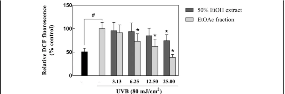
Inhibitory effect of coffee silverskin extract on intracellular ROS generation
To examine the effect of samples on intracellular ROS generation, HaCaT cells were treated with H2DCF-DA to measure the DFC content following UVB irradiation. The results showed that UVB irradiation significantly in-creased ROS production compared to that in the UVB-untreated control. In contrast, pretreatment with the EtOAc fraction of coffee silverskin drastically re-pressed the intracellular ROS level to 39% at
concentra-tions of 25μg/mL in comparison with the UVB
irradiated cells. Although a decrease trend also observed in the 50% EtOH extract treated group, it had a lesser intracellular ROS inhibitory effect than that of the EtOAc fraction (Fig.3).
Cytoprotective effect of the coffee silverskin extract
The concentration ranges of the samples used for these experiments were determined by confirming the cyto-toxicity of the coffee silverskin extract and fractions, and caffeine in human keratinocytes by the MTT method. When 70–80% confluence was reached, the cells were treated with DPBS containing various doses of the
sam-ples during the 400 mJ/cm2 UVB irradiation. Treatment
with the 50% EtOH extract and caffeine did not cause
cytotoxicity at concentrations up to 400μg/mL, and the
EtOAc fraction had no significant cytotoxic effect at
concentrations up to 200μg/mL (Fig. 4a). Therefore,
when measuring the cytoprotective effect of the samples, the cytoprotective concentration range of the samples
was determined to be 25–200μg/mL in the present
experiment.
It is known that the sunburn-inducing ability of UVB is 1000-fold higher than that of UVA. The generated ROS were able to form abnormal covalent bonds such as those in pyrimidine dimer in skin cells and induce cell death by activation of the apoptotic pathway, with caspase-3 acting
as an important regulator [19]. In this study, we analyzed
whether the 50% EtOH extract and its EtOAc fraction possessed antioxidant activity against oxidative damage by
UVB irradiation. As shown in Fig. 4, the experimental
group irradiated with 400 mJ/cm2UVB showed a survival
rate of 69.16% compared to that of the UVB-untreated group, and the cell viability did not increase significantly
at concentrations of 25–200μg/mL in the 50% EtOH
ex-tract- and caffeine-treated groups. In contrast, significant increases in cell viability of 78.94 and 84.75% were ob-served compared to that in the irradiated controls when
the cells were irradiated with 400 mJ/cm2 UVB in the
presence of the EtOAc fraction of coffee silverskin at
con-centrations of 100μg/mL and 200μg/mL, respectively
(Fig.4b and c). We also observed that the expression level
of caspase-3 decreased and a cleaved activated form has increased dramatically in HaCaT cells after exposure to
400 mJ/cm2UVB, and the EtOAc fraction of coffee
silver-skin partially counteracted this effect (Fig.4d). These data indicated that the EtOAc fraction of coffee silverskin showed a protective effect against UVB-induced cell dam-age via upregulation of caspase-3 expression in HaCaT cells.
Inhibitory effect of coffee silverskin extract on UVB-induced MMP-1 expression
To determine the concentration range of the samples to be used in this experiment, the effects of the 50% EtOH extract, EtOAc fraction, and caffeine on the viability of HaCaT keratinocytes were evaluated by the MTT assay.
When 70–80% confluence was reached, the cells were pretreated with various doses of the samples for 24 h and then cell viability was measured. Treatment with
caffeine (6.25–400μg/mL), and the 50% EtOH extract of
coffee silverskin (6.25–50μg/mL) and its EtOAc fraction
(6.25–25.00μg/mL) for 24 h had no cytotoxic effect on
HaCaT keratinocytes (Fig. 5a). Therefore, when
measuring the inhibitory effects of the samples on UVB-induced MMP-1 expression in HaCaT keratino-cytes, the concentration range for the samples was
determined to be 3.13–25.00μg/mL in the present
experiment.
ROS, which are generated excessively upon UV expos-ure, induce the expression of MMP-1, an enzyme that degrades the components of the extracellular matrix of the skin, and accelerate the aging process. The MMP-1 protein, which has a major role in the formation of wrin-kles, is generated primarily in the skin cells, especially
keratinocytes and fibroblasts [20, 21]. Therefore, in the
present study, we investigated whether the 50% EtOH extract of coffee silverskin and its EtOAc fraction have antiaging effects against UVB-induced MMP-1
produc-tion in HaCaT keratinocytes. As shown in Fig. 5, in the
experimental group irradiated with UVB, the expression level of MMP-1 increased by 310.90% compared to that in the UVB-untreated control. Treatment with the 50% EtOH extract and caffeine did not reduce the expression
of MMP-1 significantly. However, a decreasing trend in MMP-1 release was observed as the concentration of the EtOAc fraction of coffee silverskin increased, and a sig-nificant decrease of 74.27% was observed compared to that in the UVB-treated control when the cells were
pre-treated with 25μg/mL EtOAc fraction (Fig.5b).
Further-more, western blot also presented a similar result that treatment of EtOAc fraction of coffee silverskin to HaCaT cells noticeably decreased the expression of
MMP-1 (Fig. 5c). Collectively, these results showed that
the EtOAc fraction of coffee silverskin had an inhibitory effect on UVB-induced MMP-1 expression.
Inhibitory effect of coffee silverskin extract on melanin synthesis
To confirm the cytotoxicity of the samples in B16F1 mel-anoma cells, the MTT assay was used. The 50% EtOH ex-tract and its EtOAc fraction did not show any cytotoxicity
in the concentration range of 1.56 to 12.5μg/mL, rather
they increased cells viability significantly at the
concentra-tion of 12.5μg/mL. Further, at the concentration of 25μg/
mL, cell viability markedly decreased compared to that in
the untreated control (Fig. 6a). Therefore, the maximum
concentration was set to 12.5μg/mL when measuring the
melanogenesis-inhibiting activity of the 50% EtOH extract of coffee silverskin and its EtOAc fraction.
Fig. 4The effects of the 50% EtOH extract and EtOAc fraction of coffee silverskin in UVB-irradiated HaCaT cells. HaCaT cells were treated with different concentration of the samples in the absence (a) or presence (bandc) of 400 mJ/cm2UVB. The cytotoxicity was then determined by the MTT assay 24 h after UVB irradiation. The effect of coffee silverskin EtOAc fraction on caspase-3 expression was detected by western blot analysis (d). Data are presented as mean ± S.D. values. *p< 0.05 compared with the UVB-irradiated control,#p< 0.05 compared with the UVB-non-irradiated control.β-actin was used as an internal control
Fig. 5The effect of the 50% EtOH extract and EtOAc fraction of coffee silverskin on UVB-induced MMP-1 expression. Cell viability determined using the MTT assay (a). The levels of UVB-induced MMP-1 expression in HaCaT cells were measured by ELISA (b) and western blot analysis (c). Data are presented as the mean ± S.D. values. *p< 0.05 compared with the UVB-irradiated control,#p< 0.05 compared with the unirradiated control.β-actin was used as an internal control
α-MSH, which is induced by ultraviolet irradiation, is able to stimulate human melanocytes to accumulate ex-cessive melanin; it also leads to the formation of dark spots and freckles, which affects people’s quality of life
[22]. Therefore, we investigated the ability of the coffee
silverskin extract to modulate melanogenesis in
α-MSH-stimulated B16F1 melanoma cell model.
Fig-ure 5b shows that in the cells treated with α-MSH, the
production of melanin increased about 1.4 times
com-pared to that in the α-MSH-untreated control. In
con-trast, melanin synthesis decreased in groups treated with both the 50% EtOH extract of coffee silverskin and its EtOAc fraction. Combined with the above results of cell viability, both the 50% EtOH extract of coffee silverskin and its EtOAc fraction did not affect the B16F1 cell
via-bility in the concentration range of 1.56 to 12.5μg/mL,
demonstrating that the suppression of melanin synthesis on this cells are not due to the cytotoxic effect of sam-ples. In particular, the EtOAc fraction had a greater mel-anin biosynthesis inhibitory effect than that of the 50% EtOH extract. However, no statistically significant de-creasing trend was observed for melanin contents in the B16F1 melanoma cells pretreated with different caffeine concentrations, indicating that the melanogenesis inhibi-tory effect of the coffee silverskin extract does not
de-pend on caffeine (Fig.6b and c).
Discussion
UV radiation from the sun is classified as UVA (320– 400 nm), UVB (280–320 nm), and UVC (200–280 nm) according to its wavelength. UVA (95–98% of the total UV radiation) and UVB (2–5% of the total UV radiation) reach the surface of the earth, thereby affecting the hu-man body, unlike UVC, which is blocked by the ozone
layer [23]. The biological impact of UVB exposure on
the skin is much greater than that of UVA, at the same dose. UVB not only directly damages the structure of human skin cells but also leads to ROS generation, thereby affecting other biomolecules such as lipids and proteins, and the photodamage caused as a result is an important contributor to the degradation of extracellular matrix components and accumulation of excessive
mel-anin [24]. However, these events can be reduced by
treatment with antioxidants, such as phenolic
com-pounds [25, 26]. Therefore, in this study, a 50% EtOH
extract and EtOAc fraction of coffee silverskin, which is treated as waste in everyday life and causes environmen-tal pollution, were prepared and analyzed for their
antioxidant, cytoprotective, MMP-1-inhibiting, and
melanogenesis-inhibiting effects to explore the possibil-ity of using coffee silverskin as a multifunctional cosmetic material.
The antioxidative activities of the 50% EtOH extract of coffee silverskin and its EtOAc fraction were evaluated
by determining the FSC50, total antioxidant ability
(OSC50), and intracellular ROS levels. The FSC50 of the
50% EtOH extract and EtOAc fraction was 105.43 and
65.27μg/mL, respectively, and DPPH assay revealed that
the EtOAc fraction had higher radical-scavenging activity
than that shown by the 50% EtOH extract. In the Fe3
+
-EDTA/H2O2system using luminol, the total antioxidant
activity of the silverskin EtOAc fraction (15.25μg/mL)
was also higher than that of the 50% EtOH extract
(31.89μg/mL). Although the EtOAc fraction had lower
antioxidative activity than that of (+)-α-tocopherol, which is a strong lipid-soluble antioxidant, and L-ascorbic acid, a water-soluble antioxidant, it still showed a significant level of antioxidant activity. In addition, measurement of intra-cellular ROS levels also found that the silverskin EtOAc fraction was also higher than that of the 50% EtOH ex-tract. The antioxidative effect of caffeine was more than 500μg/mL in terms of both FSC50and OSC50. Therefore, the antioxidative effect of the coffee silverskin EtOAc frac-tion is not attributable to caffeine, which was the major component of the coffee silverskin extract. In addition, the total phenolic content of the EtOAc fraction was 47.84 ± 0.01 mg/g, as determined by a modified Folin-Ciocalteu assay. Therefore, the antioxidant capacity of the EtOAc fraction is not determined by caffeine, but by some com-pounds that exhibit a high antioxidant capacity even at low concentrations or the total antioxidant capacity of various phenolic compounds.
UVB radiation has been reported to cause the forma-tion of abnormal covalent bonds such as those in pyr-imidine dimers in skin cells and induce DNA damage, lipid peroxidation, and skin aging. Our results using the UVB-induced human keratinocyte injury model indi-cated that the 50% EtOH extract of coffee silverskin and caffeine did not increase the UVB-induced cell death; however, the cell viability significantly increased in the EtOAc fraction-treated group. The absorption spectra of the EtOAc fraction were determined to confirm the pos-sible causes that may account for the cytoprotective ef-fect of the coffee silverskin EtOAc fraction on human keratinocytes exposed to UVB (data not shown). As a re-sult, the maximum absorption wavelength of the EtOAc fraction was approximately 275 nm within the UVB spectra, which was consistent with the findings obtained for pure caffeine. However, treatment with pure caffeine at the same concentration as that used for treatment with the EtOAc fraction did not show a cytoprotective effect. The antioxidant test results showed that the cof-fee silverskin EtOAc fraction had a considerable antioxi-dant activity. Thus, the absorption effect of the EtOAc fraction at the wavelength tested might not contribute to its cytoprotective capacity, and the fraction likely has an indirect protective effect by eliminating UVB-induced ROS production. Moreover, our results showed that the
EtOAc fraction of coffee silverskin partially decrease the activation levels of caspase-3 in UVB-irradiated HaCaT cells. All of these findings demonstrate that the EtOAc fraction of coffee silverskin against UVB-induced cell damage by decrease of reactive oxygen species and caspase-3 activation.
ROS, which produced upon UVB exposure, can increase MMP-1 expression in keratinocytes by activating the mitogen-activated protein kinase pathway and stimulating the activator prote1 (AP-1) transcription factor. An in-crease in MMP-1 expression has been shown to cause degradation of collagen I, II, III, VII, and X type fibers,
and affect skin aging [27]. Additionally, ROS can induce
α-MSH and promote melanogenesis in melanocytes. Mel-anin is a major determinant of skin and hair color, and it is synthesized by melanocytes, which are present in the epidermal basal layer. The presence of melanin in an ap-propriate amount inhibits UV radiation-induced damage to skin cells, but excessive melanin production causes pig-mentation such as freckles and facial spots [28,29]. In the present study, it was confirmed that the EtOAc fraction of the 50% EtOH extract inhibits both UVB-induced MMP-1
expression and α-MSH-induced melanin production.
However, caffeine, a major component of the EtOAc frac-tion, showed no inhibitory effect on MMP-1 expression and melanin production. Additionally, recent studies have shown that the main phenolic compounds in the coffee
silverskin extract are caffeoylquinic acid lactone,
3-coumaroylquinic acid, 5-coumaroylquinic acid,
3-caffeoylquinic acid, 5-3-caffeoylquinic acid, 3,5-dicaffeoyl-quinic acid, and feruloyl3,5-dicaffeoyl-quinic acid [9,30]. Hence, the in-hibitory effect of the coffee silverskin EtOAc fraction on MMP-1 expression and melanin production is not attrib-utable to caffeine, but perhaps to the total activity of vari-ous phenolic compounds.
Conclusions
In conclusion, the present study demonstrated that coffee silverskin has the potential for application as a natural functional material in multifunctional cosmetics, because of its antioxidant activity, cytoprotective capacity, inhib-ition of MMP-1 expression, and anti-melanogenesis activity.
Acknowledgements Not applicable.
Funding
This research received no specific grant from any funding agency in the public, commercial, or not-for-profit sectors.
Availability of data and materials
All data and materials generated during this work are included in this article.
Authors’contributions
SHX and SNP designed the manuscript. SHX, KSL, HJJ, YMP, and JHH performed experiments. SHX analyzed data and wrote the manuscript. The author read and approved the final manuscript.
Ethics approval and consent to participate Not applicable.
Consent for publication The author consents for publication.
Competing interests
The authors declare that they have no competing interests.
Publisher’s Note
Springer Nature remains neutral with regard to jurisdictional claims in published maps and institutional affiliations.
Received: 9 October 2018 Accepted: 18 December 2018
References
1. Wlaschek M, Tantcheva-Poor I, Naderi L, Ma WJ, Schneider A, Razi-Wolf Z, Schuller J, Scharffetter-Kochanek K. Solar UV irradiation and dermal photoaging. J Photoch Photobio B. 2001;63:41–51.
2. Kammeyer A, Luiten RM. Oxidation events and skin aging. Ageing Res Rev. 2015;21:16–29.
3. Xuan SH, Park YM, Ha JH, Jeong YJ, Park SN. The effect of
dehydroglyasperin C on UVB-mediated MMPs expression in human HaCaT cells. Pharmacol Rep. 2017;69:1224–31.
4. Park SN. Skin aging and antioxidant. J Soc Cosmet Scientists Korea. 1997;23: 75–132.
5. Wohlrab J, Hilpert K, Wolff L. Epidermal aging and anti-aging strategies. Hautarzt. 2016;67:107–11.
6. Park SN, Kim SY, Lim GN, Jo NR, Lee MH. In vitro skin permeation and cellular protective effects of flavonoids isolated from Suaeda asparagoides extracts. J Ind Eng Chem. 2012;18:680–3.
7. Martini D, Del Bo C, Tassotti M, Riso P, Del Rio D, Brighenti F, Porrini M. Coffee consumption and oxidative stress: a review of human intervention studies. Molecules. 2016;21:1–20.
8. International Coffee Orcanization,http://www.ico.org/, (Accessed 10 Apr 2018).
9. Bresciani L, Calani L, Bruni R, Brighenti F, Del Rio D. Phenolic composition, caffeine content and antioxidant capacity of coffee silverskin. Food Res Int. 2014;61:196–201.
10. Rodrigues F, Palmeira-de-Oliveira A, das Neves J, Sarmento B, Amaral MH, Oliveira MB. Coffee silverskin: a possible valuable cosmetic ingredient. Pharm Biol. 2015;53:386–94.
11. Borrelli RC, Esposito F, Napolitano A, Ritieni A, Fogliano V. Characterization of a new potential functional ingredient: coffee silverskin. J Agric Food Chem. 2004;52:1338–43.
12. Furusawa M, Narita Y, Iwai K, Fukunaga T, Nakagiri O. Inhibitory effect of a hot water extract of coffee“silverskin”on hyaluronidase. Biosci Biotechnol Biochem. 2011;75:1205–7.
13. Alves RC, Costa AS, Jerez M, Casal S, Sineiro J, Nunez MJ, Oliveira B. Antiradical activity, phenolics profile, and hydroxymethylfurfural in espresso coffee: influence of technological factors. J Agric Food Chem. 2010;58: 12221–9.
14. Seong JS, Xuan SH, Park SH, Lee KS, Park YM, Park SN. Antioxidative and antiaging activities and component analysis ofLespedeza cuneataG. Don extracts fermented withLactobacillus pentosus. J Microbiol Biotechnol. 2017; 27:1961–70.
15. Silvan JM, Reguero M, de Pascual-Teresa S. A protective effect of anthocyanins and xanthophylls on UVB-induced damage in retinal pigment epithelial cells. Food Funct. 2016;7:1067–76.
16. Rice-Evans CA, Miller NJ, Paganga G. Structure-antioxidant activity relationships of flavonoids and phenolic acids. Free Radic Biol Med. 1996;20: 933–56.
enzymatic extracts from leaves ofPerilla frutescensvar. japonica. Food Sci Biotechnol. 2008;17:279–86.
18. Hwang JP, Ha JH, No GY, Jeong YJ, Park SN. Cellular protective effect of novel dimeric ferulamide derivatives against UVA and1O2and its structural mechanism. J Ind Eng Chem. 2017;53:164–70.
19. Schuch AP, Moreno NC, Schuch NJ, Menck CFM, Garcia CCM. Sunlight damage to cellular DNA: focus on oxidatively generated lesions. Free Radic Biol Med. 2017;107:110–24.
20. Fisher GJ, Wang ZQ, Datta SC, Varani J, Kang S, Voorhees JJ.
Pathophysiology of premature skin aging induced by ultraviolet light. N Engl J Med. 1997;337:1419–28.
21. Bosch R, Philips N, Suarez-Perez JA, Juarranz A, Devmurari A, Chalensouk-Khaosaat J, Gonzalez S. Mechanisms of photoaging and cutaneous photocarcinogenesis, and photoprotective strategies with phytochemicals. Antioxidants (Basel). 2015;4:248–68.
22. Chao WW, Su CC, Peng HY, Chou ST. Melaleuca quinquenervia essential oil inhibits alpha-melanocyte-stimulating hormone-induced melanin production and oxidative stress in B16 melanoma cells. Phytomedicine. 2017;34:191–201.
23. Lephart ED. Skin aging and oxidative stress: Equol's anti-aging effects via biochemical and molecular mechanisms. Ageing Res Rev. 2016;31:36–54. 24. Rittie L, Fisher GJ. Natural and sun-induced aging of human skin. Cold
Spring Harb Perspect Med. 2015;5:a015370.https://doi.org/10.1101/ cshperspect.a015370.
25. Meng TX, Irino N, Kondo R. Melanin biosynthesis inhibitory activity of a compound isolated from young green barley (Hordeum vulgareL.) in B16 melanoma cells. J Nat Med. 2015;69:427–31.
26. Perez-Sanchez A, Barrajon-Catalan E, Herranz-Lopez M, Castillo J, Micol V. Lemon balm extract (Melissa officinalis, L.) promotes melanogenesis and prevents UVB-induced oxidative stress and DNA damage in a skin cell model. J Dermatol Sci. 2016;84:169–77.
27. Hwang YP, Oh KN, Yun HJ, Jeong HG. The flavonoids apigenin and luteolin suppress ultraviolet A-induced matrix metalloproteinase-1 expression via MAPKs and AP-1-dependent signaling in HaCaT cells. J Dermatol Sci. 2011; 61:23–31.
28. Brash DE. UV-induced melanin Chemiexcitation: a new mode of melanoma pathogenesis. Toxicol Pathol. 2016;44:552–4.
29. Costin GE, Hearing VJ. Human skin pigmentation: melanocytes modulate skin color in response to stress. FASEB J. 2007;21:976–94.
30. Regazzoni L, Saligari F, Marinello C, Rossoni G, Aldini G, Carini M, Orioli M. Coffee silver skin as a source of polyphenols: high resolution mass spectrometric profiling of components and antioxidant activity. J Funct Foods. 2016;20:472–85.