RESEARCH
Evolving trends in infective endocarditis
in a developing country: a consequence
of medical progress?
Mohamed Sunil
1, Huynh Quoc Hieu
2, Ramesh Singh Arjan Singh
1, Sasheela Ponnampalavanar
1,
Kelvin S. W. Siew
1and Alexander Loch
1*Abstract
Background: Staphylococcus has replaced streptococcus as the most common cause of infective endocarditis (IE) in developed health care systems. The trend in developing countries is less clear.
Aim: To examine the epidemiological trends of infective endocarditis in a developing nation.
Methods: Single-centre, retrospective study of patients admitted with IE to a tertiary hospital in Malaysia over a 12-year period.
Results: The analysis included 182 patients (n = 153 Duke’s definite IE, n = 29 possible IE). The mean age was 51 years. Rheumatic heart disease was present in 42%, while 7.6% were immunocompromised. IE affected native valves in 171 (94%) cases. Health-care associated IE (HCAIE) was recorded in 68 (37.4%). IE admission rates increased from 25/100,000 admissions (2012) to 59/100,000 admissions (2017). At least one major complication on admission was detected in 59 (32.4%) patients. Left-sided IE was more common than right-sided IE [n = 159 (87.4%) vs. n = 18 (9.9%)]. Pathogens identified by blood culture were staphylococcus group [n = 58 (40.8%)], streptococcus group [n = 51 (35.9%)] and Enterococcus species [n = 13 (9.2%)]. staphylococcus infection was highest in the HCAIE group. In-hospital death occurred in 65 (35.7%) patients. In-In-hospital surgery was performed for 36 (19.8%) patients. At least one complication was documented in 163 (85.7%).
Conclusion: Staphylococcus is the new etiologic champion, reflecting the transition of the healthcare system. Strep-tococcus is still an important culprit organism. The incidence rate of IE appears to be increasing. The rate of patients with underlying rheumatic heart disease is still high.
Keywords: Infective endocarditis, Staphylococcus aureus, Healthcare associated infection, Mortality predictors, Malaysia
© The Author(s) 2019. This article is distributed under the terms of the Creative Commons Attribution 4.0 International License (http:// creat iveco mmons .org/licen ses/by/4.0/), which permits unrestricted use, distribution, and reproduction in any medium, provided you give appropriate credit to the original author(s) and the source, provide a link to the Creative Commons license, and indicate if changes were made. The Creative Commons Public Domain Dedication waiver (http://creativecommons.org/publicdomain/zero/1.0/) applies to the data made available in this article, unless otherwise stated.
Introduction
Infective endocarditis (IE) is a universal disease with risk factors, predispositions and outcomes considered similar across continents for decades. An epidemiological tran-sition has been reported over the past decade for devel-oped health care systems where viridans streptococcus
has been replaced by staphylococcus as the most com-mon etiological agent. Methicillin-susceptible Staphylo-coccus aureus (MSSA) is now the most common agent in native valve infective endocarditis (NVIE) and methicil-lin-resistant S. aureus (MRSA) in healthcare-associated infective endocarditis (HCAIE) [1]. This transition is thought to be a result of ageing populations, the abolition of rheumatic heart disease (RHD) as the traditional major risk factor for IE due to appropriate antibiotic treatment of rheumatic fever and advanced device management
Open Access
*Correspondence: [email protected]
1 Department of Medicine, University Malaya Medical Centre, 59100 Kuala Lumpur, Malaysia
particularly in cardiac patients [2, 3]. Moreover, reduc-tions in IE mortality rates that had been anticipated with improved medical care did not materialize due to the changing epidemiology and the coinciding higher rates of HCAIE [1, 4, 5]. It is not clear whether developing countries undergo an epidemiological transition of IE to the same extent. Available studies show notable differ-ences in developing countries with much higher rates of culture-negative IE, zoonotic infections and mortality, although with significant regional variations [6, 7]. There is a lack of data for developing countries overall and South-East Asia in particular. The aim of this study is to describe the clinical trends, characteristics and outcomes of IE patients in a Malaysian tertiary hospital.
Materials and methods
The study is a single-centre, retrospective study of patients admitted with IE to a tertiary hospital in Kuala Lumpur, Malaysia from 1st January 2005, until 31st December 2017. The hospital caters a population of 1.7 million with an annual adult admission rate of 44,000– 46,000/year. The study received approval from the insti-tutional ethics committee (No: 201782-5459).
Patient recruitment and inclusion criteria
Patients were identified from the echocardiography reporting system, the admission logbooks and the hos-pital electronic medical records (EMR). All patients fulfilling the modified Duke’s criteria [8] for definite or possible IE were included in the study. The first admis-sion during the study period was considered the index admission for patients with more than one episode of IE. Patients younger than 18 years were excluded.
Data collection
Data regarding patient demographics, comorbidi-ties and recent hospital admissions were documented. Healthcare-associated infective endocarditis (HCAI) was defined as IE in patients with health care contact before the index hospitalization. Health care contact consti-tuted a history of receiving intravenous therapy (includ-ing chemotherapy), transfer from a specialized nurs(includ-ing care facility, haemodialysis, or hospitalization for at least 2 days in the 90 days prior to the index admission.
Presenting features and examination findings were recorded. Documented laboratory variables included haemoglobin (Hb), white blood cell count (WBC), plate-let count, urine haemoglobin, serum creatinine, erythro-cytes sedimentation rate (ESR), C-reactive protein (CRP), serum albumin and troponin I. Results on admission (i.e. baseline) and results after 2 weeks of IE treatment were analysed for all those parameters. Blood results at the
time when the IE diagnosis was made were considered baseline results for those patients who developed IE dur-ing the admission.
Every patient had at least 4 blood samples taken for blood culture. Automated laboratory systems were uti-lised for pathogen identification and antimicrobial sus-ceptibility testing. Genus and species of organisms cultured and their respective sensitivities were recorded. The type of echocardiographic modality [transthoracic echocardiography (TTE) vs. trans-oesophageal echo-cardiography (TOE)] and its results were documented. All available echocardiographic cinematic loops were reviewed by two consultant cardiologists for the pres-ence of rheumatic heart disease (RHD) according to the 2012 WHF criteria for the echocardiographic diagnosis of RHD [9]. Type of antibiotics used for initial empirical treatment and subsequent definite treatment were docu-mented. All patients included in this study completed 4 to 6 weeks of intravenous antibiotics. For all patients undergoing surgery; timing, type of surgery and outcome were recorded.
Outcome measures and statistical analysis
The outcome was recorded as “death” or “discharged alive”. Results were analysed using SPSS for Windows version 24. Data are summarized as a percentage or mean ± standard deviation (SD). Categorical variables were compared using Chi-square or Fisher-Exact test as appropriate, while Student t-test was used for continuous variables. A p-value < 0.05 was considered significant.
Results
Recruitment
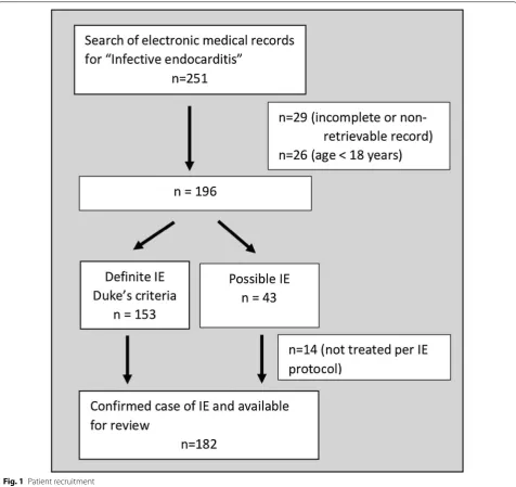
A search of the electronic medical records database and the admission logbooks yielded 251 patients with the label “IE”. The folders of 29 patients treated for IE between 2005 and 2007 were incomplete or could not be retrieved. Twenty-six patients were excluded as they were under 18 years of age. The details of the remaining 196 patients were analysed for the presence of the modified Dukes criteria for definite or possible IE [8]. The modi-fied Duke’s criteria for definite IE were fulfilled by 153 patients which were consequently included in the study. Fourteen out of 43 patients who fulfilled the criteria for possible IE did not receive antibiotic treatment as per IE protocol and were excluded from the study. A total of 182 patients remained for analysis as shown in Fig. 1.
Baseline demography
a history of previous infective endocarditis. Common co-morbidities included diabetes (29.7%), hypertension (34.6%) and ischaemic heart disease (11.5%). Underlying immunosuppression was present in 14 (7.6%) patients. Intravenous drug abuse was reported by 9 (4.9%) patients. Seventeen patients (9.3%) were on regular haemodialysis while 11 (6%) patients had central venous catheters.
Incidence
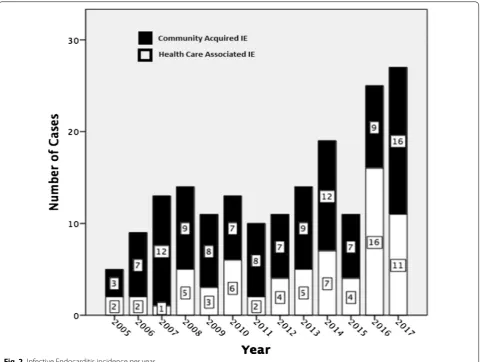
Native valves were affected by the IE in 171 (94%) cases. Only 11 (6%) patients had prosthetic valve IE. Health-care-associated IE (HCAIE) was recorded in 68 (37.4%) patients and community-acquired IE (CAIE) in 114
(62.6%) patients. The number of IE patients increased progressively over the 13-year study period with a par-ticular increase over the last 6 years Fig. 2. IE admission rates almost tripled from 25/100,000 admissions in 2012 to 59/100,000 admissions in 2017. The mean incidence of IE was 30.7/100,000 adult admissions.
Clinical presentation
Most patients 152 (83.5%) reported fever on admis-sion. The mean duration of fever was 20 (± 24) days. The mean time to establish the diagnosis of IE was 4 (± 5 days). At least one major complication at the time of admission was documented in 59 (32.4%) patients.
Complications comprised of heart failure in 38 (20.1%), systemic embolization in 21 (11.5%) and stroke in 10 (5.5%) patients. A murmur was present in 57 (31%) patients. Vascular and immunological evidence of IE was found in 48 (26.4%) and 13 (5.1%) patients respec-tively. Thirty-five (19.2%) patients had clinically spleno-megaly and 24 (13.2%) hepatospleno-megaly.
Blood parameters
Anaemia was diagnosed on admission for 152 (68.7%) patients. The mean haemoglobin was 10.6 (± 2.4) g/dl and the mean white blood cell count was 13.8 (± 7.1). Nine (4.9%) patients had leucopenia. The mean platelet count on admission was 232 (± 135). Thrombocytope-nia (< 150,000/ml) was present in 52 (28.6%) patients. Most patients 145 (79.7%) had hypoalbuminemia on admission (albumin < 35 g/dl). The mean erythrocyte sedimentation rate was 61 (± 33) and the mean CRP was 9.2 (± 6.7). Renal impairment was common with
a mean creatinine of 158 (± 183) micromol/l. Raised Troponin I was found in 80 (44%) patients with a mean level of 0.45 (± 1.6).
Echocardiographic features
All patients underwent transthoracic echocardiography (TTE) at least once, while transoesophageal echocardi-ography (TOE) was done for only 72 (39.6%) patients. TTE missed vegetations in 8 (4.4%) patients which were consequently identified by TOE. Left-sided IE was by far more common than right-sided IE [n = 159 (87.4%) vs. n = 18 (9.9%)]. Bilateral involvement was seen in 3 (1.6%) cases. Combinations of infected valves were seen in 11 (6.0%) cases. Concurrent IE affected most com-monly the aortic and mitral valves [n = 8 (4.4%)].
Fourteen (7.7%) patients had underlying congenital heart disease comprising of atrial septal defects (n = 2), ventricular septal defects (n = 9) and cyanotic heart lesions (n = 3). Another 14 (7.7%) patients had pros-thetic valve endocarditis. Echocardiographic cinematic
loops for review were available for 120 patients. 50 (42%) of those fulfilled the WHO criteria for the pres-ence of rheumatic heart disease (RHD).
Microbiology data
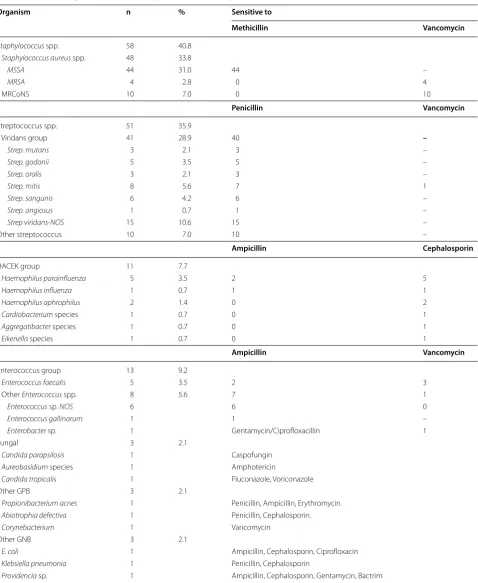
Positive blood cultures were available for 142 (78%) patients. The mean duration to achieve culture clearance was 6 (± 5) days. Pathogens identified by blood culture in descending frequency were staphylococcus group [n = 58 (40.8%)], streptococcus group [n = 51 (35.9%)], Entero-coccus species [n = 13 (9.2%)], HACEK [n = 11 (7.7%)],
Candida sp. [n = 3 (2.1%)] and other bacteria [n = 6 (4.2%)]. Staphylococcus aureus infection was most com-mon in the HCAIE group.
Among 58 Staphylococcus spp. isolated, 44 (75.8%) were methicillin-sensitive S. aureus, followed by 10 (17.2%) MRCoNS and 4 (6.9%) methicillin-resistant S. aureus. Almost all of the streptococcus species isolates were penicillin-sensitive [50 out of 51, 98%)]. A penicil-lin-resistant strain was found in only one patient ( Strep-tococcus mitis—sensitive to ceftriaxone/vancomycin). All HACEK group organisms (n = 11) were sensitive to ceftriaxone, but only 3 (27.3%) of them were sensitive to ampicillin. Nine out of 13 (69.2%) Enterococcus species were ampicillin-sensitive, while 4 (30.8%) were ampicil-lin-resistant (sensitive to vancomycin). Microbial agents and respective resistance patterns are shown in Table 1.
Antibiotic prescription
Empirical antibiotics were used while awaiting cul-ture result. The majority of patients (65%) were treated with two empirical agents, typically comprising of a combination of gentamycin and benzylpenicillin or cloxacillin. Definitive therapy consisted of 6 weeks of culture-directed antibiotics for culture positive cases. 72 (39.6%) patients were treated with a single antibiotic while 110 (60.4%) patients received a combination of two antibiotics.
Outcome and complications
The overall mortality rate was high. Sixty-five (35.7%) patients died in hospital, while 117 (64.3%) were dis-charged alive. The mean length of stay was 39.2 (± 21.4) days. Altogether, 182 patients had 7126 days of in-hospi-tal treatment.
Surgery was performed for 58 (31.9%) patients, while 124 (68.1%) underwent medical therapy only. In-hospital surgery was performed for 36 (19.8%) patients, while 22 (12.1%) patients underwent surgery at a later stage post discharge.
Most patients [n = 163 (85.7%)] had at least one com-plication: 142 (78%) had valvular dysfunction, 52 (28.6%)
patients developed heart failure, and 62 (34.1%) patients had systemic embolization. The embolic complications comprised of stroke [n = 35 (19.2%)], pulmonary infarc-tion or abscess [n = 25 (13.7%)], splenic abscess or infarc-tion [n = 6 (8.8%)]. Acute kidney injury occurred in 59 (32.4%) patients. Electrocardiographic abnormalities were documented in 30 (16.5%) patients and included atrial fibrillation [n = 17 (9.3%)], supraventricular tachy-cardia [n = 8 (4.4%)], ventricular tachycardia (n = 3) and first-degree AV block (n = 2).
Discussion
Evolving epidemiology
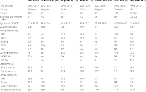
Literature from developed health care systems has been showing an epidemiological evolution of IE over the past decade Table 2. There is a lack of IE epidemiological data from developing countries, thus preventing conclusions regarding local IE trends.
Table 1 Microbial agents and resistance pattern of culture positive cases (n = 142)
MRCoNS methicillin-resistant coagulase-negative staphylococcus, NOS not otherwise specified, MSSA methicillin-sensitive Staphylococcus aureus, MRSA methicillin-resistant Staphylococcus aureus, NA not applicable
Organism n % Sensitive to
Methicillin Vancomycin
Staphylococcus spp. 58 40.8 Staphylococcus aureus spp. 48 33.8
MSSA 44 31.0 44 –
MRSA 4 2.8 0 4
MRCoNS 10 7.0 0 10
Penicillin Vancomycin
Streptococcus spp. 51 35.9
Viridans group 41 28.9 40 –
Strep. mutans 3 2.1 3 –
Strep. godonii 5 3.5 5 –
Strep. oralis 3 2.1 3 –
Strep. mitis 8 5.6 7 1
Strep. sangunis 6 4.2 6 –
Strep. angiosus 1 0.7 1 –
Strep viridans-NOS 15 10.6 15 –
Other streptococcus 10 7.0 10 –
Ampicillin Cephalosporin
HACEK group 11 7.7
Haemophilus parainfluenza 5 3.5 2 5
Haemophilus influenza 1 0.7 1 1
Haemophilus aphrophilus 2 1.4 0 2
Cardiobacterium species 1 0.7 0 1
Aggregatibacter species 1 0.7 0 1
Eikenella species 1 0.7 0 1
Ampicillin Vancomycin
Enterococcus group 13 9.2
Enterococcus faecalis 5 3.5 2 3
Other Enterococcus spp. 8 5.6 7 1
Enterococcus sp. NOS 6 6 0
Enterococcus gallinarum 1 1 –
Enterobacter sp. 1 Gentamycin/Ciprofloxacillin 1
Fungal 3 2.1
Candida parapsilosis 1 Caspofungin Aureobasidium species 1 Amphotericin
Candida tropicalis 1 Fluconazole, Voriconazole
Other GPB 3 2.1
Propionibacterium acnes 1 Penicillin, Ampicillin, Erythromycin. Abiotrophia defectiva 1 Penicillin, Cephalosporin. Corynebacterium 1 Vancomycin
Other GNB 3 2.1
E. coli 1 Ampicillin, Cephalosporin, Ciprofloxacin Klebsiella pneumonia 1 Penicillin, Cephalosporin
for health care associated infective endocarditis (HCAIE) [11]. MRSA has also overtaken coagulase-negative staphylococci as the main cause of PVIE in recent stud-ies [1, 12]. Higher staphylococcus IE incidence is typically reported in referral centres and has been shown to cor-relate with higher mortality [13, 14].
The prevalence of RHD in our IE patients (42%) is still much higher than in Western populations where degen-erative valvular pathology and valvular prostheses [1, 5] constitute the main underlying pathological substrates. The culture-negative IE rate (22%) in our centre is com-parable with those reported in developed health care sys-tems (about 10–20%). Relevant serological investigations for Coxiella burnetii, Legionella spp., Brucella spp., Bar-tonella spp., and Chlamydiae spp. should be performed where epidemiologically applicable [4, 11]. Much higher rates of culture-negative IE are commonly reported in regional studies: 30–40% in China and India; 65% in Thailand [6, 10, 15]. Often, only limited investigations can be carried out due to financial constraints when a more extensive workup would be required to accurately identify the fastidious organism in developing countries.
Echocardiography demonstrated vegetations in 99.5% of cases diagnosed as IE in our study. This high rate is most likely due to improved diagnostic imaging modali-ties [10]. However, only 39.6% of the patients had a TOE done. This contrasts with reports from developed health-care systems where the majority of patients undergo TOE to detect complications early. In our study, TTE missed vegetation in 8 (4.4%) patients emphasizing the diagnos-tic importance of TOE in suspected IE. The mitral valve was the most commonly affected valve.
IE mortality and surgery
The in-hospital mortality rate of 36.7% in our study pop-ulation was high. Recent studies conducted in developed health care systems reported persistently high mortality rates of 20–22%, 30% and 45% for the in-hospital period, at 1 year and at 5 years respectively [7, 12, 16, 17]. The root cause for the lack of improvement in IE mortal-ity in developed health care systems is believed to be the increasing numbers of older and immunocompro-mised patients with HCAIE with more resistant organ-ism [1, 4, 5, 11]. The same trend seems to apply to urban
Table 2 Major IE studies
SD standard deviation, IQR interquartile range, RHD rheumatic heart disease, CHD congenital heart disease, IVDU intravenous drug use, HCAIE health care associated IE, PVE prosthetic valve IE, CHF congestive heart failure, IE infective endocarditis, NA not available
a 90 days mortality in this study
This study Ibrahim et al. [19] Gupta et al. [15] Xu et al. [10] Poesen et al. [20] Watt et al. [6] Toyoda et al. [2]
Time of study 2005–2017 2012–2013 2005–2010 2008–2015 2003–2010 2010–2012 1998–2013
Country Malaysia Malaysia India China Belgium Thailand US
Episodes 182 50 61 174 88 132 75,829
Incidence (per 100,000
adm) 31 NA 80 NA 54 NA 7.6–9.3
Age (years) (SD/IQR) 51 ± 17.62 42 ± 16.4 49 ± 13.7 48 ± 15.7 72 IQR 59–81 47 IQR 16–85 62 ± 18.9
Male: female ratio 2.4:1 2.8:1 3.3:1 1.9:1 2:1 2.2:1 1.4:1
Predispositions (%)
RHD 42 NA 37.7 14.9 2.3 28.0 NA
CHD 7.7 6.0 22.9 29.9 5.7 8.3 4.5
Dialysis 9.3 6.0 NA 3.4 4.5 NA 18.4
IVDU 4.9 26.0 1.6 0.6 1.1 NA 12.5
HIV 2.7 4.0 NA NA NA NA 1.7
Culture positive (%) 78 68 67.2 60.3 89.8 45.5 73.0
HCAIE (%) 37.4 NA NA NA NA NA 52.9
PVE (%) 6 NA 31 5.2 8 9.9 12.9
Organisms (%)
Streptococcus 35.9 20 21.4 37.4 44.4 22 26.6
Staphylococcus 40.8 36 21.4 13.8 21.7 5.3 38.5
Complications (%)
CHF 28.6 NA 47.5 69.0 51.1 48 NA
Stroke 19.2 10.0 11.5 16.1 NA NA NA
Surgery for IE (%) 32 20.0 49.2 43.7 29.5 75.8 NA
tertiary centres of developing countries: increasing rates of HCAIE in a multimorbid, older population resulting in persistently high mortality rates despite optimal medical care.
Early surgery has been shown in RCTs to significantly reduce in-hospital death and embolic complications [16, 18]. In developed medical systems, about 40–50% of patients are managed surgically whereas the surgical intervention rate in our study was only 32% and might partially explain the rather high mortality. Mortality rates reported in other developing countries, such as China (11%) [10] and Thailand (11.4%) [6] are much lower than local figures and are associated with higher surgery rates (44% and 75% resp.).
Study limitations
As a retrospective study, this study has limitations associ-ated with retrospective data collection. 29 patients identi-fied as IE cases had to be excluded as the medical records could not be retrieved. The data may not be generalizable as the hospital is a tertiary referral centre with inherent selection and referral bias. There are strong urban/rural socio-economic disparities in developing nations with regards to standard of care and health care access. Pro-spective studies with larger sample sizes are needed to confirm our findings.
Conclusions
Staphylococcus is the new etiologic champion, likely reflecting the transition of the healthcare system to a more developed state entailing more invasive procedures and serving a generally older and more multi-morbid population with increasing HCAIE rates. Despite the epi-demiological transition to staphylococcus predominance, RHD remains as an important predisposing factor in our local IE population. Streptococcus viridans is still an important etiological agent of IE. The incidence rate of IE is increasing.
Acknowledgements Not applicable.
Authors’ contributions
MS and KSWS collected the data. HQH, AL and RSV reviewed echocardio-graphic findings and data. SP reviewed the manuscript. MS and AL prepared the manuscript and are responsible for the overall content as guarantor. All authors read and approved the final manuscript.
Funding Not applicable.
Availability of data and materials
The datasets used and/or analysed during the current study are available from the corresponding author on reasonable request.
Ethics approval and consent to participate
The study was approved by the ethics committee of University Malaya Medi-cal Centre (No: 201782-5459). The need for individual approval was waived.
Consent for publication Not applicable.
Competing interests
The authors declare that they have no competing interests.
Author details
1 Department of Medicine, University Malaya Medical Centre, 59100 Kuala Lumpur, Malaysia. 2 Tam Duc Cardiology Hospital, Ho Chi Minh City, Vietnam.
Received: 29 May 2019 Accepted: 2 December 2019
References
1. Fowler VG, Miro JM, Hoen B, Cabell CH, Abrutyn E, Rubinstein E, Corey GR, Spelman D, Bradley SF, Barsic B, Pappas PA. Staphylococ-cus aureus endocarditis: a consequence of medical progress. JAMA. 2005;293(24):3012–21.
2. Toyoda N, Chikwe J, Itagaki S, Gelijns AC, Adams DH, Egorova NN. Trends in infective endocarditis in California and New York State, 1998–2013. JAMA. 2017;317(16):1652–60.
3. Hoen B, Alla F, Selton-Suty C, Béguinot I, Bouvet A, Briançon S, Casalta JP, Danchin N, Delahaye F, Etienne J, Le Moing V. Changing profile of infective endocarditis: results of a 1-year survey in France. JAMA. 2002;288(1):75–81.
4. Cahill TJ, Prendergast BD. Infective endocarditis. Lancet. 2016;387(10021):882–93.
5. Hoen B. Epidemiology and antibiotic treatment of infective endocarditis: an update. Heart. 2006;92(11):1694–700.
6. Watt G, Pachirat O, Baggett HC, Maloney SA, Lulitanond V, Raoult D, Bhengsri S, Thamthitiwat S, Paupairoj A, Kosoy M, Ud-Ai N. Infective endocarditis in northeastern Thailand. Emerg Infect Dis. 2014;20(3):473. 7. Abdulhak AA, Baddour LM, Erwin PJ, Hoen B, Chu VH, Mensah GA, Tleyjeh
IM. Global and regional burden of infective endocarditis, 1990–2010: a systematic review of the literature. Glob Heart. 2014;9(1):131–43. 8. Li JS, Sexton DJ, Mick N, Nettles R, Fowler VG Jr, Ryan T, Bashore T, Corey
GR. Proposed modifications to the Duke criteria for the diagnosis of infec-tive endocarditis. Clin Infect Dis. 2000;30(4):633–8.
9. Reményi B, Wilson N, Steer A, Ferreira B, Kado J, Kumar K, Lawrenson J, Maguire G, Marijon E, Mirabel M, Mocumbi AO. World Heart Federation criteria for echocardiographic diagnosis of rheumatic heart disease—an evidence-based guideline. Nat Rev Cardiol. 2012;9(5):297.
10. Xu H, Cai S, Dai H. Characteristics of infective endocarditis in a tertiary hospital in East China. PLoS ONE. 2016;11(11):e0166764.
11. Martinez G, Valchanov K. Infective endocarditis. Continuing education in anaesthesia. Crit Care Pain. 2012;12:134–9.
12. Chambers J, Sandoe J, Ray S, Prendergast B, Taggart D, Westaby S, Arden C, Grothier L, Wilson J, Campbell B, Gohlke-Bärwolf C. The infective endo-carditis team: recommendations from an international working group. Heart. 2014;100(7):524–7.
13. Bergin SP, Holland TL, Fowler VG, Tong SY. Bacteremia, sepsis, and infective endocarditis associated with Staphylococcus aureus. Curr Top Microbiol Immunol. 2017;409:263–96.
14. Ben-Ami R, Giladi M, Carmeli Y, Orni-Wasserlauf R, Siegman-Igra Y. Hospi-tal-acquired infective endocarditis: should the definition be broadened? Clin Infect Dis. 2004;38(6):843–50.
15. Gupta A, Kaul U, Varma A. Infective endocarditis in an Indian setup: are we entering the ‘modern’era? Indian J Crit Care Med. 2013;17(3):140. 16. Kang DH, Kim YJ, Kim SH, Sun BJ, Kim DH, Yun SC, Song JM, Choo SJ,
Chung CH, Song JK, Lee JW. Early surgery versus conventional treatment for infective endocarditis. N Engl J Med. 2012;366(26):2466–73. 17. Prendergast BD. The changing face of infective endocarditis. Heart.
•fast, convenient online submission
•
thorough peer review by experienced researchers in your field
• rapid publication on acceptance
• support for research data, including large and complex data types
•
gold Open Access which fosters wider collaboration and increased citations maximum visibility for your research: over 100M website views per year
•
At BMC, research is always in progress.
Learn more biomedcentral.com/submissions
Ready to submit your research? Choose BMC and benefit from: 18. Netzer RO, Zollinger E, Seiler C, Cerny A. Infective endocarditis: clinical
spectrum, presentation and outcome. An analysis of 212 cases 1980– 1995. Heart. 2000;84(1):25–30.
19. Ibrahim KS, Ismail JR, Yusof Y, Yuhana Y, Saman MS, Khir NR, Lim CW, Ibrahim OZ, Rahman EA, Chua N, Abidin HA. Pattern and predictors of outcomes for infective endocarditis in North Kuala Lumpur. Heart India. 2017;5(1):12.
20. Poesen K, Pottel H, Colaert J, Niel CD. Epidemiology of infective endocarditis in a large Belgian non-referral hospital. Acta Clin Belg. 2014;69(3):183–90.
Publisher’s Note