Isolate and identified cancer stem cells by using MCF7 human breast cancer cell line,
using mammospheres formation in two different methods.
By shadia s. Alhamd
Nottingham Trent university
UNITED KINGDOM
Abstract
Cancer represents one of the most significant challenges facing biological and clinical research. A higher understanding of the theory and mechanism of cancer will allow scientists to develop an active target therapy of cancer diseases. It has been suggested in several studies that a tumour that has been derived from many cells that have the ability to rapidly reproduce many more new cells and increase the tumour. Cancer stem cells are one of these types of cells which have been isolated in different parts of the body such as in the breast, prostate and colon. Breast cancer stem cells (BCSCs) have been shown in several studies to comprise of small parts of cancer cells which lead to an increase the prognosis of breast cancer. This type of tumour cells able to develop tumour generation. The focus of this research will be in breast cancer (BC) which still the main issue of public concerns. As a result of many studies which show that BC remains the popular cancers among women in the world. In China, for example, more than million cases each year. In spite of all the concentration to discover a new, fast diagnosis method and treatment, BC still forms the second causes of death among women in the world. The majority of cancers incidence are belonged to uncontrolled on the rapid growing of the cells.
In this project, MCF7 human breast cancer cell line used to isolate cancer stem cells by using mammospheres formation in two different methods.
Introduction
Cancer is defined as a gathering of diseases
which is portrayed by the uncontrolled
development of cells. If this rapid growth of
cells is left to continue, the result is death
(American Cancer Society, 2015; Hue et al.,
2013). This malignancy represents one of the
most significant challenges in clinical,
biological and medical research. Normally
the body includes trillions of living cells
which can divide and grow quickly in
younger people to allow their body to grow
while it will grow more slowly in elderly
people. In the case of cancer, the cells begin
to grow and reproduce abnormally, without
control. (American Cancer Society, 2014,
2015; Xu et al., 2015) These cancer cells are
different in their generation due to
continuous growth of cells that form more
and more cancerous cells. These cells are
called cancer cells because of the lack of
control in the growth and invasion of other
parts of the body. This invasion is called
metastasis. Cancer cells are caused by
damage to their DNA, which, unlike normal
cells, do not repair or have apoptosis occur.
DNA damaging may occur due to genetic
causes, environment (for example, ultraviolet
radiation from the sun) or without any clear
reason at all. Because the cancer cells do not
renew or perish, they reproduce rapidly,
creating cells that the body is not in need of
(American Cancer Society, 2008, 2010;
Van-De Vijver et al., 2002).
There is a variety of different cancers that
inhabit various parts of the body. Therefore,
it requires a good diagnostic method to
discover the particular area being affected by
cancer to have an effective treatment. Cancer
is classified into two types, benign and
malignant. Benign cancer is a tumorous
growth that grows slowly and does not
spread to the rest of the body. This means is
not as dangerous as malignant cancer
because when it is removed there is no
recurrence. (American Cancer Society, 2014;
King and Robins,2006). Malignant cancer is
a tumerous growth that
can
spread to other
areas of the body and cause different cancers
(Pecorino, 2005).
spread in different parts of the body which
are known as malignant tumours or
neoplasms. Studies show that cancer can be
controlled if there is an early diagnosis and if
some of the risk factors are avoided. Some of
these risk factors are smoking, drinking
alcohol, lack of physical activity and obesity.
All of these are external factors while the
genetic mutation, changes in hormones,
immunity and race are examples of internal
factors (King and Robins, 2006; Sagar
et al
.,
2007). Data show that cancer is very popular
in the UK. Approximately 331,500 cases of
cancer were diagnosed in 2011 (Wang
et al
.,
2012). Diagnosis is difficult because there
are more than 200 types that all need
different
diagnosis
methods
(Cancer
Research UK, 2012; Sagar
et al
., 2007).
Treatment of cancer is either by surgery,
chemotherapy or by radiotherapy (American
Cancer Society, 2014; Weinberg, 2007). The
biology of cancer includes several stages that
lead to a formation of a tumour by
carcinogenesis, in which the tumour is
induced and formed. This process includes
three steps, initiation, promotion and
progression (King and Robins, 2006;
Weigelt, Kreik and Reis-Filho, 2009).
CANCER STEM CELLS
initiating cells. These cells had been isolated
for the first time in 1994 by Charles
et al
.
AL-Hajj
et al
. conducted a study that
indicated that CSCs found in solid tumours
and leukaemia can contain breast carcinoma
(Lorico and Rappa, 2010). Cancer stem cells
can be found in a different type of tumour
such as breast, brain, colon, ovary, prostate
and pancreatic (Wang
et al
., 2012). These
cancer stem cells are found in a small
percentage of tumours. It was believed that
one single cancer stem cell might proliferate
and produce a greater expansion of
heterogeneity of tumour cells. More research
needs to be done to find the ideal treatment
for these cells due to their high resistance to
drug therapy and radiotherapy. Studies have
shown that CSCs form important evidence
related to the theory of heterogeneity among
cancer cells. These cells are characterised by
a hierarchy of subpopulations of tumour and
non-tumour in cancer stem cells (Heitzer
et
al
., 2013; Ponti
et al
., 2006). Different
studies illustrate the theory of cancer stem
cells, which show they are tumorigenic, they
can differentiate and they can self-renew to
produce daughter cells (Charaf-Jauffret
et
al
., 2008). Cancer stem cells are generated
from cellular or tissue level while the tumour
proceeds from a cellular system that is
exhibited by Stemness (Max
et al
., 2006).
Cancer can be connected with CSCs due to
several cancers depending on cancer stem
cells (Max
et al
., 2006). CSCs can be an
ideal pathway of prognostic factors with
malignancy tumours (such as breast, colon
and prostate) which is assessed in the
diagnostic procedures (AL-Hajj
et al
., 2003;
Wang
et al
., 2014).
STEM CELLS
2012). Stem cells can be found in every
tissue in the body. The other main properties
of these cells are their ability to make exact
copies of themselves in a process called
self-renewal. Also, they can differentiate into
other cells types that make the organs of the
body (Reya
et al
., 2001; Cyranoski,2012).
Stem cells have been discovered for the first
time in 2003 in a solid tumour by using U-M
centre. We knew that stem cells have been
involved in cancer for more than 20 years
(Saadin and White, 2013). These cells form
ideal targets in biomedical research by
targeting molecular pathways because the
function of these cells is: self-renewal and
differentiation (Tuck and Miranker, 2010).
In addition to that, stem cells share the same
features with cancer stem cells, for that
reason the issue will be more complicated
and need a high focus on these cells (Badve
and Nakshatri, 2012).
BREAST CANCER STEM CELLS
It has been identified in several studies that
breast cancer stem cells are heterogeneous in
their morphology and function (Senbanjo,
Miller and Hawkins, 1986), which occurs
because of the self-renewal features and
EVIDENCE OF PERDENSE CANCER
STEMS CELLS
There is evidence provided by Lic
et al
.,
suggesting that pancreatic cancer cells with a
high frequency of CD44 markers have a
greater tumourgenicity. Another important
piece of proof of the existence of BCSCs
was proposed by AL-Hajj
et al
, where they
found that tumour-initiating cells can
identify from BC (CD44+/CD24- low) cells
(Lic
et al
., 2007).
Figure 1.1 the formation of cancer stem
cells
Figure1.2 shows the formation of cancer
stem cells. There are two models of how to
generate CSCs. In the first theory, the
tumour cells originate from stem cells that
are called the linear hierarchy. In the second
model, CSCs initiate from evolved EMT to
produce cancer cells or as a response to the
tumour microenvironment. (Velazquez
et al
.,
2012).
The Function of breast cancer stem cells:
SELF-RENEWAL AND DIFFRENTATION
The major important function of stem cells is
their capacity of self-renewal. Studies show
a variety of pathways that can applied in this
feature such as Notch, Hedgehog and Wnt.
Another important function of BCSCs is the
differentiation of non-stem breast cancer
cells. It has been estimated that retinoic acid
and ALDH+ form a crucial pathway to
manage on the differentiation of BCSCs. The
inducing in retinoid signalling trigger to
reduce mammospheres formation. Then will
lead to induce gene expression in the
distinguishable breast cancer cells and
down-regulated those genes (Velazquez
et al
.,
2012; McDermott and Wicha, 2010).
DRUGS RESISTANCE
example, Notch-1 overexpression is related
mainly
to
the
drug
resistance
and
radioresistance of breast cancer stem cells.
Notch pathway has the capacity to raise the
anti-apoptotic gene and D1. The latter will
improve the Notch-1 activity by inhibition
the expression of cell regulation. Different
studies proof that D1 is imperative as a target
to expand stem cells (Velazquez
et al
.,
2011).
METASTASIS
The breast cancer initial tumour
is
responsible for the metastasis. This process
refers to the invasion of cancer from one part
to each part in the body. This procedure
remains till now under debates. There is
evidence proposed that BCSCs form an
essential roles in metastasis because it will
increase the cell movements, infestation and
gene expression. All of these roles need to
the end to metastasis (Strati
et al
., 2011).
THE MOLECULAR SIGNALLING
PATHWAYS OF CANCER STEM CELLS
BCSCs are growing very slowly, therefore, it
is required a signalling patterns for the
reproduction and self-renewal (Nguyen
et
al
., 2010; Wicha, 2009). Research suggest
that it is worthy to determine the main
signalling pathways in CSCs that consider as
the main cellular target for treat cancer stem
cells. This includes the identification of the
cell surface markers which related to cancer
stem cells (Foltz
et al
., 2009). In the
experiments, research shows that CSCs
markers such CD44, CD133 represent the
fundamental process of the isolation breast
cancer stem cells (Kim
et al
., 2005). A study
by AL-Hajj
et al
. was the first step to isolate
solid tumour indicating that reverse and
forward
cancer
rely
on
the
microenvironment of BCSCs (AL-Hajj
et al
.,
2003). There are three important signalling
pathways
which is involved in
the
reproduction of stem cells and cancer stem
cells
such
as
Shh-Sonic
hedgehog
Wnt/Catenin, Notch, Hox gene and Bmi-1
which help in the transformation of CSCs
from stem cells and normal stem cells
organization (Nguyen
et al
.,2010; Bjerkvig
estimated that Wnt signalling important
pathway to maintaining cancer stem cells
and it is originated from Drosophila gene
wingless. Also, it was suggested that the
dysregulation of this pathway leads to
produce cancer (Nguyen
et al
.,2010). More
recently, studies show the high connection
between Wnt signalling, FGF, Hedgehog and
TGF-β, which lead to stopping the regulation
of EMT, which involves snail and TWIST.
Another pathway was Hedgehog, which
increase spheres formation (Saadin and
White, 2013). In addition, this pathway
includes different ligands which allow for
this signalling such as; sonic Hedgehog
(Shh), Desert Hedgehog (Dhh) and Indian
Hedghog (Ihh). All of these ligands can bind
to the receptor and the latter will form
essential pathway in stem cells reproduction.
Moreover, the inhibition of these pathways
signalling will lead to induce the reduction
of the generation of BCSCs (Nguyen
et al
.,
2010). For all these function, Notch
pathways as a several papers indicate its
involvement in normal cells and cancer stem
cells self-renewal (Wicha,2009; Morel
et
al
.,2008).
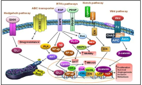
Figure 1.2 the main signalling patterns that
indicate the Wnt, Hh and Nothch pathways
that are related to breast cancer stem cells
(Vera-Ramirez et al., 2010)
HETEROGENITY OF CANCER STEM
CELLS
expressed and used in isolating CSCs
(Li-Heidt
et al
., 2007). This isolated cells can
generate phenotypic heterogeneity from the
parental
cells
(Reya
et
al
.,
2001).
Intratumour heterogeneity is frequently
observed in across multiple tumour types.
There is evidence suggest that Increase in the
heterogeneity result to resistance to many
targeted therapies (Heitzer
et al
., 2013; Dick
et al
., 2003). The theory of heterogeneity in
breast cancer remains unclear till now. It is
believed that cancer stem cells have
contributed to tumour heterogeneity (Badve
and Nackshatri, 2012). Heterogeneity clearly
refers to a tumour which involve in different
types of cells. The majority of studies
supported the idea of heterogeneity of breast
cancer stem cells because they found that
different group BCSCs express a variety of
cell surface markers such as CD133 which is
important markers in breast cancer stem cells
(Lassus
et al
.,2010;Wei
et al
.,2011). In study
carried out by Murohashi et al., they used
different cell lines to detect the expression of
genes. The co workers, found that these cell
lines had a variety of CD44 markers to
identify breast cancer stem cells (Murohashi
et al.,2010; Ababneh et al.,2013).
Figure 1.3 models of cells which are initiated
cancer stem cells. (Velazquez et al.,2009)
ISOLATION OF CANCER STEMS CELLS
able to produce new cells (Razmkayshi et
al.,2012). The number of inhabitants in
CSCs could remain untouched. Studies have
demonstrated that undeveloped tumour cells
had
been
impervious
to
illumination
treatment and chemotherapy medications, for
example, It is an anthracycline drug that acts
by intercalating DNA strands, bringing about
complex development which hinders DNA
and RNA combination. In this way, ideal
strategies must be looked into to target
undifferentiated
disease
organisms
particularly, by going for their multiplication
and development pathways. Hindering these
pathways might thus stop their quick rate of
tumour proliferation (Phue
et al
., 2011).
Isolation and characterization of CSCs
considered as the main challenges for
clinical and biological researchers (Jaggupilli
and Elkord, 2012).There are three techniques
have been used widely to identify BCSCs.
Firstly,
fluorescence-activated
cell
sorting(FACS), which rely on CD44markers,
CD133 and other markers. Secondly,
identification of side population (SP) that
one effluxes Hoechst 33342. Finally,
mammosheres assay (Pham
et al
., 2012;
Saadin
and
White,
2013;
Yan
et
cells have been approved by several studies
in vivo and in vitro.it is assumed that CSCs
cause imitation, metastasis of cancer. In
order to study cancer stem cells in details, it
is required to identify the biomarkers of
CSCs that form an essential target for
treatment. Using molecular methods allow
research to investigate different types of
protein which used as markers specific for
cancer stem cells. It is noteworthy to
understand that BCSCs have been enhanced
by characterising breast cancer cells for
CD44 by culturing of cells in non-adherent
conditions to produce mammospheres (Wang
et al
., 2014). Using serum free media
provides an ideal method to enhance cancer
stem cells. Several studies have been used
low attachment surface and serum-free
media which lead to producing clusters of
stem cells. One of these studies was used
neural cells that generate neurospheres after
using the conditions above. Dontu
et al
., was
the first were used non-adherent culture and
serum free media. The result were BCSCs
can be used to enrich by using this
technique. The concept behind that is the
cells will die in these conditions and the
other
cells
that
survive
called
- Methodology
Materials and chemicals
Material for tissue culture
This equipment has been used in the tissue culture method, as presented in a table (3.1).
Laboratory materials for tissue culture
The laboratory material Suppliers
Class-II safety cabinet Microflow Biological Safety, UK
Centrifuge (refrigerated) Eppendorf, UK
Humid 37°C CO2 incubator Forma Scientific, UK
Micropipets P10, P20,p100 and p1000 Gilson, UK
Water distillation unit Millipore, USA
Inverted microscope & imaging system Nikon, Japan
Inverted microscope Olympus, Japan
Tubes size 25cm3 SARSTEDT, Germany(reference
number: 62.547.254)
T75cm3 and tissue culture flasks(red) SARSTEDT 83.3911.002
Germany
Water bath Grant, UK (Fisherbrand)
75cm3 ultra low adherent tissue culture
flasks(ORANGE)
Corning, USA LOT 3814
5mL, 10mL and 25mL disposable pipettes Sarstedt, Germany
10ul, 200ul and 1000ul Micropipettes tips Sarstedt, Germany
Haemocytometer Minisart Sartorius, Germany
Table 3.1 laboratory material for tissue culture.
Chemical and reagents for tissue culture
Chemical Suppliers References
Dulbecco’s Modified Eagle Medium
(DMEM) Lonza, UK
Epidermal Growth Factor Sigma-Aldrich,
UK
(Kreso and O'Brien
2008)
Foetal Calf Serum (FCS) HyClone, UK
Trypan blue Sigma-Aldrich,
UK
Trypsin-EDTA Lonza, UK
DMSO (Dimethyl Sulphoxide, 100ml Sigma-Aldrich,
UK RNBB7017
bFGF (Fibroblast Growth
Factor Basic
Sigma-Aldrich,
UK
Table 3.2 chemicals and reagents for tissue culture
METHODS
BREAST CANCER CELL LINE
The human breast cancer cell lines MCF-7 that were provided kindly by Dr Selman Ali from Nottingham Trent
CELLS REVIVAL AND MAINTENCE
Initially, the cells were harvested after being in the freezing media which contains Nitrogen. They were removed
and then centrifuged at 1500rpm for 5 minutes. The cells were planted in Dulbecco’s Modified Eagle Medium
(DMEM), which contains insulin 5mg/ml and epidermal growth factor (EGF) long/ml, in adherent tissue culture
flasks size T75. The new culture was incubated at 37◦c with 5% CO2. The cells were regularly checked every
two days to see the percentage of the growth if cell splitting is possible. Normally, the cells take two days to
reach 70 to 80% confluence. After the growth of cells had been checked under the microscope, the cells were
collected, halved and replanted in new flasks and fresh media.
CELLS CULTIVATION TO PRODUCE SPHERE
Culturing of cells in non-adherent, non-differentiate growth condition
When the cells have a greater confluence than 80%, they need to be reseeded. The media was removed from the
flask gently and the cells were washed twice with PBS. Trypsin was then added (1-2 ml depending on the flask
size) to the cells and incubated at 37ᵒC for 5 minutes. The reason for this is to help the cells float in the flask and
stop attachment to the flasks surface. By adding serum- containing a medium, the effect of trypsin was
attenuated. The cells were suspended and centrifuged and a white cells pellet was seen at the bottom of the new
tube. The cells were planted as pellet and the supernatant was discharged. The fresh media that was added
contains (DMEM) and 10% of (FCS). Lastly, the cells were incubated at 37'C in 2 flasks each time, one as
normal culturing and the other for producing spheres. This method was repeated for several (5-10) times to
confirm the results.
Stage 1 Prenatal MCF7 cells where grown in adherent condition 10%
FCS
stage 2.Adherent flask SARSTED, Germany
Trypsnization and single cells suspension. then
incubated
stage 3.Sphere Cells where collected after 72
Figure 3.1 Direct Sphere Formation Assay Using Adherent Conditions
3.2.3.2 Culturing cells in Ultra-low Attachment surface polystyrene e (U shape with Vent cap)
The cells were cultured using a low attachment surface (Corning Flask). The cells were split into two and
washed by PBS and trypsin. The cells were grown in a Corning Flask with new media by adding 15% serum
free media which includes DMEM/F-12 1µl, BFGF 10ng, EGF 20ng, Insulin 5µg and the percentage of foetal
calf serum FCS was increased.
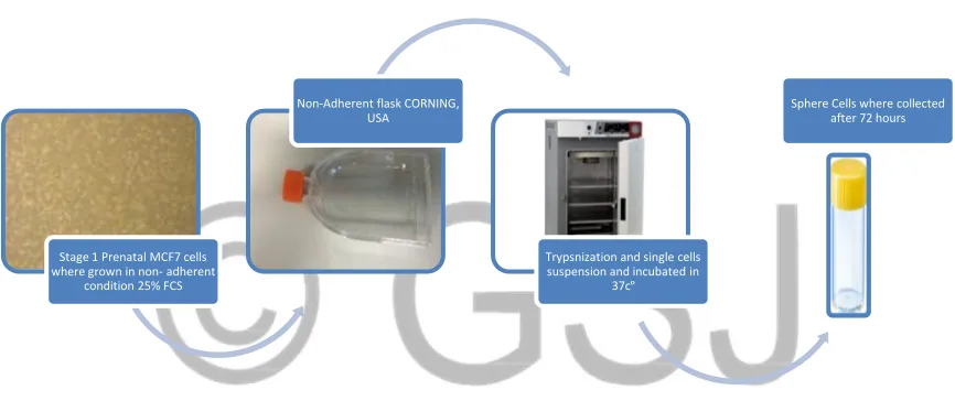
Figure 3.2 Direct Sphere Formation Assay Using Non-Adherent Conditions
COUNTING THE CELLS
This step was applied in order to know the number of the cells by using a haemocytometer. The pellet was taken
from the cultured cells and suspended in 1ml of media then 100ml of the suspension was diluted with 900ml of
trypan blue in a 1 to 2 dilution. The last dilution was then loaded to the haemocytometer. The cells were counted
under the microscope with an x10 magnification (when the cells were not painted with trypan blue). The number
was timed by 105 to calculate the total of cells per 1ml of suspension. The total number of cells in a large flask
size T75 was around 1.2 * 106 cells/ml.
Stage 1 Prenatal MCF7 cells where grown in non- adherent
condition 25% FCS
Non-Adherent flask CORNING, USA
Trypsnization and single cells suspension and incubated in
37cº
FREEZING THE CELLS
The cells were prepared for freezing, to be kept for a long time. The cells were collected and suspended in
1million cells for each 1ml of freezing cell media. The latter was prepared by adding 10% Dimethyl disulfoxide
(DMSO) to keep the cells alive and DMEM media with 10% of FCS. Then the cells were filtered and collected
in small freezing sterile tubes and were maintained at -80 ᵒC for a long period of time.
RESULTS
TISSUE CULTURE RESULTS
Producing mammosheres of MCF7 cells by using standard conditions (normal flask and normal media)
Figure 4.1spheres formation assay of MCF7 cells by using normal culturing condition (media with serum and
adherent flasks).
Figure 4.1 Phenotypic morphology of MCF7 cells by using standard conditions.
This experiment includes several stages. The first phase, the parental MCF7 cells were grown under standard
conditions for five days (normal conditions such as normal flask and normal media). In stage 2, the parental
MCF7 were split into two groups. The first population were normal cells and the second one was spheres in the
normal flask. In stage 3, the spheroid was separated from the single cells. The RNA was isolated from parental,
single and spheres cells respectively.
• Stage 1 Prenatal MCF7 cells where grown
for 2 – 3 days
MCF7 Parent cells •Stage 2 planted in Adherent Flask
normal media
•Stage 3 Spheroid cells where
spread from single cells and total RNA was isolated from
Figure 4.1 shows a trial to isolate spheres from parental MCF7 cells by using mammospheres assay. Several
steps have been applied in this attempt. First of all, the MCF7 parental cells were grown to the level of
over-growth of 70-80% and the total RNA was isolated at this stage. In stage 2, the spheroid is in a different shape
and has formed adherent cells. In stage 3, the total RNA was extracted from spheres cells after 72 hours and 96
hours. MCF7 single cells were separated from the spheres cells and the total mRNA was isolated from these
cells as well. MCF7 cells were planted in standard conditions that included normal culturing flasks and normal
media and 10% of foetal calf serum (FCS). In this part, the cells clustered as a group of cells to produce spheres
that are assumed to be mammospheres.
Figure 4.2 shows spheres generated from MCF7 cells grown in normal conditions (flask and media) indicating
the changes during 24, 72 and 96 hours.
Spheroid after 24 hours
4 X magnification
Spheroid after 72 hours
10 X magnification
Spheroid after 96 hours
Table 4.2 Sphere formation by culturing MCF7 cells in normal flasks and normal media.
Phenotypic changes in MCF7 sphere cells are produced by the direct formation of parental MCF7 cell lines.
During different times 24, 48, 72 and 96 hours under the microscope by using a camera (ZEISS, Primo Vert) in
a variety of power magnifications. MCF7 cells were cultured in normal media and normal flasks for 24, 48, 72
and 96 hours.10 5cells/ml the number of cells in each large flask sizeT75. The cells were incubated at 37°c for
several days and from 2-3 days the cells were regularly checked. This experiment has been done several times.
4.1.2 Direct sphere formation assay of MCF7 cells by using non-adherent non- differentiate conditions (serum free
media and CORNING flasks)
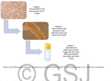
Figure 4.02 Phenotypic changes of parental, single and spheres MCF7 cells grown under non- adherent conditions
This experiment included several stages. The first phase involves MCF7 parental cells that were cultured under
standard conditions for 2-3 days. In stage 2, the MCF7 parental cells were cultured under the non-adherent
condition, using flasks (CORNING, USA) which are characterised as having a U shape, a vent cap and a low
attachment surface. Also, serum free media was used and there was an increase in the percentage of FCS to 25%
and growth factor. This was done for 96 hours.
Figure 4.3 shows an attempt to investigate mammospheres from MCF7 cells line of parental, single and sphere
cells by culturing them in non- adherent conditions (new flasks CORNING and new media). In level 1, the cells
were grown to a 90% confluence in normal media and the appearance of the cells were coherent with each other
and formed a small tree. Total RNA was isolated from MCF7 parental, single and spheroid cells. In the second
stage of this assay, the parental MCF7 cells were seeded in a non-adherent flask with 25% of FCS, free serum
media. The results show a lot of large spheroids and this is proposed to be mammospheres.
Single cells were isolated from the spheres by transferring the cells from the flask to the universal tube and
leaving the cells 5 minutes to settle down. Because the spheres are bigger in size and numbers, they will
concentrate in the bottom of the tube and the single cells will be floating at the top of the tube.
•Stage 1
Prenatal MCF7 cells where grown for 2 –
3 days
MCF7 Parent cells •Stage 2 planted in Non-Adherent Flask and
Serum-free media
•Stage 3 Spheroid cells where spread from single cells and total RNA was isolated from both cells after
Figure 4.4 shows morphological changes of producing mammospheres by using new culturing flask
(CORNING, USA) and a new media:
Table 4.4 indicates a comparison between the phenotype changes by direct sphere assay.
Morphological differences of producing spheres by using non-adherent conditions that include new media
(serum free media, GF and 25% FCS) and Corning Flasks. These phenotypic changes in MCF7 sphere cells
happen by applying direct spheres formation from parental cells. These results show that these spheres are more
defined and bigger than spheres that were formed from the normal culture conditions. After one day, the sphere
was very clear and big in size.
DISCUSSION
For long time the human cancer was considered as a big issue in the world wild. However, in more recent years,
many investigation reported that there is highly decreased in mortality of cancer (Vera-Ramirz et al., 2010;
Kobayshi et al.,2009). More recent studies the focus on CSCs which considers as alternative treatment therapy
due to it is resistance, self-renewal and differentiation (Liu et al., 2015). Therefore, the current work was
conducted to investigate the ideal isolation tools of BCSCs using biomarkers. This experiment includes the
direct formation assay which used under different circumstances. The data shows information about the
characteristics of sphere formation assay and the concept behind this procedure. This technique is used widely to
method, such as increasing the death of normal cells, a reduction in stem cell self-renewal and a decrease in a
number of ancestor cells. Sphere assay is still a vital method for the isolation and quantification of breast cancer
stem cells, in spite of these limitations (Shaw et al., 2012). BCSCs are used in different studies because of the
importance of how breast cancer originates from it (Pham et al., 2012; Clevers,2011; Staveren et al.,2009).
Therefore, the aim of this project was to isolate breast cancer stem cells.
A small population of tumorous breast cancer stem cells have been isolated from MCF7 cell lines using direct
mammospheres assay. The CD44++ cells highly adhesive features were isolated from BCSCs (AL-Hajj et al.,
2003). The main goal of this study was to enrich an MCF7 (CD44++) sub-population of cells, and then identify BCSC features by increasing their tumorsphere growth ability in vitro. Although there are several techniques that have been used in the past, there is a need for more specific methods and indicators, and that is the reason for this study.
Optimisation of spheroid cultures
Similar to several methods which have used in different experiments, BCSCs were cultured in two separate
methods:
Producing spheroids in normal flask and normal media
In this part of the work, MCF7 cells were successfully cultured in a normal flask (SARSTEDT, Germany) with
normal media with only 10% of FCS added. It is estimated that the MCF7 cells that were planted under these
conditions had a different morphology when making spheres. The latter was slowly produced, one to two
clusters after four to five days. The appearance of the spheroid was not very clear and it was small in size. It had
a few cells and an irregular shape. There were only one, two and three spheres after 24, 72 and 96 hours
respectively [Figure4.1, 4.2]. Therefore, we can say that MCF7s production of spheres in these circumstances
were not in as good a condition as how we expect the isolated bulk of BCSCs to be at this stage.
Producing spheroids in a non-adherent flask and serum free media(Corning, US)
MCF7 cells were cultured in non-adherent conditions in serum free media with an increased percentage of FCS
to 25% and the addition of growth factors to produce a group of cells called mammospheres. Under these
conditions, it was noticed that the spheres were able to grow very quickly after 24 hours to be bigger in size and
quantity and clearer than the spheroid in the normal conditions [Figure4.3, 4.4]. For this reason, BCSCs show
because of the self-renewal abilities of CSCs. This result is similar to a study carried out by Abboodi (2014). In
their findings, the clusters of cells were collected after one day (24hours) by using new media and a new flask
(Corning, US). Normally the cells need four days to make spheres in ideal conditions; the images were shown
under the microscope X4, X10, X20 and X40 magnification [Figure 4.3]. It is believed that spheroids are
generated from single cells and this proved the ability of these cells to differentiate and initiate tumours (Ponti et
al., 2006). Similar result were displayed by Ponti et al., (2005) when they cultured MCF7 cells under
non-adherent conditions in serum free media with added growth factors resulted in producing spheroids. It is
assumed that this formation of mammospheres might be occurred due to the presence of GF which shares CSCs
in the same microenvironment. It was noticed that under these conditions using low attachment flask,
non-adherent culture, MCF7 cells produce a large spheres compared with spheroid which generate from non-adherent
conditions. Also, their observation was indicate the high quality of the morphology of spheres and quickness in
producing spheres.
Wang et al., pointed out that serum free culture presents a crucial means to generate cancer stem cells. In their experiment they used MCF7 cells in non-adherent condition (which included using an ultralow attachment flask (Corning),
DMEM/F12, 10ng/ml b-FGF, 20ng/ml of EGF) and these cells were attached to the flask and it was highly difficult to produce a sphere. This finding clarified that culturing MCF7 cells can help to enrich BCSCs in adherent non-differentiated circumstances to form a spheroid (Wang et al., 2014).
Summary
This study was aimed to eradicate the use of human breast cancer cell line(MCF7) as a model to simplify the
nature of BCSCs. Different culturing methods were applied to achieve this target. Based on several kinds of
literature mammospheres can be produced in breast cancer cell lines under different circumstances such as
adherent flask, normal media and non-adherent, low attachment flask with serum-free media; but the results will
be different in the morphology of spheres in both of the conditions. MCF7 spheres cells in CORNING flask
showed higher tumorigenic than parental, single cells (Wang et al., 2014). Recently, mammospheres technique
has been provided a relative pathway to study cancer in general and CSCs in specific in order to provide an ideal
solution to clarify CSCs theory (Saadin and White, 2013).
1.
Ababneh, N., Alshaer, W., Allozi, O., Mahafzah, A., El-Khateeb, M., Hillaireau, H., Noiray, M., Fattal,E and Ismail, S.2013.In Vitro Selection of Modified RNA Aptamers Against CD44 Cancer Stem Cell
Marker. NUCLEIC ACID THERAPEUTICS. 23(6), 401-407.
2.
Al-Hajj, M., Wicha, M.S., Hernandez, A.B., Morrison, S.J. and Clarke, M.F., 2003.Prospectiveidentification of tumorigenic breast cancer cells. PNAS.100 (11), 6891- 3988.
3.
American cancer Society. Cancer Facts and Figures2014.Atlanta, Ga: American Cancer Society; 2014.4.
American cancer Society. Cancer Facts and Figures2015. Atlanta, Ga: American Cancer Society; 2015.5.
American joint committee on cancer. Breast. In: AJCC Cancer staging Mammal, 7th ed. New York:springer; 2010:347-369.
6.
Badve, S., and Naskhatri, H.,2012. Breast cancer stem cells- beyond semantics. Lancet Oncology.132012, e43-48.
7.
Baker, M., 2012.Cancer stem cells tracked. Nature. 488,13-14.8.
BC Sun, T.L., Zhao, X., Zhao, X., Sun, T., Gu, Q., Yao, Z., Dong, X., Zhao, N. and Liu, N.,2013.CD133+cells with cancer stem cell characteristics associated with vasculogenic mimicry in
triple-negative breast cancer. Oncogene (2013)32,544 – 553.
9.
Brugnoli, F., Grassilli, S., Piazzi, M., Palomba, M., Nika, E., Bavelloni, A., Capitani, S. and Bertagnolo,V.,2013. In triple negative breast tumour cells, PLC-β2promotes the conversion of CD133high
toCD133lowphenotype and reduces theCD133-related invasiveness. Molecular Cancer.12 (165).1-15.
10.
Chaves, K.J., Garimella, S.V. and Lipkowize, S.,2010. Triple Negative Breast Cancer Cell lines: OneTool in the Search for Better Treatment of Triple Negative Breast Cancer. NIH Public Access. 32(1-2),
35-48.
11.
Chilet,M.P., Martínez,M.T., Pérez-Fidalgo,J.A., Peiró-Chova,L., Oltra,S.S., Tormo,E., Alonso-Yuste,E.,Martinez-Delgado, B., Eroles,P., Climent,J., Burgués,O., Ferrer-Lozano,J., Bosch,A., Lluch,A. and
Ribas, G.,2014. MicroRNA profile in very young women with breast cancer. BMC Cancer. 14(529),
1471-2407.
12.
Conley, S.J., Gheordunescu, E., Kakarala, P., Newman, B., Korkaya, Heth, A.N., Clouthier, S.G. andWhicha, M.S.,2012. Antiangiogenic agents increase breast cancer stem cells via the generation of tumour
hypoxia.PNAS.109 (8), 2784-2789.
14.
Fillmore, C.M and Kuperwasser, C. 2008.Human Breast Cancer Cell Lines Contain Stem-like Cells ThatSelf-renew, Give Rise to Phenotypically Diverse Progeny and Survive Chemotherapy. Breast Cancer
Res. 10(2), 303.
15.
Gerlinger,M., Rowan,A.J., Horswell,S., Larkin,J., Endesfelder,D., Gronroos,D.M.E., Martinez,P.,Matthews,N., Stewart,A., Tarpey,P., Varela, L., Phillimore, B., Begum, S., McDonald,N.Q., Butler,A.,
Jones,D., Raine, K., Latimer,C., Santos,C.R., Nohadani,M., Eklund,A.C., Spencer-Dene,B., Clark,G.,
Pickering,L., Stamp,G., Gore,M., Szallasi,Z., Downward,J., Futreal,A. and Swanton,c.,2012. Intratumor
Heterogeneity and Branched Evolution Revealed by Multiregion Sequencing. The new England Journal
of medicine. 366(10).833-892.
16.
Germain,A.R.,Carmody,L.C.,Morgan,B.,Fernandez,C.,Forbeck,E.,Lewis,T.A.,Nag,P.P.,Ting,A.,Verplank,L.,Feng,Y., Perez, J.R., Dandapani, S., Palmer, M., Lander, E.S., Gupta, P.B. and Schreiber, S.L
.,2012. Identification of selective small molecule inhibitor of breast cancer stems cells. Bioorganic and
Medicinal Chemistry Letters. 22(2012), 3571-3574.
17.
Graham, N, and Greaber, T.G. 2014 Complexity of metastasis- associated SDF-1 ligand signalling in breast cancer stem cells. PNAS. 111(21), 7503-7504.18.
Hermann, P.C., Bhaskar, S., Cioffi, M. and Heeschen, C., 2010.Cancer stems cells in solid tumours.Seminar in Cancer Biology. 20(2010), 77-84.
19.
http://www.cancer.org/research/cancerfactsstatistics/breast-cancerfacts-figures.20.
Hu, F., Meng, X., Tong, Q., Liang, L., Xiang, R., Zhu, T. and Yang, S.,2013. Biochimica et Biophysica
Acta. 1832(2013), 2379-2390.
21.
Hu,F.,Ru,N.,Xiao,H.,Chaturbedi,A.,Hoa,N.T.,Tian,X.J.,Zhang,H.,Ke,C.,Yan,F.,Nelson,J.,Li,Z.,Gramer,R.,Yu,L.,Siegal,E.,Zhang,X.,Jia,Z.,Jadus,M.R.,Limoli,C.L.,Linskey,M.E.,Xing,J. and Zhou,Y.H.
2013.Tumor-Specific Chromosome Mis-Segregation Controls Cancer Plasticity by Maintaining Tumour
Heterogeneity. PLOS ONE.8 (11), 1-16.
22.
Jaggupilli, A., and Elkord, E., 2012. Significance of CD44 and CD24 as cancer stem cell markers: AnEnduring Ambiguity. Clinical and Developmental Immunology.2012, 1-11.
23.
KING, R.J.B., and ROBINS, M.W. (2006). Cancer biology. 3rdEdition. England, Pearson EducationLimited.
24.
Lassus, Heini; Liang, Shun; Kaur, Sippy; Ye, Qunrui; Li, Chunsheng; Wang, Li-Ping; Roby, KatherineF.; Orsulic, Sandra; Connolly, Denise C.; Zhang, Youcheng; Montone, Kathleen; Bützow, Ralf; Coukos,
George; Zhang, Lin. 2010. Distinct Expression Levels and Patterns of Stem Cell Marker, Aldehyde
25.
Li, J., Wang, K., Li, S., Wilenga, V.T., Rank, F., WiUF, C., Zhang, X., Yang, X. and Boland, L., 2009. DNA Copy Number Aberrations in Breast Cancer by Array Comparative Genomic Hybridization.Genomics Proteomics Bioinformatics. 7(1-2).1-24.
26.
Liang,Y.J.,Ding,Y.,Levery,S.B.,Lobaton,M.,Handa,k.,Hakomori,S.I2013. Differential expressionprofiles of glycosphingolipids in human breast cancer stem cells vs. cancer non-stem cells. PNAS.
110(13), 4968-4973.
27.
LiC, Heidt DG, Dalerba P, Burant CF, Zhang L, Adsay V, Wicha M, Clarke MF, Simeone DM2007.Identification of pancreatic cancer stems cells. Cancer Research 67(3), 1030-1037.
28.
LiC, Heidt DG, Dalerba P, Burant CF, Zhang L, Adsay V, Wicha M, Clarke MF, Simeone DM2007.Identification of pancreatic cancer stems cells. Cancer Research 67(3), 1030-1037.
29.
Llipoulous, D., Hirsch, H.A., Wang, G and Struhi, K (2011). Inducible formation of breast cancer stemcells and their dynamic equilibrium with non- stem cancer cells via IL6 secretion. PNAS. 108(4),
1397-1402.
30.
Lorico, A., and Rappa, G., 2010.Phenotypic Heterogeneity of Breast Cancer Stem Cells. Journal of Oncology.2011, 1-6.31.
McDermott, S.P., and Whica, M.S.,2010. Targeting breast cancer stem cells. Molecular Oncology.4(2010), 404-419.
32.
Morel, A.P., Lievre, M., Thomas, C., Hinkle, G., Ansieau, S. and Puisieux, A.,2008. Generation ofBreast cancer stem cells through Epithelial-Mesenchymal Transition. PLOS: ONE. 3(8), 1-7.
33.
MUROHASHI, M., HINOHARA, K., KURODA, M., ISAGAWA, T., TSUJI, S., KOBAYASHI, S.,UMEZAWA, K., TOJO, A., ABURATANI, H. and GOTOH, N., (2010).Gene enrichment set analysis
provides insight into novel signalling pathways in breast cancer stem cells. British Journal of Cancer,
102, 206-212.
34.
Nguyen, N.P., Almeida, F.S., Chi, A., Nguyen, L.M., Cohen, D., Karlsson, U. and Vinh-Hung, V.,2010.Molecular biology of breast cancer stem cells: Potential clinical applications. Cancer Treatment Reviews
36 (2010), 485–491.
35.
Owens, T.W and Naylor, M.J., 2013 Breast cancer stem cells. Physiology. 4(225).1-10.36.
Pecorino, L. (2005). Molecular biology of cancer – Mechanisms, targets and therapeutics. New York,37.
Phuc, P.V., Keng, S.C., Nguyet, N.T.M., Thuy, D.T. and Ngoc, P.K., 2011. POPULATIONS FROMTHE MCF-7 BREAST CANCER CELL LINE BASED ON CD44 AND CD24 MARKERS. Research
Gate.9 (1), 13-19.
38.
Razmkhah,M.,Jaberipour,M.,Ahmed,H.,Safaei,A.,Khalatbari,B. and Chaderi, A.,2010. Expressionprofile of IL-8 and growth factors in breast cancer cells and adipose-derived stem cells (ASCs) isolated
from breast canceinoma. Cellular Immunology.265 (2010), 80-85.
39.
Reya, T., Morrison, F.C., Clarke, M.F and Weissman, I.L. 2001 Stem cells, cancer, and cancer stemcells.Nature.414, 105-111.
40.
Saadin, K., and White, M.I.,2013. Breast cancer stems cell enrichment and isolation by mammospheresculture and its potential diagnostic applications. Expert Rev. Mol.Diagn.13 (1), 49 – 60.
41.
Sagar,J.,Chaib,B.,Sales,K.,Winslet,M and Seifalian,A (2007). Roles of stem cells in cancer therapy andcancer stem cells: a review. Cancer Cell International.7 (9), 1-11.
42.
Sahlberg, S.H., Spiegelberg, D., Glimelius, B., Stenerlow, B. and Nestor, M.,2014. Evaluation of CancerStem Cell Markers CD133, CD44, CD24: Association with AKT Isoforms and Radiation Resistance in
Colon Cancer Cells. PLOS ONE 9(14).1-12.
43.
Sehrawat, A., Arlotti, J.A., Murakami, A. and Singh, S.V.,2012. Zerumbone causes Bax andBak-mediated apoptosis in human breast cancer cells and inhibits orthotopic xenograft growth in vivo. Breast
Cancer Res Treat. 136(2), 429–441.
44.
Senbanjo, R.O., Miller, W.R and Hawkins, R.A. 1986. Variations in steroid receptors and cyclicAMP-binding proteins across human breast cancers: Evidence for heterogeneity. Br. J. Cancer. 54, 127-130.
45.
Senbanjo, R.O., Miller, W.R and Hawkins, R.A. 1986. Variations in steroid receptors and cyclicAMP-binding proteins across human breast cancers: Evidence for heterogeneity. Br. J. Cancer. 54, 127-130.
46.
Shaw, F.L., Harrison, H., Spence, K., Ablett, M.P., Simões, B.M., Farnie, G. and Clarke, R.B.,2012. ADetailed Mammospheres Assay Protocol for the Quantification of Breast Stem Cell Activity. J
Mammary Gland Biol Neoplasia. 2012(17), 111–117.
47.
Sheridan, C., Kishimoto, H., Fuchs, R.K., Mehrotra, S., BhatNakshatri, P., Turner, C.H., Goulet, R.,Badve, S. and Nakshatri, H., 2006.CD44+/CD24-breast cancer cells exhibit enhanced invasive
properties: an early step necessary for metastasis. Breast Cancer Research. 8 (5), 1-13.
48.
Strati, A., Markou, A., Parisi, C., Politaki, E., Mavroudis, D., Georgoulias, V. and Lianidou, E.,2011.Gene expression profile of circulating tumour cells in breast cancer by RT-Qpcr. BMC Cancer. 11(422),
49.
Swanton, C., 2012.Intratumor Heterogeneity: Evolution through Space and Time. American Association for Cancer Research.72 (19), 4875-4882.50.
Tuck, D.P. and Miranker, W., 2010. Modelling the clonal heterogeneity of stem cells. TheoreticalBiology and Medical Modelling. 7(44), 2-27.
51.
VAN DE-VIJVER, M.J.V., He,Y.D., VAN’TVEER,L.J., Dai,H., HART,A.A.M., VOSKUI,D.W., SCHREIBER,G.J., PETERSE,J.L., ROBERTS,C., MARTON,M.J., PARRISH,M., ATSMA,D.,WITTEVEEN,A., GLAS,A., DELAHAYE,L., VANDERVELDE,T., BARTELINK,H., RODENHUIS,S., RUTGERS,E.T., FRIEND,S.H. and BERNARDS,R.,2002. A GENE-EXPRESSION SIGNATURE AS A PREDICTOR OF SURVIVALIN BREAST CANCER. The New England Journal of medicine. 347(25), 1999-2009.
52.
VAN DE-VIJVER, M.J.V., He,Y.D., VAN’TVEER,L.J., Dai,H., HART,A.A.M., VOSKUI,D.W.,SCHREIBER,G.J., PETERSE,J.L., ROBERTS,C., MARTON,M.J., PARRISH,M., ATSMA,D.,
WITTEVEEN,A., GLAS,A., DELAHAYE,L., VANDERVELDE,T., BARTELINK,H.,
RODENHUIS,S., RUTGERS,E.T., FRIEND,S.H. and BERNARDS,R.,2002. A GENE-EXPRESSION
SIGNATURE AS A PREDICTOR OF SURVIVALIN BREAST CANCER. The New England Journal
of medicine. 347(25), 1999-2009.
53.
VELASCO-VELÁZQUEZ, M., Yu, Z., Jiao, X. and Pestell, R.G.,2009. Cancer stem cells and the cellcycle: targeting the drive behind breast cancer. Anticancer Ther.9 (3),275-279.
54.
VELASCO-VELÁZQUEZ, M.A., HOMSI, N., DE LA FUENTE, M. and PESTELL, R.G., 2012. Breastcancer stem cells. The International Journal of Biochemistry & Cell Biology, 44 (4), 573-577.
55.
Vera-Ramirez,L.,Sanchez-Rovira, P.,Tortosa-Ramirez,,C.L.,Quiles,J.L., Ramirez-Tortosa,M.C.,Alvarez,J.C.,Navarro- Fernandez,M. and Lorenzo, J.A.,20-10. ANTI-TUMOUR
TREATMENT Gene-expression profiles, tumour microenvironment, and cancer stem cells in breast
cancer: Latest advances an integrated approach. Cancer Treatment Reviews. 36(6), 477-484.
56.
Wang, N., Shi, L., Li., H., Hu.,Y.,Du., W., Liu.,W.,Zheng.,J and Huang., S. 2012 Detection ofcirculating tumour cells and tumour stem cells in patients with breast cancer by using flow cytometry.
Tumour Biol. 33(2012), 561-569.
57.
Wei,W.,Hu,H.,Tan,H.,Chow,L.W.,Yip,A.Y.,Loo,W.T.2011 Relationship of CD44+ CD24-/low breastcancer stem cells and axillary lymph node metastasis. Journal of Translational Medicine. 10(1), 1-6.
58.
Weigelt, B., Kreike, B. and Reis-Filho, J.S.,2009. Metaplastic breast carcinomas are basal-like breastcancers: a genomic profiling analysis. Breast Cancer Res Treat. 117:273–280.
59.
Weigelt, B., Kreike, B. and Reis-Filho, J.S.,2009. Metaplastic breast carcinomas are basal-like breast60.
WEINBERG, R.A. 2007. The biology of cancer. New York, Garland Science. WHO, 2014. Cancer data, Abril, 2014. Available on WHO, 2014.61.
Wicha, M.S., Targeting breast cancer stem cells. The Breast 18 (2009) S3, S56–S58.62.
Xu, G., Shen, J., Yang, X.O., Sasahara, M. and Su, X.,2013. Cancer stem cells: the ‘heartbeat’ of gastriccancer. J Gastroenterol. (2013) 48,781–797.
63.
Yae ,T., Tsuchihashi ,K., Ishimoto ,T., Motohara ,T., Yoshikawa,M., Yoshida1 G.J., Wada ,T., Masuko,T., Mogushi ,K., Tanaka ,H., Osawa ,T., Kanki ,Y., Minami,T., Aburatani, H., Ohmura ,M., Kubo ,A.,
Suematsu ,M., Takahashi ,K., Saya ,H.and Nagano ,O., 2012. Alternative splicing of CD44mRnA by