Antigen-Specific IgG Antibodies in Stage IV Long-Time Survival
Breast Cancer Patients
Mona H. Hansen, Bjørn Østenstad, and Mouldy Sioud
Department of Immunology, Molecular Medicine Group, The Norwegian Radium Hospital (M.H.H., M.S.) and the Department of Oncology, Ullevål University Hospital (B.Ø.), Oslo, Norway
Accepted January 1, 2001.
Abstract
Background:Profiling the immune responses in patients with cancer is expected to facilitate the design of diagnos-tic tests and therapeudiagnos-tic vaccines. Such studies usually re-quire the parental antigens. We attempted to profile the immune responses in patients with breast cancer using a peptide phage display selection strategy, which identifies antibody specificities whether or not the antigens are known.
Materials and Methods: A panel of random peptide phage libraries was panned on serum IgG antibodies from breast cancer patients with stage IV, seeking for disease specific IgG epitopes. ELISA, immunoscreening, and Western blotting techniques were the main approaches used.
Results: Phage-displayed peptides were specifically enriched for binding to IgG antibodies from patients with breast cancer. Several peptides have been identi-fied, in particular the SQRIPARIHHFPTSI peptide, which was recognized by IgG antibodies from breast
Address correspondence and reprints to: Dr. Mouldy Sioud, The Institute for Cancer Research, Department of Immunology, Molecular Medicine Group, The Norwegian Radium Hospital, Montebello, N-0310 Oslo, Norway. Phone: 47 22 93 45 63. Fax: 47 22 50 07 30. E-mail: mosioud@ulrik.uio.no
cancer patients, but not from normals (p 0.0004). In patients who responded to the selected peptides, in par-ticular the SQRIPARIHHFPTSI peptide, antibodies against a 66 kDa cellular protein were found. Interest-ingly, three out of six patients with the strongest im-munoreactivity are still alive, with a mean survival time from first recurrence until now of 2553 days. In contrast, all the nonresponders (n10) are deceased. The mean survival time of these patients was 784 days, whereas the mean survival time of the three deceased responders was 1050 days (p 0.02).
Conclusions.The data provide the first example in which panning of peptide phage display libraries on patient IgG antibodies results in the isolation of breast cancer specific IgG epitopes, some of which correlate with patient sur-vival time. Thus, the identified B-cell epitopes should be of great interest in vaccine development.
Key Words.Breast cancer, phage display, autoantibodies, serological markers.
Introduction
Several human diseases, including cancer, autoim-mune diseases, and infectious diseases, are con-cerned with the immune responses against self and nonself proteins (1,2). Therefore, defining the speci-ficities of B and T cells in these diseases should fa-cilitate the design of diagnostic tests and vaccines. Unfortunately, the analysis of B- and T-cell epitopes requires structural information about the parental antigens, which is a major limitation if the antigens are not known.
In recent years, peptide phage display libraries have been developed and proved useful tools for the identification of monoclonal antibody specifities, whether or not the parental antigens are known (3–5). This strategy is based on the ability of fila-mentous bacteriophage to display foreign peptides on their surfaces (6). In contrast to many other mol-ecular methods, the phage display selection strategy relies on the physical link between the displayed
antigen-specific immune responses in patients with breast cancer, with the emphasis on selecting dis-ease specific IgG epitopes.
Materials and Methods
Sera
Serum samples from patients with breast cancer with stage-VI lymphoma, prostate cancer, and vol-unteer blood donors were obtained from Ullevål University Hospital. Sera from patients with os-teosarcoma were obtained from The Norwegian Ra-dium Hospital. All sera were aliquoted and stored at 70C until use. Pooled IgG antibodies from pa-tients and controls were affinity purified using protein G columns according to the manufacturer’s instructions (Pharmacia).
Affinity Selection
The peptide phage libraries were biopanned on pa-tient IgG antibodies as described by Dybwad et al. (7). In brief, 1011transduction units from an
ampli-fied 15-mer library expressed on pVIII (4) were incubated overnight at 4C with patient IgG, prein-cubated with the fd phage. After incubation, biotin-conjugated anti-human IgG, preincubated with the fd phage, was added to the mixture and incubation was continued for 3 hr at room temperature. Fol-lowing incubation, streptavidin-coated magnetic beads were added to the mixture and incubation was continued for 30 min. Antibody/phage-binding beads were attracted to a magnet, while unbound phages were removed. Beads were washed 10 times with PBS/0.2% Tween over a period of 1 hr and streptavidin-binding phages were acid eluted and neutralized by Tris Base. The eluted phages were amplified in E. coli K91 cells and then used for fur-ther rounds of selection. The enrichment of specific phage-displayed peptides was monitored by im-munoscreening. In the case of the 7- and 12-mer pIII peptide libraries, the eluted phages were amplified in ER 2537 E. colicells as described by the manufac-turer (New England Biolabs).
Immunoscreening
Serum IgG antibody-binding phages were identi-fied using an immunoscreening technique (12,13). In brief, E. coliK91 cells were infected with the se-lected phages. Various dilutions of the infected bac-teria were plated on NZY agar plates containing tetracycline (50 g/mL) and IPTG (1 mM) and incu-bated overnight at 37C. Following transfer to the nitrocellulose membranes, the membranes were washed twice with TNT buffer (10 mM Tris HCl, 150 mM NaCl, 0.05% Tween 20) for a total of 30 min and then blocked for 30 min with 20% FCS in TNT buffer. After blocking, membranes were incubated with 8 g/mL pooled IgG in FCS/TNT for 2 hr and then washed once with TNT buffer with 0.1% BSA and 0.1% NP-40 for 10 min and then twice with
TNT buffer with 0.1% BSA (10 min each). Alkaline phosphatase-conjugated anti-human IgG (1/5000) was added to the membranes and incubated for 2 hr. After washing with TNT buffer, the immunoreactive phage clones were detected by BCIP/NBT premixed substrate (Sigma). In the 7- and the 12-mer peptide libraries, random clones from the fourth round of se-lection were picked up and characterized.
Phage Preparation and Sequencing
Phages from liquid culture were obtained by poly-ethylene glycol 6000/NaCl precipitation as described elsewhere (7). The phages were resuspended in PBS buffer, filtered through 0.45 mm filter and then stored at 4C until use. For DNA sequencing, phage DNA was prepared and the DNA inserts encoding for the peptides were sequenced by the dideoxy chain termi-nation method. The sequencing reaction was carried out using the Sequenase version II Kit (Amersham).
Enzyme-Linked Immunosorbent Assay (ELISA)
ELISA experiments were performed as described previously (7). In brief, 96-well microtitre plates were coated overnight at 4C with the purified phages (107/well). After blocking with 0.5% BSA in PBS buffer for 1 hr at room temperature and subse-quent washing, the plates were incubated with sera samples (1/100 dilution in PBS/0.2% BSA/0.2% Tween 20) for 1 hr at 37C. After additional washing, plates were incubated with alkaline phosphatase-conjugated anti-human IgG (1/5000) for 1 hr at 37C. The immune complexes were detected by adding
p-nitrophenyl phosphate as substrate. The reactivity of each serum to the phage displaying the SQRI-PARIHHFPTSI peptide was obtained by subtracting its reactivity to a phage displaying a random peptide (YLRNVSRVLIQSSSN). A result was considered as positive when the serum reactivity was equal to or greater than 3.5-fold of the mean reactivity of normal individuals.
Competition Experiments
Various concentration of the phage displaying the SQRIPARIHHFPTSI peptide or the wild-type phage was incubated with each patient serum diluted 1/500 in PBS/0.2% BSA/0.2% Tween 20. After incubation overnight at 4C, the mixtures were added to the 96-well microtiter plates coated with the phage display-ing SQRIPARIHHFPTSI peptide (106 TU/well ). In the experiments with the synthetic peptides, increas-ing concentrations of the synthetic peptide or a con-trol peptide were incubated with each patient serum (diluted 1/500), incubated overnight at 4C and then added to the 96-well microtiter plates coated with the phage displaying the SQRIPARIHHFPTSI peptide (106TU/well). The results are expressed as
Statistics
Differences between patients and controls were tested for significance with an unpaired t-test.
Results
Affinity Enrichment of Breast Cancer Patient IgG Antibody-Binding Phages
The ability to probe specific immune responses in patient sera with peptide phage display libraries (7–9) prompted us to evaluate this novel technology in patients with cancer. In these experiments, a pooled IgG fraction derived from seven breast cancer patients with stage IV was epitope-mapped by a panel of random peptide phage libraries as described in Materials and Methods. After six rounds of affinity enrichment, the immuno reactivity of the selected phages was investigated by an im-munoscreening technique. This method allows a di-rect identification of IgG-binding phages after a transfer to nitrocellulose membrane filters. To test for specificity, pooled IgG fractions from patients with lymphoma (n6), osteosarcoma (n8), and normals (n7) were also tested (Figure 1A). A strong reactivity was found with pooled IgG anti-bodies from breast cancer patients (b), but no signif-icant reactivity was found with IgG antibodies from normals (n), patients with lymphoma (l), or those with osteosarcoma (o). Notably, positive clones are clearly distinguishable from negative clones (b,
ar-Western Blotting
Aliquots of 40 g cell proteins were subjected to electrophoresis in SDS 10% polyacrylamide gels, followed by protein electrotransfer to nitrocellulose membranes and immunoblotting with patient and normal sera at a dilution of 1/1000. Antibody bind-ing was detected through the application of HRP-conjugated anti-human IgG antibodies. Immunore-active bands were visualized with ECL detection reagents (Amersham).
Cytosolic, Nuclear, and Membrane Protein Preparation
PMI cells (ATCC) were washed twice with PBS and then resuspended in buffer A (5 mM Tris HCl pH 8.8, 0.5 mM EDTA and 75 mM sucrose with proteinase inhibitors) and then sonicated 6 times (20 seconds each). After sonication, the nuclei were pelleted by centrifugation at 2000 rpm for 5 min and then the supernatant was centrifuged at 30,000 rpm for 30 min. The supernatant from this step was saved as the cytosol fraction. The pellet, which con-tains the membrane fraction, was washed 4 times with PBS, resuspended in buffer A containing 1% Triton X-100, and incubated on ice for 10 min. After incubation, the sample was centrifuged at 15,000 rpm for 10 min, and the supernatant was saved as the membrane fraction. The nuclei pellet was resus-pended in 10% NP-40 and then sonicated. Protein concentration was determined using the protein as-say kit (BioRad Laboratories, Hercules, CA).
Fig. 1. Immunoreactivity of the selected phages with patients and controls. (A)Reactivity of breast cancer patient sera and controls to random phages derived from the 6th round of selection. Following transfer to nitrocellulose and blocking, the membranes were incubated with (8 g/mL) pooled IgG fraction from breast cancer patients (b), lymphoma (l), osteosarcoma (o), or normal (n). The use of alkaline phosphatase conjugated anti-human IgG antibodies as described in Materials and Methodsdetected the im-munoreactive phage clones. (B)Reactivity of phage particles with individual serum as detected by immunospot assays. In these ex-periments purified phage particles (106TU/spot) were spotted onto nitrocellulose, blocked and then processed as in A. Each serum
rowheads), confirming the specificity of the im-munoreaction.
To identify the most relevant peptides, phage particles from 63 random positive phage clones were purified and their immunoreactivity to 40 individual serum samples was tested by immunospot assays. A representative example is shown in Figure 1B. A high proportion of breast cancer patients showed strong reactivity to at least one of the selected phages. In our experimental conditions, none of the positive phages reacted with normal serum samples (n40) or sera from patients with osteosarcoma (n10). In immunospot assay, some of the selected phage-displayed peptides reacted with at least 15% of patients with breast cancer.
Structural Characteristic of Peptides Displayed By the IgG Antibody-Selected Phages
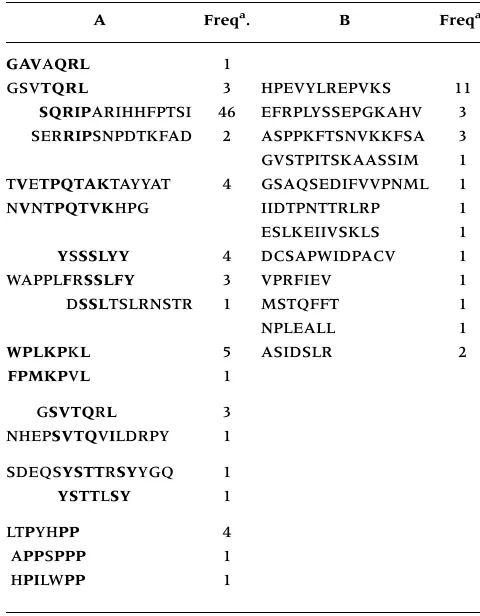
To determine the structural characteristic of the peptides displayed by the positive phages from three independent experiments using 15-, 12-, and 7-mer peptide phage display libraries, the DNA sequences encoding for the peptides were deter-mined (Table 1). The sequences were arranged into two groups. The peptides in group A showed differ-ent consensus core motifs (bolded amino acids). Group B consisted of peptide with no obvious ho-mology. The phage-displayed peptides which showed reactivity with at least 15% of breast cancer patients in immunospot assay were found to belong mainly to group A (e.g., SQRIPARIHHFPTSI, TV ET-PQTAKTAYYAT, SDEQSYSTTRSYYGQ, GSVTQRL). Notably, the SQRIPARIHHFPTSI peptide was se-lected 46 times.
Humoral Immune Response of Patients and Controls to the SQRIPARIHHFPTSI Peptide
Although most of the selected B-cell epitopes need further characterization, we have focused on the phage displaying the SQRIPARIHHFPTSI peptide sequence as its reactivity with patient IgG antibod-ies was the strongest in comparison with the reac-tivity obtained with other selected phage-displayed peptides. Sera from patients with breast cancer with stage IV (n16) and normal adults (n35) were tested by ELISA. The relative reactivity of each pa-tient serum to the phage-displayed SQRIPARIHHF-PTSI peptide was obtained by substracting its ab-sorbency to a phage displaying a random peptide (Figure 2A). There were significantly increased pep-tide IgG antibodies in patients (mean0.507 OD units) compared to normals (mean0.156 OD units) (p 0.0004). Seven out of the 16 patients had antibody levels at least 3.5-fold greater than the mean of the normals. Under our ELISA conditions no significant reactivity was obtained with sera from os-teosarcoma (n14) and lymphoma patients (n10) (data not shown).
To further confirm that the binding seen in ELISA is the result of the interaction of the serum
IgG antibodies with the peptide displayed by the phage, we performed competition assays. Represen-tative examples are shown in Figure 2 Band C. In competitive ELISA, the binding of the serum IgG was inhibited by the phage itself, whereas no sig-nificant effect was obtained with a phage displaying an irrelevant peptide (Figure 2B). The binding of the phage displaying the SQRIPARIHHFPTSI peptide to the patient sera was also inhibited with a syn-thetic peptide corresponding to the peptide dis-played by the phage, but not with an irrelevant pep-tide (Figure 2 C), thus confirming the specificity of the IgG binding.
Antibodies to a 66 kDa Cellular Protein in Patients Who Strongly Reacted with the SQRIPARIHHFPTSI Peptide
The overall evidence presented above suggests that in some patients with breast cancer, specific humoral immune response against tumor cells has been acti-vated. As the first step toward the identification of cellular antigens, protein extracts from a breast cancer cell line (PMI) was prepared, separated by SDS
Table 1. Sequences of phage-displayed peptides obtained from three independent experiments using 15-, 12- and 7-mer peptide phage display libraries. The amino acid se-quence of each clone is expressed by single-letter abbrevia-tion. Consensus core motifs are shown in bold.
A Freqa. B Freqa.
GAVAQRL 1
GSVTQRL 3 HPEVYLREPVKS 11
SQRIPARIHHFPTSI 46 EFRPLYSSEPGKAHV 3 SERRIPSNPDTKFAD 2 ASPPKFTSNVKKFSA 3 GVSTPITSKAASSIM 1 TVETPQTAKTAYYAT 4 GSAQSEDIFVVPNML 1 NVNTPQTVKHPG IIDTPNTTRLRP 1
ESLKEIIVSKLS 1
YSSSLYY 4 DCSAPWIDPACV 1
WAPPLFRSSLFY 3 VPRFIEV 1
DSSLTSLRNSTR 1 MSTQFFT 1
NPLEALL 1
WPLKPKL 5 ASIDSLR 2 FPMKPVL 1
GSVTQRL 3
NHEPSVTQVILDRPY 1
SDEQSYSTTRSYYGQ 1 YSTTLSY 1
LTPYHPP 4
APPSPPP 1
HPILWPP 1
aThe frequency refers to the number of times each phage was
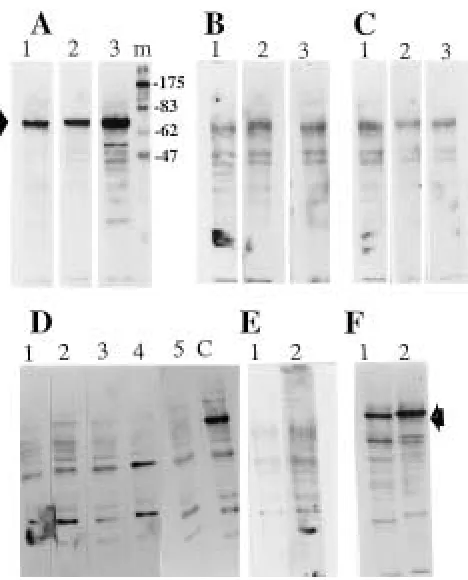
PAGE, transferred to nitrocellulose membranes, and then incubated with patient sera which strongly re-acted with the SQRIPARIHHFPTSI peptide. These patients also reacted with most of the peptides in group A as tested by the immunospot assay. Interest-ingly, a band with an apparent molecular mass of 66 kDa was strongly recognized by IgG antibodies from only the patients who reacted strongly with the pep-tide (Fig. 3A, lanes 1–3). No significant reactivity was obtained with IgG serum antibodies from patients who did not react with the peptide (Figure 3B, lanes 1–3), from normals (Figure 3C, lanes 1–3 ), from pa-tients with prostate cancer (Figure 3D, lanes 1–5), and from patients with osteosarcoma (Figure 3E, lanes 1 and 2). The 66 kDa protein was found to be also expressed by peripheral blood mononuclear cells (Figure 3F, lane 1) and by other cancer cell lines such as the breast cancer cell lines MCF-7 and T47D, and the prostate cancer cell line DU145 (data not shown). Thus, it is more likely to be an autoantigen.
Clinical Significance of the IgG Antibodies to the 66 kDa Protein
To evaluate the role of the described IgG antibodies against the selected SQRIPARIHHFPTSI peptide and the 66 kDa protein in disease duration, we have an-alyzed the patients’ clinical data. It is important to note that the clinical data of the patients were ob-tained after the experimental component of this study was completed. Thus, the present study should be considered as a blind investigation. Surprisingly, all patients with stage IV who did not respond to the phage-displayed peptides (n10), in particular the SQRIPARIHHFPTSI peptide, and to the 66 kDa pro-tein were deceased. The mean survival time of these patients from the first recurrence until the death was 784 days. Three out of six patients with the strongest reactivity are still alive, with a mean survival time from the first recurrence until now of 2553 days. The mean survival time of the three deceased responders
Fig. 2. (Continued)
was 1050 days. There is a difference in survival time between deceased responders and nonresponders (p 0.02).
Following this clinical observation, a second serum sample from a long-time survival patient with 14 years’ duration of disease was obtained, and its reactivity was investigated. Western blot analysis of the patient’s serum at a dilution of 1:1000 showed positive reaction with the 66 kDa protein at both oc-casions (Figure 4, lanes F and S). The antibodies are detectable even at dilution 1/5000, suggesting that a very high concentration of high-affinity antibodies were produced in this patient. This patient has re-sponded to most of the selected phage-displayed peptides (Figure 1B, patient F1). This obervation would indicate that the immune response in this pa-tient is more likely to be directed against different antigens. This is further supported by the fact that phage-displayed peptides with different consensus sequences (Table 1, group A) did not compete with each other in ELISA experiments (e.g., SQRIP ARI-HHFPTSI vs NHEPSVTQVILDRPY). To address whether the SQRIPARIHHFPTSI peptide and the 66 kDa protein are physically related, we have in-vestigated whether affinity purified SQRIPARIHHF-PTSI peptide antibodies would react with the 66 kDa protein. No significant reactivity was obtained with the purified antibodies, indicating that the antigen containing the peptide is not the 66 kDa protein. However, it is worth noting that the anti-66 kDa antibodies were found only in patients with high IgG titer against the peptide.
To determine the cellular localization of the pro-tein, cellular fractions containing the cytosolic, mem-brane and nuclear proteins were prepared from PMI cells and then analyzed by Western blotting. The 66 kDa protein was found to be associated with the cytosolic fraction (Figure 4, lane C).
Discussion
The current study shows that peptide phage display libraries can be used to trace specific immune re-sponses in patients with breast cancer whether or
not the parental antigens are known. Indeed, IgG antibodies to the selected peptides were found in patients with breast cancer and not in patients with lymphoma, osteosarcoma or normal individuals (Figure 1, A and B). Therefore, the selected peptides should constitute a valuable source for the develop-ment of serological markers. Three patients with high titer IgG antibodies against the SQRIPARIH-HFPTSI peptide are still alive despite the stage IV of the disease, indicating that high-titer antibody re-sponses in some patients is indeed translated into an improved outcome.
As shown in Table 1, a variety of peptide core motifs have been selected. Most important, similar, if not identical, peptide sequences were selected from three phage libraries displaying peptides of different lengths. The selection of such core motifs from different libraries is consistent with the pres-ence of specific immune response in the pooled IgG fraction from patients with breast cancer (14). Indeed, the presence of specific IgG antibodies against, for example, the SQRIPARIHHFPTSI pep-tide, was consistent with the competition phage ELISA (Figure 2, B and C). Most of the other phage-displayed peptides in group A also reacted specifically with serum from individual patients. In this respect, 32% and 27% of breast cancer patients reacted with the phage displaying the TVETPQTAKTAYYAT or NHEPSVTQVILDRPY, respectively. In contrast, no significant reactivity was found with normal sera. Thus, phage display-based approaches will un-doubtedly be important for probing immune re-sponses in patients with cancer.
The portion of an antigen molecule that is specifically recognized by an antibody combining site (paratope) is generally referred to as an anti-genic determinant (epitope). Protein epitopes have been divided into two major categories (15). Con-formational epitopes involve residues widely spaced in the primary sequence but brought into spatial proximity by protein folding. These epitopes are ab-sent in denatured proteins. Continuous epitopes con-sist of residues adjacent in the primary sequences, and are therefore detectable in denatured protein. The immunizing epitopes may not represent the op-timal binding motif for the antibodies. Indeed, high affinity mimotopes with no apparent homology to the immunogen have been described (4). The phage display strategy allows the antibodies to select their own ideal peptides. Therefore, some of the selected peptides may not be homologous in sequence to the parental immunogens, yet they perfectly mimic the immunogen structures. These mimic peptides (mi-motopes) are extremely useful as diagnostic and prognostic tools as well as a novel form of vaccine (16,17), especially when protective immunity can not be obtained with the parental antigens. In addi-tion, linear epitopes could be valuable leads for the identification of the parental immunogens (9). In this respect, homology search with the
mune system (23). Humoral and cellular responses against intracellular proteins such as p53 and Ras have been described in patients with cancer (20). Whatever the mechanism that initiated the immune response, the anti-66 kDa antibodies do not seem to cause other clinical features. This observation, to-gether with the expression of the protein in PBMC cells, suggest that the described antibodies are not pathogenic. From an immunological standpoint, both B- and T-cell responses may help to produce anti-tumor response in the long-time survival patients. In addition to being markers for screening, antibodies also have the potential to control tumor growth by antibody-dependent cell mediated cytotoxicity. No-tably, the anti-66 kDa antibodies are IgG which pre-dicts the coexistence of helper T-cell (24).
Regarding vaccines, intracellular and membrane-bound proteins have been used (25). In the case of in-tracellular antigens, T-cell response against tumors has been elicited by immunization with Ras peptides in animal models for cancers (26). In addition, im-munization with MHC class I restricted Ras peptides generated specific T-cell response in patients with pancreatic cancer (27). Notably, tumor-infiltrating lymphocytes have been found in many tumors. These cells can be primed by intracellular proteins released during tumor necrosis. After specific B- and T-cell activation, however, it remains to be deter-mined whether the immune response spreads to other peptide epitopes on the same protein and to different epitopes on different tumor proteins. In principle, this concept of determinant spreading (22) should enhance the eradication of tumors. Many factors, including the nature of the initial anti-gen and perhaps the extent of the “danger” (28) may control the activation of this process. The immune response in the responders who are still alive, in particular patient f1, seems to be directed against more than one antigen. Therefore, the identification of immunogenic antigens that can initiate the deter-minant spreading process should facilitate the de-sign of therapeutic cancer vaccines.
Some studies have evaluated the immunity in breast cancer patients to defined antigens (29–36). However, none of the described antigens appears to be the 66 kDa protein. Notably, antibodies to puri-fied human heat shock protein hsp 90 have been de-tected by ELISA in 37% of patients with breast can-cer (29,30). The presence of these antibodies was found to correlate with patient survival and metasta-tic occurrence (30, 31). Between 10% and 25% of patients with breast cancer have been found to have antibodies against the p53 protein (32). However, their clinical significance was not established (33). Antibodies to epithelial mucin have also been de-tected in patients with breast cancer and found to protect against disease progression (34). High-titer antibodies against Her-2/neu protein were detected in patients with early-stage breast cancer (35) and antibodies to a 128 kDa synaptic protein were found HFPTSI peptide core motif (SQRIP) indicated that it
is present in human CMV coat protein, suggesting a possible molecular mimicry with human pro-tein(s). CMV infection has been implicated in breast cancer (18). However, two patients who responded to the peptide were found to be CMV negative. Thus, the immune response to the SQRIPcore motif peptide is not CMV-related, at least not in these pa-tients. It is worth noting that the phage display strategy identifies both linear and conformational epitopes. Therefore, sequences identified by homol-ogy search should be considered only as leads that need further investigation. Furthermore, the induc-tion of the immune response against the selected peptides is likely to be unrelated to chemotherapy as evidenced by one of the responders, who is still alive but did not receive chemotherapy.
Spontaneous regressions of human tumors have been reported in some types of cancer, including breast cancer (19). This process is more likely to be the result of the interaction of the immune system with tumor cells. Therefore, in some patients, the immune system seems to be activated against tumor cells. Immune responses against some cellular pro-teins such as Ras, p53 and heat shock propro-teins have been described in patients with cancer (20). In the present study, high IgG antibody titers against a 66 kDa cellular protein were found in patients who strongly reacted with group A peptides (Table 1), in particular, the SQRIPARIHHFPTSI peptide. In one patient, the 66 kDa antibodies are detectable even at serum dilution 1/5000. Notably, no physical link was found between the SQRIPARIHHFPTSI peptide and the 66 kDa protein. Thus, it appears that the patients who developed a strong immune response towards the antigen-containing peptides also devel-oped immune response against the 66 kDa protein. This observation is not surprising, as immune re-sponses against different antigens have been de-scribed in patients with cancer. Indeed, sera from melanoma patients reacted with both NY-ESO-1 and MAGE-1 antigens (21). It is worth noting that under some unknown conditions, the immune response to self has the potential to spread in an intra- and in-termolecular fashion (22). This phenomenon, known as “epitope spreading,” is expected to increase the power of the immune response toward target tissues such as tumors. Therefore, it is perhaps not surpris-ing that the patients who strongly responded to most of the selected peptides in group A (Table 1) and the 66 kDa protein had the longest survival. In particu-lar patient f1 who is still doing well.
im-in three women with the stiff-man syndrome and breast cancer (36).
Why do some patients develop such high titers of anti-66 kDa protein IgG antibodies while others do not? Two major sources may account for the initiation of the immune response in these patients. One form of induction of the immune response would be mol-ecular mimicry. Indeed, a similarity between, for example, virus proteins and endogenous protein may lead to autoreactive immune response at the level of antibodies and T cells (37–41). The second form is endogenous immunization. In this case, we can spec-ulate that tolerance against the 66 kDa and the selected peptide-containing antigens is broken in some patients with breast cancer. Notably, it is be-coming clear that many antigenic determinants on self-proteins have not induced self-tolerance (42), and these peptide determinants can be target struc-tures for autoimmune diseases and could provide potential targets for immune response against tu-mors. Thus, understanding the mechamisms that modulate the immune response against these pep-tide determinants (cryptic epitopes) in patients with cancer may reveal what needs to be overcome in the cancer patients to allow the killing of tumors. We believe that in the long-time survival patients, in particular patient f1, an anti-tumor autoimmunity is likely to be responsible for the improved outcome. We are in the process of validating our findings in a prospective study of breast cancer patients that analyses the level of IgG antibody against the se-lected peptides and the 66 kDa protein in serum samples taken at various time points (e.g., diagno-sis) and relates the presence of these antibodies to the outcome of the disease.
In conclusion, our data identify specific IgG an-tibodies in breast cancer patients towards the se-lected peptides, in particular the SQRIPARIHHF-PTSI peptide. High titers of IgG antibodies against a 66 kDa protein was found in some patients who re-sponded to the peptide and have long survival time. This observation should have therapeutic potential. The identity of the 66 kDa protein is under investi-gation in our laboratory. Finally, the demonstration that specific immune responses in patients with can-cer can be traced by phage display should facilitate the study of tumor immunology.
Acknowledgements
This work is supported in part by the Norwegian Foundation for Health and Rehabilitation to Dr. Sioud. We thank Dr. Øyvind Melien and Anne Dybwad for critical reading of the manuscript. We are indebted to the treating physicians and the patients at Ullevål University Hospital for their cooperation.
References
1. Houghton AN. (1994) Cancer antigens: immune recognition of self and altered self. J. Exp. Med.180:1–4.
2. Sahin U, Tureci O, Schmitt H, Cochlovius B, Johannes T, Schmits R, Stenner F, Luo G, Schobert I, Pfreundschuh M. (1995) Human neoplasms elicit multiple specific immune responses in the autologous host. Proc. Natl. Acad. Sci. USA92: 11810–11813.
3. Scott JK, Smith GP. (1990) Searching for peptide ligands with an epitope library. Science249:386–390.
4. Bonnycastle LLC, Mehroke JS, Rashed M, Gong X, Scott JK. (1996) Probing the basis of antibody reactivity with a panel of constrained peptide libraries displayed by filamentous phage. J. Mol. Biol.258:747–762.
5. Stephen CW, Lane DP. (1992) Mutant conformation of p53. Precise epitope mapping, a filamentous phage epitope li-brary. J. Mol. Biol.225:577–583.
6. Smith GP. (1985) Filamentous fusion phage: Novel expres-sion vectors that display cloned antigens on the virion sur-face. Science228:1315–1316,.
7. Dybwad A, Førre Ø, Kjeldsen-Kragh J, Natvig JB, Sioud M. (1993) Identification of new B cell epitopes in the sera of rheumatoid arthritis patients using a random nanopeptide phage library. Eur J. Immunol.23:3189–3193.
8. Sioud M, Dybwad A, Jespersen L, Suleyman S, Natvig JB, Førre Ø. (1994) Characterization of naturally occurring autoantibodies against tumour necrosis factor-alpha (TNF-a): In vitrofunction and precise epitope mapping by phage epi-tope library. Clin. Exp. Immunol.98: 520–525.
9. Dybwad A, Bogen B, Natvig JB, Førre Ø, Sioud M. (1995) Peptide phage libraries can be an efficient tool for identifying antibody ligands for polyclonal antisera. Clin. Exp. Immunol.
102:438–442.
10. Folgori A, Tafi R, Meola A, Felici F, Galfre G, Cortese R, Monaci P, Nicosia A. (1994) A general strategy to identify mi-motopes of pathological antigens using only random peptide libraries and human sera. EMBO J.13:2236–2243.
11. Bartoli F, Nuzzo M, Urbanelli L, Bellintani F, Prezzi C, Cortese R, Monaci P. (1998) DNA-based selection and screening of peptide ligands. Nature Biotech16:1068–1073.
12. Sambrook J, Fritsch EF, Maniatis T (eds.) “Molecular Cloning: A Laboratory Manual.” Cold Spring Harbor, NY: Cold Spring Harbor Laboratory Press, 1982.
13. Christian RB, Zuckermann RN, Kerr JM, Wang L, Malcolm BA. (1992) Simplified methods for construction, assessment and rapid screening of peptide libraries in bacteriophage. J. Mol. Biol.227:711–718.
14. Felici F, Galfre G, Luzzago A, Monaci P, Nicosia A, Cortese R. (1996) Phage-displayed peptides as tools for characterization of human sera. Methods Enzymol.267:116–129.
15. Berzofsky JA. (1985) Intrinsic and extrinsic factors in protein antigenic structure. Science229:932–940.
16. Meola A, Delmastro P, Monaci P, Luzzago A, Nicosia A, Felici F, Cortese R, Galfre G. (1995) Derivation of vaccines from mi-motopes: immunogenic properties of human hepatitis b virus surface antigen mimotopes displayed on filamentous phage. J. Immunol.154:3162–3172.
17. Apostololoulos V, Sandrin MS, McKenzie IF. (1999) Mimics and cross reactions of relevance to tumour immunotherapy. Vaccine18:268–275.
18. Richardson A. (1997) Is breast cancer caused by late exposure to a common virus? Med. Hypotheses48:491–497.
19. Balch CM, Houghton AN, Milton GW, Soong SJ. Cutaneous melanoma. Philadelphia: Lippincott, 1992.
20. Disis ML, Cheever MA. (1996) Oncogenic proteins as tumor antigens. Curr. Opin. Immunol.8:637–642.
21. Stockert E, Jager E, Chen Y-T, Scanlan MJ, Gout I, Karbach J, Arand M, Knuth A, Old LJ. (1998) A survey of the humoral immune responses of cancer patients to a panel of human tu-mor antigens. J. Exp. Med.187:1349–1354.
22. Kaufman DL, Clare-Salzler M, Tian J, Forsthuber T, Ting GSP, Robinson P, Atkinson MA, Sercarz EE, Tobin AJ, Lehmann PV. (1993) Spontaneous loss of T-cell tolerance to glutamic acid decarboxylase in murine insulin-dependent diabetes. Nature366:69–72.
intracellu-lar proteins: clinical and biological significance. Am. J. Med.
80:53–61.
24. Stevens TL, Bossie A, Sanders VM, Fernandez-Botram R, Coffman LR, Mosmann RT, Vitetta ES. (1988) Regulation of antibody isotype secretion by subsets of antigen-specific helper T cells. Nature334:255.
25. Knuth A, Wolfel T, Meyer zum, Buschenfelde KH. (1991) Cel-lular and humoral responses against cancer: implication for cancer vaccines. Curr. Opin. Immunol.3:659–664.
26. Fenton RG, Keller CJ, Hanna N, Taud DD. (1995) Induction of T-cell immunity against Ras oncoprotein by soluble protein or Ras-expressing E. Coli. J. Natl. Cancer Inst.87:1853–1861. 27. Gjertsen MK, Bakka A, Breivik J, Saeterdal I, Solheim BG,
Soreide O, Thorsby E, Gaudernack G. (1995) Vaccination with mutant Ras peptides and induction of T-cell responsive-ness in pancreatic carcinoma patients carrying the corre-sponding Ras mutation. Lancet346:1399–1400.
28. Matzinger P. (1994) Tolerance, danger, and the extended fam-ily. Annu. Rev. Immunol.12:991–1045.
29. Conroy SE, Latchman DS. (1996) Do heat shock proteins have a role in breast cancer? Brit. J. Cancer74:717–721.
30. Conroy SE, Gibson SL, Brunstrom G, Isenberg D, Luqmani Y, Latchman DS. (1995) Autoantibodies to the 90kD heat shock protein in sera of breast cancer patients. Lancet345:126. 31. Conroy SE, Sasieni PD, Fentiman I, Latchman DS. (1998)
Au-toantibodies to the 90 kD heat shock protein and poor sur-vival in breast cancer patients. Eur. J. Cancer34:942–943. 32. Crawford LV, Pim DC, Bulbrook RD. (1982) Detection of
an-tibodies against the cellular protein P53 in sera from patients with breast cancer. Int. J. Cancer30:403–408.
33. Vojtesek B, Kovarik J, Dolezalova H, Nenutil R, Havlis P,
Brentani RR, Lane DP. (1995) Absence of P53 autoantibodies in a significant proportion of breast cancer patients. Br. J. Cancer
71:1253–1256.
34. von Mensdorff-Pouilly S, Gourevitch MM, Kenemans P, Ver-straete A, Litvinov SV, van Kamp GJ, Meijer S, Vermorken J, Hilgers J. (1996) Humoral immune response to polymorphic epithelial mucin 1 in patients with benign and malignant breast tumours. Eur. J. Cancer32:1325–1331.
35. Disis ML, Pupa SM, Gralow JR, Dittadi R, Menard S, Cheever MA. (1997) High-titer HER-2/neu protein specific antibody can be detected in patients with early-stage breast cancer. J. Clin. Oncol.15:3363–3367.
36. Folli F, Solimena M, Cofiell R, Austoni M, Tallini G, Fassetta G, Bates D, Cartlidge N, Bottazzo GF, Piccolo G. (1993) Autoan-tibodies to a 128 kDa synaptic protein in three women with the stiff-man syndrome and breast cancer. N. Engl. J. Med.238: 546–551.
37. Oldstone MB. (1987) Molecular mimicry and autoimmune disease. Cell 50:819–820.
38. von Herrath MG, Oldstone MB. (1996) Virus-induced au-toimmune disease. Curr. Opin. Immunol.8:878–885.
39. Davies JM. (1997) Molecular mimicry: can epitope mimicry induce autoimmune disease? Immunol. Cell Biol.75:113–126. 40. Wucherpfennig KW, Strominger JL. (1995) Molecular mimicry in T-cell-mediated autoimmunity: viral peptides activate human T cell clones specific for myelin basic protein. Cell80:695–705. 41. Talbot PJ, Paquette JS, Ciurli C, Antel JP, Ouellet F. (1996)
Myelin basic protein and human coronavirus 229E cross-reactive T cells in multiple sclerosis. Ann. Neurol.39:233–240. 42. Lanzavecchia A. (1995) How can cryptic epitopes trigger