R E V I E W
Open Access
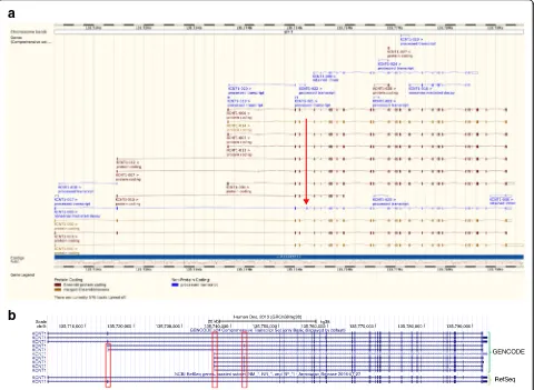
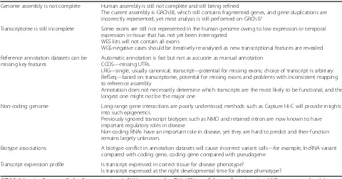
Genome annotation for clinical genomic
diagnostics: strengths and weaknesses
Charles A. Steward
1,2*, Alasdair P. J. Parker
3, Berge A. Minassian
4,5, Sanjay M. Sisodiya
6,7, Adam Frankish
2,8and Jennifer Harrow
2,9Abstract
The Human Genome Project and advances in DNA sequencing technologies have revolutionized the
identification of genetic disorders through the use of clinical exome sequencing. However, in a considerable
number of patients, the genetic basis remains unclear. As clinicians begin to consider whole-genome
sequencing, an understanding of the processes and tools involved and the factors to consider in the
annotation of the structure and function of genomic elements that might influence variant identification is
crucial. Here, we discuss and illustrate the strengths and weaknesses of approaches for the annotation and
classification of important elements of protein-coding genes, other genomic elements such as pseudogenes
and the non-coding genome, comparative-genomic approaches for inferring gene function, and new
technologies for aiding genome annotation, as a practical guide for clinicians when considering pathogenic
sequence variation. Complete and accurate annotation of structure and function of genome features has the
potential to reduce both false-negative (from missing annotation) and false-positive (from incorrect
annotation) errors in causal variant identification in exome and genome sequences. Re-analysis of unsolved
cases will be necessary as newer technology improves genome annotation, potentially improving the rate of
diagnosis.
Background
Advances in genomic technologies over the past 20 years
have provided researchers with unprecedented data
re-lating to genome variation in different diseases [1].
How-ever, even after whole-exome sequencing (WES), the
genetic basis for a particular phenotype remains unclear
in a considerable proportion of patients. Here, we
exam-ine how genomic annotation might influence variant
identification, using examples mostly from both
com-mon and rarer neurological disorders. We highlight why
the present technology can fail to identify the pathogenic
basis of a patient
’
s disorder, or produce an incorrect
re-sult where the wrong variant is labelled as causative. For
these reasons, we believe it is important to re-analyse
unresolved cases as newer technology and software
im-prove gene and genome annotation. The aim of this
paper is to make common genomic techniques
access-ible to clinicians through the use of figures and examples
that help to explain genome sequencing, gene
classifica-tion and genome annotaclassifica-tion in the context of
patho-genic sequence variation. Finally, we discuss how new
genomic techniques will improve our ability to identify
pathogenic sequence variation.
Genome sequencing
The Human Genome Project (HGP) was launched
offi-cially in 1987 by the US Department of Energy to
se-quence the approximately 3 billion base-pairs (bp) that
constitute the human genome [2]. The first draft
se-quence was published in 2001 and computational
anno-tation, a process that attributes a biological function to
the genomic elements, described 30,000 to 40,000
protein-coding genes across 22 pairs of autosomes and
the X and Y sex chromosomes in a genome of 2.9 billion
bases (gigabases, Gb) [2]. The precise size and gene
count of the reference human genome remains uncertain
to this day because sequence gaps remain, while the
* Correspondence:charles.steward@congenica.com1
Congenica Ltd, Wellcome Genome Campus, Hinxton, Cambridge CB10 1DR, UK
2The Wellcome Trust Sanger Institute, Wellcome Genome Campus, Hinxton,
Cambridge CB10 1SA, UK
Full list of author information is available at the end of the article
classification of genes becomes more refined [3].
Conse-quently, additions are continually made to the genome
to fill sequence gaps [4]. The most recent published
esti-mates suggest that just under 20,000 protein-coding
genes [5] are present in a genome of approximately 3.1
Gb [6]. The HGP enabled initial research examining
se-quence variation on chromosome 22 [7], to more recent
medical advances that now see DNA sequencing used
routinely in large-scale research programs, such as the
Deciphering Developmental Disorders (DDD) study [8,
9]. Sequencing for the HGP used the chain terminator
method [10], more commonly known as
‘Sanger
sequen-cing’
, and owing to the better-quality sequence data and
read-length associated with Sanger sequencing
com-pared with current sequencing technologies, Sanger
se-quencing is still used to confirm sequence variants [11].
Current methods for producing the raw sequence data
for whole-genome sequencing (WGS) are placed into two
categories based upon the length of the nucleotide
se-quence produced, or sese-quence
‘read’. Short-read technology
comes from Illumina Inc. [12] and uses well-established
chemistry to identify the sequence of nucleotides in a given
short segment of DNA. Illumina sequencing platforms such
as the HiSeq X produce base-pair reads of lengths from
150 to 250 bp in a given DNA segment and are used to
read sequences from both ends of a DNA fragment. This
‘next-generation’
technology is a dramatic improvement
over older Sanger sequencing methods that produced
lon-ger reads but at much higher cost [13]. More recently,
‘third-generation’
technologies from Pacific Biosciences
(PacBio) and Oxford Nanopore are gaining users and
mak-ing an impact. These third-generation methods generate
longer reads, up to tens of thousands of base-pairs per read,
but with higher error rates.
The speed of DNA sequencing, the amount of
se-quence that can be produced and the number of
ge-nomes that can be sequenced have increased massively
with next-generation sequencing (NGS) techniques
[14]. Such advances have enabled large collaborative
projects that look at variation in a population, such as
the 1000 Genomes Project [15], as well as those
investi-gating the medical value of WGS, such as the UK
100,000 Genomes Project [16]. It is hoped that WGS
will facilitate the research, diagnosis and treatment of
many diseases.
Once a patient genome has been sequenced, it needs
to be aligned to the reference genome and analysed for
variants. Typically, software algorithms such as the
Burrows-Wheeler Aligner (BWA) are used for
short-[17] and long-read [18] alignment and the Genome
Ana-lysis Toolkit (GATK) is used to identify or
‘
call
’
sequence
variants [19]. Figure 1 illustrates a typical genome
ana-lysis pipeline, describing the different file formats
com-monly used
—
FASTQ [20], BAM [21] and VCF [22].
Pathogenic sequence variation can range in size from
single-nucleotide variants (SNVs), small insertions and
deletions (
‘
indels
’
) of fewer than 50 base-pairs in length,
to larger structural variants (SVs) [23], which are
gener-ally classified as regions of genomic variation greater
a
b
c
Fig. 1The genome analysis pipeline. Note that, for clarity, some steps have been omitted. Figure illustrations are not to scale and are only meant to be illustrative of the differences between short- and long-read sequencing.aUnaligned reads from sequencing machines are stored as FASTQ file formats. This is a text-based format for storing both a DNA sequence and its corresponding quality scores.bReads are aligned to the genome. Short reads provide deep coverage, whereas reads that have been sequenced from both ends (blue arrows) help to orientate unaligned contigs. It is difficult to align short reads confidently across repetitive sequences when the repeating genome sequence is longer than the sequence read. Long-read sequences help to order contigs across larger regions, particularly with repetitive sequences, but do not provide the necessary depth needed to be confident of calling a base at a certain position. Note that there is a large region where there is no read coverage at all. This is indicative of structural variation. Here, the patient has a large deletion with respect to the reference genome. Once the reads have been aligned to the reference genome they are stored in a BAM file. A BAM file (.bam) is the binary version of a sequence alignment map (SAM file format). The latter is a tab-delimited text-based format for storing DNA sequences aligned to a reference sequence.cThe Variant Call Format (VCF) specifies the format of a text file used in bioinformatics for storing genetic sequence variations. VCF files are much smaller than FASTQ and BAM files. Note that single-nucleotide variants (SNVs) and small insertions and deletions (‘indels’) are illustrated asredandpurple blocks, whereas a much larger structural variant is indicated by an
than 1 kb, such as copy-number variants (CNVs),
inser-tions, retrotransposon elements, inversions, segmental
duplications, and other such genomic rearrangements
[24, 25]. Currently, the consequence of non-synonymous
variants of the protein-coding elements only can be
rou-tinely automatically predicted by algorithms such as
SIFT and PolyPhen [26], yet many different types of
vari-ants are implicated in disease. As sequencing techniques
begin to move away from
‘gene panel’
testing to WGS, it
is crucial to understand the structure of genes and any
regulatory features that might lie within intra/intergenic
regions as changes in any of these regions might have a
crucial impact on the function of a gene.
Recently, the American College of Medical Genetics
and Genomics (ACMG) recommended a set of
stan-dards and guidelines to help medical geneticists assign
pathogenicity using standardized nomenclature and
evidence used to support the assignment for
Mendel-ian disorders [27]. For example, the terms
‘mutation’
and
‘polymorphism’
have often been used
mislead-ingly, with assumptions made that
‘mutation’
is
patho-genic, whereas
‘polymorphism’
is benign. As such, one
recommendation that ACMG makes is that both these
terms are replaced by
‘variant’
, with the following
modifiers (1) pathogenic, (2) likely pathogenic, (3)
un-certain significance, (4) likely benign, or (5) benign
[27]. As such, here, we use the term variant. A
stand-ard gene-variant nomenclature is maintained and
ver-sioned by the Human Genome Variation Society
(HGVS) [28]. Both ACMG and HGVS examples are
il-lustrated in Table 1.
Classifying genes and other genomic elements
Current gene sets identify under 20,000 protein-coding
genes and over 15,000 long non-coding RNAs (lncRNAs)
[29, 30]. In this section, for clinicians who might not be
fa-miliar with gene structure and function, we present the
important elements of different parts of protein-coding
genes, and other categories of genomic elements, such as
pseudogenes and elements of the non-coding genome
such as lncRNAs, and we highlight their potential
func-tionality, illustrated with examples of their roles in disease.
We demonstrate the importance of classifying such
re-gions correctly and why incorrect classification could
im-pact the interpretation of sequence variation.
Important elements of protein coding genes
A eukaryotic gene is typically organized into exons and
in-trons (Fig.
2), although some genes, for example
SOX3,
which is associated with X-linked mental retardation [31],
can have a single exon structure. The functional regions of
protein-coding genes are typically designated as the
coding sequence (CDS) and the 5′
and 3′
untranslated
re-gions (UTRs) (Fig.
2).
The 5′
UTR of a transcript contains regulatory regions.
For example, some upstream open reading frames
(uORFs; which are sequences that begin with an ATG
codon and end in a stop codon, meaning that they have
the potential to be translated) in the 5′
UTR are translated
to produce proteins that could enhance or suppress the
function of the main CDS [32]. Experimental techniques
such as cap-analysis gene expression (CAGE) [33] are
used to identify transcription start sites (TSSs) (Fig.
2a).
Table 1
Examples of disease-causing variation with associated HGVS nomenclature
Location Gene Variation HGVS nomenclature ACMG clinical significance
Associated disorder Reference
5′UTR FMR1 Expansion NM_002024.5(FMR1):c.-128_-126(200)
Pathogenic Fragile X syndrome [186]
CDS GRIN2A Nonsense NM_000833.4(GRIN2A):c.2041C > T (p.Arg681Ter)
Pathogenic Idiopathic focal epilepsy (IFE) with rolandic spikes is the most common childhood epilepsy
[187]
CDS GABRB3 Missense NM_021912.4(GABRB3):c.745C > A (p.Gln249Lys)
Pathogenic Early infantile epileptic encephalopathy (EIEE) [188]
CDS WDR62 Deletion/
frameshift
NM_001083961.1(WDR62) :c.3839_3855del17 (p.Gly1280Alafs)
Pathogenic Malformations of cortical development [189]
3′UTR MECP2 SNV NM_004992.3(MECP2):c.*2956G > A Uncertain significance
Rett syndrome [190]
Promoter CRH SNV NC_000008.11:g.66178947G > T Pathogenic Familial autosomal dominant nocturnal frontal lobe epilepsy
[191]
Splice site
ATP6AP2 SNV NM_005765.2(ATP6AP2):c.321C > T (p.Asp107=)
Pathogenic X-linked mental retardation and epilepsy due to inefficient inclusion of exon 4
[192]
Poly(A) ARSA SNV NM_000487.5(ARSA):c.*96A > G Pathogenic Metachromatic leukodystrophy [193]
NMD SNRPB SNV NM_003091.3(SNRPB):c.-72C > A Pathogenic Cerebro-costo-mandibular syndrome [194]
lncRNA ATXN8OS Insertion NR_002717.2(ATXN8OS) :n.1103_1105CTG(15_40)
Pathogenic Spinocerebellar ataxia type 8 [195]
ACMGAmerican College of Medical Genetics and Genomics,CDScoding sequence,HGVSHuman Genome Variation Society,lncRNAlong non-coding RNA,NMD
Variants in the CDS are generally the most well
stud-ied and understood area of pathogenic sequence
vari-ation. For example, approximately 700 pathogenic CDS
variants have been reported in the epilepsy-associated
gene
SCN1A
[34].
The 3′
UTR of a transcript can contain regions
control-ling regulatory proteins such as RNA binding proteins
(RBPs) and microRNAs (miRNAs) (Fig.
2a). Interestingly,
the 3′
UTR has been linked to overall translation
effi-ciency and stability of the mRNA [35]. The 5′
and 3′
UTRs can also interact with each other to regulate
translation through a closed-loop mechanism [36].
Im-portant sequence motifs involved in controlling the
ex-pression of a gene include promoters, enhancers and
a
b
c
d
e
silencers, which are found in exonic, intragenic and
intergenic regions (Fig.
2a).
A multi-exonic eukaryotic gene can produce different
disease phenotypes through alternative protein isoforms
that result from the use of alternative splice site/exon
combinations (Fig.
3) [37]. Canonical splice sites are
generally conserved at the 5′
(donor) and 3′
(acceptor)
ends of vertebrate introns. The GT–intron–AG
config-uration is the most common, although other, rarer
in-stances of splice sites are found, such as GC–intron–AG
and AT–intron–AC [38].
Although there can be an abundant transcript that is
expressed in a particular cell, the same transcript might
not dominate elsewhere, and, even if a dominant
tran-script is identified, the trantran-script might not be functional
[39]. Differential expression can be both tissue- and
age-specific [40], can occur in response to different
environ-mental signals [41, 42], and an exon expressed in one
tissue might not be relevant to further analysis if it is
not expressed in the tissue where a disease phenotype is
present. For example, genes expressed in brain generally
have longer 3′
UTRs than those in other tissues, and
such differences could impact miRNA binding sites and
other regulatory regions [43]. Studies have shown that
retained introns have an important role in brain gene
ex-pression and regulation [44, 45].
Polyadenylation (poly(A)), which involves addition of
the poly(A) tail, is important for nuclear export to the
cytosol for translation by the ribosome and also helps
with mRNA stability (Fig. 2d). Many annotated genes
also have more than one poly(A) site, which can be
func-tional in different tissues or different stages of
develop-ment [42].
After translation, the polypeptide chain produced by
the ribosome might need to undergo posttranslational
modification, such as folding, cutting or chemical
modi-fications, before it is considered to be a mature protein
product (Fig. 2e). Noonan syndrome is believed to result
from the disruption of the phosphorylation-mediated
auto-inhibitory loop of the Src-homology 2 (SH2)
do-main during post-translational modification [46].
Transcripts that contain premature stop codons
(perhaps as a result of using an alternative splice
donor, splice acceptor, or inclusion/exclusion of an
alter-native exon, which causes a CDS frameshift) are degraded
through the nonsense-mediated decay (NMD) cellular
surveillance pathway (Fig. 4) [47, 48]. NMD was originally
believed to degrade erroneous transcripts, but much
evi-dence has been found to suggest it is also an active
regula-tor of transcription [49, 50]. Several NMD facregula-tors have
been shown to be important for the regulation of
neuro-logical events such as synaptic plasticity and neurogenesis
[51
–
53].
Two other types of cellular surveillance pathways are
known to exist: non-stop decay and no-go decay.
Non-stop decay is a process that affects transcripts that have
poly(A) features but do not have a prior stop codon in
the CDS. The translation of such transcripts could
pro-duce harmful peptides with a poly-lysine amino acid
se-quence at the C-terminal end of the peptide—therefore,
these transcripts are subject to degradation. Similar to
NMD transcripts, either aberrant splicing or SNVs can
cause the generation of these transcripts [54]. Finally,
no-go decay is triggered by barriers that block ribosome
movement on the mRNA [55].
The functional importance of pseudogenes
Pseudogenes are traditionally regarded as
‘broken’
cop-ies of active genes. Freed of selective pressure, they
have typically lost the ability to encode functional
pro-teins through the occurrence of nonsense variations,
frameshifts, truncation events, or loss of essential
regu-latory elements. The majority of pseudogenes fall into
one of two categories: processed and unprocessed
(Fig. 5, Table 2) [56].
Processed pseudogenes represent back-integration or
retrotransposition of an RNA molecule into the genome
sequence, and, although they generally lack introns, they
frequently incorporate the remains of the poly(A) tail.
Processed pseudogenes are often flanked by direct
re-peats that might have some function in inserting the
pseudogene into the genome, and are often missing
se-quence compared with their parent gene (Fig. 5) [57]. By
contrast, unprocessed pseudogenes are defunct relatives
of functional genes that arise through faulty genomic
du-plication resulting in missing (parts of ) exons and/or
flanking regulatory regions (Fig. 5).
Fig. 4The nonsense-mediated decay (NMD) pathway. Under normal cellular circumstances, exon–exon junction complexes (EJCs) that are in place after splicing are removed by the ribosome during the first round of translation. However, when a transcript contains a premature termination codon (PTC), perhaps as a result of an single-nucleotide variant (SNV), indel or inclusion of an out-of-frame exon upstream of one or more EJCs, these EJCs remain in place because the ribosome complex disassociates at the premature stop codon and thus cannot remove the downstream EJC. This triggers the NMD pathway, and the transcript is degraded
a
b
Computational annotation of pseudogenes tends to
suffer from significant false positives/negatives and can
cause problems that result from the misalignment of
NGS data. Specifically, identification of transcribed
pseu-dogenes and single-exon pseupseu-dogenes can be a challenge
[58]. Such difficulties were demonstrated where it was
found that more than 900 human pseudogenes have
evi-dence of transcription, indicating functional potential
[58, 59]. Consequently, the ability to distinguish between
pseudogenes and the functional parent gene is essential
when predicting the consequence of variants.
MacArthur and colleagues [60] reported that reference
sequence and gene annotation errors accounted for
44.9% of candidate loss-of-function (LoF) variants in the
NA12878 genome, which belongs to the daughter from
a trio of individuals belonging to the CEPH/Utah
pedi-gree whose genomes were sequenced to high depth as
part of the HapMap project [61]. The NA12878 genome
sequence and transformed cells from the same individual
(the GM12878 cell line) are often used as a reference in
other projects [62, 63]. After reannotation of
protein-coding genes harbouring 884 putative LoF variants, 243
errors in gene models were identified, 47 (19.3%) of
which were updated from protein-coding to pseudogene,
removing a significant source of false-positive LoF
anno-tation [60].
Table 2
GENCODE annotation biotypes (2017)
Biotype Description
Protein coding Contains an ORF that has strong coding potential
Known coding 100% identical to known RefSeq protein or Swiss-Prot entry
Novel coding Shares >60% length with known coding sequence from RefSeq, or Swiss-Prot, or has cross-species/family support or domain evidence
Putative coding Shares <60% length with known coding sequence from RefSeq, or Swiss-Prot, or has an alternative first or last coding exon
Nonsense-mediated decay If the coding sequence (following the appropriate reference) of a transcript finishes >50 bp from a downstream splice site, then it is tagged as NMD. If the variant does not cover the full reference coding sequence, then it is
annotated as NMD if NMD is unavoidable—i.e. no matter what the exon structure of the missing portion is, the transcript will be subject to NMD
Non-stop decay Transcripts that have poly(A) features (including signal) without a prior stop codon in the CDS—i.e. a non-genomic poly(A) tail attached directly to the CDS without a 3′UTR; these transcripts are subject to degradation
Retained intron Alternatively spliced transcript believed to contain intronic sequence relative to other, coding, variants
Processed transcript Cannot assign an ORF, but is part of a coding locus
lncRNA Long non-coding RNA—lacks protein-coding potential and is of length >200 bp
Bidirectional promoter Transcription start sites of the lncRNA model and the protein-coding model are on opposite strands and within 200 bp of one another, or are found in the same CpG island
3-Prime overlapping Transcription start site and/or published experimental data support independent transcription from the 3′UTR of a coding gene
Antisense At least one variant overlaps a protein-coding locus on the opposite strand, or evidence of antisense regulation of a coding gene has been published
lincRNA Long intergenic ncRNA: does not overlap (neither sense nor antisense) a coding gene
Sense intronic In an intron of a coding gene; no exonic overlap
Sense overlapping Contains a coding gene in an intron; no exonic overlap.
Pseudogene Matches to protein, but ORF disrupted by frameshifts and/or premature stop codons
Processed Lacks introns and arose from retrotransposition of parent gene mRNA
Unprocessed Can contain introns and is produced by genomic duplication
Transcribed Locus-specific transcripts indicate transcription; these can be classified into‘processed’and‘unprocessed’
Translated Locus-specific protein mass spectroscopy data suggest translation; the connection is maintained with the pseudogene biotype until the experimental community validates it as a coding gene
Polymorphic Pseudogene owing to a single-nucleotide variant (SNV), or insertion-deletion variant (indel); but the same gene is translated in other individuals/haplotypes/strains
Unitary Species-specific unprocessed pseudogene, without a parent gene, that has an active orthologue in another species
Data sourced from GENCODE project [196]
Transcripts derived from the pseudogene locus
PTENP1
have been shown to regulate the parent
PTEN
locus [64].
Deletion of
PTENP1
has been reported to downregulate
PTEN
expression in breast and colon cancer [64] and
mel-anoma [65], and downregulation of
PTENP1
through
methylation of its promoter sequence in clear-cell renal cell
carcinoma suppresses cancer progression [66]. Although
PTENP1
has not yet been associated with any neuronal
dis-orders, both
PTEN
and
PTENP1
are expressed in multiple
brain tissues [67, 68].
The non-coding genome
Most of the genome is non-coding, and therefore most
variation occurs in non-coding regions. To understand
the effect of a sequence variant in such regions, the
non-coding elements need to be classified. Non-non-coding
ele-ments consist of
cis-regulatory elements such as
pro-moters and distal elements (for example, enhancers) [69]
and non-coding RNAs (ncRNAs). Large collaborative
initiatives, such as ENCODE [63] and RoadMap
Epige-nomics [70], have been tasked to create comprehensive
maps of these regions. The Ensembl regulatory build
[71] and Variant Effect Predictor (VEP) [72] are able to
determine whether variants fall within such regions, but
are not yet able to determine pathogenicity, although
tools that do so are beginning to emerge, such as
Fun-Seq [73] and Genomiser [74].
The ncRNAs are generally divided into two groups,
small RNAs (sRNAs) and lncRNAs. sRNAs include
miR-NAs, Piwi-interacting RNAs (piRNAs), short interfering
RNAs (siRNAs), small nucleolar RNAs (snoRNAs) and
other short RNAs [75]. The sRNAs can be predicted using
tools such as Infernal [76] and Rfam [77], which makes
the interpretation of sequence variation and consequence
easier, especially when compared with the analysis of
lncRNAs. However, correctly discriminating functional
copies from pseudogenes remains a challenge.
Of particular interest to the study of neurological
dis-ease are microRNAs (miRNAs), which are small
(approxi-mately 20 nucleotides) ncRNAs that are involved in the
regulation of post-transcriptional gene expression [78].
miRNAs can trigger transcript degradation, modify
trans-lational efficiency and downregulate gene expression by
triggering epigenetic changes (DNA methylation and
his-tone modifications) at the promoter of target genes, and
are the best-understood of the ncRNAs. Studies have
shown that variants in miRNA binding sites are associated
with some neurological diseases, and there is evidence
for a role in epilepsy, suggesting that miRNAs might
be good candidates for the development of novel
molecu-lar approaches for the treatment of patients with epilepsy
[79, 80]. For example, miRNA
MIR328
binds to the 3
′
UTR of
PAX6
to regulate its expression. However,
vari-ation in the miRNA binding site reduces the binding
affin-ity of
MIR328
, which in turn results in an increase in the
abundance of
PAX6
transcripts, which is associated with
electrophysiological features of Rolandic epilepsy [81].
The EpiMiRNA consortium is investigating the role of
miRNAs in the development, treatment and diagnosis of
temporal lobe epilepsy [82].
The classification of lncRNAs is increasingly used to
convey functional information, despite the fact that we
know relatively little about the role or mechanism of the
vast majority of them (Fig. 6). The term lncRNA was itself
established to distinguish longer ncRNAs from the small
ncRNAs that were initially separated using an experimental
threshold of >200 nucleotides, which remains the simplest
definition of a lncRNA [63]. RNA sequencing (RNA-Seq)
assays predict that potentially tens, if not hundreds, of
thousands of lncRNA transcripts have now been identified
[83], which has inevitably led to the naming of many
pro-posed subclasses of lncRNA [84, 85]. Without any
inter-national agreement on the classification of lncRNAs,
proposed subclasses have been classified based on either
length, function, sequence or structural conservation, or
Fig. 6Different classifications of long non-coding RNAs (lncRNAs). The classification of lncRNAs is based on their position with respect to coding genes. lncRNAs are illustrated here with onlyredexons, whereas the coding genes are shown asredandgreen. ASantisense,BDPbi-directional promoter,lincRNAlong-intergenic RNA (not overlapping a protein-coding locus on either strand),OSoverlapping sense,O3′overlapping 3′,SI
association with either protein-coding genes, DNA
ele-ments, subcellular location or a particular biological state.
They are hard to predict owing to their size, but also
be-cause they are expressed at low levels and lack a known
ter-tiary structure, unlike miRNAs. A recent study by Nitsche
and colleagues showed that >85% of lncRNAs have
con-served splice sites that can be dated back to the divergence
of placental mammals [86].
lncRNAs, such as
XIST
[87], have been studied for some
time, yet little is known about the function of most.
How-ever, they are gaining interest within the scientific and
medical community [63] owing to their potential
involve-ment in disease [88, 89]. Experiinvolve-ments in mouse models
have demonstrated that dysregulation of certain lncRNAs
could be associated with epilepsy [90], and a role in gene
regulation is proposed for the vast number of unstudied
cases [91], which makes them interesting candidates for
new targeted therapies and disease diagnostics [92]. For
example, experiments in a knock-in mouse model of
Dra-vet syndrome have shown that the upregulation of the
healthy allele of
SCN1A
by targeting a lncRNA improved
the seizure phenotype [93].
CNVs also play an important role in human disease
and can affect multiple coding genes, resulting in dosage
effects, truncation of single genes or novel fusion
prod-ucts between two genes. CNVs have also been shown to
be pathogenic in non-coding regions [94]. Talkowski and
colleagues [95] observed a CNV causing disruption in
the
long-intergenic
non-coding
RNA
(lincRNA)
LINC00299
in patients with severe developmental delay,
raising the possibility that lincRNAs could play a
signifi-cant role in developmental disorders. More recently,
Turner et al. [96] reported WGS of 208 patients from 53
families with simplex autism and discovered small
dele-tions within non-coding putative regulatory regions of
DSCAM
, implicated in neurocognitive dysfunction in
Down syndrome. These CNVs were transmitted from
the mother to the male proband.
Repetitive sequences and transposable elements are
known to be involved in disease and are believed to make
up more than two-thirds of the human genome. They also
have a strong association with genomic CNVs [97]. Long
interspersed nuclear elements (LINEs) and
Alu
repeats
(which are types of retrotransposons) have been associated
with increased genomic instability through non-allelic
homologous recombination events and can lead to
patho-genic duplications and deletions [98].
Alu
–
Alu
repeat
re-combinations within the introns of
ALDH7A1
have been
associated with pyroxidine-dependent epilepsy [99]. The
ability to accurately detect repetitive sequences is of great
importance due to the problems they can cause during
the aligning or assembling of sequence reads [100], and
the human genome is commonly analysed for repeats
using Repbase annotation [101] and computational
algorithms, such as the hidden Markov model
(HMM)-de-rived database Dfam [102].
Genome annotation
The ability to understand the function of a gene and how
variation might affect its function is dependent upon
un-derstanding its structure, which can be elucidated by
gen-ome annotation. Gengen-ome annotation in its simplest form
proceeds by ab initio gene prediction algorithms that
search a genome for putative gene structures [103–105]
such as signals associated with transcription,
protein-coding potential and splicing [106]. Although these
gene-prediction algorithms were used in the early analysis of
the human genome [107, 108], they are limited in both
accuracy and coverage [29]. The current automated
gene-annotation tools, such as Ensembl, provide fast
computa-tional annotation of eukaryotic genomes using evidence
derived from known mRNA [109], RNA-Seq data [110]
and protein sequence databases [111].
Computational annotation systems are essential for
providing an overview of gene content in newly
se-quenced genomes and those with fewer resources
assigned to annotation, yet manual annotation is still
regarded as the
‘gold standard’
for accurate and
compre-hensive annotation (Table 3) [112]. As part of the
EN-CODE project, which was established to investigate all
functional elements in the human genome [113], a
genome-annotation assessment project was developed to
assess the accuracy of computational gene annotation
compared with a manually annotated test-set produced
by the Human and Vertebrate Analysis and Annotation
(HAVANA) team [29]. Although the best computational
methods identified ~70% of the manually annotated loci,
prediction of alternatively spliced transcript models was
significantly less accurate, with the best methods
achiev-ing a sensitivity of 40
–
45%. Conversely, 3.2% of
tran-scripts only predicted by computational methods were
experimentally validated.
Only two groups, HAVANA and Reference Sequence
(RefSeq) [30], produce genome-wide manual transcript
annotation. The HAVANA team is based at the Wellcome
Trust Sanger Institute, UK, and provides manual gene and
transcript annotation for high-quality, fully finished
‘
refer-ence
’
genomes, such as that of human [3]. HAVANA
man-ual annotation is supported by computational and wet lab
groups who, through their predictions, highlight regions
of interest in the genome to be followed up by manual
an-notation, identify potential features missing from
annota-tion and experimentally validate the annotated transcripts,
then provide feedback to computational groups to help
improve the analysis pipelines.
Although many RefSeq transcripts are completely manually
annotated, a significant proportion are not: for example in
NCBI Homo sapiens Annotation Release 106,
approxi-mately 45% of transcripts were classified as being
computa-tionally annotated [114]. Furthermore, unlike HAVANA
transcripts, which are annotated on the genome, RefSeq
transcripts are annotated independently of the genome and
based upon the mRNA sequence alone, which can lead to
difficulty mapping to the genome.
The GENCODE [58] gene set takes advantage of the
benefits of both manual annotation from HAVANA and
automated annotation from the Ensembl gene build
pipeline by combining the two into one dataset.
GEN-CODE describes four primary gene functional categories,
or biotypes: protein-coding gene, pseudogene, lncRNA
and sRNA. The adoption of further biotypes, at both the
gene level and transcript level, has enriched annotation
greatly (Table 2). The final gene set is overwhelmingly
manually annotated (~100% of all protein-coding loci
and ~95% of all transcripts at protein-coding genes are
manually annotated). Computational annotation
predic-tions of gene features are provided to give hints to
man-ual annotators and direct attention to unannotated
probable gene features, and are also used to quality
con-trol (QC) manual annotation to identify and allow
cor-rection of both false-positive and false-negative errors.
GENCODE and RefSeq collaborate to identify agreed
CDSs in protein-coding genes and to try and reach
agreement where there are differences as part of the
col-laborative Consensus CoDing Sequence (CCDS) project
[115, 116]. These CDS models, which do not include 5
′
or 3
′
UTRs, are frequently used in exome panels
along-side the full RefSeq and GENCODE gene sets that form
the majority of the target sequences in exome panels.
The GENCODE gene set improves on the CCDS set
as it is enriched with additional alternatively spliced
transcripts at protein-coding genes as well as
pseudo-gene and lncRNA annotation, and as such is the most
detailed gene set [117]. GENCODE is now incorporated
into the two most widely used commercial WES kits
[118, 119], with fewer variants of potential medical
im-portance missed [120].
To present genome annotation in a meaningful and
use-ful manner, publicly available, web-based interfaces for
viewing annotation have been provided
—
for example, the
Ensembl Genome Browser [71] and the UCSC browser
[121] (Fig. 7), both of which display the GENCODE
models. The GENCODE genes are updated twice a year,
whereas CCDS is updated at least once a year. All
tran-scripts are assigned a unique stable identifier, which only
changes if the structure of the transcript changes, making
the temporal tracking of sequences easy.
A great deal of functionality is provided by genome
browsers, such as: displaying and interrogating
gen-ome information by means of a graphical interface,
which is integrated with other related biological
data-bases; identifying sequence variation and its predicted
consequence using VEP; investigating phenotype
in-formation and tissue-specific gene expression; and
searching for related sequences in the genome using
BLAST. Figure 7 presents by way of example the
gene
KCNT1
, which is associated with early infantile
Table 3
Comparison of computationally derived annotation versus manually derived annotation
Annotation procedure Automatic annotation—for example, Ensembl Manual annotation—for example, HAVANA
Genome analysis Very quick Very slow and labour intensive
Annotation consistency Consistent Risk of subjectivity—achieving consistency requires
careful training and monitoring
Sequence quality Flexible; can use unfinished, short-read NGS sequence, shotgun assembly
Best results on high-quality sequence, but can offer great insight into lower-quality assembly
Functional annotation Limited, lacking comprehensive detail of manual annotation—frequently misassign related
sequences—i.e. protein-coding loci and pseudogenes
Extensive use of biotypes, such as coding, pseudogene, lncRNA, NMD, etc.
Complex genomic regions Limited in ability to represent complex structures and other nonstandard features
Superior representation and resolution of gene families and able to define CDS regions of complicated gene structures
Gene annotation Many false-positive and false-negative calls at locus level in all gene biotypes
Better coverage of loci and alternatively spliced transcripts
Pseudogenes Limited Able to predict pseudogenes and differentiate from
genuine coding genes
Poly(A) features Limited Annotates poly(A) features
Flexibility Error prone, forces problems such as non-canonical splicing and can only look at sequences more or less in isolation
Deals with inconsistencies in data, consults literature and other databases, can compare paralogues and orthologues and rapidly integrate new sequencing technologies
CDScoding sequence,HAVANAHuman and Vertebrate Analysis and Annotation,lncRNAlong non-coding RNA,NGSnext-generation sequencing,NMD
epileptic encephalopathies [122] displayed in both the
Ensembl and UCSC genome browsers.
Using comparative genomics to confirm gene
functionality
Sequence data from other organisms are essential for
interpreting the human genome owing to the functional
conservation of important sequences in evolution [123]
that can then be identified by their similarity [124]. The
zebrafish, for example, has a high genetic and
physio-logical homology to human, with approximately 70% of
human genes having at least one zebrafish orthologue.
This means that the zebrafish model can provide
inde-pendent verification of a gene being involved in human
disease. Zebrafish also develop very quickly and are
transparent, and so the fate, role and life cycle of
individual cells can be followed easily in the developing
organism. This makes the zebrafish a highly popular
ver-tebrate model organism with which to study complex
brain disorders [125, 126], and it has been essential for
modelling disease in the DDD study [127].
Likewise, owing to a combination of experimental
acces-sibility and ethical concerns, the mouse is often used as a
proxy with which to study human disease [128, 129], and
this justified the production of a high-quality, finished,
ref-erence mouse genome sequence, similar to that of the
hu-man sequence [130]. Murine behavioural traits, tissues,
physiology and organ systems are all extremely similar to
those of human [131], and their genomes are similar too,
with 281 homologous blocks of at least 1 Mb [132] and
over 16,000 mouse protein-coding genes with a
one-to-one orthology to human [133]. The large number of
a
b
Fig. 7Examples of genome browsers.aScreenshot of Ensembl genome browser showing the transcript splicing variants for the geneKCNT1
encoding a potassium channel subunit.Gold-colouredtranscripts are those that are found by both manual and computational annotation.Black
knockout mouse models available can be used to study
many neurological diseases in patients [128], such as the
Q54 transgenic mouse used to study
Scn2A
seizure
disor-ders [134]. Recent studies in rodent models of epilepsy
have identified changes in miRNA levels in neural tissues
after seizures, which suggests that they could be key
regu-latory mechanisms and therapeutic targets in epilepsy
[135]. It is therefore important that high-quality
annota-tion for these model organisms is maintained, so that
genes and transcripts can be compared across these
or-ganisms consistently [136]. With the advent of CRISPR–
Cas9 technology, it is now possible to engineer specific
changes into model organism genomes to assess the
ef-fects of such changes on gene function [137].
Nevertheless, model organism genomes and human
ge-nomes differ. For example, the laboratory mouse is highly
inbred, whereas the human population is much more
het-erogeneous [138]. Furthermore, many environmental and
behavioral components are known to affect disease in
cer-tain mouse strains, which are factors that are not clearly
understood in human disease [139]. Although
compara-tive genomics helps to build good gene models in the
hu-man genome and understand gene function and disease,
basing predictions in clinical practice upon animal models
alone might lead to misdiagnosis.
New techniques to improve functional annotation
of genomic variants
NGS technologies facilitate improvements in gene
anno-tation that have the potential to improve the functional
annotation and interpretation of genomic variants. The
combination of both long and short NGS reads [140]
will change the scope of annotation. While short-read
RNA-Seq assays may be able to produce hundreds of
millions of reads and quantify gene expression, they are
generally unable to represent full-length transcripts,
which makes the assembly of such transcripts incredibly
difficult [141]. However, the greater read lengths
pro-duced by new sequencing technologies such as PacBio
and synthetic long-read RNA-Seq (SLR-Seq), which uses
Illumina short-read sequencing on single molecules of
mRNA, have the potential to produce sequence for
complete transcripts in a single read. In addition,
utiliz-ing longer-read technologies such as that from PacBio
has already been shown to improve resolution of regions
of the genome with SVs [142], and emerging
technolo-gies, such as 10X genomics [143], promise further
im-provements. This is especially important because WES is
unable to represent structural variation reliably. The
im-portance of representing such regions through WGS has
been demonstrated by numerous neurological diseases
associated with SVs, including cases of severe intellectual
disability [144]. Other examples of SV-induced
neuro-logical disease include Charcot–Marie–Tooth disease,
which is most commonly caused by gene-dosage effects
as a result of a duplication on the short arm of
chromo-some 17 [145], although other causes are known [146];
Smith–Magenis syndrome, caused by copy-number
variants on chromosome 17p12 and 17p11.2 [147]; and
Williams–Beuren syndrome, caused by a hemizygous
microdeletion involving up to 28 genes on chromosome
7q11.23 [148].
Together, NGS data will also lead to the discovery of
new exons and splice sites that both extend and truncate
exons in a greater diversity of tissues and cell types.
Whether the variants identified that are associated with
novel exons or splice sites belong to protein-coding
tran-scripts, or potential regulatory trantran-scripts, or are transcripts
likely to be targets of the NMD pathway, such technologies
will permit better functional annotation of these
overlap-ping variants. An example is the re-annotation of variants
that were previously called intronic as exonic sequences.
Similarly, a previously described synonymous substitution,
or benign non-synonymous substitution, could affect core
splice-site bases of a novel splice junction. RNA-Seq assays
are able to discern expression of individual exons, allowing
prioritisation of variants expressed in appropriate tissues
for a disease. In the future, clinical investigation could
tar-get the genome in conjunction with the transcriptome
—
for
example, using patient tissue as the basis for RNA-Seq
assays
—
to identify regions where genes are expressed
irregularly.
Transcriptomics datasets, such as CAGE [33],
RAM-PAGE [149] and polyA-seq [150], aid the accurate
identifi-cation of the 5
′
(for the two former) and 3
′
(for the latter)
ends of transcripts. This knowledge allows researchers to
better annotate the functionality of a biotype, specifically
enabling the addition of CDS where this was not
previ-ously possible, and enriching the functional annotation of
overlapping variants. Furthermore, knowledge of termini
allows the confident annotation of 5
′
and 3
′
UTRs that
could harbor important regulatory sequences such as
uORFs and miRNA target sites.
Other datasets, such as mass spectrometry (MS) [151]
and ribosome profiling (RP, or Riboseq) [152], indicate
translation, either by directly identifying proteins (MS) or
by identifying translation on the basis of ribosomal
bind-ing to mRNA transcripts (RP), which aids the accurate
identification of the presence and extent of expression of
the CDS. Combining these datasets with cross-species
conservation of protein coding potential found by
Phy-loCSF [153] allows annotators to identify previously
unan-notated protein-coding loci and confirm lncRNAs as
lacking in protein-coding potential.
nucleotides directly, as part of their sequencing reaction
[156]. Other well-described genetic marks are the DNase
hypersensitivity sites that are often found in regions of
active transcription [63]. However, before these marks
are considered in the process of annotation, we will
re-quire better experimental datasets that validate them. To
put such marks into context and aid validation, gene
an-notation must be as accurate and comprehensive as
pos-sible so that potential
cis
(local) and
trans
(distant)
interactions can be identified. Regulatory regions such as
enhancers are features that can be described as part of
the extended gene and represent the next frontier for
gene annotation using data such as Capture Hi-C [157]
and ChIA-PET [158] to identify physical connections
be-tween regulatory regions affected by variation and the
genes they regulate, which can often be located a great
distance away. This could mean that variants that were
previously considered to be benign could in future be
re-classified as pathogenic. For example, variants in
evolu-tionarily conserved transcription factor binding sites are
believed to have a role in narcolepsy [159].
Computational and manual genome-annotation methods
that have been described have relied almost exclusively on
traditional transcriptional evidence to build or extend
models of genes and their transcripts. While the number of
sequences in public databases continues to increase, genes
expressed at very low levels, or with restricted expression
profiles (such as many non-coding loci), are likely to remain
either under-represented or incomplete when relying on
such evidence [160, 161].
New technologies and software will help assess the
complexity of loci much more thoroughly through the
investigation of alternative splicing/translation start
sites/poly(A) sites [162], alternative open reading frames,
and so on. They will also allow the revisiting of the
hu-man genome
—
for example, to investigate evolutionarily
conserved regions and regulatory features for
functional-ity and to identify new non-coding loci structures as well
as new coding transcripts.
Conclusions
We have reviewed how important regions of the genome
that harbor pathogenic sequence variation can lie outside
the CDS of genes. We have discussed how researchers can
better understand why an incorrect interpretation of a
pathogenic variant could arise. Such reasons can range
from the human reference genome being incomplete, not
all exons being represented in public databases, to
incor-rect annotation of transcripts/exons owing to their
expres-sion in a different tissue or at a different developmental
stage to the disease phenotype. Table 4 gives a summary
of such examples. As such, considerable efforts continue
to be made to increase the catalogue of new genes
in-volved in diseases, such as neurological disease [127].
However, even well-studied genes should be revisited
itera-tively to identify novel features that previous technology
could not detect. For example, a recent publication by
Dje-mie and colleagues [163] revisited patients who had
pre-sented with Dravet syndrome, typically associated with
SCN1A
variants, but had been
SCN1A
variant-negative after
clinical sequencing. By re-testing with NGS, it was possible
to identify 28 variants that were overlooked with Sanger
se-quencing. Around 66% of the reported false-negative results
were attributed to human error, whereas many of the others
were a result of poor base-calling software [164].
It is important to remember that the full human
tran-scriptome has yet to be annotated across all tissues of the
human genome. Clearly, while gene panels and
whole-exome sequences are a great start to getting a diagnosis,
they are not perfect as they are snapshots of sequence at a
particular point in time, meaning that pathogenic
se-quence variants that lie in yet-to-be-annotated exons will
not be detected. This emphasizes the power of
whole-genome sequences as, unlike exomes, they can be
re-analysed again at any point in the future as new gene
structures are found [165]. To identify such features, it
will be important to update the annotation of disease
genes using the most relevant experimental methods and
tissue to help identify transcripts that might be expressed
at low levels or only at certain developmental stages.
Similarly, improvements in the understanding and
an-notation of gene structures can lead to reclassification of
variants as less pathogenic than previously believed, with
implications for treatment strategies. For example, de la
Hoya and colleagues demonstrated that improvements to
understanding of native alternative splicing events in the
breast cancer susceptibility gene
BRCA1
show that the
risk of developing cancer is unlikely to be increased for
carriers of truncating variants in exons 9 and 10, or indeed
other alleles that retain 20
–
30% tumour-suppressor
func-tion, even where such variants had been previously
char-acterized as pathogenic [166].
A solution to this problem would be to identify all the
high-confidence transcripts and call variants against these
transcripts, highlighting variants that might have severe
effects against one or more such transcripts. To improve
sensitivity, these findings could be weighted by transcript
expression level in the disease-relevant tissue(s) (Fig. 8).
To improve sensitivity even further, RNA-Seq assays from
different developmental stages could be interrogated to
see whether exons are expressed at the correct
develop-mental stage as that of the disease phenotype [63].
Table 4
Important areas to consider for genome annotation
Genome assembly is not complete Human assembly is still not complete and still being refined
The current assembly is GRCh38, which still contains fragmented genes, and gene duplications are incorrectly represented, yet most analysis is still performed on GRCh37
Transcriptome is still incomplete Some exons are still not represented in the human genome owing to low expression or temporal expression in tissue that has not yet been interrogated
WES kits will not contain all exons
WGS-negative cases should be iteratively re-analysed as new transcriptional features are revealed
Reference annotation datasets can be missing key features
Automatic annotation is fast but not as accurate as manual annotation CCDS—missing UTRs
LRG—single, usually canonical, transcript—potential for missing exons; choice of transcript is arbitrary RefSeq—based on transcriptome, potential for missing exons and problems with inconsistent mapping to reference assembly
Annotation does not necessarily determine which transcripts are the most likely to be functional, and the longest one might not be the major one
Non-coding genome Long-range gene interactions are poorly understood; methods such as Capture Hi-C will provide insights into such epigenetics
Previously ignored transcript biotypes such as NMD and retained intron are now known to have important regulatory roles in disease
Non-coding RNAs have an important role in disease, yet they are hard to predict and their function remains largely unknown.
Biotype associations A biotype conflict in annotation datasets will cause incorrect variant calls—for example, lncRNA variant compared with coding gene, coding gene compared with pseudogene
Transcript expression profile Is transcript expressed in correct tissue for disease phenotype?
Is transcript expressed at the right developmental time for disease phenotype?
CCDSCollaborative Consensus Coding Sequence project,lncRNAlong non-coding RNA,LRGLocus Reference Genomic project,NMDnonsense-mediated decay,
WESwhole-exome sequencing,WGSwhole-genome sequencing
Fig. 8The importance of multiple alternative transcripts for variant interpretation. This hypothetical example of gene‘AGENE’expressed in brain highlights how the same variant could have different outcomes in different transcripts. We illustrate this further using hypothetical HGVS nomenclature. Note that when there are multiple transcripts for a gene, this can have an effect on amino acid numbering of variants as different transcripts can have different exon combinations, meaning that the same exon in two different transcripts can have a different translation and can also result in different lengths for the amino acid sequence. Note too that the untranslated region is represented byorange boxes. Green boxesrepresent the coding sequence (CDS), whereas
Also of interest and concern is where genes thought to
be implicated in a specific disease are now thought to
have insufficient evidence for their role in disease. For
example, the following genes were previously thought to
be associated with epilepsy:
EFHC1
[168],
SCN9A,
CLCN2,
GABRD,
SRPX2
and
CACNA1H
[169]. The
Epi-lepsy Genetics Initiative (EGI) attempts to address such
problems by iteratively re-analysing WES and WGS of
epilepsy cases every 6 months.
The overwhelming amount of sequence variation that
is generated by WES and WGS means that many
vari-ants produced will have no role in disease. Therefore,
the use of databases that contain sequence variants from
global sequencing projects, such as ExAC [170] and the
1000 Genomes Project [171] can help filter out common
variants to help identify rare variants [60, 172]. Such
da-tabases can be used to identify those genes that are
in-tolerant of any variation in their sequence, and, when
variants in such genes are identified in patients, this
could be an indicator of pathogenic sequence variation
[173]. Other variant databases, such as The Human
Gene Mutation Database (HGMD) [174] and ClinVar
[175], provide information on inherited disease variants
and on relationships between variants and phenotype.
Genomic interpretation companies are now providing
increasingly quick pathogenic variant interpretation
turnaround times [176–179]. However, the value of such
interpretation will only be as good as the gene
annota-tion that is used for genome analysis and interpretaannota-tion,
demonstrating the need for continual updating and
im-provement of current gene sets.
Genome annotation is also increasingly seen as
essen-tial for the development of pharmacological
interven-tions, such as drug design. Typically, drug design targets
the main transcript of a gene (the choice of such a
tran-script is not necessarily informed by biological data, but
is generally based upon the longest transcript), yet, as
mentioned previously, it is now understood that certain
transcripts can be expressed in different tissues, or at
certain developmental times [180]. For example, the
onconeural antigen Nova-1 is a neuron-specific
RNA-binding protein, and its activity is inhibited by
paraneo-plastic antibodies. It is encoded by
NOVA1
, which is
only expressed in neurons [181]. The alternative splicing
of exon 5 of the epilepsy-associated gene
SCN1A
gener-ates isoforms of the voltage-gated sodium channel that
differ in their sensitivity to the anti-epileptic medications
phenytoin and lamotrigine [180]. Finally, isoform
switch-ing in the mouse gene
Dnm1
(encoding dynamin-1), as a
result of alternative splicing of exon 10 during
embry-onic to postnatal development, causes epilepsy [182].
With new drugs having a high failure rate and
associ-ated financial implications [183
–
185], it is not
unreason-able to suggest that identifying tissue-specific exons and
transcripts through annotation has the potential to
re-duce such failure rates significantly. New methods of
generating genomic data must therefore be adopted
con-tinually and interrogated by annotators to facilitate the
translation of genomic techniques into the clinic in the
form of genomic medicines.
Such advances will begin to address some of the
con-troversies and challenges for clinicians that the fast
ad-vances in genomics bring. They will help to understand
why current technology can fail to identify the
patho-genic basis of a patient
’
s disorder, or, more worryingly,
why it can produce an incorrect result where the wrong
variant is labelled as causative. This understanding will
help clinicians to explain the advantages and limitations
of genomics to families and healthcare professionals
when caring for patients. The implication is that it will
empower them to request reanalysis of unsolved cases as
newer technology improves the annotation of gene
structure and function. It will also encourage clinicians
to request referral for disease modification when therapy
becomes available for a clinical disease caused by
spe-cific genomic alterations.
Abbreviations
ACMG:American College of Medical Genetics and Genomics; CAGE: Cap-analysis gene expression; CCDS: Consensus coding sequence; CDS: Coding sequence; CNV: Copy-number variant; DDD: Deciphering Developmental Disorders; HAVANA: Human and Vertebrate Analysis and Annotation; HGP: Human Genome Project; HGVS: Human Genome Variation Society; indel: Insertion and deletion; lincRNA: Long-intergenic non-coding RNA; lncRNA: Long non-coding RNA; LoF: Loss-of-function; miRNA: MicroRNA; NCBI: National Center for Biotechnology Information; ncRNA: Non-coding RNA; NGS: Next-generation sequencing; NMD: Nonsense-mediated decay; ORF: Open reading frame; PacBio: Pacific Biosciences; RefSeq: Reference Sequence; RNA-Seq: RNA sequencing; sRNA: Small RNA; TSS: Transcription start site; UTR: Untranslated region; VEP: Variant effect predictor; WES: Whole-exome sequencing; WGS: Whole-genome sequencing
Acknowledgements
We thank Jane Rogers for her guidance, Eugene Bragin for his informatics input, and Imogen Steward, who is still awaiting her genetic diagnosis, but instrumental in the undertaking of this manuscript. We hope that this paper will support patients such as Imogen, now and in the future.
Funding
This work is funded by the National Institutes of Health grant U41HG007234 to the GENCODE project, and Wellcome Trust grant (WT098051) to the Sanger Institute and the European Molecular Biology Laboratory. Part of this work was undertaken at University College London Hospitals, which received a proportion of funding from the NIHR Biomedical Research Centres funding scheme. We are grateful for support from the Epilepsy Society.
Authors’contributions
All authors wrote and proof-read the manuscript and then read and approved the final manuscript. CAS managed the writing and organized the article and produced all figures and tables after mutual discussion with all authors. APJP, BAM and SMS provided clinical input and structure.
Competing interests
JH is Program Manager for Population Sequencing at Illumina Inc. The other authors declare that they have no competing interests.
Publisher
’
s Note
Springer Nature remains neutral with regard to jurisdictional claims in published maps and institutional affiliations.
Author details
1Congenica Ltd, Wellcome Genome Campus, Hinxton, Cambridge CB10 1DR,
UK.2The Wellcome Trust Sanger Institute, Wellcome Genome Campus,
Hinxton, Cambridge CB10 1SA, UK.3Addenbrooke’s Hospital and University
of Cambridge, Cambridge CB2 0QQ, UK.4Department of Pediatrics (Neurology), University of Texas Southwestern, Dallas, TX, USA.5Program in
Genetics and Genome Biology and Department of Paediatrics (Neurology), The Hospital for Sick Children and University of Toronto, Toronto, Canada.
6
Department of Clinical and Experimental Epilepsy, UCL Institute of Neurology, London WC1N 3BG, UK.7Chalfont Centre for Epilepsy, Chesham
Lane, Chalfont St Peter, Buckinghamshire SL9 0RJ, UK.8European Molecular
Biology Laboratory, European Bioinformatics Institute, Wellcome Genome Campus, Hinxton, Cambridge CB10 1SD, UK.9Illumina Inc, Great Chesterford, Essex CB10 1XL, UK.
References
1. EpiPM Consortium. A roadmap for precision medicine in the epilepsies. Lancet Neurol. 2015;14:1219–28.
2. Lander ES, Linton LM, Birren B, Nusbaum C, Zody MC, Baldwin J, et al. Initial sequencing and analysis of the human genome. Nature. 2001;409:860–921. Erratum in: Nature. 2001;411:720. Szustakowki, J [corrected to Szustakowski, J]. Nature 2001 Aug 2;412(6846):565.
3. International Human Genome Sequencing Consortium. Finishing the euchromatic sequence of the human genome. Nature. 2004;431:931–45. 4. Church DM, Schneider VA, Graves T, Auger K, Cunningham F, Bouk N, et al.
Modernizing reference genome assemblies. PLoS Biol. 2011;9:e1001091. 5. GENCODE. Human GENCODE version 24. 2016. http://www.gencodegenes.
org/stats/current.html. Accessed 14 Feb 2017.
6. Ensembl. Ensembl Human, release 83, GRC38. 2016. http://www.ensembl. org/Homo_sapiens/Info/Annotation. Accessed 14 Feb 2017.
7. Mullikin JC, Hunt SE, Cole CG, Mortimore BJ, Rice CM, Burton J, et al. An SNP map of human chromosome 22. Nature. 2000;407:516–20.
8. Firth HV, Wright CF. The Deciphering Developmental Disorders (DDD) study. Dev Med Child Neurol. 2011;53:702–3.
9. Deciphering Developmental Disorders Study. Prevalence and architecture of de novo mutations in developmental disorders. Nature. 2017;542:433–8. 10. Sanger F, Nicklen S, Coulson AR. DNA sequencing with chain-terminating
inhibitors. Proc Natl Acad Sci U S A. 1977;74:5463–7.
11. Papandreou A, McTague A, Trump N, Ambegaonkar G, Ngoh A, Meyer E, et al. GABRB3 mutations: a new and emerging cause of early infantile epileptic encephalopathy. Dev Med Child Neurol. 2016;58:416–20. 12. Illumina. Illumina Inc. https://www.illumina.com/. Accessed 26 Apr 2017. 13. Bentley DR, Balasubramanian S, Swerdlow HP, Smith GP, Milton J, Brown
CG, et al. Accurate whole human genome sequencing using reversible terminator chemistry. Nature. 2008;456:53–9.
14. McPherson JD. A defining decade in DNA sequencing. Nat Methods. 2014; 110:1003–5.
15. 1000 Genomes Project Consortium, Abecasis GR, Altshuler D, Auton A, Brooks LD, Durbin RM, et al. A map of human genome variation from population-scale sequencing. Nature. 2010;467:1061–73.
16. 100K Genomes. Sequencing 100000 Genomes. 2014. http://www. genomicsengland.co.uk/. Accessed 14 Feb 2017.
17. Li H, Durbin R. Fast and accurate short read alignment with Burrows-Wheeler transform. Bioinformatics. 2009;25:1754–60.
18. Li H, Durbin R. Fast and accurate long-read alignment with Burrows-Wheeler transform. Bioinformatics. 2010;26:589–95.
19. McKenna A, Hanna M, Banks E, Sivachenko A, Cibulskis K, Kernytsky A, et al. The Genome Analysis Toolkit: a MapReduce framework for analyzing next-generation DNA sequencing data. Genome Res. 2010;20:1297–303. 20. Cock PJ, Fields CJ, Goto N, Heuer ML, Rice PM. The Sanger FASTQ file
format for sequences with quality scores, and the Solexa/Illumina FASTQ variants. Nucleic Acids Res. 2010;38:1767–71.
21. Li H, Handsaker B, Wysoker A, Fennell T, Ruan J, Homer N, et al. The Sequence Alignment/Map format and SAMtools. Bioinformatics. 2009;25:2078–9. 22. Danecek P, Auton A, Abecasis G, Albers CA, Banks E, DePristo MA, et al. The
variant call format and VCFtools. Bioinformatics. 2011;27:2156–8. 23. Chiang C, Scott AJ, Davis JR, Tsang EK, Li X, Kim Y, et al. The impact of
structural variation on human gene expression. Nat Genet. 2017;9:692–9. 24. Sudmant PH, Rausch T, Gardner EJ, Handsaker RE, Abyzov A, Huddleston J,
et al. An integrated map of structural variation in 2,504 human genomes. Nature. 2015;526:75–81.
25. Freeman JL, Perry GH, Feuk L, Redon R, McCarroll SA, Altshuler DM, et al. Copy number variation: new insights in genome diversity. Genome Res. 2006;16:949–61.
26. Frousios K, Iliopoulos CS, Schlitt T, Simpson MA. Predicting the functional consequences of non-synonymous DNA sequence variants–evaluation of bioinformatics tools and development of a consensus strategy. Genomics. 2013;102:223–8.
27. Richards S, Aziz N, Bale S, Bick D, Das S, Gastier-Foster J, et al. Standards and guidelines for the interpretation of sequence variants: a joint consensus recommendation of the American College of Medical Genetics and Genomics and the Association for Molecular Pathology. Genet Med. 2015; 17:405–24.
28. HGVS. HGVS nomenclature. 2017. http://www.hgvs.org/mutnomen. Accessed 24 Apr 2017.
29. Guigo R, Flicek P, Abril JF, Reymond A, Lagarde J, Denoeud F, et al. EGASP: the human ENCODE genome annotation assessment project. Genome Biol. 2006;7 Suppl. 1:S2. 1–31.
30. Pruitt KD, Brown GR, Hiatt SM, Thibaud-Nissen F, Astashyn A, Ermolaeva O, et al. RefSeq: an update on mammalian reference sequences. Nucleic Acids Res. 2014;42(Database issue):D756–63.
31. Bauters M, Frints SG, Van Esch H, Spruijt L, Baldewijns MM, de Die-Smulders CE, et al. Evidence for increased SOX3 dosage as a risk factor for X-linked hypopituitarism and neural tube defects. Am J Med Genet A. 2014;164A: 1947–52.
32. Araujo PR, Yoon K, Ko D, Smith AD, Qiao M, Suresh U, et al. Before it gets started: regulating translation at the 5′UTR. Comp Funct Genomics. 2012; 2012:475731.
33. Shiraki T, Kondo S, Katayama S, Waki K, Kasukawa T, Kawaji H, et al. Cap analysis gene expression for high-throughput analysis of transcriptional starting point and identification of promoter usage. Proc Natl Acad Sci U S A. 2003;100:15776–81.
34. Parihar R, Ganesh S. The SCN1A gene variants and epileptic encephalopathies. J Hum Genet. 2013;58:573–80.
35. Beaudoing E, Freier S, Wyatt JR, Claverie JM, Gautheret D. Patterns of variant polyadenylation signal usage in human genes. Genome Res. 2000;10:1001–10.
36. Kang MK, Han SJ. Post-transcriptional and post-translational regulation during mouse oocyte maturation. BMB Rep. 2011;44:147–57.
37. Black DL. Mechanisms of alternative pre-messenger RNA splicing. Annu Rev Biochem. 2003;72:291–336.
38. Burset M, Seledtsov IA, Solovyev VV. Analysis of canonical and non-canonical splice sites in mammalian genomes. Nucleic Acids Res. 2000;28:4364–75. 39. Gonzalez-Porta M, Frankish A, Rung J, Harrow J, Brazma A. Transcriptome
analysis of human tissues and cell lines reveals one dominant transcript per gene. Genome Biol. 2013;14:R70.
40. Jaffe AE, Shin J, Collado-Torres L, Leek JT, Tao R, Li C, et al. Developmental regulation of human cortex transcription and its clinical relevance at single base resolution. Nat Neurosci. 2015;18:154–61.
41. Wang Z, Burge CB. Splicing regulation: from a parts list of regulatory elements to an integrated splicing code. RNA. 2008;14:802–13. 42. Lianoglou S, Garg V, Yang JL, Leslie CS, Mayr C. Ubiquitously transcribed
genes use alternative polyadenylation to achieve tissue-specific expression. Genes Dev. 2013