R E V I E W
Open Access
The tale of histone modifications and its
role in multiple sclerosis
Hui He, Zhiping Hu, Han Xiao, Fangfang Zhou and Binbin Yang
*Abstract
Epigenetics defines the persistent modifications of gene expression in a manner that does not involve the corresponding alterations in DNA sequences. It includes modifications of DNA nucleotides, nucleosomal remodeling, and post-translational modifications (PTMs). It is becoming evident that PTMs which act singly or in combination to form“histone codes”orchestrate the chromatin structure and dynamic functions. PTMs of histone tails have been demonstrated to influence numerous biological developments, as well as disease onset and progression. Multiple sclerosis (MS) is an autoimmune inflammatory demyelinating and neurodegenerative disease of the central nervous system, of which the precise pathophysiological mechanisms remain to be fully elucidated. There is a wealth of emerging evidence that epigenetic modifications may confer risk for MS, which provides new insights into MS. Histone PTMs, one of the key events that regulate gene activation, seem to play a prominent role in the epigenetic mechanism of MS. In this review, we summarize recent studies in our understanding of the epigenetic language encompassing histone, with special emphasis on histone acetylation and histone lysine methylation, two of the best characterized histone modifications. We also discuss how the current studies address histone acetylation and histone lysine methylation influencing pathophysiology of MS and how future studies could be designed to establish optimized therapeutic strategies for MS.
Keywords:Histone modifications, Multiple sclerosis, Immune-mediated injury, Myelin destruction, Neurodegeneration
Background
Epigenetic modifications is the ensemble of mechanisms of concurrent chromatin modification to modulate glo-bal patterns in gene expression and phenotype in a herit-able manner, without affecting the DNA sequence itself, which can be classified into DNA modifications (methy-lation and hydroxymethy(methy-lation) [1], (PTMs) [2], ex-change of histone variants (e.g., H1, H3.3, H2A.Z, H2A.X) [3], and as non-coding RNA [4]. Unlike genes, which remain largely stable across a person’s lifetime, the epigenome is highly dynamic. To get a better under-standing of how this works, in 2008, the NIH invested in an exploration of the epigenome, launching its Roadmap Epigenomics Mapping Consortium. The project set out to produce a public resource of human epigenomic data that would help fuel basic biology and disease research.
Up to now, the most intensely studied epigenetic modification is DNA methylation; however, the most di-verse modifications are on histone proteins. There are at least eight distinct types of modifications found on his-tones, including acetylation, methylation, phosphoryl-ation [5], ubiquitylation [6], sumoylation [7], ADP ribosylation [8], deamination [9], and prolineisomeriza-tion [10]. Histone acetylation and histone methylation are among the most prevalent histone modifications. Re-searches in the last decades has greatly advanced our knowledge of not only histone modification but also modification of non-histone proteins, providing func-tional diversity of protein-protein interactions, as well as protein stability, localization and enzymatic activities. Given the complexity of the topic, in the current review, we will concentrate specifically on histone acetylation and histone lysine methylation, of which we now have the most information.
MS is a chronic debilitating disease that affects the brain and spinal cord. Familial clustering is one of * Correspondence:[email protected]
Department of Neurology, 2nd Xiangya Hospital, Central South University, No 139, Renmin Road, Changsha, Hunan Province, China
important characteristics of MS, suggesting that a her-editary factor involved in determining the risk of MS [11]. However, twin studies showed that monozygotic twins are genetically identical, but a monozygotic twin whose co-twin afflicted with MS has only 25% risk of de-veloping the disease [12]. This suggests that the disease phenotype results from genetic code itself, as well as the regulation of this code by other factors. Increasing evi-dence suggests that epigenetic modifications may hold the keys to explain the partial heritability of MS risk [13]. In addition, it is believed that epigenetic mecha-nisms mediate the response to many environmental in-fluences including geographic location, month of birth, Epstein-Barr virus (EBV) infection [14], smoking [15], and latitude/vitamin D [16], which ultimately affect dis-ease development. In this review, we propose a view of MS pathogenesis that specifically involves histone modulations.
Post-translational histone modifications
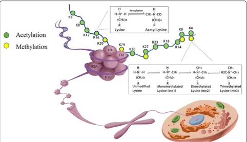
Histones are among the most highly conserved proteins that act as building blocks of the nucleosome, the funda-mental structural and functional unit of chromatin. The nucleosome is an octamer, which is wrapped by147 bp of DNA, consisting of two copies of four core histone
(H) H2A, H2B, H3, and H4 around, tied together by linker histone H1 [17]. These five classes of histone pro-teins, bearing over 60 different residues, constitute the major protein components of the chromatin and provide a tight packing of the DNA. Meanwhile, the histones contain a flexible N-terminus, often named the “histone tail” [17], which can undergo various combinations of PTMs, dynamically allowing regulatory proteins access to the DNA to fine tune almost all chromatin-mediated processes including chromatin condensation, gene tran-scription, DNA damage repair, and DNA replication [18] (Fig. 1). Transcriptionally active and silent chromatin is characterized by distinct post-translational modifications on the histones or their combinations. H3K27ac and H3K4me1 are associated with active enhancers [19], and high levels of H3K4me3 and H3 and H4 acetylation are found at the promoters of active genes [20, 21]. The bodies of active genes are enriched in H3 and H4 acetyl-ation [22], H3K79me3 [23], H2BK120u1, and a progres-sive shift from H3K36me1 to H3K36me3 between the promoters and the 3′ ends [24]. The methylation of H3K27 and H3K9 have emerged as hallmarks of repres-sive chromatin and are often found at silent gene loci. H3K27me3 are associated with the formation of faculta-tive heterochromatin, whereas H3K9me2/3 has
Fig. 1Schematic presentation of a nucleosome. A nucleosome functions as the fundamental packing unit of chromatin, with a stretch of
double-stranded DNA wrapped around a histone octamer of two H2A–H2B dimers and a (H3–H4) 2 tetramer. Different possible histone
important roles in the formation of constitutive hetero-chromatin [25]. H4K20m3 is a novel hallmark of peri-centric heterochromatin, whereas H4K20m1 regulates transcription both positively as well as negatively [26], suggesting that specific histone modifications can have dual functions. There are many combinations of modifica-tions that are either occurring together or mutually exclu-sive, suggesting crosstalk between these marks. Combinations of PTMs, thus, may be associated with tran-scription in a manner that was not simply related to their individual effects. For example, Fischer et al. indicated that single-code histone acetylation, in particular H3 acetylation (H3ac), are better predictors of increased transcript levels than domains containing further modifications [27]. Single-code H3K4dimethylation (H3K4m2) or its combin-ation with H3K4 tri-methylcombin-ation (H3K4m3) showed no positive correlation with transcript levels [27]. It is interest-ing given that H3K4m3 is known to be associated with transcription-start sites of actively transcribed genes. The results from Fischer and his colleague suggested that H3K4me3 is actually not an optimal marker of active pro-moters and that the activating effect mainly results from its frequent colocalization with acetylations [27].
Histone proteins can undergo post-translational modifi-cations by“writers”and“erasers,”a set of enzymes respon-sible for the deposition and removal of the chemical modifications. Through different combinations and patterns of histone PTMs, they can form the“histone code” [28]. Then, how are these codes interpreted? There are several mechanisms that are not mutually exclusive. First, direct nucleosome-intrinsic effects, particularly by neutralization or addition of charge, PTMs weaken histone-DNA interaction and enable generation of a stably remodeled nucleosome with increased mobility [29]. Such conventional allosteric regulation usually relies on a highly specialized population of molecular interactions [30]. Second, in direct nucleosome-extrinsic effects, H4K16 has been demonstrated to be such a unique histone tail, the acetylation of which impedes the higher-order chromatin formation as a result of its modulation of internucleosomal contacts [31]. Third, the emerging effector-mediated para-digm posits that histone PTMs are“read”by protein mod-ules termed as effectors, which translate them into patterns of gene activation or repression recruiting transcriptional or chromatin-remodeling machinery [30]. In the past decade, a wealth of conserved protein-interaction domains has been characterized as histone effectors, which recognize and bind histone PTMs specifically in a modification- and site-specific way. By covalent combinations of PTMs for binding, modified histone tails may function as integrating platforms for different chromatin-associated complexes, permitting them to receive inputs from upstream signaling cascades and transmit them to the downstream effectors [32].
Histone acetylation
Histone acetylation has been shown to be reversible. The N-terminal domains of histones bear a dozen of ly-sine residues subject to acetylation, with the exception of a lysine within the globular domain of H3K56, which was found to be acetylated in human by GCN5 [33]. This K residue is facing towards the major groove of the DNA within the nucleosome, so it is in good position to modulate nucleosome assembly by altering histone-DNA interactions when acetylated [34].
Readers of acetyl-lysines
The combinatorial effect of histone acetylation can be deciphered by two distinct, yet overlapping mechanisms— direct and effector-mediated readout mechanisms. In the direct mechanism, histone acetylation neutralizes the posi-tive charge on lysine residues, thus destabilizing the DNA-histone interaction [35]. This results in an open, loosely packed chromatin structure (euchromatin) and consequently allows access for specific transcription fac-tors and the general transcription machinery [31].
Writers and erasers of acetylation
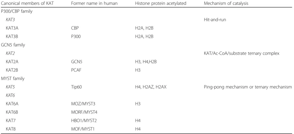
KATs, formally named as histone acetyltransferaces (HATs), can be generally classified into two categories based on sub-cellular localization. Type A KATs are located in the nu-cleus, involved in the acetylation of histones in chromatin, whereas type B KATs, predominantly cytoplasmic, acetylate newly translated histones to facilitate their transfer to the chromatin assembly factors [41]. In eukaryotes, the major-ity of canonical type A KATs has been grouped into three major families including p300/CBP, GCN5/PCAF, and MYST proteins [42] (Table 1). Two subfamilies of histone deacetylases (HDACs) have been identified in humans so far—Zn2+-dependent (classes I, II, and IV) and Zn2+-inde-pendent and NAD-deZn2+-inde-pendent (class III). Generally speak-ing, class I HDACs are ubiquitously expressed and exhibit strongest enzymatic activity. Class II HDACs have sequence similarity to the yeast Hda1 protein which seems to be expressed in a more cell-specific manner [43]. They possess unique 14-3-3 binding sites at their N-termini. Following phosphorylation, the N-terminal regions recruit 14-3-3, with consequent export of the HDAC/14-3-3 complex from the nucleus to the cytoplasm [44, 45]. Thus, phosphoryl-ation of class II HDACs provides a mechanism for coupling external signals to the genome. The class III HDACs, or sirtuins, display NAD+-dependent deacetylase activity and may specifically interact with and modify dozens of distinct substrates in various the biological processes.
Histone lysine methylation
Histone methylation occurs at lysine and arginine resi-dues. In this review, we only focus on histone lysine methylation due to its prominence and its array of well-established roles in epigenetic gene control and
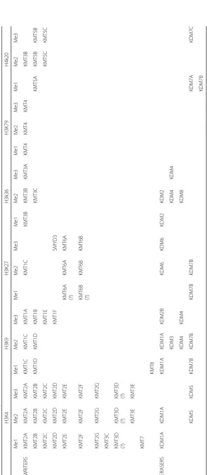
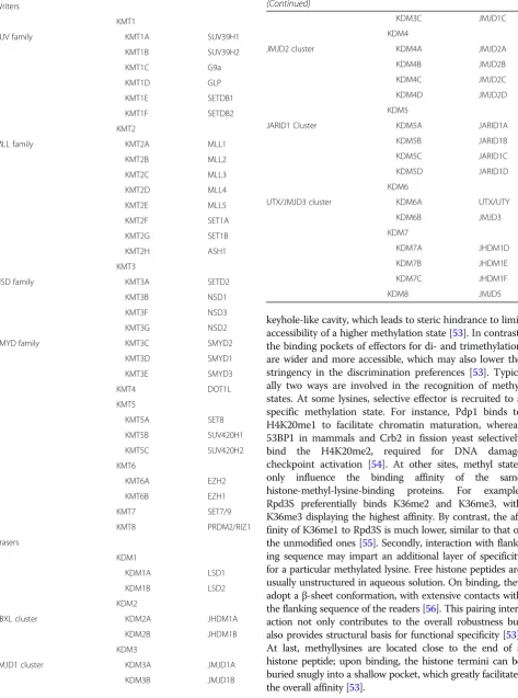
chromatin domains organization. Histone lysine methyla-tions have been found on a range of lysine residues in vari-ous histones, including K4, K9, K27, K36, and K79 residues in histone H3, K20 in histone H4, K59 in the globular domain of histone H4 [46], and K26 in histone H1B [47]. Instead of influencing the net charge of the his-tone tails, methylation of hishis-tone tails contributes to regu-lation of the transcriptional activity by functioning as a recognition template to recruit effector proteins to local chromatin [48]. Thus, histone lysine methylation can be associated with either activation or repression of tran-scription ultimately determined by the effectors. When compared with acetylation, histone lysine methylation is a relatively stable modification with a generally low turnover [49]. Moreover, methylation is controlled by histone meth-yltransferases (KMTs) and demethylases (KDMs) that pos-sess strong substrate specificity (Table2) (Table3).
Readers of methylysines
Chromodomain is the founding member of “readers” of histone methyllysine [50], Besides the well-known methy-lysine-binding family of chromodomain, a large family of reader proteins including Tudor, MBT, PWWP, plant homeodomain (PHD) finger, Ankyrin repeats, and WD repeats make up the so-called Royal family [51, 52]. Three elements determine the strength and specificity of a particular methylated lysine reader. The foremost trait of the methyllysine readers is the presence of an aromatic cage structure in their binding to methyllysines, consisting two to four aromatic residues. The exact composition and size of the pocket make the readers selective in recogniz-ing mono-, di-, or trimethylated state of lysine. Effectors for mono- and dimethylation tend to have a small
Table 1Enzymatic mechanisms used for histone acetylation
Canonical members of KAT Former name in human Histone protein acetylated Mechanism of catalysis
P300/CBP family
KAT3 Hit-and-run
KAT3A CBP H2A, H2B
KAT3B P300 H2A, H2B
GCN5 family
KAT2 KAT/Ac-CoA/substrate ternary complex
KAT2A GCN5 H3, H4,H2B
KAT2B PCAF H3
MYST family
KAT5 Tip60 H4, H2AZ, H2AX Ping-pong mechanism or ternary mechanism
KAT6
KAT6A MOZ/MYST3 H3
KAT6B MORF/MYST4
KAT7 HBO1/MYST2 H4
Table 2 Substrate specificity of KMTs and KDMs H3K4 H3K9 H3K27 H3k36 H3K79 H4k2 0 Me1 Me2 Me3 Me 1 Me2 Me3 Me1 Me2 Me3 Me1 Me2 Me3 Me1 Me2 Me3 Me 1 Me2 Me3 WRITER S KMT2A KMT 2A KMT2 A KMT 1C KMT1 C KMT1 A KMT1C KMT3 B KMT3 B KMT3 A KMT4 KMT4 KMT 4 KMT3 B KMT2B KMT 2B KMT2 B KMT 1D KMT1 D KMT1 B KMT3 C K MT5A KMT5 B KMT5 B KMT2C KMT 2C KMT2 C KMT1 E KMT5 C KMT5 C KMT2D KMT 2D KMT2 D KMT1 F SMY D3 KMT2E KMT 2E KMT2 E KMT6 A (?) KMT6A KMT6 A KMT2F KMT 2F KMT2 F KMT6 B (?) KMT6B KMT6 B KMT2G KMT 2G KMT2 G
KMT3C KMT3D (?)
keyhole-like cavity, which leads to steric hindrance to limit accessibility of a higher methylation state [53]. In contrast, the binding pockets of effectors for di- and trimethylation are wider and more accessible, which may also lower the stringency in the discrimination preferences [53]. Typic-ally two ways are involved in the recognition of methyl states. At some lysines, selective effector is recruited to a specific methylation state. For instance, Pdp1 binds to H4K20me1 to facilitate chromatin maturation, whereas 53BP1 in mammals and Crb2 in fission yeast selectively bind the H4K20me2, required for DNA damage checkpoint activation [54]. At other sites, methyl states only influence the binding affinity of the same histone-methyl-lysine-binding proteins. For example, Rpd3S preferentially binds K36me2 and K36me3, with K36me3 displaying the highest affinity. By contrast, the af-finity of K36me1 to Rpd3S is much lower, similar to that of the unmodified ones [55]. Secondly, interaction with flank-ing sequence may impart an additional layer of specificity for a particular methylated lysine. Free histone peptides are usually unstructured in aqueous solution. On binding, they adopt aβ-sheet conformation, with extensive contacts with the flanking sequence of the readers [56]. This pairing inter-action not only contributes to the overall robustness but also provides structural basis for functional specificity [53]. At last, methyllysines are located close to the end of a histone peptide; upon binding, the histone termini can be buried snugly into a shallow pocket, which greatly facilitates the overall affinity [53].
Table 3Histone methyltransferases and demethyltransferases
Writers
KMT1
SUV family KMT1A SUV39H1
KMT1B SUV39H2
KMT1C G9a
KMT1D GLP
KMT1E SETDB1
KMT1F SETDB2
KMT2
MLL family KMT2A MLL1
KMT2B MLL2
KMT2C MLL3
KMT2D MLL4
KMT2E MLL5
KMT2F SET1A
KMT2G SET1B
KMT2H ASH1
KMT3
NSD family KMT3A SETD2
KMT3B NSD1
KMT3F NSD3
KMT3G NSD2
SMYD family KMT3C SMYD2
KMT3D SMYD1
KMT3E SMYD3
KMT4 DOT1L
KMT5
KMT5A SET8
KMT5B SUV420H1
KMT5C SUV420H2
KMT6
KMT6A EZH2
KMT6B EZH1
KMT7 SET7/9
KMT8 PRDM2/RIZ1
Erasers
KDM1
KDM1A LSD1
KDM1B LSD2
KDM2
FBXL cluster KDM2A JHDM1A
KDM2B JHDM1B
KDM3
JMJD1 cluster KDM3A JMJD1A
KDM3B JMJD1B
Table 3Histone methyltransferases and demethyltransferases (Continued)
KDM3C JMJD1C
KDM4
JMJD2 cluster KDM4A JMJD2A
KDM4B JMJD2B
KDM4C JMJD2C
KDM4D JMJD2D
KDM5
JARID1 Cluster KDM5A JARID1A
KDM5B JARID1B
KDM5C JARID1C
KDM5D JARID1D
KDM6
UTX/JMJD3 cluster KDM6A UTX/UTY
KDM6B JMJD3
KDM7
KDM7A JHDM1D
KDM7B JHDM1E
KDM7C JHDM1F
Writers and erasers of histone lysine methylation
KMTs catalyze methylation of lysine residues with high site- and methyl-level specificity (Table2). In the last de-cades, numerous KMTs have been identified and crystal-lized, which use S-adenosylmethionine (SAM) as a methyl group donor [57]. Except for KMT4/DOT1L, all known KMTs contain a conserved SET domain harbor-ing the enzymatic activity [58]. Based on the similarities in the sequence within and around the catalytic SET do-main, as well as homology to other protein modules and their domain architectures, SET-containing KMTs have traditionally been categorized into distinct subfamilies [59].
Histone lysine methylation was previously considered static and enzymatically irreversible until the first his-tone KDM—LSD1/KDM1A identified by Shi et al. [60], which changed our view of histone methylation regula-tion and ushered in the identificaregula-tion of numerous his-tone demethylases. Subsequent to the discovery of KDM1A, a new class of KDM enzymes which comprises the JmjC domain-containing protein was discovered. While KDM1A is unable to catalyze the dimethylation of trimethylated lysine residues owing to its requirement for imine formation for catalytic activity, the JmjC-driven demethylase have demethylation activity for mono-, di-, and trimethylated histone lysine residues. In-deed, most of the JmjC histone demethylases identified so far are capable of efficiently catalyzing demethylation of trimethylated lysines, and in most cases, they prefer-entially bind the trimethylated substrates [61,62].
Histone modifications in MS
A core of pathogenetic functions common to both the immune and neurodegenerative processes of MS has been characterized by deregulation of MS-risk genes and resulting dysfunction of their encoding proteins [63]. Epigenetic transcription-regulating mechanisms in nu-cleated cells including cells of the CNS have been widely accepted. Therefore, MS-specific alterations in epigen-etic regulation of chromatin may play a central role in gene expression and be essential for the initiation and development of MS. Among which, histone modification is an important epigenetic mechanism.
Histone modifications in MS susceptibility
Twin studies have established that susceptibility to MS is partly genetic. One family of major histocompatibility complex (MHC) genes, the human leukocyte antigen (HLA) alleles, has identified as a genetic determinant for MS [64]. In particular, carriage of HLA-DR/DQ serotype has been identified as a major MS risk allele. Notably, expression of HLA-DR has been shown to be suppressed by HDAC1 [65], which suggests that MS susceptibility loci have histone regulation links.
Histone modifications in autoimmunity-related mechanisms
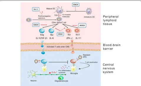
The hallmark of MS and experimental autoimmune en-cephalomyelitis (EAE) is that myelin injury and axonal damage driven by an immune-mediated inflammatory response begins at disease onset. Autoreactive myelin-specific CD4+ T cells are believed to play a cru-cial pathogenic role [66]. Upon encountering myelin antigen, antigen-presenting cells (APCs) acquire a ma-ture phenotype and migrate to lymph nodes where they present exogenous antigens to naïve CD4+ T cells. Naive CD4+ T cells may then differentiate into diverse func-tional subsets, including the T helper (Th) 1, Th2, Th17 cells, and Treg cells [67]. Once activated, CD4+ T cells are translocated into the CNS by crossing the brain-blood barrier (BBB) and then are reactivated by resident APCs (such as microglia) [68], which in turn initiate the recruitment of other inflammatory cells, resulting in demyelination and axon injury. While interferon-γ (IFN-γ)-associated Th1 and interleukin-17 (IL-17)-associated Th17 cells are considered to lead to disease progression and worsening of symptoms, IL-4-associated Th2 and transforming growth factor-β (TGF-β)-associated Treg have been indicated to associ-ate with inflammation reduction and improvement of symptoms in MS patients [69].
It is widely accepted that the activation of CD4+ auto-reactive T cells and their differentiation into a Th1 or Th17 phenotype are crucial events in the initial steps of MS, though many studies have shown that monocytes and monocyte-derived macrophages are also the primary cell types responsible for cellular pathology and tissue damage. In MS pathology, activated monocytes, which facilitate the migration of T cells across the blood-brain barrier (BBB), largely represent the inflammatory infil-trate [70]. Knowledge on the features of blood mono-cytes in MS, however, are little understood. Circulating monocytes, as an important source of cytokines, have been hypothesized to play a key role in regulating crucial immune functions. The M1/M2 paradigm is currently used to categorize the monocyte/macrophage functions [71], and M1/M2 macrophage balance polarization gov-erns the fate of an organ in inflammation. Generally, M1 monocytes/macrophages are generally characterized by an IL-12hi, IL-23hi, tumor necrosis factor (TNF)-αhi, and IL-10lo phenotype, which produce abundant react-ive oxygen species and shift the immune response to-wards a Th1 profile [72]. M2 monocytes/macrophages typically have IL-12lo, IL-23lo, TNF-αlo, and IL-10hi re-sponses to stimulation, which are thought to drive Th2 responses [73].
chromatin structure which then influence gene expres-sion. Correspondingly, HDAC inhibitors have also been demonstrated to elicit control over the immune re-sponse, which in turn suppress systemic and local in-flammation [74]. Several recent studies have shown the potential for the use of HDAC inhibitor therapy to in-hibit the proliferative response of CD4+ T cells and ab-rogated IFN-γ production [75]. A growing literature indicated that HDAC inhibitors inhibit the proinflamma-tory cytokine IL-2 expression, which is secreted by Th1 cells, and IL-2 mediated gene expression as well. More-over, HDAC inhibitors reduce macrophage production of pro-demyelinating cytokines involved in T helper (Th) cell differentiation, including IL-12, IL-6, and TNF-α. Consequently, HDAC inhibitors cause a Th1 to Th2 dominance shift [76], and expanding Tregs, which by virtue of its immunosuppressive role, may help ameli-orate MS.
Actually, dysregulated Th cell responses are not unique for MS pathology, but also a characteristic of a wide variety of several other inflammatory diseases, in-cluding inflammatory bowel disease, arthritis, diabetes, asthma, and allergies [77]. Therefore, compounds that inhibit HDACs, especially, class I, II, and IV enzymes, have been pursued as therapeutic agents for a wide range of inflammatory diseases. However, treating cells with HDAC inhibitors has also been shown to increase the expression of cytokines IL-10 [76], contributing to pro-humoral and protective role in EAE, which, in sys-temic lupus erythematosus (SLE) cells, actually downreg-ulated expression of IL-10 and other anti-inflammatory cytokines [78]. The contrasting effects might reflect disease-specific effects of these compounds and further studies are needed.
It is suggested that chromatin remodeling, via histone lysine methylation, is mechanistically important in the acquisition of the M2-macrophage phenotype. Ishii et al. demonstrated that at the promoters of the M2 marker genes, H3K4me3 was significantly upregulated, whereas H3K27me2/3 was significantly decreased. Increased Jmjd3 contributes to the decrease of H3K27me2/3 marks and skews macrophages to an M2 phenotype [79]. Therefore, target gene regulation by histone Lysine methylation is a dynamic process that modulates inflam-matory responses in the development of a variety of autoimmune diseases, including MS.
Recent studies demonstrated that KDM6 modulate immune functions by determining Th cell maturation and egress from the thymus [80], as well as CD4+ Th cell lineage differentiation [66], thereby significantly af-fecting immune responses in multiple biological systems. It is reported that Jmjd3 positively regulate the differen-tiation of Th17 cells, which play critical roles in proin-flammatory reactions in autoimmue disorders, such as
rheumatoid arthritis and systemic lupus erythematosis [81]. Jmjd3-deficient mice were demonstrated to be re-sistant to the induction of EAE [66]. Correspondingly, H3K27 demethylase-specific inhibitor GSK-J4 markedly inhibited Th17 cell differentiation in vitro [66]. However, another independent research demonstrated that while Th1 and Th17 differentiation were not affected, 10 or 25 nM GSK-J4 significantly increased differentiation of anti-inflammatory Treg cells in vivo, which could partly explain the beneficial effects of GSK-J4 on EAE. GSK-J4 promoted Treg differentiation was proposed to be dependent on its direct effect on the maturation status of dendrite cells (DCs). DCs, the professional APC, be-ing the key players in maintainbe-ing immune tolerance, now have gained increasing attention [82]. Specifically, H3K27me3 demethylase activity would skew DC differ-entiation towards a tolerogenic phenotype [83]. Accord-ingly, through altering the permissive H3K4me3/ repressive H3K27me3 ratio at specific gene promoters, GSK-J4 induced a tolerogenic phenotype on DCs and subsequently inhibited the development of EAE [83].
Moreover, T cell anergy is thought to be a critical mechanism for preventing autoimmunity and failure of this tolerance mechanism causes MS [84]. The upregu-lated Sirt1 protein has been demonstrated to suppress T cell activation and lead to anergy induction in mice. Conversely, Sirt1 deficiency was reported to result in in-creased T cell activation and failed to maintain CD4+ T cell tolerance and increased susceptibility to EAE [85]. Mice with DC-specific deletion of SIRT1 showed re-markable resistance to EAE through enhanced IL-27 and IFN-β activation, two anti-inflammatory cytokines that negatively regulate Th17 cell differentiation [86]. These findings make the role of HDAC in MS quite controver-sial (Fig.2).
Histone modifications in myelin destruction
acetylation has been detected in oligodendrocyte lineage cells within normal-appearing white matter (NAWM) in the brain of patients with chronic MS [89]. The data suggested negative correlations between histone deacety-lation efficiency and duration of disease.
Histone modifications in neurodegeneration
For decades, MS research has heavily focused on inflam-matory white matter pathology. However, recent studies have discovered neurodegenerative components of the disease such as insidious axonal degeneration and neur-onal atrophy, which seem to be the histopathological correlates of progressive clinical disability in MS patients [90]. Mitochondrial injury and subsequent energy failure are indicated as key factors in the induction of neurogeneration. Betaine, a methyl donor, was found to be de-creased in MS cortex, which was correlated with decreased H3K4me3 in neuronal (NeuN+) nuclei in MS cortex, in comparison to controls [91]. Mechanistic studies demonstrated that reduced methylation of H3K4me3 may result in the downregulation of oxidative phosphorylation genes and defects of respiratory chain enzymes in MS cortex [91]. A recent study showed that variant carriers of certain HDAC genes, including mitochondrial-related gene variants in SIRT4 and SIRT5, have been linked to more pronounced brain volume loss
(atrophy) during the clinical course of MS [92]. These results indicate that the histone modifications might be centrally linked with neurodegenerative processes in MS.
Potential treatment methods based on epigenetic mechanisms
Disturbance of transcriptional balance may promote dys-regulation of immune system and neurodegeneration, both of which contribute to the clinical profile of MS. Animal model experiments support that deliberate epi-genetic reprogramming for oligodendrocyte, immune cells, and neurons to perform properly may be a poten-tial therapeutic strategy for MS.
KATs, has been shown to ameliorate EAE through sup-pression of inflammatory cells infiltration in the spinal cord [95]. As previously mentioned, systemic administra-tion with the epigenetic drug GSK-J4 prevented the de-velopment of EAE in mice [83]. Thus, the inhibitors of histone deacetylation or demethylation may be promis-ing agents for MS treatment. However, systemic use of HDACis negatively affects the generation of new myelin since histone deacetylation is important for progenitor cell differentiation into myelin-forming oligodendrocytes [96] and is critical for remyelination efficiency in adults [88], as we reviewed previously. The potential detrimen-tal consequence on myelin might counteract the beneficial effects, thus cautioning against the use of broad inhibitors of histone deacetylases in MS. Therefore, more targeted therapy that specifically epigenetically modifies certain pathogenic loci need to be developed. In the recent years, the CRISPR-dCas9 system is poised to become the most promising targetable epigenome-editing tools. The results of two recent seminal studies have strongly supported the capability of epigenome editing by a CRISPR-Cas9 to acti-vate or silence transcription by targeting histone PTMs [97,98]. Moreover, CRISPR-dCas9 epigenome-editing ap-proach has been demonstrated to produce long-lasting changes in expression of targeted genes both in vitro and in vivo. Its simplicity and efficiency may facilitate the clinical application of this technology by avoiding repeti-tive or chronic administration. However, the research on CRISPR-mediated technology is still in its early stage, and it is important to continue to probe for its feasibility and safety for clinical purposes. An additional challenge for treating MS with these inhibitors is the lack of specificity, which would cause a relatively high risk of adverse effects. Correspondingly, successful epigenetic therapy would be the tissue specificity of the therapeutic effect. Receptor-coated nanoparticles or microvesicles as highly effective drug carriers pertaining to BBB may hold great promise in MS therapy. Several studies have recently demonstrated that treatment of mice with nanoparticles effectively decreased EAE progression [99]. Collectively, translational use of epigenetics might offer hope for a new class of therapeutics to treat MS and the development of targeted epigenetic therapies open new avenues for effective personalized treatment of patients with MS.
Conclusion
MS is the most prevalent autoimmune disease with highly variable clinical course and disease progression, in which the main common pathogenetic pathway involves an immune-mediated cascade [100]. Recently, huge steps have been made in the field of MS immunotherapy. More-over, emerging evidence has shed light on the epigenetic mechanisms contributing MS. Several epigenetic drugs which are in clinical trials or under investigation in human
diseases have been proven to have immunomodulatory ef-fects [101]. In addition, other expected changes also may occur in response to epigenetic treatment. In particular, histone PTMs in regulation of myelination and degener-ation gene associated with MS and ameliordegener-ation of EAE symptoms by drugs with PTM effects, such as HDAC in-hibitors and KDM inin-hibitors, all emphasize the critical role of histone PTMs in the pathogenesis of MS. The amalgamation and crystallization of histone PTMs re-search and MS promises novel pleiotropic treatment strategies. However, given the potential for off-target po-tential to cause deleterious effects from HDAC and KDM inhibitors with broad activity, the endeavor to completely understand molecular mechanisms governing histone modifications and their precise molecular targets will hold the key to successfully translate the drug candidates to clinical practice.
Abbreviations
Ac-CoA:Acetyl coenzyme A; APC: Antigen-presenting cells; BBB: Brain-blood barrier; BRD: Bromodomain; DCs: Dendrite cells; EAE: Experimental autoimmune encephalomyelitis; EBV: Epstein-Barr virus; HATs: Histone acetyltransferaces; HDACs: Histone deacetylases; HLA: Human leukocyte antigen; IFN: Interferon; IL: Interleukin; MHC: Major histocompatibility complex; NAWM: Normal-appearing white matter; PHD: Plant homeodomain; PTMs: Post-translational modifications; TAF1: TATA-binding protein-associated factor-1; TGF: Transforming growth factor; Th: T helper
Acknowledgements
The authors would like to acknowledge Dr. Shiyu Chen for the artwork.
Authors’contributions
BY conceived and planned the review. ZH and BY drafted the manuscript. HH revised it critically for important intellectual content with support from FZ, HX, and BY. All authors contributed to the final manuscript.
Ethics approval and consent to participate Not applicable.
Consent for publication Not applicable.
Competing interests
The authors declare that they have no competing interests.
Publisher’s Note
Springer Nature remains neutral with regard to jurisdictional claims in published maps and institutional affiliations.
Received: 28 March 2018 Accepted: 8 June 2018
References
1. Laird PW. The power and the promise of DNA methylation markers. Nat Rev
Cancer. 2003;3:253–66.
2. Jenuwein T, Allis CD. Translating the histone code. Science. 2001;293:1074–80.
3. Pusarla RH, Bhargava P. Histones in functional diversification: core histone
variants. FEBS J. 2005;272:5149–68.
4. Mattick JS, Makunin I V. Non-coding RNA. Hum Mol Genet 2006; 15 Spec No
1:R17–R29.
5. Nowak SJ, Corces VG. Phosphorylation of histone H3: a balancing act
between chromosome condensation and transcriptional activation. Trends
6. Li W, Nagaraja S, Delcuve GP, Hendzel MJ, Davie JR. Effects of histone acetylation, ubiquitination and variants on nucleosome stability. Biochem J.
1993;296:737–44.
7. Shiio Y, Eisenman RN. Histone sumoylation is associated with transcriptional
repression. Proc Natl Acad Sci U S A. 2003;100:13225–30.
8. Boulikas T. DNA strand breaks alter histone ADP-ribosylation. Proc Natl Acad
Sci U S A. 1989;86:3499–503.
9. Cuthbert GL, Daujat S, Snowden AW, Erdjument-Bromage H, Hagiwara T,
Yamada M, et al. Histone deimination antagonizes arginine methylation.
Cell. 2004;118:545–53.
10. Nelson CJ, Santos-Rosa H, Kouzarides T. Proline isomerization of histone H3
regulates lysine methylation and gene expression. Cell. 2006;126:905–16.
11. Sadovnick AD, Baird PA, Ward RH, Optiz JM, Reynolds JF. Multiple sclerosis:
updated risks for relatives. Am J Med Genet. 1988;29:533–41.
12. Willer CJ, Dyment DA, Risch NJ, Sadovnick AD, Ebers GC, Canadian
Collaborative Study Group. Twin concordance and sibling recurrence rates
in multiple sclerosis. Proc Natl Acad Sci U S A. 2003;100:12877–82.
13. Huynh JL, Casaccia P. Epigenetic mechanisms in multiple sclerosis: implications
for pathogenesis and treatment. Lancet Neurol. 2013;12:195–206.
14. Niller HH, Wolf H, Minarovits J. Epigenetic dysregulation of the host cell genome
in Epstein-Barr virus-associated neoplasia. Semin Cancer Biol. 2009;19:158–64.
15. Wan ES, Qiu W, Baccarelli A, Carey VJ, Bacherman H, Rennard SI, et al.
Cigarette smoking behaviors and time since quitting are associated with differential DNA methylation across the human genome. Hum Mol Genet.
2012;21:3073–82.
16. Pereira F, Barbáchano A, Singh PK, Campbell MJ, Muñoz A, Larriba MJ.
Vitamin D has wide regulatory effects on histone demethylase genes. Cell
Cycle. 2012;11:1081–9.
17. Luger K, Mäder W, Richmond RK, Sargent DF, Richmond TJ. Crystal structure of
the nucleosome core particle at 2.8 A resolution. Nature. 1997;389:251–60.
18. Önder Ö, Sidoli S, Carroll M, Garcia BA. Progress in epigenetic histone
modification analysis by mass spectrometry for clinical investigations. Expert
Rev Proteomics. 2015;12:499–517.
19. Creyghton MP, Cheng AW, Welstead GG, Kooistra T, Carey BW, Steine EJ, et
al. Histone H3K27ac separates active from poised enhancers and predicts
developmental state. Proc Natl Acad Sci U S A. 2010;107:21931–6.
20. Deckert J, Struhl K. Histone acetylation at promoters is differentially affected
by specific activators and repressors. Mol Cell Biol. 2001;21:2726–35.
21. Liang G, Lin JCY, Wei V, Yoo C, Cheng JC, Nguyen CT, et al. Distinct
localization of histone H3 acetylation and H3-K4 methylation to the transcription start sites in the human genome. Proc Natl Acad Sci U S A.
2004;101:7357–62.
22. Myers FA, Evans DR, Clayton AL, Thorne AW, Crane-Robinson C. Targeted
and extended acetylation of histones H4 and H3 at active and inactive
genes in chicken embryo erythrocytes. J Biol Chem. 2001;276:20197–205.
23. Ng HH, Ciccone DN, Morshead KB, Oettinger MA, Struhl K. Lysine-79 of
histone H3 is hypomethylated at silenced loci in yeast and mammalian cells: a potential mechanism for position-effect variegation. Proc Natl Acad
Sci U S A. 2003;100:1820–5.
24. Pokholok DK, Harbison CT, Levine S, Cole M, Hannett NM, Tong IL, et al.
Genome-wide map of nucleosome acetylation and methylation in yeast.
Cell. 2005;122:517–27.
25. Zhang T, Cooper S, Brockdorff N. The interplay of histone
modifications—writers that read. EMBO Rep. 2015;16:1467–81.
26. Nishioka K, Rice JC, Sarma K, Erdjument-Bromage H, Werner J, Wang Y, et al.
PR-Set7 is a nucleosome-specific methyltransferase that modifies lysine 20 of
histone H4 and is associated with silent chromatin. Mol Cell. 2002;9:1201–13.
27. Fischer JJ, Toedling J, Krueger T, Schueler M, Huber W, Sperling S.
Combinatorial effects of four histone modifications in transcription and
differentiation. Genomics. 2008;91:41–51.
28. Latham JA, Dent SY. Cross-regulation of histone modifications. Nat Struct
Mol Biol. 2007;14:1017–24.
29. Cosgrove MS, Boeke JD, Wolberger C. Regulated nucleosome mobility and
the histone code. Nat Struct Mol Biol. 2004;11:1037–43.
30. Seet BT, Dikic I, Zhou MM, Pawson T. Reading protein modifications with
interaction domains. Nat Rev Mol Cell Biol. 2006;7:473–83.
31. Shogren-Knaak M, Ishii H, Sun JM, Pazin MJ, Davie JR, Peterson CL. Histone
H4-K16 acetylation controls chromatin structure and protein interactions.
Science. 2006;311:844–7.
32. Cheung P, Allis CD, Sassone-Corsi P. Signaling to chromatin through histone
modifications. Cell. 2000;103:263–71.
33. Kenseth JR, Coldiron SJ. High-throughput characterization and quality
control of small-molecule combinatorial libraries. Curr Opin Chem Biol.
2004;8:418–23.
34. Kouzarides T. Chromatin modifications and their function. Cell. 2007;128:693–705.
35. Tessarz P, Kouzarides T. Histone core modifications regulating nucleosome
structure and dynamics. Nat Rev Mol Cell Biol. 2014;15:703–8.
36. Tamkun JW, Deuring R, Scott MP, Kissinger M, Pattatucci AM, Kaufman TC,
et al. Brahma: a regulator of Drosophila homeotic genes structurally related
to the yeast transcriptional activator SNF2SWI2. Cell. 1992;68:561–72.
37. Filippakopoulos P, Picaud S, Mangos M, Keates T, Lambert JP,
Barsyte-Lovejoy D, et al. Histone recognition and large-scale structural analysis of
the human bromodomain family. Cell. 2012;149:214–31.
38. Jacobson RH, Ladurner AG, King DS, Tjian R. Structure and function of a
human TAFII250 double bromodomain module. Science. 2000;288:1422–5.
39. Morinière J, Rousseaux S, Steuerwald U, Soler-López M, Curtet S, Vitte AL, et
al. Cooperative binding of two acetylation marks on a histone tail by a
single bromodomain. Nature. 2009;461:664–8.
40. Miller TCR, Simon B, Rybin V, Grötsch H, Curtet S, Carlomagno T, et al. A
bromodomain-DNA interaction facilitates acetylation-dependent bivalent nucleosome recognition by the BET protein BRDT. Nat Commun. 2016;7:13855.
41. Richman R, Chicoine LG, Collini MP, Cook RG, Allis CD. Micronuclei and
the cytoplasm of growing Tetrahymena contain a histone acetylase activity which is highly specific for free histone H4. J Cell Biol.
1988;106:1017–26.
42. Hodawadekar SC, Marmorstein R. Chemistry of acetyl transfer by histone
modifying enzymes: structure, mechanism and implications for effector
design. Oncogene. 2007;26:5528–40.
43. Seto E, Yoshida M. Erasers of histone acetylation: the histone deacetylase
enzymes. Cold Spring Harb Perspect Biol. 2014;6:a018713.
44. Vega RB, Harrison BC, Meadows E, Roberts CR, Papst PJ, Olson EN, et al. Protein
kinases C and D mediate agonist-dependent cardiac hypertrophy through
nuclear export of histone deacetylase 5. Mol Cell Biol. 2004;24:8374–85.
45. McKinsey TA, Zhang CL, Lu J, Olson EN. Signal-dependent nuclear
export of a histone deacetylase regulates muscle differentiation. Nature.
2000;408:106–11.
46. Zhang L, Eugeni EE, Parthun MR, Freitas MA. Identification of novel histone
post-translational modifications by peptide mass fingerprinting.
Chromosoma. 2003;112:77–86.
47. Cai Y, Jin J, Swanson SK, Cole MD, Choi SH, Florens L, et al. Subunit
composition and substrate specificity of a MOF-containing histone acetyltransferase distinct from the male-specific lethal (MSL) complex. J Biol
Chem. 2010;285:4268–72.
48. Cloos PAC, Christensen J, Agger K, Helin K. Erasing the methyl mark: histone
demethylases at the center of cellular differentiation and disease. Genes
Dev. 2008;22:1115–40.
49. Trojer P, Reinberg D. Histone lysine demethylases and their impact on
epigenetics. Cell. 2006;125:213–7.
50. Blus BJ, Wiggins K, Khorasanizadeh S. Epigenetic virtues of chromodomains.
Crit Rev Biochem Mol Biol. 2011;46:507–26.
51. Kim J, Daniel J, Espejo A, Lake A, Krishna M, Xia L, et al. Tudor, MBT and
chromo domains gauge the degree of lysine methylation. EMBO Rep.
2006;7:397–403.
52. Nameki N, Tochio N, Koshiba S, Inoue M, Yabuki T, Aoki M, et al. Solution
structure of the PWWP domain of the hepatoma-derived growth factor
family. Protein Sci. 2005;14:756–64.
53. Yun M, Wu J, Workman JL, Li B. Readers of histone modifications. Cell Res.
2011;21:564–78.
54. Greeson NT, Sengupta R, Arida AR, Jenuwein T, Sanders SL. Di-methyl H4
lysine 20 targets the checkpoint protein Crb2 to sites of DNA damage. J
Biol Chem. 2008;283:33168–74.
55. Li B, Jackson J, Simon MD, Fleharty B, Gogol M, Seidel C, et al. Histone H3
lysine 36 dimethylation (H3K36me2) is sufficient to recruit the Rpd3s histone deacetylase complex and to repress spurious transcription. J Biol
Chem. 2009;284:7970–6.
56. Klein BJ, Lalonde ME, Côté J, Yang XJ, Kutateladze TG. Crosstalk between
epigenetic readers regulates the MOZ/MORF HAT complexes. Epigenetics.
2014;9:186–93.
57. Dillon SC, Zhang X, Trievel RC, Cheng X. The SET-domain protein
superfamily: protein lysine methyltransferases. Genome Biol. 2005;6:227.
58. Van Leeuwen F, Gafken PR, Gottschling DE. Dot1p modulates silencing in
59. Cheng X, Collins RE, Zhang X. Structural and sequence motifs of protein (histone)
methylation enzymes. Annu Rev Biophys Biomol Struct. 2005;34:267–94.
60. Shi Y, Sawada J, Sui G, Affar EB, Whetstine JR, Lan F, et al. Coordinated
histone modifications mediated by a CtBP co-repressor complex. Nature.
2003;422:735–8.
61. Whetstine JR, Nottke A, Lan F, Huarte M, Smolikov S, Chen Z, et al. Reversal
of histone lysine trimethylation by the JMJD2 family of histone
demethylases. Cell. 2006;125:467–81.
62. Tsukada Y, Fang J, Erdjument-Bromage H, Warren ME, Borchers CH, Tempst
P, et al. Histone demethylation by a family of JmjC domain-containing
proteins. Nature. 2006;439:811–6.
63. Van Den Elsen PJ, Van Eggermond MCJA, Puentes F, Van Der Valk P, Baker
D, Amor S. The epigenetics of multiple sclerosis and other related disorders.
Mult Scler Relat Disord. 2014;3:163–75.
64. Hillert J. Human leukocyte antigen studies in multiple sclerosis. Ann Neurol.
1994;36(Suppl):S15–7.
65. Gray SG, Dangond F. Rationale for the use of histone deacetylase inhibitors as
a dual therapeutic modality in multiple sclerosis. Epigenetics. 2006;1:67–75.
66. Liu Z, Cao W, Xu L, Chen X, Zhan Y, Yang Q, et al. The histone H3 lysine-27
demethylase Jmjd3 plays a critical role in specific regulation of Th17 cell
differentiation. J Mol Cell Biol. 2015;7:505–16.
67. Zhu J, Paul WE. CD4 T cells: fates, functions, and faults. Blood. 2008;112:1557–69.
68. Furtado GC, Marcondes MCG, Latkowski J-A, Tsai J, Wensky A, Lafaille JJ.
Swift entry of myelin-specific T lymphocytes into the central nervous system in spontaneous autoimmune encephalomyelitis. J Immunol.
2008;181:4648–55.
69. Seder RA, Ahmed R. Similarities and differences in CD4+ and CD8+ effector
and memory T cell generation. Nat Immunol. 2003;4:835–42.
70. Larochelle C, Alvarez JI, Prat A. How do immune cells overcome the
blood-brain barrier in multiple sclerosis? FEBS Lett. 2011;585:3770–80.
71. Gordon S. Alternative activation of macrophages. Nat Rev Immunol. 2003;3:23–35.
72. Gordon S, Taylor PR. Monocyte and macrophage heterogeneity. Nat Rev
Immunol. 2005;5:953–64.
73. Sica A, Mantovani A. Macrophage plasticity and polarization: in vivo veritas.
J Clin Invest. 2012;12:787–95.
74. Camelo S, Iglesias AH, Hwang D, Due B, Ryu H, Smith K, et al. Transcriptional
therapy with the histone deacetylase inhibitor trichostatin A ameliorates
experimental autoimmune encephalomyelitis. J Neuroimmunol. 2005;164:10–21.
75. Su R-C, Becker AB, Kozyrskyj AL, Hayglass KT. Epigenetic regulation of
established human type 1 versus type 2 cytokine responses. J Allergy Clin
Immunol. 2008;121:57–63.e3.
76. Säemann MD, G a B, Osterreicher CH, Burtscher H, Parolini O, Diakos C, et al.
Anti-inflammatory effects of sodium butyrate on human monocytes: potent inhibition
of IL-12 and up-regulation of IL-10 production. FASEB J. 2000;14:2380–2.
77. Antignano F, Zaph C. Regulation of CD4 T-cell differentiation and inflammation
by repressive histone methylation. Immunol Cell Biol. 2015;93:245–52.
78. Mishra N, Reilly CM, Brown DR, Ruiz P, Gilkeson GS. Histone deacetylase
inhibitors modulate renal disease in the MRL-lpr/lpr mouse. J Clin Invest.
2003;111:539–52.
79. Satoh T, Takeuchi O, Vandenbon A, Yasuda K, Tanaka Y, Kumagai Y, et al.
The Jmjd3-Irf4 axis regulates M2 macrophage polarization and host
responses against helminth infection. Nat Immunol. 2010;11:936–44.
80. Manna S, Kim JK, Baugé C, Cam M, Zhao Y, Shetty J, et al. Histone H3 lysine
27 demethylases Jmjd3 and Utx are required for T-cell differentiation. Nat Commun. 2015;6:8152.
81. Singh RP, Hasan S, Sharma S, Nagra S, Yamaguchi DT, Wong DTW, et al. Th17
cells in inflammation and autoimmunity. Autoimmun Rev. 2014;13:1174–81.
82. Kushwah R, Hu J. Dendritic cell apoptosis: regulation of tolerance versus
immunity. J Immunol. 2010;185:795–802.
83. Doñas C, Carrasco M, Fritz M, Prado C, Tejón G, Osorio-Barrios F, et al.
The histone demethylase inhibitor GSK-J4 limits inflammation through the induction of a tolerogenic phenotype on DCs. J Autoimmun.
2016;75:105–17.
84. Waldner H, Collins M, Kuchroo VK. Activation of antigen-presenting cells by
microbial products breaks self tolerance and induces autoimmune disease. J
Clin Invest. 2004;113:990–7.
85. Zhang J, Lee SM, Shannon S, Gao B, Chen W, Chen A, et al. The type III
histone deacetylase Sirt1 is essential for maintenance of T cell tolerance in
mice. J Clin Invest. 2009;119:3048–58.
86. Yang H, Lee SM, Gao B, Zhang J, Fang D. Histone deacetylase sirtuin 1
deacetylates IRF1 protein and programs dendritic cells to control Th17
protein differentiation during autoimmune inflammation. J Biol Chem. 2013;
288:37256–66.
87. Marin-Husstege M, Muggironi M, Liu A, Casaccia-Bonnefil P. Histone
deacetylase activity is necessary for oligodendrocyte lineage progression. J
Neurosci. 2002;22:10333–45.
88. Shen S, Sandoval J, Swiss VA, Li J, Dupree J, Franklin RJM, et al.
Age-dependent epigenetic control of differentiation inhibitors is critical for
remyelination efficiency. Nat Neurosci. 2008;11:1024–34.
89. Pedre X, Mastronardi F, Bruck W, Lopez-Rodas G, Kuhlmann T, Casaccia P.
Changed histone acetylation patterns in normal-appearing white matter
and early multiple sclerosis lesions. J Neurosci. 2011;31:3435–45.
90. Mahad DH, Trapp BD, Lassmann H. Pathological mechanisms in progressive
multiple sclerosis. Lancet Neurol. 2015;14:183–93.
91. Singhal NK, Li S, Arning E, Alkhayer K, Clements R, Sarcyk Z, et al. Changes
in methionine metabolism and histone H3 trimethylation are linked to
mitochondrial defects in multiple sclerosis. J Neurosci. 2015;35:15170–86.
92. Inkster B, Strijbis EMM, Vounou M, Kappos L, Radue EW, Matthews PM, et al.
Histone deacetylase gene variants predict brain volume changes in multiple
sclerosis. Neurobiol Aging. 2013;34:238–47.
93. Ge Z, Da Y, Xue Z, Zhang K, Zhuang H, Peng M, et al. Vorinostat, a histone
deacetylase inhibitor, suppresses dendritic cell function and ameliorates
experimental autoimmune encephalomyelitis. Exp Neurol. 2013;241:56–66.
94. Zhang Z, Zhang ZY, Wu Y, Schluesener HJ. Valproic acid ameliorates
inflammation in experimental autoimmune encephalomyelitis rats.
Neuroscience. 2012;221:140–50.
95. Xie L, Li XK, Funeshima-Fuji N, Kimura H, Matsumoto Y, Isaka Y, et al.
Amelioration of experimental autoimmune encephalomyelitis by curcumin treatment through inhibition of IL-17 production. Int Immunopharmacol.
2009;9:575–81.
96. Shen S, Li J, Casaccia-Bonnefil P. Histone modifications affect timing of
oligodendrocyte progenitor differentiation in the developing rat brain. J Cell
Biol. 2005;169:577–89.
97. Hilton IB, D’Ippolito AM, Vockley CM, Thakore PI, Crawford GE, Reddy TE, et
al. Epigenome editing by a CRISPR-Cas9-based acetyltransferase activates
genes from promoters and enhancers. Nat Biotechnol. 2015;33:510–7.
98. Thakore PI, D’Ippolito AM, Song L, Safi A, Shivakumar NK, Kabadi AM, et al.
Highly specific epigenome editing by CRISPR-Cas9 repressors for silencing
of distal regulatory elements. Nat Methods. 2015;12:1143–9.
99. Ghalamfarsa G, Hojjat-Farsangi M, Mohammadnia-Afrouzi M, Anvari E,
Farhadi S, Yousefi M, et al. Application of nanomedicine for crossing the
blood–brain barrier: theranostic opportunities in multiple sclerosis. J
Immunotoxicol. 2016;13:603–19.
100. O’Brien K, Gran B, Rostami A. T-cell based immunotherapy in experimental
autoimmune encephalomyelitis and multiple sclerosis. Immunotherapy.
2010;2:99–115.
101. Dunn J, Rao S. Epigenetics and immunotherapy: the current state of play.