R E S E A R C H
Open Access
Protein Structure Idealization: How accurately
is it possible to model protein structures with
dihedral angles?
Xuefeng Cui
1, Shuai Cheng Li
2, Dongbo Bu
3, Babak Alipanahi
1and Ming Li
1*Abstract
Previous studies show that the same type of bond lengths and angles fit Gaussian distributions well with small standard deviations on high resolution protein structure data. The mean values of these Gaussian distributions have been widely used as ideal bond lengths and angles in bioinformatics. However, we are not aware of any research done to evaluate how accurately we can model protein structures with dihedral angles and ideal bond lengths and angles. Here, we introduce the protein structure idealization problem. We focus on the protein backbone structure
idealization. We describe a fastO(nm/)dynamic programming algorithm to find an idealized protein backbone structure that is approximately optimal according to our scoring function. The scoring function evaluates not only the free energy, but also the similarity with the target structure. Thus, the idealized protein structures found by our algorithm are guaranteed to be protein-like and close to the target protein structure.
We have implemented our protein structure idealization algorithm and idealized the high resolution protein structures with low sequence identities of the CULLPDB PC30 RES1.6 R0.25 data set. We demonstrate that idealized backbone structures always exist with small changes and significantly better free energy. We also applied our algorithm to refine protein pseudo-structures determined in NMR experiments.
Keywords: Protein structure idealization, Ideal bond length and angle, Dihedral angle space
Background
When studying the functions of a protein, it is crucial to know the three-dimensional structure consisting of the Cartesian coordinates of all the atoms of the protein. These atoms are bonded together by inter-atomic forces called chemical bonds. It has been observed that the bond lengths and angles of the same type assume a Gaussian distribution with a small standard deviation (STDEV) in high resolution protein structure data. Typically, the bond lengths on protein backbones have STDEVs between
0.019 ˚A and 0.033 ˚A and the bond angles on protein
back-bones have STDEVs between 1.5◦ and 2.7◦ [1,2]. These
results suggest the possibility of modeling protein struc-tures with the mean values of bond lengths and angles,
which are often referred to asideal values.
*Correspondence: [email protected] 1University of Waterloo, Ontario, Canada
Full list of author information is available at the end of the article
Ideal bond lengths and angles have been widely used in nuclear magnetic resonance (NMR) protein structure determination [3,4] and in protein structure prediction [5-9]. Moreover, stereochemical restraints are also used in X-ray protein structure determination [10,11]. In protein structure prediction, the main advantage of using ideal bond lengths and angles is a reduction in the search space for the target protein structure [12,13]. Specifically, if the
target protein hasnamino acids, the number ofN,Cαand
C atoms on the backbone is 3n, and thus the Cartesian
search space for the idealized backbone structure has a
degree of freedom of 9n [12,13]. However, if all bond
lengths and angles have ideal values, the protein backbone structure can be represented by a series of bond torsion angles in the feasible bond torsion angle space. In this case, the degree of freedom is reduced to approximately one tenth of that in the Cartesian space [12,13].
Although ideal bond lengths and angles have been widely used and accepted, we are not aware of any research done to evaluate how accurately it is possible
to model protein structures with dihedral angles. This
motivates us to solve what we call the protein structure
idealization problem: Given the coordinates of the target protein structure, find the coordinates of the optimal ide-alized protein structure. Here, an ideide-alized protein struc-ture is a protein strucstruc-ture with bond lengths and angles that are ideal with respect to a given scoring function; the function depends on the resultant structure’s free energy, as well as its similarity with the target structure. Thus, the idealized protein structure is taken to be a protein-like structure that is close to the target protein structure.
First, we solve the protein structure idealization prob-lem by idealizing the backbone structure and then ide-alizing the side-chain structure. This approach is widely accepted because previous research suggests that the backbone conformation is archived before the side-chain
conformations are archived [14]. In our work,dihedral
angles are rounded to be either 0◦or 180◦. Some
discus-sions on the properness of idealizing dihedral angles
can be found in [15,16].
We introduce a novel dynamic programming algorithm
with a run-time complexity ofO(n/8), whereis a small
constant, to find the optimal idealized protein backbone structure according to our scoring function. In practice, we observed that it is unnecessary to remember the entire dynamic programming table. Thus, with a filtering tech-nique, the run-time complexity is further reduced to
O(nm/), wheremis a constant integer.
In our initial study on the protein structure idealiza-tion problem, side-chain structures are determined using an exhaustive search which assumes that side-chain struc-tures of different residues are independent from each other. The scoring function is similar to the one we used for backbone structure idealization. In practice, we observe that it is fast to regenerate idealized structures that are similar to a given idealized structure. We also refine the idealized backbone and side-chain structures according to our scoring functions iteratively.
We use our algorithm to evaluate how accurately it is possible to model protein structures with dihedral angles. We idealize all the X-ray protein structures from PDB [17] which satisfy the high resolution and the low sequence identity constraints downloaded on June 6, 2008 [18,19]. The results show that such idealized structures always exist and that they are very similar to the target structures
in terms of the root mean square deviation (RMSD) ofCα
or all atoms. Moreover, the idealized backbone structures tend to have dDFIRE free energy scores [20,21], which are significantly better than the target structures. The results support our conclusion that it is possible to model pro-tein structures accurately with dihedral angles on all high resolution protein backbone structures.
One application of the protein structure idealization algorithm is to refine protein pseudo-structures either
determined in experiments or predicted by computers. We have demonstrated one such case to improve poor
(,)dihedral angles of protein structures determined
by NMR. The experiment result is also consistent with the previous experiment showing that the idealized struc-ture has a small RMSD and better backbone free energy. We discuss several potential applications for our protein structure idealization algorithm in the conclusion.
Protein backbone structure idealization
Given the target protein backbone structure, we would like to find the optimal idealized backbone structure. For an idealized protein backbone structure, the coordinates
ofO,H andCβ backbone atoms can be calculated from
the coordinates ofN,CαandCbackbone atoms. Thus, we
specifically describe how to generate coordinates ofN,Cα
andCatoms in this section. For simplicity, a structure is
always referred to as a protein backbone structure unless strictly specified.
Idealized backbone structure generation
Given the target structure, we would like to generate ide-alized structures fulfilling two generation goals. First, the idealized structures should be similar to the target struc-ture. Second, each pair of idealized structures should be at least some distance away to avoid redundant computation. Furthermore, we are interested in generating as many of these idealized structures as possible.
Before describing how we fulfill the generation goals, we describe a simple distance metric to measure the distance between two sets of coordinates representing the target
protein. LetPibe a set of coordinates representing the
tar-get protein, andPij∈Pibe the coordinate of thej-th atom
of the target protein. Thus, there isPi = {Pi1,P2i, ...,P3in},
wherenis the number of amino acids of the target protein.
For simplicity, letP0always represent the target structure,
andPirepresent a generated idealized structure fori>0.
LetD(Pik,Pjk) be the Euclidean distance betweenPki and
Pkj. We describe the distance between Pi and Pj as the
bottleneck distance:
D(Pi,Pj)=max k D(P
k
i,Pkj). (1)
Using this distance metric, we fulfill both generation goals by satisfying the following generation constraints:
D(P0,Pi)≤r ∀i>0
D(Pi,Pj)≥ ∀i,j>0 . (2)
The first generation constraint assumes that the accuracy of the coordinates of the target structure is reasonably
good, and no-worse thanr. If this constraint is satisfied,
the distance between the target coordinate and any gen-erated coordinate representing the same atom is upper
idealized structurePi to be considered similar to target
structureP0. If the second generation constraint is
satis-fied, for each pair of generated idealized structures, there exists a pair of coordinates, one from each structure
rep-resenting the same atom, such that they are at least
distance away from each other. Therefore, both generation goals are achieved.
These generation constraints suggest limiting the search
space inside a sphere with radius r, and discreting the
search space with grids of size . When = 0.001 ˚A,
the accuracy of X-ray crystallography [22] and PDB (pro-tein database) format [23] is reached. Thus, this method is capable of generating all possible idealized structures at the accuracy of X-ray crystallography and PDB format.
Given the limited and discrete search space of each atom, one can generate idealized structure coordinates from the first atom to the last atom. For the first atom, an idealized coordinate lies within a sphere. Thus, the
number of generated coordinates is bounded byO(1/3).
For each generated coordinate P1i of the first atom, an
idealized coordinate of the second atom lies on a ball
sur-face with a constant distance toPi1. Thus, the number of
generated coordinates is bounded by O(1/2). For each
generated coordinate pair(P1i,Pi2)of the first two atoms,
an idealized coordinate of the third atom lies on a circle
with a constant distances toP1i andP2i. Thus, the number
of generated coordinates is bounded byO(1/). Similarly,
the number of generated coordinates for any of the
follow-ing atoms is also bounded byO(1/). Moreover, since we
rounddihedral angles to either 0◦or 180◦, the
coordi-nate of anyCαatom is unique and can be calculated from
the coordinates of the previous three atoms.
Therefore, the total number of coordinates generated
for all atoms is bounded by O(1/2n+4) by induction.
Here, it is acceptable to assume that r is a constant
because it is only related to the first atom. For subsequent atoms, we did not limit the search space to be inside the
sphere with radius r as described above, and thus the
actual number of generated coordinates should be much smaller in practice.
Idealized backbone structure scoring function
Given the generated idealized structures {Pi}, we need
a scoring functionSBB(Pi) to find the optimal idealized
structure. The scoring function should evaluate not only
the similarity between generated idealized structure Pi
and target structureP0, but should also evaluate the free
energy ofPi, to ensure that Pi is protein-like. Thus, we
define our scoring function as follows:
SBB(Pi)=Sf(Pi)−w1Dα(Pi,P0)−w2Dβ(Pi,P0)
−w3DH(Pi,P0)−w4D,(Pi,P0),
(3)
where wa are the weighting parameters, Sf(Pi) is the
free energy score, Dα(Pi,P0) is the root mean square
divergence (RMSD) ofCα atoms,Dβ(Pi,P0)is the RMSD
of Cβ atoms, DH(Pi,P0) is the RMSD of the hydrogen
and oxygen atoms participating in hydrogen bonds, and
D,(Pi,P0)is the RMSD of(,)dihedral angles.
In our scoring function, the free energy is evaluated by
a(,)dihedral angle log-odd score as the free energy
scoreSf(Pi). Specifically, we discrete the Ramachandran
plot into grids of 360 by 360, and draw one plot for each type of amino acid. Then, we calculate the log-odd score
Sf(P1,it)of idealized structureP1,itof the firsttatoms:
Sf(Pi1,t)=
5≤i≤t,Ai=Cα
logPAAi−3(i−3,i−3)
Pnull(i−3,i−3)
, (4)
where one log-odd score is calculated at eachCα atom
(by checking that atom type Ai isCα) for the previous
amino acid (represented by the previousCα atom at i−
3), PAAi−3(i−3,i−3)is the probability of the grid
con-taining(i−3,i−3)on the Ramachandran plot of amino
acid typeAAi−3, andPnull(i−3,i−3) is the probability
of the null model with a uniform distribution such that Pnull(i−3,i−3)= 3601 3601 .
Structure similarity is evaluated by other distance
matri-ces in our scoring function. We use Dα(Pi,P0) and
D,(Pi,P0)to serve as distance metrics to conserve the
backbone structures, andDβ(Pi,P0)to serve as a distance
metric to conserve the side-chain structure
compatibili-ties; we also useDH(Pi,P0) to serve as a distance
met-ric to conserve the hydrogen bonds. Thus, some global dependencies are addressed implicitly by distance matri-cesDβ(Pi1,t,P01,t)andDH(Pi,P0).
Dynamic programming algorithm
Theoretically, one can calculate scores for all generated idealized structures and find the optimal one with the maximum score. This method works well as long as sim-ilar structures always have simsim-ilar scores. More formally,
the method requires the assumption that D(Pi,Pj) ≤
=⇒ |SBB(Pi)−SBB(Pj)| ≤s, which is reasonable for
small. Note that, since the total number of generated
ide-alized structures is bounded byO(1/2n+4), this method is
computationally expensive. Thus, we introduce a dynamic programming algorithm with a filtering technique to find the optimal idealized structure efficiently.
The dynamic programming algorithm has two assump-tions. One assumption is that given two generated
ide-alized structures P1,i t−1 and Pj1,t−1 of the first t − 1
atoms, such thatD(Pti−k,t−1,Ptj−k,t−1) ≤ , for any
gen-erated coordinatePti of thet’th atom, there always exists
a generated coordinatePtj, such thatD(Pit,Ptj) ≤ . The
other assumption is that the scoring function satisfies the
additive property, such that SBB(P1,it) = SBB(P1,it−k) ⊕
We observed that counter examples of the first
assump-tion when k ≥ 5 are rare, though counter examples
do exist theoretically. The second assumption holds for
our scoring function. Distance matrices Dα(P1,it,P1,0t),
Dβ(Pi1,t,P01,t),DH(Pi1,t,P01,t)andD,(Pi1,t,P1,0t)satisfy the
additive property because RMSDDRMS(P1,i t,P01,t)satisfies
the additive property:
DRMS(P1,it,P
1,t
0 )
=DRMS(P1,it−k,P1,0t−k)⊕DRMS(Pit−k+1,t,Pt−k+0 1,t)
=
D2RMS(P1,it−k,P1,0t−k)(t−k)+D2RMS(Pit−k+1,t,Pt−k+0 1,t)k
t .
(5)
Moreover, the free energy score Sf(P1,it) satisfies the
additive property as follows:
Sf(Pi1,t) = Sf(Pi1,t−k)⊕Sf(Pti−k+1,t) = Sf(Pi1,t−k)+Sf(Pit−k+1,t).
(6)
The second assumption is fundamental to our dynamic programming algorithm. By induction, the first
assump-tion implies that if D(Pti−k,t−1,Ptj−k,t−1) ≤ , for
any generated idealized structure Pti,n, there always
exists a generated idealized structure Pjt,n such that
D(Pti,n,Pjt,n) ≤ . Recall that the scoring function
assumes that D(Pit,n,Ptj,n) ≤ =⇒ |SBB(Pti,n) −
SBB(Pjt,n)| ≤ s, and thus there isSBB(Pit,n) ≈ SBB(Pjt,n).
If SBB(Pi1,t−1) ≥ SBB(P1,jt−1), there is approximately
SBB(Pi) = SBB(Pi1,t−1) ⊕ SBB(Pit,n) ≥ SBB(P1,jt−1) ⊕
SBB(Pjt,n) = SBB(Pj). Therefore, ifD(Pit−k,t−1,Pjt−k,t−1) ≤
and SBB(Pi1,t−1) ≥ SBB(P1,jt−1), there is no need to
generatePtj,nto find an approximately optimal solution.
Based on this observation, we developed a novel dynamic programming algorithm. Idealized structures are still generated as previously described, but the genera-tion process is stopped for some idealized structures if we know it cannot lead us to the optimal one. First, the search space for each atom of the target protein is
dis-cretized to grids of size. When generating coordinates
for atomt, ifPit−k+1,tandPtj−k+1,tare located in the same
grid setGtg−k+1,t, we know that there is no need to
con-tinue the generation process on the lower scoring one of
Pi1,tandP1,jt. Thus, we define the dynamic programming
tableTBB(t,Gtg−k+1,t)to be the optimal idealized structure
for each observed tail grid setGtg−k+1,tas follows:
⎧ ⎪ ⎨ ⎪ ⎩
TBB(t,Ggt−k+1,t)=maxi,jTBB(t−1,Git−k,t−1) ⊕SBB(Ptj)
TBB(k,G1,gk)=maxiSBB(P1,ik)
, (7)
whereGtg−k+1,t−1 = Git−k+1,t−1,P t−k+1,t
j ∈ G
t−k+1,t
g and
SBB(P1,jt−1)⊕SBB(Pjt)=SBB(P1,jt). Thus, the dynamic
pro-gramming table can be calculated from the first atom to the last atom. Finally, the optimal idealized structure is the
one with the highest score maxgG3gn−k+1,3n.
The run-time complexity of our dynamic programming
algorithm depends on the value ofk. To keep all possible
(,)dihedral angles of the previous residue when
gen-eratingCα atoms, we have to choosek ≥ 5. For speed,
we choose k = 5 in our implementation. In this case,
the number of score calculations required to calculate
TBB(t,Gtg−4,t)is no more than the maximum number of
coordinates sampled for six consecutive backbone atoms.
Recall that there are exactly two Cα atoms in six
con-secutive backbone atoms, and the dihedral angle is
rounded. Thus, the coordinate of oneCαatom can be
cal-culated from the coordinates of the otherCαatom and the
two atoms between them. For this reason, the maximum
number of sampled coordinates is bounded byO(1/8).
Moreover, the number of score calculations required to
calculate TBB(k,G1,gk) is no more than the maximum
number of possible coordinates sampled for five
consec-utive backbone atoms, which is alsoO(1/8). Therefore,
the run-time complexity of our dynamic programming
algorithm isO(n/8).
To increase the speed for the dynamic programming algorithm, we applied an additional filtering technique to remember only the highly scored idealized structures. Specifically, the algorithm only remembers the optimal
idealized structure for the topmscored tail configurations
instead of all possible conformations. Thus, the run-time
complexity is reduced toO(nm/). This approach works
well in practice because an optimal idealized structure with a long poorly scored fragment is rare. Thus, we assumed that the local quality of the idealized structure
should be reasonably high (in the topmscore list).
Protein side-chain structure idealization
After the backbone structure of the target protein has been idealized, we begin to idealize the side-chain struc-tures. When doing this, the idealized backbone structure is considered to be rigid. This approach is widely accepted because previous research suggests that the backbone conformation is archived before the side-chain conforma-tions are archived [14]. After the side-chain idealization, we should have a complete idealized protein structure with all of the backbone and the side-chain structures idealized.
space and for evaluating the structure similarity score for generated idealized side-chain structures. To address this, we perform an exhaustive search on the entire feasible tor-sion angle space, instead of the limited tortor-sion angle space, around the target side-chain structure.
Our side-chain idealization method assumes that the side-chain conformations of different residues are inde-pendent of each other. Otherwise, all residues with depen-dencies have to be generated together and the run-time complexity increases exponentially to the number of
atoms involved. Moreover, theNη1−Cζ−N−Cδand the
Nη2−Cζ−N−Cδtorsion angles of arginine residues are
rounded to be either 0◦or 180◦. Then, the degree of
free-dom of the search space for each residue is at most four and it is now practical to perform an exhaustive search for each residue independently.
To find the optimal idealized side-chain structure, we design a new scoring function involving the similarity among the generated idealized side-chain structures and the target side-chain structures, and the free energy of
the generated idealized side-chain structures. Let P0 be
the target side-chain structure of some residue, andPifor
alli > 0 be a generated idealized side-chain structure of
the same residue. Then, the scoring function SSC(Pi) is
defined:
SSC(Pi)=Sf(Pi)−w1DH(Pi,P0)−w2Dχ(Pi,P0),
wherewk are the weighting parameters,Sf(Pi)is the free
energy score,DH(Pi,P0) is the root mean square
diver-gence (RMSD) of all non-hydrogen atoms, andDχ(Pi,P0)
is the RMSD ofχtorsion angles.
In our scoring function, the free energy scoreSf(Pi)is
defined as a simpleχ torsion angle log-odd score, which
is similar to the free energy score of our backbone scor-ing function. Moreover, the log-odd score is based on the popular backbone dependent rotamer library downloaded from Dunbrack’s lab [24]. Certainly, other local free energy scores can be adopted here. Similar to the
back-bone scoring function,DH(Pi,P0)andDχ(Pi,P0)serve as
distance metrics to conserve the side-chain structure.
Result
To study the protein structure idealization problem and its applications, we implemente our protein structure ide-alization algorithm. In our implementation, we use the mean bond lengths and angles that had been reported in [2] as the ideal bond lengths and angles, respectively. When idealizing the protein backbone structure, we set
the search space radius of an atom asr = 1.6 ˚A and the
discrete grid size as = r/5. We find thatm = 50, 000
had a reasonable balance between speed and accuracy. When idealizing the protein side-chain structure, we set the search space of a rotamer dihedral angle to be within
3σ distance from the mean value, whereσ is the STDEV
of the rotamer dihedral angle, and we set the discrete grid
size to be 10◦. We also refine the idealized structure by
iteratively reducing the search space and the discrete grid size by a constant factor of 0.5. Since finding the best scor-ing function for the protein structure idealization is out of
the scope of this paper, we set all weightswa =1.0 for all
ain our scoring function.
PDB protein structure idealization
In this experiment, we addressed how accurately it is possible to model protein structures with dihedral angles. We idealized high resolution protein structures with low sequence identities of the CULLPDB PC30 RE
0.1 0.2 0.3 0.4 0.5 0.6 0.7 0.8 0.9 1 1.1
0 200 400 600 800 1000 1200
RMSD
0 0.2 0.4 0.6 0.8 1
0 0.2 0.4 0.6 0.8 1
Alpha-Helix RMSD
Overall RMSD
Figure 2 Cα-RMSD of all regions v.s.α-helix regions.
S1.6 R0.25 data set [18,19]. In fact, the CULLPDB PC 30 RES1.6 R0.25 data set is the complete set of X-ray pro-tein structures in PDB [17] with a sequence identity cutoff
of 30%, a resolution cutoff of 1.6 ˚A, and anRfactor cutoff
of 0.25. In summary, the data set contains 1898 proteins with an average length of 227 residues, as downloaded on June 6, 2008.
To show that the idealized and the target backbone
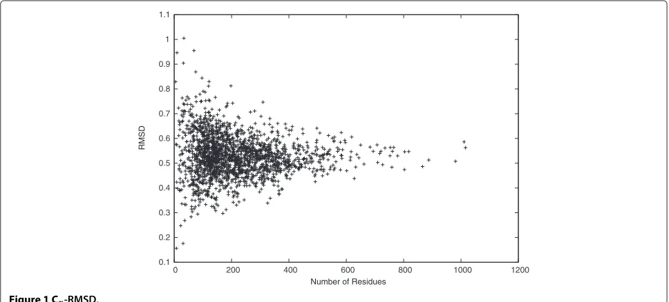
structures are very similar, we calculated theCα-RMSD as
shown in Figure 1. The Cα-RMSD is a popular distance
metric to evaluate the backbone distance between two protein backbone structures. The result shows that most
distances between the idealized and the target backbone
structures are small with mean 0.53 ˚A and STDEV 0.08 ˚A.
Specifically, the smallest Cα-RMSD reaches 0.16 ˚A, and
90% of theCα-RMSDs are smaller than 0.63 ˚A. Moreover,
the Cα-RMSD is upper bounded by 1.00 ˚A, although the
search space radius for each atom is set to be 1.6 ˚A. This
result is consistent with the result of checking(,)
dihe-dral angles, where the average difference between the
ide-alized and the target(,)dihedral angles is as small as
0.08◦. Therefore, it is possible to model protein backbone
structures in CULLPDB PC30 RES1.6 R0.25 accurately
using onlyanddihedral angles.
0 0.2 0.4 0.6 0.8 1
0 0.2 0.4 0.6 0.8 1
Beta-Sheet RMSD
Overall RMSD
0 0.2 0.4 0.6 0.8 1
0 0.2 0.4 0.6 0.8 1
Outter RMSD
Inner RMSD
Figure 4 Cα-RMSD of inner regions v.s. outer regions.
We studied theCα-RMSD further in different regions
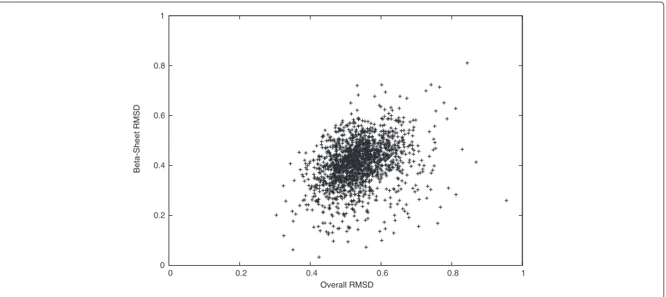
of the target protein structures. In Figures 2 and 3, we
see that the Cα-RMSD of the α-helix and the β-sheet
regions are smaller than that of the complete protein
by 0.28 ˚A and 0.12 ˚A, respectively. Indeed, these regions
are more restricted because of using DH(Pi,P0) to
con-serve hydrogen bonds of α-helices and β-sheets in our
scoring function. We also observe that the Cα-RMSD
of residues that are closer to the geometric center of
a target protein structure is 0.13 ˚A smaller on average
than the Cα-RMSD of the other residues that are
far-ther, as shown in Figure 4. Thus, the inner residues tend to be closer to the idealization state than are the outer
residues. We did not observe any significant differences
on the Cα-RMSD between the buried and the exposed
regions.
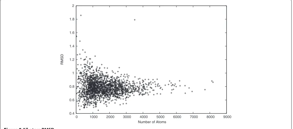
We also calculated the all-atom RMSD to show that the idealized and the target structures are very similar. In Figure 5, we see that most distances between the ide-alized and the target structures are small, with mean
0.79 ˚A and STDEV 0.13 ˚A. Moreover, the smallest all-atom
RMSD reaches 0.45 ˚A, and 90% of the all-atom RMSDs
are smaller than 0.94 ˚A. Note that both the Cα-RMSD
and the all-atom RMSD between the idealized and the target structures tend to be stable when the target pro-tein is long. Therefore, it is possible to model propro-tein
0.4 0.6 0.8 1 1.2 1.4 1.6 1.8 2
0 1000 2000 3000 4000 5000 6000 7000 8000 9000
RMSD
-400000 -350000 -300000 -250000 -200000 -150000 -100000 -50000 0 50000
0 200 400 600 800 1000 1200
Difference on dDFIRE Free Energy
Number of Residues Figure 6Protein backbone free energy (calculated by dDFIRE).
structures accurately with only , , and χ dihedral
angles.
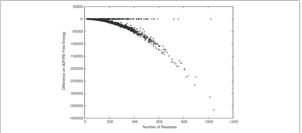
The idealized backbone structures are also favored in terms of free energy. This is shown by checking the free energy differences between the idealized and the target protein backbone structures in Figure 6. Here, we calcu-late the free energy using dDFIRE [20,21], and observe that the dDFIRE free energy of most idealized backbone structures are significantly better than are those of the target backbone structures. For the rest without signifi-cant improvements, the difference is close to zero. This may be the result of some tight thereochemical restraints used in existing X-ray structure refinement programs
[15,16]. It is also interesting that the observed free energy improvements are clearly not independent from the protein length. The figure suggests that the free energy difference has a square dependence on the protein length. After idealizing the side-chain structures, the free energy is either improved by a relatively biger amount or worsened by a relatively smaller amount as shown in Figure 7. Unfortunately, in most cases, the free energy is worsened slightly but is still in a stable state with negative values. Again, here we used dDFIRE [20,21] to calculate the free energy. We observed that the dDFIRE free energy is improved for 90 or 4.74% of the idealized protein struc-tures and is worsened slightly by 44 on average. Moreover,
-1600 -1400 -1200 -1000 -800 -600 -400 -200 0 200 400
0 200 400 600 800 1000 1200
Difference on dDFIRE Free Energy
Table 1 DSSP hydrogen bond differences before and after idealization
Type Count difference Percent difference
Parallel Bridge 9 0.04%
Antiparallel Bridge -211 -0.37%
27Helix 7080 26.46%
310Helix -1018 -2.35%
αHelix -1644 -1.48%
πHelix -82 -1.27%
All 5183 1.85%
the dDFIRE free energy is improved by 1585 in the best case, and worsened by 293 in the worst case. The figure also suggests that the free energy difference has a linear dependence on the protein length.
Several side-chain prediction tools have been proven to predict accurate side-chain structures from native backbone structures [8,9,25,26]. However, these tools does not perform well when predicting side-chain struc-tures from predicted backbone strucstruc-tures. To address this, we compared the predicted side-chain structures given the native backbone structures and those given the predicted backbone structures in terms of free energy. Here, we treat the idealized backbone struc-tures of the CULLPDB PC30 RES1.6 R0.25 data set as those which are best possibly predicted. Moreover, we used SCWRL4 [9] to predict side-chain structures and dDFIRE [20,21] to calculate free energies. The result shows that the free energy is worsened slightly by 43 if the predicted backbone structures are used. We do
not think this difference is significant to side-chain pre-diction. Certainly, more experiments will show if this is conclusive.
Finally, we study the effects of idealization on hydro-gen bonds. As shown in Table 1, we compare the num-ber of hydrogen bonds detected by the DSSP program [27,28]. Here, only differences of the most popular types of hydrogen bonds are included. We note that the total num-ber of hydrogen bonds is increased by 1.59% or 0.012 per residue after idealization. Specifically, the effects of
ideal-ization on hydrogen bonds ofβbridges is minor, and the
loss of the hydrogen bonds onαhelices is reasonably
con-trolled under 1.48%. Interestingly, the idealized backbone
structures have significantly more 27ribbons. The reason
behind this observation remains open.
In summary, we demonstrate that using dihedral angles with ideal bond lengths and angles is capable of modeling protein structures that are highly similar to the ones in CULLPDB PC30 RES1.6 R0.25 [18,19]. Since CULLPDB PC30 RES1.6 R0.25 is the complete set of PDB protein structures satisfying the high resolution and the low sequence identity constraints, it is reasonable to extend the conclusion to all protein backbone struc-tures. A positive side effect is that idealization improves backbone free energy, while most hydrogen bonds are conserved.
NMR protein structure refinement
In this experiment, we demonstrate an application of the protein structure idealization problem in NMR by idealizing 32 NMR protein structures. The NMR pro-tein structures were randomly chosen from PDB [17]
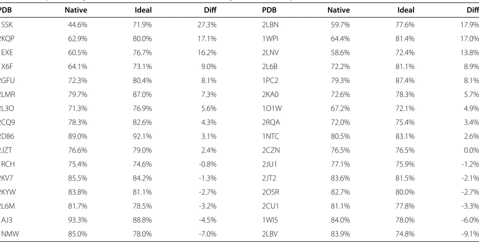
Table 2 The percentages of the favored(,)dihedral angles of32NMR protein structures before and after idealization
PDB Native Ideal Diff PDB Native Ideal Diff
1SSK 44.6% 71.9% 27.3% 2LBN 59.7% 77.6% 17.9%
2KQP 62.9% 80.0% 17.1% 1WPI 64.4% 81.4% 17.0%
1EXE 60.5% 76.7% 16.2% 2LNV 58.6% 72.4% 13.8%
1X6F 64.1% 73.1% 9.0% 2L6B 72.2% 81.1% 8.9%
2GFU 72.3% 80.4% 8.1% 1PC2 79.3% 87.4% 8.1%
2LMR 79.7% 87.0% 7.3% 2KA0 72.6% 78.3% 5.7%
2L3O 71.3% 76.9% 5.6% 1O1W 67.2% 72.1% 4.9%
2CQ9 78.3% 82.6% 4.3% 2RQA 72.0% 75.4% 3.4%
2D86 89.0% 92.1% 3.1% 1NTC 80.5% 83.1% 2.6%
2JZT 76.6% 79.0% 2.4% 2CZN 76.5% 76.5% 0.0%
1RCH 75.4% 74.6% -0.8% 2JU1 77.1% 75.9% -1.2%
2KV7 85.5% 84.2% -1.3% 2JT2 83.6% 81.5% -2.1%
2KYW 83.8% 81.1% -2.7% 2OSR 82.7% 80.0% -2.7%
2L6M 81.7% 78.5% -3.2% 2CU1 81.1% 77.8% -3.3%
1AJ3 93.3% 88.8% -4.5% 1WI5 84.0% 78.0% -6.0%
with a sequence identity cutoff of 30% and a gapless fragment length cutoff of 80 residues. In cases of multi-ple chains or models of some NMR protein structures, only the first chain from the first model is used in this experiment. This addition to the conclusion of the
previous experiment shows that poor (,) dihedral
angles of the NMR protein structures are improved by idealizing them.
To demonstrate this, we compared the percentage of
favored(,)dihedral angles calculated by PROCHECK
[29] in Table 2. After idealization, we see that 19 out of 32
NMR protein structures have more favored(,)
dihe-dral angles. Overall, the percentage is increased by 4.34% on average and 27.30% in the best case, which is closer to the minimum percentage of 90% expected in a good quality model [29].
Note that for those NMR protein structures that already
have more than approximately 75% of favored (,)
dihedral angles, idealization harms the percentage by
−0.85% on average. There are at least two reasons for
this. First, our free energy score Sf(Pi) is calculated
from a data set that is different from the one used by PROCHECK. In fact, we used 1898 protein structures of the CULLPDB PC30 RES1.6 R0.25 data set [18,19], while PROCHECK used 118 protein structures, with a
resolution cutoff of 2.0 ˚A and an R factor cutoff of 0.20
[29]. Although the percentages of favored(,)dihedral
angles are decreased in Table 2, our free energy scores of proteins 1WI5, 1NMW, and 2LBV are increased by 0.22, 1.35, and 0.31, respectively, after idealization. Sec-ond, our implementation is trying to optimize our
scor-ing functionSBB(Pi), instead of optimizing only the free
PROCHECK
B
A
L b
a
l
p ~p ~b
~a
~l
b ~b
b ~b
~b
-180 -135 -90 -45 0 45 90 135 180 -135
-90 -45 0 45 90 135 180
Ramachandran Plot
1wpi.native
Phi (degrees)
Psi (degrees)
ASP 20 (A)
ILE 21 (A)
ALA 22 (A) ASP 37 (A)
HIS 40 (A) ARG 41 (A)
LYS 82 (A)
LYS 84 (A)
ASP 87 (A) MET 92 (A)
ARG 100 (A)
Plot statistics
Residues in most favoured regions [A,B,L] 76 64.4% Residues in additional allowed regions [a,b,l,p] 31 26.3% Residues in generously allowed regions [~a,~b,~l,~p] 8 6.8% Residues in disallowed regions 3 2.5% ---- ---Number of non-glycine and non-proline residues 118 100.0%
Number of end-residues (excl. Gly and Pro) 2 Number of glycine residues (shown as triangles) 5 Number of proline residues 8 ---- Total number of residues 133
Based on an analysis of 118 structures of resolution of at least 2.0 Angstroms and R-factor no greater than 20%, a good quality model would be expected
to have over 90% in the most favoured regions.
energy score. Thus, it is possible to see decreased free energy scores after idealization, especially when the target
protein structure has a high percentage of favored(,)
dihedral angles.
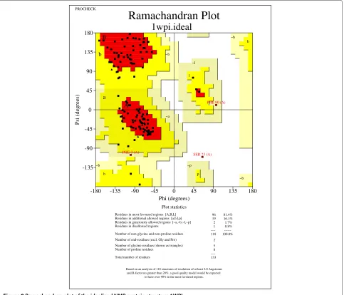
Our conclusion is further supported by the case study of the NMR structure with PDB ID 1WPI [30]. From the Ramachandran plots drawn by PROCHECK [29] in
Figures 8 and 9, we find that (,) dihedral angles
tend to move towards favored regions. Specifically, the
native structure contains only 64.4% of (,) dihedral
angles in favored regions, while the idealized structure contains a significantly improved percentage of 81.4% of
(,)dihedral angles in favored regions. Moreover, the
native structure contains three (,) dihedral angles
that are not in any feasible areas of the Ramachandran plot. However, there is only one such case found in the
idealized structure. Thus, two infeasible(,)dihedral
angles are fixed by the (,) dihedral angle log-odd
score. Here, we did not, but certainly can, implement a
hard constraint to disallow any infeasible(,)dihedral
angles.
In summary, we have demonstrated that protein
struc-ture idealization can be used to improve poor(,)
dihe-dral angles of protein pseudo-structures. These protein pseudo-structures can either be predicted or be exper-imentally determined. More applications of the protein structure idealization problem will be studied.
Conclusion
We have introduced the protein structure idealization problem and performed our first attempt to solve it. The experiment results show that idealized structures always exist with small changes on the coordinates. Further-more, the idealized backbone structures have significantly
PROCHECK
B
A
L b
a
l
p ~p ~b
~a
~l
b ~b
b ~b
~b
-180 -135 -90 -45 0 45 90 135 180 -135
-90 -45 0 45 90 135 180
Ramachandran Plot
1wpi.ideal
Phi (degrees)
Psi (degrees)
PHE 3 (A)
SER 23 (A) PHE 90 (A)
Plot statistics
Residues in most favoured regions [A,B,L] 96 81.4% Residues in additional allowed regions [a,b,l,p] 19 16.1% Residues in generously allowed regions [~a,~b,~l,~p] 2 1.7% Residues in disallowed regions 1 0.8% ---- ---Number of non-glycine and non-proline residues 118 100.0%
Number of end-residues (excl. Gly and Pro) 2 Number of glycine residues (shown as triangles) 5 Number of proline residues 8 ---- Total number of residues 133
Based on an analysis of 118 structures of resolution of at least 2.0 Angstroms and R-factor no greater than 20%, a good quality model would be expected
to have over 90% in the most favoured regions.
better free energy and (,) dihedral angle distribu-tions. Therefore, protein structures can be modeled accu-rately with dihedral angles and ideal bond lengths and angles, and it is feasible to predict protein backbone and side-chain structures by searching the dihedral angle space.
Our protein structure idealization algorithm may be improved in several ways. Since our scoring functions
are very simple with all weights wa = 1.0 in the
cur-rent implementation, there is space for improvements. We are also looking forward to adding protein-ligand inter-action energy to our scoring function and to studying the effect of idealization on protein-ligand interactions. Moreover, since some atoms are more flexible than oth-ers, we can also set different search spaces for different atoms in our algorithm. For example, when idealizing X-ray protein structures, the search space of each atom could be selected according to its B-factor. We can also adopt a divide-and-conquer algorithm in our algorithm to find the global, rather than local, optimal idealized structure. Specifically, we can divide the protein structure into small fragments, idealize each fragment separately, and merge idealized fragments. The key is to divide the protein struc-ture by a tree decomposition of the interaction graph and to remember the optimal idealized fragment for each pos-sible configuration of atoms with interactions to external atoms. Similar ideas have already been used successfully to improve the speed and the accuracy of backbone and side-chain structure predictions [8,9,25,26,31,32].
Our protein structure idealization algorithm can also correct modelling errors of protein structures in PDB [17]. In fact, previous research indicates that many bond con-formations and side-chain rotamers are likely incorrect in PDB, and it is useful to have an automated mechanism to fix these problems [33,34]. Thus, we can address these problems by idealizing all protein structures in PDB with our protein structure idealization algorithm and using our specially tuned scoring functions.
The idealized version of the PDB [17] provides new protein structure references to study protein structures and functions. For example, we can rebuild fragment and rotamer libraries based on the idealized PDB. It would then be more intuitive to use the idealized fragment or rotamer libraries in the protein backbone or side-chain structure prediction algorithms searching the dihedral angle space. Thus, we expect to see some improvements of the accuracy of these algorithms with the idealized fragment and rotamer libraries. Therefore, we also pro-vide a new approach for discovering unusual atoms and bonds by comparing the idealized and the original PDB structures. Although most of these unusual atoms and bonds are due to errors, we expect to discover some biochemical insights that assist in understanding protein functions.
Competing interests
The authors declare that they have no competing interests.
Authors’ contributions
The algorithm implementation and all experiments were done by XC; the protein structure idealization algorithm was designed by XC and SCL; the CULLPDB PC30 RES1.6 R0.25 experiment was designed by XC, SCL and DB; the NMR experiment was designed by XC and BA; the project was directed by ML; All authors read and approved the final manuscript.
Acknowledgements
This work was supported by a Startup Grant at City University of Hong Kong [7002731], National Basic Research Program of China [2012CB316500], an NSERC Grant [OGP0046506], Canada Research Chair program, an NSERC Collaborative Grant, OCRiT, Premier’s Discovery Award, Killam Prize and SHARCNET.
Author details
1University of Waterloo, Ontario, Canada.2City University of Hong Kong, Hong
Kong, China.3Chinese Academy of Sciences, Beijing, China.
Received: 20 December 20121 Accepted: 5 February 2013 Published: 25 February 2013
References
1. Engh RA, Huber R:Accurate bond and angle parameters for X-ray protein structure refinement.Acta Crystallogr Sect A1991,47:392–400. 2. Engh RA, Huber R:Structure quality and target parameters.Vol F Int
Tables Crystallogr2006:382–392. chap. 18.3.
3. G ¨untert P, W ¨uthrich K:Improved efficiency of protein structure calculations from NMR data using the program DIANA with redundant dihedral angle constraints.J Biomol NMR1991,
1(4):447–456.
4. G ¨untert P, Mumenthaler C, W ¨uthrich K:Torsion angle dynamics for NMR structure calculation with the new program DYANA.J of Mol Biol1997,273:283–98. [http://www.ncbi.nlm.nih.gov/pubmed/9367762] 5. Simons KT, Kooperberg C, Huang E, Baker D:Assembly of protein
tertiary structures from fragments with similar local sequences using simulated annealing and Bayesian scoring functions.J Mol Biol 1997,268:209–225.
6. Simons KT, Strauss C, Baker D:Prospects for ab initio protein structural genomics.J Mol Biol2001,306:1191–1199.
7. Li SC, Bu D, Xu J, Li M:Fragment-HMM: a new approach to protein structure prediction.Protein Sci2008,17(11):1925–1934.
8. Canutescu AA, Shelenkov AA, Dunbrack RL:A graph-theory algorithm for rapid protein side-chain prediction.Protein Sci2003,
12(9):2001–2014. [http://dx.doi.org/10.1110/ps.03154503] 9. Krivov GG, Shapovalov MV, Dunbrack RL:Improved prediction of
protein side-chain conformations with SCWRL4.Proteins: Struct Funct Bioinformatics2009,77(4):778–795. [http://dx.doi.org/10.1002/prot. 22488]
10. Kuszewski J, Gronenborn AM, Clore GM:Improving the quality of NMR and crystallographic protein structures by means of a
conformational database potential derived from structure databases.Protein Sci1996,5(6):1067–1080.
11. Kuszewski J, Gronenborn AM, MClore G:Improvements and extensions in the conformational database potential for the refinement of NMR and X-ray structures of proteins and nucleic acids.J Magn Reson1997,
125:171–177.
12. Rice LM, Br ¨unger AT:Torsion angle dynamics: Reduced variable conformational sampling enhances crystallographic structure refinement.Proteins: Struct Funct Genet1994,19(4):277–290. [http://dx. doi.org/10.1002/prot.340190403]
13. Stein EG, Rice LM, Br ¨unger AT:Torsion-angle molecular dynamics as a new efficient tool for NMR structure calculation.J Magn Reson1997,
124:154–164.
14. Dunbrack RL, Cohen FE:Bayesian statistical analysis of protein side-chain rotamer preferences.Protein Sci1997,6(8):1661–1681. [http://www.proteinscience.org/cgi/content/abstract/6/8/1661] 15. Evans PR:An introduction to stereochemical restraints.Acta
16. Jaskolski M, Gilski M, Dauter Z, Wlodawer A:Stereochemical restraints revisited: how accurate are refinement targets and how much should protein structures be allowed to deviate from them?.Acta Crystallogr D2007,63(5):611–620.
17. Berman HM, Westbrook J, Feng Z, Gilliland G, Bhat TN, Weissig H, Shindyalov IN, Bourne PE:The Protein Data Bank.Nucleic Acids Res2000,
28:235–242.
18. Wang G, Dunbrack RL:PISCES: a protein sequence culling server.
Bioinformatics2003,19(12):1589–1591.
19. Wang G, Dunbrack RL:PISCES: recent improvements to a PDB sequence culling server.Nucleic Acids Res2005,
33(Web-Server-Issue):94–98.
20. Yang Y, Zhou Y:Ab initio folding of terminal segments with secondary structures reveals the fine difference between two closely related all-atom statistical energy functions.Protein Sci2008,17(7):1212–1219. 21. Yang Y, Zhou Y:Specific interactions for ab initio folding of protein
terminal regions with secondary structures.Proteins2008,
72(2):793–803.
22. Lattman E, Loll PJ, Loll P:Protein Crystallography: A Concise Guide: Johns Hopkins University Press; 2008.
23. Rutgers UCSD:Protein data bank contents guide.2011. http://www. wwpdb.org/documentation/format33/v3.3.html.
24. Shapovalov MV, Dunbrack RL:A smoothed backbone-dependent rotamer library for proteins derived from adaptive kernel density estimates and regressions.Structure London England 19932011,
19(6):844—858. [http://www.ncbi.nlm.nih.gov/pubmed/21645855] 25. Xu J:Rapid protein side-chain packing via tree decomposition: Springer;
2005.
26. Xu J, Jiao F, Berger B:A tree-decomposition approach to protein structure prediction.Fourth Int IEEE Comput Soc Comput Syst Bioinformatics Conf2005:247–256. [http://view.ncbi.nlm.nih.gov/ pubmed/16447982]
27. Kabsch W, Sander C:Dictionary of protein secondary structure: Pattern recognition of hydrogen-bonded and geometrical features.
Biopolymers1983,22(12):2577–2637.
28. Joosten RP, te Beek TAH, Krieger E, Hekkelman ML, Hooft RWW, Schneider R, Sander C, Vriend G:A series of PDB related databases for everyday needs.Nucleic Acids Res2011,39(suppl 1):D411–D419.
29. Laskowski RA, MacArthur MW, Moss DS, Thornton JM:PROCHECK: a program to check the stereochemical quality of protein structures.
J of Appl Crystallogr1993,26(2):283–291. [http://dx.doi.org/10.1107/ S0021889892009944]
30. Jung JW, Yee A, Wu B, Arrowsmith CH, Lee W:Solution structure of YKR049C, a putative redox protein from Saccharomyces cerevisiae.
J Biochem Mol Biol2005,38(5):500–504.
31. Xu Y, Xu D:Protein threading using PROSPECT: Design and evaluation.Proteins: Struct Funct Genet2000,40(3):343–354. [http://dx. doi.org/10.1002/1097-0134(20000815)40:3]
32. Kim D, Xu D, Guo JT, Ellrott K, Xu Y:PROSPECT II: protein structure prediction program for genome-scale applications.Protein Eng2003,
16(9):641–650. [http://view.ncbi.nlm.nih.gov/pubmed/14560049] 33. Hooft RWW, Vriend G, Sander C, Abola EE:Errors in protein structures.
NATURE1996,381(6580):272.
34. Joosten RP, Joosten K, Cohen SX, Vriend G, Perrakis A:Automatic rebuilding and optimization of crystallographic structures in the Protein Data Bank.Bioinformatics2011,27(24):3392–3398. [http://dx. doi.org/10.1093/bioinformatics/btr590]
doi:10.1186/1748-7188-8-5
Cite this article as:Cuiet al.:Protein Structure Idealization: How accurately is it possible to model protein structures with dihedral angles?.Algorithms for Molecular Biology20138:5.
Submit your next manuscript to BioMed Central and take full advantage of:
• Convenient online submission
• Thorough peer review
• No space constraints or color figure charges
• Immediate publication on acceptance
• Inclusion in PubMed, CAS, Scopus and Google Scholar
• Research which is freely available for redistribution