INTRODUCTION
Type 1 diabetes is an autoimmune dis-ease caused by destruction of insulin-producing pancreatic islet cells. Cur-rently, islet transplantation is considered a less effective treatment modality for Type 1 diabetes than pancreas transplan-tation, especially from the viewpoint of long-term graft survival. There are two major impediments to the clinical appli-cation of islet transplantation: immune destruction of transplanted islets (1) and the limited supply of islet tissue (2–4). The islet rejection process is character-ized by rapid infiltration of immune cells, followed by antigen-specific T-cell responses. Among the strategies used to
overcome immune rejection are the use of novel immunosuppressive agents and regimens and donor-specific induction of immune tolerance in the host. Mesenchy-mal stem cells (MSCs) are self-renewing, multipotent progenitor cells with the ca-pacity to differentiate into several dis-tinct mesenchymal lineages. It has been suggested that MSCs escape the immune system because they possess a cell sur-face phenotype that is poorly recognized by T cells. MSCs also mediate their im-munosuppressive action through the se-cretion of cytokines (5). In line with their immunosuppressive capacities in vitro, MSCs have also been shown to display immunosuppressive capacities in vivo, as
evidenced by the demonstration that al-logeneic MSCs prolonged allograft sur-vival in immunocompetent mice (6). On the basis of their capacity to modulate immune responses and to promote tissue repair in experimental models, MSCs have been proposed as a treatment for autoimmune diseases, such as diabetes, rheumatoid arthritis, systemic lupus ery-thematosus and multiple sclerosis. The use of MSCs to prevent rejection of allo-geneic grafts is still mostly limited to ani-mal models, and the results obtained to date are conflicting. In the present study, we evaluated the therapeutic potential of autologous MSCs for prevention of graft rejection after allogeneic islet transplan-tation. In addition, we evaluated the ex-pression profiles of pro- and antiinflam-matory cytokine genes in the grafted site, serum cytokine levels, immune re-sponses in splenocytes of recipients after allostimulation and the frequency of T-regulatory cells (Tregs) in secondary
IL-10-Activated Regulatory T Cells Play a Role in Immune
Modulation of Mesenchymal Stem Cells in Rat Islet Allografts
Yang-Hee Kim, Yu-Mee Wee, Monica-Y Choi, Dong-Gyun Lim, Song-Cheol Kim, and Duck-Jong Han
Department of Surgery, University of Ulsan College of Medicine, and Asan Medical Center, Seoul, Korea
Mesenchymal stem cells (MSCs) are suggested to be immune modulators because of their therapeutic potential in transplan-tation. In the present study, we evaluated the therapeutic potential of autologous MSCs for preventing graft rejection after allo-geneic rat islet transplantation. We assessed the ability of MSCs to elicit an antiproliferative response in alloreactive lymphocytes and tested the immunosuppressive effect of MSCs in allogeneic islet transplantation. In islet allotransplantation, injection of autol-ogous MSCs or a subtherapeutic dose of cyclosporine A (CsA; 5 mg/kg) alone did not prolong allograft survival. However, graft survival was attained for >100 d in 33% of autologous MSC-plus-CsA–treated recipients, indicating that graft acceptance was achieved in a subgroup of allograft recipients. Splenocytes from autologous MSC-plus-CsA–treated rats exhibited a reduced mixed lymphocyte reaction (MLR)-proliferative response to donor stimulators and increased interleukin (IL)-10 release. Interestingly, after excluding host CD11b+cells, splenic T cells from autologous MSC-plus-CsA–treated rats did not produce IL-10 or did not in-hibit proliferative responses under the same conditions. The use of autologous MSC-plus-CsA downregulated immune responses, inducing donor-specific T-cell hyporesponsiveness by reducing the production of proinflammatory cytokines and inducing antiin-flammatory cytokine production, especially that of IL-10, during the early posttransplantation period. T-regulatory cells made a contribution at a later phase. In conclusion, the combined use of autologous MSCs and low-dose CsA exerted a synergistic im-munosuppressive effect in an islet allograft model, suggesting a role for autologous MSCs as an immune modulator.
© 2011 The Feinstein Institute for Medical Research, www.feinsteininstitute.org Online address: http://www.molmed.org
doi: 10.2119/molmed.2010.00098
lymphoid organs. The objective of these measurements was to determine whether any of these factors played a role in modulating the immune response after MSC injection, thereby influencing islet allograft tolerance.
MATERIALS AND METHODS
Animals
Male Lewis (RT11) and Fisher (RT11v1) rats <8–10 wks of age were used as donors and recipients, respectively. Fisher rats were purchased from Charles River Japan (Kanagawa, Japan). Lewis rats were bred and maintained at Asan Medical Center Animal Facilities. Recipi-ents were rendered diabetic by a single injection of streptozotocin (35 mg/kg body weight in citrate buffer, pH 4.5; Sigma, St. Louis, MO, USA) 21 d before transplantation. An animal was consid-ered diabetic when its blood glucose level exceeded 200 mg/dL in two consec-utive measurements. This experiment was approved by the Institutional Ani-mal Care and Use Committee of Asan In-stitute for Life Sciences, Asan Medical Center (Seoul, South Korea; review num-ber 2008-13-091), and conducted in accor-dance with the guidelines of the Asan In-stitute for Life Sciences for Experimental Animal Care and Use.
Islet Isolation
Lewis pancreatic islets were isolated and purified using the Ficoll purification method (7). Briefly, the pancreas of each rat was distended with a 10-mL intra-ductal injection of collagenase type XI (800 units/mL; Sigma), and pancreas di-gestion was performed at 37°C. Digested pancreatic tissue was mechanically dis-rupted by filtering through a mesh (400-μm pore size). Islets were purified by discontinuous density gradient cen-trifugation using Ficoll (Sigma). Islet numbers and purity were determined by dithizone staining. Before transplanta-tion, islets in RPMI-1640 medium supple-mented with 10% (v/v) fetal bovine serum (FBS) were cultured overnight in a 37°C, 5% CO2environment.
Isolation and Culture of MSCs Bone marrow (BM)-derived MSCs were harvested from 8-week-old (200–250 g) male Fisher rats. After re-moving epiphyses and gaining access to the marrow cavities, whole BM plugs were flushed out from tibial and femoral bones with low-glucose Dulbecco’s mod-ified Eagle’s medium (DMEM-LG; Invit-rogen, Carlsbad, CA, USA) using a 10-mL syringe with a 22-gauge needle. The cell suspension was filtered through a nylon sieve (100 μm) and centrifuged for 5 min at 450g. Collected cells were then resuspended in supplemented medium. After counting, cells in DMEM-LG supplemented with 10% (v/v) FBS and 1% (v/v) GibcoΤΜ Antibiotic-Antimycotic (Invitrogen) were plated in 25- to 75-cm2flasks (BD Bio-sciences, San Jose, CA, USA) at a concentration of 5 ×10⁶nucleated cells/mL/cm2and incubated in 5% CO2 at 37°C. After 24 h, the medium was changed, unattached cells were dis-carded, and the adherent cells were cul-tured until they reached 80% confluence. Cells were then detached from the bot-tom using a 0.25% Trypsin-EDTA (Invit-rogen). The resulting suspension was then expanded by plating at 6,000 cells/ cm2in 75-cm2flasks. The same condi-tions were used for subsequent passages. At least two independently isolated batches of MSCs were used in each experiment.
Phenotypic Analysis of MSCs
MSCs (passage 3–5) were resuspended in phosphate-buffered saline (PBS), pH 7.2, containing 1.0% (w/v) bovine serum albumin (BSA; Sigma) and 0.1% (w/v) sodium azide (Sigma). The suspensions were incubated with monoclonal antibod-ies (mAbs) for 30 min at 4°C. The follow-ing mAbs, purchased from Dickinson Pharmingen (San Diego, CA, USA), were used: fluorescein isothio-cyanate (FITC)-conjugated antirat RT1A, CD90.1, CD 29, CD25 or CD11b; phyco-erythrin (PE)-conjugated antirat RT1B, CD4 or CD54; and cychrome- conjugated antirat CD45. Each fluorescence analysis
included the appropriate FITC-, PE- or cychrome-conjugated isotype Ab controls. Cells were separated using a flow cytom-etry (FACSCalibur; Becton- Dickinson, San Diego, CA, USA) and not sorted.
In Vitro Differentiation
Rat BM-MSCs were also assessed for adipogenic and osteogenic differentiation in vitroby using the Trevigen’s rat mes-enchymal stem cell differentiation kit fol-lowing the manufacturer’s protocol (Trevigen, Gaithersburg, MD, USA).
Adipogenic differentiation.For adi-pogenic differentiation, the MSCs were plated in 24-well plates in adipogenic medium at a cell density of 5 ×10³cells per well. The adipogenic medium was composed of DMEM with low glucose supplemented with 10% FBS, 0.1 mmol/L indomethacin, 0.5 mmol/L isobutyl-methylxanthine and 10–6mol/L dexa -methasone (9). Undifferentiated MSCs were grown for 14 d in complete growth medium. The media were replaced every 3 d for 14 d. Adipogenic differentiation was assessed by oil red O staining at 3 wks after initial adipogenic induction. For oil red O staining, the cells were rinsed in PBS and fixed in 10% formalin followed by incubation of the cells in 2% (w/v) oil red O reagent for 5 min at room temperature. The cells were rinsed in isopropanol followed by several changes of distilled water and were then examined under a light microscope and photographed. Cells stained were dis-solved in 100% isopropanol for 10 min with shaking, and 200 μL of the sample were transferred to a 96-well plate. Ex-tracted oil red O was measured using a SpectraMax 190 Absorbance Microplate Reader (Molecular Devices) at 500 nm and calculated the oil red O present in each sample relative to the standard curve (50–500 μg/mL).
Mixed Lymphocyte Reaction Assay
Immunosuppressive effect of MSCs on mixed lymphocyte reaction (MLR).
Fisher splenocytes (responder; 2.5 × 10⁵/well) and irradiated Lewis spleno-cytes (stimulator; 5 ×10⁵/well) were co-cultured with or without MSCs for 3 d. Autologous or allogeneic MSCs were plated in triplicate onto round-bottom 96-well plates (BD Biosciences) in decreasing numbers (25 ×104, 12.5 ×10⁴, 6.25 ×10⁴ and 3.13 ×10⁴cells/well) and allowed to adhere to the plate for 1–2 h. The same procedure was followed in transwell chambers (0.2 μm; Corning, Corning, NY, USA). MSCs (2.5 ×10⁵/well) were seeded in the lower chamber and allowed to ad-here for 1–2 h. Equal numbers of Fisher splenocytes and irradiated Lewis
spleno-cytes were then cultured in the upper transwell chamber for 3 d. For secondary MLRs, Fisher splenocytes (2.5×10⁵/well), obtained at the end of the primary MLR performed with either MSCs or MSCs and cyclosporine A (CsA) (300 nmol, Sandimmune; Novartis Pharmaceuticals Corporation, East Hanover, NJ, USA) for 7 d, were washed twice with PBS to elim-inate any trace of MSCs and CsA. They were then restimulated with irradiated splenocytes (5 ×10⁵/well) from Lewis rats (the same allogeneic condition as for primary MLR), Wistar Furth rats (WF, RT1u, third party) or concanavalin A (ConA; 1 μg/well) for 3 d.
T-cell proliferation in splenocytes of rats transplanted. Stimulator cells were prepared from splenocytes of Lewis rat donors. The cells were irradiated and seeded into 96-well U-bottom culture plates (NUNC, Roskilde, Denmark). Splenocytes isolated from each treatment recipient were used as responder cells and purified by centrifugation on Ficoll density gradients (Histopaque 1077; Sigma) and stored in liquid nitrogen until used. In another experiment, T cells were purified from splenocytes of trans-planted rat by negative selection with anti–major histocompatibility complex (MHC) class II (OX6) microbeads (Mil-tenyi Biotec, Auburn, CA, USA). Thawed splenocytes (2.5 ×10⁵cells/well) were mixed with irradiated (25 Gy; Cs irradia-tor, Cisbio International, Bedford, MA, USA) allogeneic splenocytes (5 ×10⁵/ well) and plated on 96-well U-bottom culture plates (NUNC). To investigate cy-tokine interactions in MLR, neutralizing Abs directed against rat IL-10 or trans-forming growth factor (TGF)-β(R&D Systems, Minneapolis, MN, USA) were added at a concentration of 0.5 μg/mL on day 0 to block IL-10 or TGF-β. Normal goat IgG and IgG1 Abs (R&D Systems) at the same concentration were used as negative controls. Twenty-four hours be-fore termination of the culture, 1 μCi [3H]-thymidine (Amersham Bioscience, Arlington Heights, IL, USA) was added to each well and harvested 18 h later. Thymidine uptake was quantified in a
β-counter (TopCount NXT; PerkinElmer, Waltham, MA, USA). T-cell proliferation is expressed as mean cpm ± standard de-viation (SD) of three wells.
Islet Transplantation
Islets (4,000 islet equivalents/rat) and MSCs (2 ×10⁶cells/rat) were trans-planted into recipient rat via the portal vein. Four groups of diabetic Fisher rats, with four to six animals per group, were transplanted with islets. CsA was diluted in PBS and administered once daily for 14 consecutive d at 5 mg/kg, intraperi-toneally (i.p.). Group 1 received donor islets alone, group 2 received donor islets with MSCs, group 3 was treated with CsA (5 mg/kg/day) for 2 wks, and group 4 was treated with both MSCs and CsA (5 mg/kg/day, for 2 wks). Graft function was monitored by daily mea-surement of blood glucose and at least twice thereafter. Islet rejection was de-fined as blood glucose values >200 mg/ dL on 2 consecutive days.
Immunohistochemistry
Glostrup, Denmark), anti-CD4 (1:200; Serotec, Oxford, UK) and anti-CD8 (1:200, Serotec). Slides were counter-stained with hematoxylin and eosin (H&E).
Intracellular Staining for Foxp3 and IL-10
Peripheral blood mononuclear cells (PBMCs), graft-infiltrating lymphocytes, splenocytes and draining lymph node (LN) cells were preincubated with Fc block to prevent nonspecific binding to Fcγreceptors, and cell surface proteins were stained using FITC-labeled anti-CD25 (OX39) and PE-labeled anti-CD4 (OX-38) mAbs. Intracellular Foxp3 was stained using allophycocyanin labeled anti-Foxp3 mAb (FJK-16s; eBio-science, San Diego, CA, USA), according to the manufacturer’s instructions. For intracellular IL-10 staining, several sam-ples were stimulated with alloantigen for 24 h; Golgi-Stop was included for the last 6 h of stimulation. The harvested cells were stained with FITC-labeled anti-CD4 and anti-CD11b mAbs and then
processed for intracellular staining with PE-labeled anti–IL-10 mAb (JES5-16E3). Anti-CD4, anti-CD11b, anti-CD25 and anti–IL-10 mAbs, except those for anti-Foxp3 mAb, Fc block and isotype IgGs, were purchased from BD Biosciences. Samples were assessed on a FACSCal-ibur instrument using CellQuest soft-ware (BD Biosciences).
RNA Extraction and Quantitative Reverse Transcription–Polymerase Chain Reaction (RT-PCR)
After transplantation, liver samples were snap-frozen in liquid nitrogen and stored at –80°C until used. Total RNA was isolated using TRIzol (Invitrogen), and cDNA was synthesized from a 1-μg RNA template using random hexamers and AccuPower®RT PreMix (Bioneer, Daejeon, South Korea). One-twentieth of the RT re-action mixture was added to the PCR. Rat primer sequences for IL-2 (NM_053836.1), IL-4 (NM_201270.1), interferon (IFN)-γ (NM_138880), IL-10 (X60675), TGF-β1 (NM_021578.2), Foxp3 (NM_001108250.1)
and GAPDH (BC059110) were as follows: glyceraldehyde-3-phosphate dehydroge-nase (GAPDH) sense, 5′-TCA TGA CCA CAG TCC ATG CCA-3′, GAPDH anti-sense, 5′-GGG AGT TGC TGT TGA AGT CAC-3′; IL-2 sense, 5′-TTG CAC TGA CGC TTG TCC TCC TTG TCA ACA-3′, IL-2 antisense, 5′-CCA TCT CCT CAG AAA TTC CAC CAC AGT TGC-3′; IL-4 sense, 5′-AGG TCA ACA CCA CGG AGA AC-3′, IL-4 antisense, 5′-AGT TCA GAC CGC TGA CAC CT-3′; IFN-γsense, 5′ -TAC ACG CCG CGT CTT GGT TTT-3′, IFN-γantisense, 5′-AGC CTA AGG AAG CGG AAA AGG-3′; IL-10 sense, 5′-GGT GAC AAT AAC TGC ACC CAC-3′, IL-10 antisense, 5′-GTG TCA CGT AGG CTT CTA TGC-3′; TGF-β1 sense, 5′-GCC TCC GCA TCC CAC CTT TG-3′, TGF-β1 anti-sense, 5′-GCG GGT GAC TTC TTT GGC GT-3′; and Foxp3 sense, 5′-GCA CAA GTG CTT TGT GCG AGT-3′, Foxp3 anti-sense, 5′-TGT CTG TGG TTG CAG ACG TTG-3′. PCR was performed at 95°C for 5 min, followed by 30 cycles at 95°C for 30 s, 60°C for 45 s and 72°C for 1 min. Amplified products were analyzed on 1.5% agarose gels. Band densities were quantified using the Bio-Rad Quantity One program (Bio-Rad, Hercules, CA, USA). GAPDH was used as an endoge-nous control to normalize RNA amounts.
Statistical Analysis
Data were expressed as means ± SD. Statistical comparisons among groups were performed by Student ttest or anal-ysis of variance (ANOVA) followed by Bonferroni posttests for multiple com-parisons. Differences were considered statistically significant at P values <0.05.
RESULTS
Characterization of MSCs from Adult Rat Bone Marrow
Adherent MSCs had a spindle-shaped fibroblastic morphology (Figure 1A, a and b). To test whether multilineage differentiation of BM-derived MSCs is possible, MSCs were induced to differen-tiate into osteoblast and adipocyte (Fig-ure 1A). When MSCs were tested for their
potential to differentiate into adipocytes at passage 3, morphologic changes in the cells as well as the formation of neutral lipid vacuoles were noticeable as early as day 7 after induction. At day 14, accumu-lation of lipid vacuoles within and around the cells was visualized by staining with oil red O (Figure 1A, e). Osteogenic differ-entiation was studied by alizarin red S staining for each substrate in control and osteogenic media. Alizarin red staining is an indicator of mineralization of the ex-tracellular matrix. Undifferentiated MSCs were used as a negative control, and staining for mineralization was negative. In contrast, MSCs cultured in the osteo -genic medium were positive in alizarin staining. BM-MSCs exhibited classic min-eralization nodules, and alizarin red staining was more intense for MSCs than for fibroblasts (Figure 1A, h). After adi-pogenic or osteo genic induction, BM-MSCs markedly increased the amount of oil red O or alizarin red stain compared with primary fibroblast (Figure 1B). These data confirm that BM-MSCs can differentiate into osteocytes and adipocytes. A phenotypic analysis of MSCs and fibroblast by flow cytometry showed that fibroblasts were positive for MHC class I molecule (RT1A), CD29 and CD54; but were negative for MHC class II molecule (RT1B), CD45, CD11b, CD25 , CD90 and CD106 (Figure 1C). In contrast, MSCs were positive for MHC class I mol-ecule (RT1A), Thy1 (CD90), CD29, CD54 and CD106; but were negative for MHC class II molecule (RT1B), CD45, CD11b and CD25 (Figure 1D).
Rat BM-Derived MSCs Inhibit the Alloantigen-Dependent Proliferative Response of T Cells
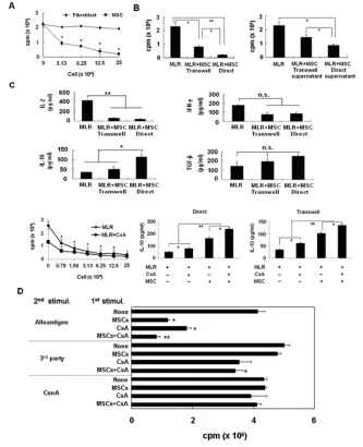
of T-cell proliferation (see Figure 2A). MLR assays using MSCs were also per-formed in transwell cultures to assess the influence of humoral substances released from MSCs and the role played by cell–cell contact between responder cells and MSCs. As shown in Figure 2B, the in-hibitory effect of MSCs was partially di-minished when cells were physically sep-arated by the transwell membrane, indicating that the inhibition is mediated by both cell–cell contact and soluble fac-tor. A comparison of the production of these cytokines during MLR showed that IL-10 secretion in cocultures with MSCs was significantly increased compared with that in cultures without MSCs (P< 0.05). Furthermore, IL-10 levels were higher under coculture conditions that al-lowed cell contact than in cultures with-out cell contact (Figure 2C; P< 0.005). CsA, added at the MLR, inhibited allo-geneic lymphocyte proliferation in the dose-dependent manner of MSCs (see Figure 2C). IL-10 secretion in cocultures with MSCs was significantly increased compared with that in cultures without MSCs. When adding CsA to MLR mixed with MSCs, IL-10 secretion was signifi-cantly increased compared with other groups (see Figure 2C; P< 0.005). There was also a trend toward increased TGF-β secretion in MSC-containing cocultures, although this did not attain statistical sig-nificance. Next, to evaluate whether the inhibitory effect of MSCs was reversible, we cocultured recipient splenocytes with MSCs or MSC-plus-CsA for 7 d in a pri-mary MLR and then restimulated with ir-radiated donor, third-party splenocytes or ConA (1 μg/mL) for 3 d. Cells cocul-tured with MSCs responded weakly to restimulation with donor splenocytes. Moreover, splenocytes preexposed to CsA or MSC-plus-CsA showed impaired responses to restimulation with alloanti-gen, compared with unexposed spleno-cytes. However, the preexposed cells showed normal responses to third-party antigens or nonspecific mitogens, show-ing that the immunosuppressive activity of MSCs was antigen specific and par-tially irreversible (Figure 2D).
MSC-plus-CsA Administration Prolongs Islet Allograft Survival
To assess the in vivofunction of MSCs, we investigated whether MSCs alone, or
MSCs in combination with a subthera-peutic dose of CsA (5 mg/kg) over 2 wks, had any effect on islet graft sur-vival after islet transplantation into the
liver. In the untreated group, graft sur-vival (mean ± SD) was 5.0 ± 0.52 d. The mean graft survival times in the allo-geneic and autologous MSC-treated groups were 3.7 ± 0.6 and 7.8 ± 1.26 d, respectively. Treatment with MSCs alone had no beneficial effect on graft sur-vival. The mean graft survival time in rats receiving a subtherapeutic dose of CsA alone was 9.2 ± 4.02 d, similar to controls. In animals treated with allo-geneic MSC-plus-CsA, mean graft survival time was 13.75 ± 12.3 d, not significantly different from CsA alone (Figure 3A). In contrast, treatment with autologous MSC-plus-CsA dramatically improved islet graft survival, increasing mean graft survival time to more than 89.3 ± 77.4 d (P< 0.05, compared with CsA alone). Collectively, these results demonstrate that allogeneic MSCs did not show an immunosuppressive effect in our rat islet transplantation model, and that treatment with autologous MSC-plus-CsA significantly enhances rat islet allograft survival compared with that after CsA alone (Figure 3B). In normoglycemic rats receiving MSCs and a short course of CsA, islets remained viable in the livers, and immunohisto-logical analyses confirmed that insulin was generated by transplanted islets (Figure 3C). To examine cellular infiltra-tion of the graft, we conducted a second series of islet transplants. Grafts were harvested 5 d after transplantation and analyzed immunohistochemically for in-sulin, CD4 and CD8 (n = 3 per treat-ment group). Control animals showed poor insulin staining at the graft site and a scattered infiltrate of CD8+T cells. No differences in CD4+T-cell infiltration were observed between the groups. However, the number of CD8+T cells was higher in the control group than in MSC-plus-CsA groups on day 5. Grafts from MSC-plus-CsA– treated recipients showed positive staining for insulin and limited CD4 and CD8 cellular infiltra-tion (Figure 3D). Histological analyses of pancreases from long-term surviving recipients confirmed destruction of islets.
MSC-plus-CsA Administration Results in Production of Low Levels of Th1-Type Cytokines and High Levels of Th2-Type Cytokines in the Grafted Liver Tissue and Serum
To elucidate potential mechanisms un-derlying the immunomodulatory func-tion of MSCs on allografts, we analyzed grafted livers on posttransplantation day 10, 30 and 100 for the expression of Th1-and Th2-type cytokines, Th1-and Foxp3, by RT-PCR. In grafted livers of the
MSC-plus-CsA group, the expression of genes encoding the antiinflammatory Th2-type cytokines, IL-4 and IL-10, was signifi-cantly increased compared with controls (P< 0.05), whereas expression of the proinflammatory Th1-type cytokines, IL-2 and IFN-γ, was reduced compared with the MSC alone or control group (Figure 4A, B). The antiinflammatory cy-tokine IL-10 was upregulated continu-ously, beginning early after transplanta-tion, whereas TGF-βand Foxp3 were
Figure 3.MSC-plus-CsA administration pro-longs rat islet allograft survival. (A) Islets (4,000 islet equivalents/rat) and allogeneic MSCs (2 ×10⁶cells/rat) were transplanted into recipient rats. (B) Islets (4,000 islet equivalents/rat) and autologous MSCs (2 × 10⁶cells/rat) were transplanted into recipi-ent rat via the portal vein. Group 1 re-ceived donor islets without MSCs, group 2 received donor islets with MSCs, group 3 was treated with CsA (5 mg/kg/day) for 2 wks, and group 4 was treated with both MSCs and CsA (5 mg/kg) for 2 wks. *P < 0.001, group 4 versus groups 1, 2 or 3. POD, postoperative day. (C) Immunohistochemi-cal staining for insulin (magnification ×200) and H&E (magnification ×200) in liver tissue from islet-transplanted rats on day 100 after transplantation. (D) Immunohisto-chemical staining for CD4 (magnification ×200) and CD8 (magnification ×200) in liver tissue from islet-transplanted rats on day 5 after transplantation.
highly expressed at a later phase. We also measured serum IL-10 levels on day 10, 30 and 100 after transplantation and found that IL-10 serum levels in the MSC-plus-CsA group were higher than those in other groups (Figure 4C). Serum IFN-γwas below the level of detection in all groups.
MSC-plus-CsA Administration Induces T-Cell Hyporesponsiveness to Donor Antigen by IL-10-Secreting CD11b+ Cells
To verify that MSC-plus-CsA treatment affects the capacity of T cells from trans-planted animals to mount an appropriate response upon stimulation, we evaluated the T-cell response to alloantigen from transplanted and nontransplanted con-trol animals. In MSC-plus-CsA–treated rats, the proliferative response to allo -antigen was inhibited to an extent com-parable to that of control rats, whereas the proliferative response to third-party antigen was not inhibited in any of the groups (Figure 5A, left). This Tcell hypo -responsiveness was donor specific, since splenocytes from long-term surviving animals were still able to proliferate in response to third-party stimulation. To identify the soluble factors involved in suppressing MLR responses, we investi-gated the levels of cytokines in culture medium by enzyme-linked immunosor-bent assay (ELISA) and found that IL-10 secretion by long-term surviving spleno-cytes was increased after incubation in the presence of donor antigen (Figure 5B). To confirm that IL-10 was involved in mediating suppression in this model, we performed MLR assays using spleno-cytes from long-term surviving, rejected and normal animals in the presence of mAbs that neutralize IL-10. The suppres-sive effect of splenocytes from MSC-plus-CsA–treated animals was clearly re-versed by neutralizing anti–IL-10 Abs (Figure 5A, right). To determine the source of IL-10, which is one of the key factors prolonging survival of grafted islets in the MSC-plus-CsA group, we stained intracellular cytokine by confocal microscopy after splenocytes from
trans-Figure 5.MSC-plus-CsA administration induces T cells hyporesponsive to donor antigen in vivo and in IL-10-secreting CD11b+cells and Tregs. (A) Isolated splenocytes (2.5
planted rats were stimulated with al-loantigen and found that the number of IL-10-producing cells in MSC-plus-CsA–treated animals was significantly higher than in the other groups. In addi-tion, cells that were positive for intracel-lular IL-10 after allo antigen stimulation were determined to be CD11b+cells and not T cells (Figure 5C). To demonstrate whether IL-10-producing cells are host-derived cells, responder T cells were neg-atively selected from spleen of trans-planted rats and normal rats by depletion of B cells, dendritic cells and most monocytes and macrophages. These cells were stimulated with irradi-ated allogeneic splenocytes for 3 d. Whole-splenocyte proliferation was sig-nificantly decreased and IL-10 produc-tion was increased in long-term surviv-ing animals. Interestsurviv-ingly, MLR responses and IL-10 production in the condition of CD11b+cells excluded was similar in both rejected and long-term surviving animals (Figure 5D).
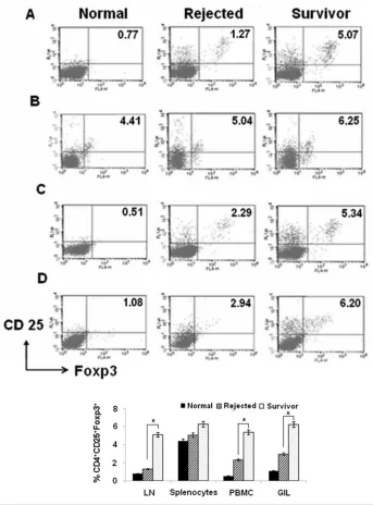
MSC-plus-CsA Administration Increases the Frequency of CD4+CD25+Foxp3+T Cells
As CD4+CD25+Foxp3+proportions had previously been reported to increase on coculturing naive T cells with MSCs in vitro(10), we examined whether
CD4+CD25+Foxp3+T cells contributed to islet allograft acceptance in MSC-plus-CsA–treated allograft recipients. Accord-ingly, we tested for the presence of Tregs in LN, spleen, graft-infiltrating cells and PBMCs in the MSC-plus-CsA group, by flow cytometry. We observed a signifi-cant increase in the proportion of CD4+CD25+Foxp3+T cells within the total CD4+population recovered from LN, graft infiltrating cells and PBMCs, but not from spleens, in the MSC-plus-CsA group compared with untreated controls on day-30 posttransplantation (Figure 6). Whereas in the frequency of CD4+CD25+Foxp3+T cells in lymph node lymphocytes, splenocytes, PBMCs and graft-infiltrating cells, there was no dif-ference between any groups on day-10 posttransplantation (data not shown).
DISCUSSION
Most studies suggest that MSCs inhibit alloreactive/autoreactive T-cell responses and promote therapeutic effects in exper-imental models (5,11–13). Controversy surrounds the optimal source of MSCs for use in immune modulation. Although the immunoregulatory aspects of MSCs
are well established, other studies have indicated that these cells might not be able to completely evade the immune system. This result was initially indicated by Djouad et al. (11), showing that al-though allogeneic murine MSCs could engraft and form bone in immunocom-petent mice, lymphocytic infiltrates were
seen at the periphery of the newly formed tissue. Subsequently, several studies questioned the efficacy of allo-geneic MSCs in vivo(14–20). In particu-lar, Nauta et al.demonstrated that infu-sion of donor MSCs, in conjunction with allogeneic bone marrow transplantation, is associated with enhanced rejection of the latter cells, indicating that allogeneic MSCs might induce a memory T-cell re-sponse (14,17). Indeed, our data show that injection of allogeneic MSCs alone or with CsA showed islet graft survival similar to the control group, although the strong suppressive reactivity was exhib-ited in vitro. Furthermore, allogeneic MSCs combined with low-dose CsA failed to prolong islet allograft survival. Allogeneic MSCs are likely recognized by host T cells and might not be able to evade immune surveillance in allogeneic recipients. In a recent report, Fiorina et al. (21) found that allogeneic MSCs are ca-pable of protecting islet mass and of delaying the onset of disease when in-jected into prediabetic NOD mice, but monotherapy often fails to provide a du-rable favodu-rable outcome. In addition, the Fiorina et al. study was conducted in an NOD setting, where alloimmune and au-toimmune responses are both involved, whereas our study was performed in streptozotocin (STZ)- induced diabetic rats. STZ has been widely used to induce hyperglycemia by specifically destroying the insulin-producing βcells of the islets of Langerhans in experimental models of Type I diabetes. Against this backdrop, we sought to determine whether autolo-gous MSCs downregulate the immune response and prolong graft survival. Im-portantly, autologous MSCs coadminis-tered with a subtherapeutic dose of CsA suppressed the early activation of T cells and synergistically prolonged allograft survival (Figure 3). Induction of trans-plantation tolerance in experimental sys-tems using CsA (22,23) typically requires treatment for at least 7–14 days. Two pre-vious studies reported that autologous MSC-plus-CsA exerted a synergistic ef-fect in vitro, an observation that could have clinical implications in transplant
recipients (24,25). Although a number of studies have documented the immuno-suppressive activities of MSCs, the mech-anisms underlying this effect have not been fully explained. Generally, contact-dependent mechanisms and soluble fac-tors are thought to collaborate to induce MSC-mediated immunosuppression. Re-cently, Kathryne Wood’s group sug-gested that one of the principal explana-tions for the immunosuppressive capacity of MSCs is the cleavage of CD25 from the T-cell surface by MSC- secreted matrix metalloproteinases in vitroand in vivo. Moreover, several soluble immuno-suppressive factors, either produced con-stitutively by MSCs or released after cross-talk with target cells, were reported to be involved in MSC-mediated im-munoregulation (5,10,13, 26–29). Previ-ously, we reported that transfection of the vIL-10gene into islets effectively ele-vated IL-10 levels within the grafts of re-cipients and proved beneficial in rodent allogeneic islet transplantation (30). We therefore tested whether this cytokine plays a role in MSC-mediated inhibition and further assessed the involvement of cell–cell contact in this process. We found that the conditioned medium from MSC–T-cell cocultures modulated T-cell alloreactivity, suggesting a role for solu-ble factors. We further found that addi-tion of MSCs to MLRs inhibited IL-2 and IFN-γproduction while increasing the levels of IL-10, suggesting that IL-10 is important in mediating the suppressive capacity of MSCs (Figure 2C). We found that in grafted livers from the allo–MSC-plus-CsA group, the expression of IL-2, IFN-γ, IL-4 and IL-10 was comparable to other groups at 20 days after transplanta-tion. However, autologous MSC-plus-CsA treatment decreased IL-2 and IFN-γ mRNA levels but increased IL-4 and IL-10 mRNA levels in grafted livers (Fig-ure 4). Interestingly, IL-10 was upregu-lated continuously from the early post-transplantation period, whereas TGF-β and Foxp3 were upregulated later. These changes in the cytokine environment were restricted to the autologous MSC-plus-CsA–treated group and were closely
related to suppression of rejection and long-term allograft survival. The prolifer-ation of splenocytes from rats trans-planted with autologous MSC-plus-CsA was suppressed upon restimulation with allogeneic splenocytes in vitro. These splenocytes also produced a low level of IL-2 and a high level of IL-10 compared with other groups. The functional contri-bution of IL-10 to the immunomodula-tory function of MSCs in autologous MSC-plus-CsA–treated rats was con-firmed by addition of a neutralizing anti–IL-10 antibody in MLR, which re-sulted in recovery of the MLR response in long-term surviving splenocytes (Fig-ure 5A). TGF-βwas readily detected in all groups, but the inhibitory activity of splenocytes from MSC-plus-CsA–treated animals was slightly abolished by addi-tion of a neutralizing anti–TGF-βAb (data not shown). These data support the idea that TGF-βdoes not play a major role in the long-term survival of grafted islets in animals treated with the combi-nation of MSCs and low-dose CsA. Sev-eral reports demonstrated that TGF-β1 can either enhance (31,32) or inhibit (33,34) effector T-cell proliferation. These results suggest that immunoregulatory effects of TGF-β1 depend on strength of stimulation of effector T cells and could change tolerogenic property of TGF-β1 in vivo. In our study, TGF-βand Foxp3 mRNA were upregulated late in the post-transplantation period in grafted liver. TGF-βwas recognized as a critical regu-lator in immune responses and greatly dampens T-cell responses in particular (35). TGF-βwas shown to convert pe-ripheral CD4+
CD25–T cells into Tregs and thus could promote peripheral toler-ance (36). We suggest that TGF-βmay af-fect the generation of Tregs in later stages after transplantation and thus contribute to improved graft survival.
ALS + CsA treatment, but not autologous MSC + CsA treatment, was associated with the generation of IL-10-secreting CD4+T cells. This result suggests that ALS treatment may increase IL-10 ex-pression (38) and contribute to long-term islet allograft survival. In our present study, autologous MSCs plus low-dose CsA prolonged islet allograft survival and induced production of IL-10 from host-derived CD11b+cells after trans-plantation (Figure 5D). In the recent re-ports, IL-10 produced by recipient cells other than T lymphocytes was required for Treg function and maintenance of Foxp3 expression, despite the ability of the donor Tregs to secrete IL-10. Tregs transferred into Il10–/–Rag1–/–recipient mice expanded in number in vivoand homed to various tissues, but these cells failed to maintain Foxp3 expression and suppressive activity in the absence of IL-10 (39). In our study, the production of IL-10 itself by host-derived CD11b+cells is certainly one mechanism by which au-tologous MSCs may exert their suppres-sive activity in the early phase. Then we determined the frequency of Tregs (CD4+CD25+Foxp3+cells) harvested from islet transplanted rats. Interestingly, rela-tive to other T-cell subsets, the number of Tregs in long-term surviving rats was in-creased in LNs, PBMCs and graft at a later stage (around 30 days), but was rel-atively unchanged in splenocytes (see Figure 6). Thus, increased Tregs presum-ably will maintain the suppressive effect by host cell–derived IL-10 and then sup-port long-term graft function.
In conclusion, we have demonstrated that MSCs administered with low-dose CsA prolong graft survival compared with MSCs or CsA alone, and combined treatment with MSCs and CsA prevents rejection of islet allografts by suppression of local proinflammatory cytokine pro-duction. MSCs are known to produce high levels of IL-10. Our study shows that MSCs induce monocytes to produce IL-10 in in vivoas well in vitro experi-ments. Furthermore, MSCs indirectly af-fected the Treg via IL-10 secreted from CD11b+cells and TGF-βat a later phase.
Thus, upregulation of IL-10 synthesis by combined autologous MSC-plus-CsA treatment causes donor antigen-specific T-cell hyporesponsiveness during the early posttransplantation phase and maintains suppressive activity by Tregs later. The immunomodulatory effect of combined autologous MSC-plus-CsA treatment in islet transplantation, re-ported here, suggests a new strategy for preventing and treating rejection after transplantation and provides a prelimi-nary experimental basis for the applica-tion of MSCs in clinical islet transplanta-tion as well as other solid organ
transplantation.
ACKNOWLEDGMENTS
This work was supported by a grant from the Ministry of Education, Science and Technology (2009-0074341) and the Asan Institute for Life Science (2011-050).
DISCLOSURE
The authors declare that they have no competing interests as defined by Molecu-lar Medicine, or other interests that might be perceived to influence the results and discussion reported in this paper.
REFERENCES
1. Wee YM, et al.(2008) Cell surface modification by activated polyethylene glycol prevents allosensi-tization after islet transplantation. Cell Transplant.
17:1257–69.
2. Rother KI, Harlan DM. (2004) Challenges facing islet transplantation for the treatment of type 1 diabetes mellitus. J. Clin. Invest.114:877–83. 3. Lakey JR, Mirbolooki M, Shapiro AM. (2006)
Current status of clinical islet cell transplanta-tion. Methods Mol. Biol.333:47–104.
4. Mabley JG, et al.(2003) Inosine protects against the development of diabetes in multiple-low-dose streptozotocin and nonobese diabetic mouse models of type 1 diabetes. Mol. Med.9:96–104. 5. Di Nicola M, et al.(2002) Human bone marrow
stromal cells suppress T-lymphocyte prolifera-tion induced by cellular or nonspecific mitogenic stimuli. Blood.99:3838–43.
6. Bartholomew A, et al.(2002) Mesenchymal stem cells suppress lymphocyte proliferation in vitro and prolong skin graft survival in vivo. Exp. Hematol.30:42–48.
7. Kim SC, et al.(2005) Comparative study on biologic and immunologic characteristics of the pancreas islet cell between 24 degrees C and 37 degrees C culture in the rat. Transplant. Proc.37:3472–5.
8. Zuk PA, et al.(2001) Multilineage cells from human adipose tissue: implications for cell-based therapies. Tissue Eng.7:211–28.
9. Lee OK, Kuo TK, Chen WM, Lee KD, Hsieh SL, Chen TH. (2004) Isolation of multipotent mes-enchymal stem cells from umbilical cord blood.
Blood.103:1669–75.
10. Aggarwal S, Pittenger MF. (2005) Human mes-enchymal stem cells modulate allogeneic im-mune cell responses. Blood.105:1815–22. 11. Djouad F, et al.(2003) Immunosuppressive effect
of mesenchymal stem cells favors tumor growth in allogeneic animals. Blood.102:3837–44. 12. Tse WT, Pendleton JD, Beyer WM, Egalka MC,
Guinan EC. (2003) Suppression of allogeneic T-cell proliferation by human marrow stromal cells: implications in transplantation. Transplanta-tion.75:389–97.
13. Meisel R, Zibert A, Laryea M, Gobel U, Daubener W, Dilloo D. (2004) Human bone marrow stromal cells inhibit allogeneic T-cell responses by in-doleamine 2,3-dioxygenase-mediated tryptophan degradation. Blood.103:4619–21.
14. Eliopoulos N, Stagg J, Lejeune L, Pommey S, Galipeau J. (2005) Allogeneic marrow stromal cells are immune rejected by MHC class I- and class II-mismatched recipient mice. Blood.
106:4057–65.
15. Sudres M, et al.(2006) Bone marrow mesenchy-mal stem cells suppress lymphocyte proliferation in vitro but fail to prevent graft-versus-host dis-ease in mice. J. Immunol.176:7761–7.
16. Inoue S, et al.(2006) Immunomodulatory effects of mesenchymal stem cells in a rat organ trans-plant model. Transplantation.81:1589–95. 17. Nauta AJ, Westerhuis G, Kruisselbrink AB,
Lurvink EG, Willemze R, Fibbe WE. (2006) Donor-derived mesenchymal stem cells are im-munogenic in an allogeneic host and stimulate donor graft rejection in a nonmyeloablative set-ting. Blood.108:2114–20.
18. Wan CD, Cheng R, Wang HB, Liu T. (2008) Im-munomodulatory effects of mesenchymal stem cells derived from adipose tissues in a rat ortho-topic liver transplantation model. Hepatobiliary Pancreat. Dis. Int.7:29–33.
19. Nauta AJ, Fibbe WE. (2007) Immunomodulatory properties of mesenchymal stromal cells. Blood.
110:3499–506.
20. Kotobuki N, Katsube Y, Katou Y, Tadokoro M, Hirose M, Ohgushi H. (2008) In vivo survival and osteogenic differentiation of allogeneic rat bone marrow mesenchymal stem cells (MSCs).
Cell Transplant.17:705–12.
21. Fiorina P, et al.(2009) Immunomodulatory func-tion of bone marrow-derived mesenchymal stem cells in experimental autoimmune type 1 dia-betes. J. Immunol.183:993–1004.
22. Tufveson G, Gannedahl G, Johnsson C, Olausson M, Wanders A, Ekberg H. (1993) New immuno-suppressants: testing and development in animal models and the clinic: with special reference to DSG. Immunol. Rev.136:99–109.
tautomycetin on cyclosporine A–mediated im-munosuppression in a rodent islet allograft model. Mol. Med.16:298–306.
24. Le Blanc K, et al.(2004) Mesenchymal stem cells in-hibit the expression of CD25 (interleukin-2 recep-tor) and CD38 on phytohaemagglutinin- activated lymphocytes. Scand. J. Immunol.60:307–15. 25. Maccario R, et al.(2005) Human mesenchymal
stem cells and cyclosporin a exert a synergistic suppressive effect on in vitro activation of al-loantigen-specific cytotoxic lymphocytes. Biol. Blood Marrow Transplant.11:1031–2.
26. Augello A, et al.(2005) Bone marrow mesenchy-mal progenitor cells inhibit lymphocyte prolifera-tion by activaprolifera-tion of the programmed death 1 pathway. Eur. J. Immunol.35:1482–90. 27. Krampera M, et al.(2006) Role for
interferon-gamma in the immunomodulatory activity of human bone marrow mesenchymal stem cells.
Stem Cells.24:386–98.
28. Selmani Z, et al.(2008) Human leukocyte antigen-G5 secretion by human mesenchymal stem cells is required to suppress T lymphocyte and natural killer function and to induce CD4+CD25highFOXP3+ regulatory T cells. Stem Cells.26:212–22.
29. Chabannes D, et al.(2007) A role for heme oxyge-nase-1 in the immunosuppressive effect of adult rat and human mesenchymal stem cells. Blood.
110:3691–4.
30. Kim YH, et al.(2008) Viral IL-10 gene transfer prolongs rat islet allograft survival. Cell Trans-plant.17:609–18.
31. Kim HP, Kim BG, Letterio J, Leonard WJ. (2005) Smad-dependent cooperative regulation of inter-leukin 2 receptor alpha chain gene expression by T cell receptor and transforming growth factor-beta. J. Biol. Chem.280:34042–7.
32. Sung JL, Lin JT, Gorham JD. (2003) CD28 stimulation regulates the effect of transforming growth factor-beta1 on the proliferation of naive CD4+ T cells. Int. Immunopharmacol.3:233–45. 33. Gunnlaugsdottir B, Maggadottir SM, Ludviksson
BR. (2005) Anti-CD28-induced co-stimulation and TCR avidity regulates the differential effect of TGF-beta1 on CD4+ and CD8+ naive human T-cells. Int. Immunol.17:35–44.
34. Kehrl JH, et al.(1986) Production of transforming growth factor beta by human T lymphocytes and its potential role in the regulation of T cell growth.
J. Exp. Med.163:1037–50.
35. Li MO, Flavell RA. (2008) TGF-beta: a master of all T cell trades. Cell.134:392–404.
36. Chai JG, et al.(2005) Regulatory T cells, derived from naive CD4+CD25- T cells by in vitro Foxp3 gene transfer, can induce transplantation toler-ance. Transplantation.79:1310–16.
37. Solari MG, et al.(2009) Marginal mass islet trans-plantation with autologous mesenchymal stem cells promotes long-term islet allograft survival and sustained normoglycemia. J. Autoimmun.
32:116–24.
38. Simon T, et al.(2003) The effect of ATG on