www.impactjournals.com/oncotarget/
Oncotarget, Vol. 7, No. 7
Targeting Notch to overcome radiation resistance
Sanaz Yahyanejad
1, Jan Theys
1and Marc Vooijs
11 Department of Radiotherapy (MAASTRO)/GROW, School for Developmental Biology and Oncology, Maastricht University,
Maastricht, The Netherlands
Correspondence to: Marc Vooijs, email: marc.vooijs@maastrichtuniversity.nl
Keywords: Notch, radiotherapy, treatment resistance, Notch inhibitor, personalized treatment
Received: September 13, 2015 Accepted: December 07, 2015 Published: December 21, 2015
ABSTRACT
Radiotherapy represents an important therapeutic strategy in the treatment of cancer cells. However, it often fails to eliminate all tumor cells because of the intrinsic or acquired treatment resistance, which is the most common cause of tumor recurrence. Emerging evidences suggest that the Notch signaling pathway is an important pathway mediating radiation resistance in tumor cells. Successful targeting of Notch signaling requires a thorough understanding of Notch regulation and the context-dependent interactions between Notch and other therapeutically relevant pathways. Understanding these interactions will increase our ability to design rational combination regimens that are more likely to be safe and effective. Here we summarize the role of Notch in mediating resistance to radiotherapy, the different strategies to block Notch in cancer cells and how treatment scheduling can improve tumor response. Finally, we discuss a need for reliable Notch related biomarkers in specific tumors to measure pathway activity and to allow identification of a subset of patients who are likely to benefit from Notch targeted therapies.
INTRODUCTION
Cancer is one of the major causes of mortality
worldwide. More than half of all cancer patients receive
radiation therapy as part of a curative or palliative
treatment often in combination with surgery or
chemotherapy. While most tumors initially respond to
treatment, they often acquire resistance to therapy and
eventually recur. The varied clinical responses observed
between and within patients are in part the result of tumor
heterogeneity and both acquired and intrinsic treatment
resistance often caused by deregulation of signaling
pathways that control normal cell renewal in adult tissues.
The Notch signaling pathway is one of these frequently
altered pathways in many tumors.
The involvement of ionizing radiation in the
majority of cancer treatments, the pivotal role of Notch
signaling in many fundamental processes such as
self-renewal and differentiation, together with the fact that
Notch signaling is often deregulated in cancer, suggest
that targeting the Notch pathway may be beneficial for
many cancer patients. Here, we review the opportunities
and challenges of targeting Notch signaling to improve
treatment response to radiation therapy.
RADIATION RESISTANCE OF CANCER
CELLS
Resistance to radiation is a common phenomenon
and a major obstacle in cancer therapy [1]. Intrinsic
determinants of radiation resistance include pathways
regulating survival and apoptosis, cell cycle status as
well as DNA repair capability. Extrinsic factors including
extracellular matrix molecules, cytokines, hypoxia and
angiogenesis also influence radiosensitivity.
Additionally, biological heterogeneity within the
tumor population leads to differential radiation response.
Pre-clinical models as well as clinical observations have
demonstrated substantial genotypic and phenotypic
heterogeneity between (inter-tumor) and within
(intra-tumor) tumors [2]. This tumor heterogeneity poses a
challenge to both cancer diagnostics and therapy. First
because small tumor biopsies are unlikely to capture
the complete genomic landscape of a patient’s tumor
and thereby fail to identify (all) treatment-resistant
cancer genotypes [3]. Second, while the sensitive tumor
population will shrink, treatment-resistant tumor cells
invariably will take over and tumors recur. Understanding
factors underlying this heterogeneous treatment response
will help to overcome treatment resistance.
Heterogenic treatment response may arise
from influences of the microenvironment and genetic
instability generating (epi)genetic changes. Malignant
cell populations may alternatively (or complementary)
consist of a developmentally defined hierarchy of
heterogeneous phenotypes derived from a small subset
of so-called cancer stem cells (CSC) [4]. Such cells have
been best characterized in hematological [5], breast [6-7]
and CNS malignancies [8]. Mounting evidence indicates
that CSC populations in solid tumors are resistant to
radiation. Mechanisms to explain this intrinsic resistance
include efficient repair of DNA damage, lower numbers
of DNA breaks, redistribution of cells in the cell cycle,
repopulation, and reoxygenation of hypoxic tumor areas.
These studies have been extensively reviewed elsewhere
[9-10]. If these CSC rely on specific pathways for their
survival after treatment, identification of such pathways
would provide opportunities for the targeted and selective
killing of treatment resistant cancer cells [11]. One of the
promising candidates in this context is Notch signaling,
a pathway active in many developmental as well as adult
stem cell pathways and frequently altered in human
cancers [12-13]
Here, we will discuss the role of Notch signaling
in the radiation response of human tumors and highlight
the opportunities to exploit inactivation of Notch using
pharmacological inhibitors in conjunction with radiation
therapy.
NOTCH SIGNALING AND RADIATION
RESISTANCE
Notch proteins are short-range cell-cell signaling
receptors that have key roles during development and in
adult tissue self-renewal, proliferation and differentiation.
In mammals, the Notch pathway consists of 4 Notch
receptors (Notch1-4) that are present in signal receiving
cells and 5 ligands on adjacent signal sending cells.
In the canonical pathway, Notch receptor activation
via ligand interaction leads to a consecutive series of
proteolytic cleavages finally resulting in the release of
the Notch intracellular domain (NICD) that translocates
to the nucleus to act as transcription regulator. The list
of target genes regulated by Notch is cell type dependent
and includes genes involved in cell cycle regulation [14],
cellular differentiation [15] and stem cell maintenance
[16].
Consistent with its fundamental role in many
aspects of vertebrate development, deregulation of the
Notch pathway is implicated in various developmental
syndromes. In adult tissues, deregulation or mutation
of NOTCH proteins is observed in many cancer types
and has been shown to contribute to carcinogenesis and
treatment resistance [13]. Notch inhibitors have been
under pre-clinical investigation for over a decade and
shown strong responses in many cancer models. Several
clinical trials of Notch pathway inhibitors in patients with
leukemia have been reported and several are ongoing in
solid cancers [17]. Here, we focus specifically on the role
of Notch in resistance to radiotherapy and the different
intrinsic and extrinsic mechanisms involved.
Intrinsic resistance
a) Targeting DNA repair
It has recently been demonstrated that Notch has a
direct role in DNA damage response (DDR). The activity
of Notch1 and ataxia-telengiectasia mutated kinase (ATM,
the primary DNA sensor kinase in DDR) were shown to
be inversely correlated in C.elegans and in human cell
lines. ATM is activated specifically upon double strand
(ds) DNA breaks induced by ionizing radiation. Notch1
directly binds to ATM thereby inactivating its kinase
activity. Importantly, inactivation of ATM via Notch
activation contribute to the survival of Notch driven human
leukemia (T-ALL). Blocking Notch using a γ-secretase
inhibitor (GSI) in the presence of DNA damage leads
to increased radiation sensitivity in an ATM-dependent
manner [18]. Activated Notch1 and pATM levels were
also significantly inversely correlated in human primary
breast cancer, validated by immunohistochemistry and in
expression microarray datasets [18]. This result suggests
that cancer cells treated with DNA-damaging agents
such as radiation may undergo more robust cell death if
treated with a Notch inhibitor. Another very recent and
interesting observation came from a study by Deng et al.
[19] in which they show that inactivation of homologous
recombination in human Notch-driven cancer results in
significant radiosensitization. This provides a basis for
Notch-directed cancer therapy via blocking of
homology-directed dsDNA break repair.
b) Targeting cancer stem cells (CSC)
There is increasing evidence supporting the role
of Notch in maintenance and self-renewal of CSC in
T-ALL [20-22], brain [23-24], breast [11, 25], lung [26]
and colon tumors [27]. In glioma, it has been reported
that blocking Notch using GSI depleted CD133+ glioma
CSCs, attenuated neurosphere formation and lowered
tumorigenicity [28]. In line with this, Notch inhibition
selectively impaired clonogenic survival of the glioma
CD133+ CSCs sub-population thereby enhancing its
radiation sensitivity. The intrinsic radioresistance may be
caused by alteration of DNA damage checkpoints [8] or
through up-regulation of the pro-survival factors Akt and
Mcl-1 in CSCs [29]. Hovinga et al. reported that Notch
inhibition enhances the response to radiation by reducing
proliferation and self-renewal of CSCs in tumor explants
only when endothelial cells were present, suggesting a
critical role for Notch not only in tumor cells but within
the entire microenvironment [28]. Consistently, the use of
Notch inhibitors in an orthotopic glioma model slowed
tumor growth and prolonged survival by decreasing the
number of the CD133+ CSCs [30-31].
Also in breast cancer, inhibition of Notch decreased
breast CSCs (ESA+/CD44+/CD24low) activity and
reduced tumor initiation in vivo [32]. Notch inhibition
after radiotherapy prevented up-regulation of
radiation-induced expression of Notch2, Notch3, Dll1, Dll3, Jag1
and was associated with a reduction in breast CSCs
[33]. Radiation resistance in breast CSC has also been
associated with lower levels of DNA damaging reactive
oxygen species (ROS) due to increased production of free
radical scavengers such as of glutathione [34]. Although
a role for Notch signaling in regulating ROS in CSCs has
not yet been reported, Notch inhibition in endothelial cells
has been shown to increase ROS generation, proliferation,
migration and adhesion, suggesting that increased ROS
production upon Notch inhibition after radiotherapy
could also reduce the number of breast CSCs in a non-cell
autonomous manner. Also, in airway basal stem cells ROS
regulates self-renewal in a Notch dependent manner [35],
yet a direct relationship with the response to radiation and
Notch has not been established.
In non-small cell lung cancer (NSCLC), cells
with stem cell properties have also been shown to be
dependent on Notch activity. These cells are more
treatment resistant and tumorigenic in vivo, whereas
GSI-treated xenografts failed to regenerate tumors upon
re-implantation in suitable hosts [26]. A similar role for
Notch in the maintenance and renewal of colon cancer
initiating cells has been described [27]. Although in these
studies, the direct role of Notch in response to radiation
has not been investigated, the data suggest that Notch
inhibition in these malignancies may result in improved
tumor radiation sensitivity by inhibiting the viability of the
cancer initiating cells.
c) Cross-talk with other signaling pathways
Cancers are driven by the interaction of multiple
signaling pathways. Inhibiting an individual pathway
will thus almost never be sufficient to cure cancer. Even
in tumors that are “oncogene addicted” (referring to the
dependency of some cancers on one or few genes for
proliferation and survival) [36], targeting the specific
genes that are critical for maintenance of the malignant
phenotype will eventually result in tumor recurrence due
to emergence of therapy-resistance [37]. The effect of
Notch signaling on radiation response most likely also
occurs through cross-talk with other signaling pathways.
In glioma, Notch activity increased the radiation resistance
of glioma CSCs by activating the Akt pathway. Notch
blockade prior to irradiation was shown to inhibit Akt
activation, an effect that was rescued by ectopic expression
of the active form of Notch1 and Notch2 [29]. Likewise,
in glioma spheres, Notch inhibition significantly decreased
Akt and STAT3 phosphorylation and reduced survival of
glioma CSCs [23]. These studies suggest that Notch can
promote radiation resistance by activating Akt signaling.
In NSCLC, one of the most important overexpressed
cellular targets is epidermal growth factor receptor
(EGFR). Increased EGFR expression has been associated
with radiation resistance [38] and combination of radiation
with EGFR inhibition have yielded in relatively small
but statistically significant radiosensitizing effects [39].
Others have shown that pharmacological inhibition
of EGFR using erlotinib increased the stem like-cells
(ALDH+) in EGFR-mutated NSCLC cell lines and that
Notch transcriptional activity was increased in these
cells. Strikingly, Notch inhibition eliminated the ALDH+
population, an effect attributed specifically to
Notch3-dependent signaling [40]. As stem-like cells have been
shown to contribute to radioresistance [41], combined
EGFR/Notch targeting in lung cancer cells bearing
activating mutations in EGFR could offer a very powerful
approach to reduce the radiation resistant populations.
K-RAS is one of the most commonly mutated
oncogenes in human cancer. Activated RAS oncogene was
shown to increase radiation resistance in human cells [45].
Notch has been demonstrated to cooperate with the RAS
pathway to promote carcinogenesis in various tumor types.
For example, Notch1 activity was shown to be upregulated
in RAS-transformed cells. Genetic or pharmacological
down-regulation of Notch signaling was sufficient to
abolish the RAS-induced neoplastic phenotype including
proliferation and anchorage-independent growth in vitro
and in vivo [46]. Taking into account the role of oncogenic
K-RAS in radiation resistance, these data support a rational
for targeting both pathways. However, caution should be
taken and application of such an approach not generalized.
Indeed, in a K-RAS driven NSCLC mouse model
opposing tumorigenic functions of Notch1 and Notch2
were reported. In these mice, Notch1 ablation resulted in
decreased levels of the Notch target gene expression and
of pERK1/2, resulting in reduced tumor formation, while
Notch2 ablation showed an increase in HES1 expression
and resulted in increased carcinogenesis [47].
Several reports describe a direct effect of Notch
signaling on the cell cycle. This may be exploited
in the context of fractionated radiation, as it is well
established that cells in different phases of the cell cycle
exhibit different radiation sensitivity. Notch can directly
induce cyclin D1 and cyclin-dependent kinase2 activity
[48-49] and in breast epithelial cells Notch promotes
transformation by inducing cyclin D1 [50]. c-Myc, an
oncogene and potent driver of cell cycle entry, is a direct
target of Notch and essential for cell cycle progression in
T-ALL [51-52] and mouse mammary tumors [53].
d) Regulating EMT
There is mounting evidence showing that Notch
signaling contributes to the acquisition of the EMT
phenotype, for instance by up-regulating Snail and Slug,
both transcriptional repressors of E-cadherin [54-55].
During EMT, epithelial cells undergo a morphological
change resulting in increased motility, invasion and
stemness [56] a process associated with chemo- and
radiation therapy resistance [57-60]. For example,
radioresistant NSCLC have been shown to share many
phenotypical properties with cells that have undergone
EMT [61]. Notch1 signaling was shown to enhance the
EMT process in EGFR inhibitor resistant lung cancer cells
[62]. In lung adenocarcinoma, a population of
metastasis-prone cells with significantly enriched expression of Notch
receptors and ligands that drive EMT were identified [63].
While the majority of the metastatic lung cancer cells were
shown to be radioresistant [64], Notch targeting suggests a
role in increasing radiation sensitivity by inhibiting genes
involved in the EMT process.
In NSCLC, c-MET amplification is shown to direct
invasion and metastasis [65]. Others have shown that
co-expression of c-MET and Notch1 induces EMT in
NSCLC patients and promotes invasion [66], likely due
to their interaction by cross-talk. Given the role of Notch
and c-MET expression in poor radiation response [67]
as well as direct interaction between c-MET and Notch
[68], Notch blocking in combination with c-MET targeted
therapy could be critical to inhibit the aggressive behavior
of NSCLCs and increase the radiation sensitivity.
The association between EMT and radioresistance
and the prominent role of Notch signaling as driving force
in the EMT process, suggest that Notch inhibition will
result in radiosensitization of tumors that underwent EMT.
CROSS-TALK WITH MICRORNAS
Increasing evidence implicates microRNAs
(miRNA) in the regulation of drug and radiation
resistance [69-73]. The most extensively studied miRNA
in the context of Notch signaling and cancer is the tumor
suppressive miR-34. In glioma, miR-34 was shown to
inhibit tumor growth in vivo by down-regulating Notch1
and Notch2 expression [73]. Similarly, in colorectal
cancer, high miR-34a levels inhibited colon CSCs
self-renewal in vitro as well as xenograft tumor formation by
suppressing Notch signaling whereas low miR-34a levels
up-regulated Notch and promoted a CSC phenotype [74].
Also in NSCLC the expression of miR-34 was reported
to be low [75] and its ectopic expression enhanced the
radiation sensitivity of lung cancer cells [76]. Kang
et al. showed that this effect is Notch1-dependent and
demonstrated that miR-34 induced Notch1 downregulation
thereby promoting apoptosis resulting in a radiosensitizing
effect [77]. Similarly, in P53-deficient gastric and
pancreatic cancer cells, restoration of miR-34 reduced
the expression of Notch pathway members and was
associated with reduced in vivo tumor formation as well
as increased treatment sensitization [78-79]. Overall, these
studies provide insight on the role of miR-34 in radiation
resistance, partly mediated via regulation of Notch.
Extrinsic resistance
a) Angiogenesis
Notch signaling is important in pro-angiogenic role
in tumor vasculature. Endothelial cells (ECs) express
several Notch receptors (Notch1, 4) and ligands (Delta-like
1, 4 and Jagged1). VEGF acts as a proliferative driver of
angiogenesis, while Dll4/Notch signaling helps to control
vessel sprouting and branching [80-81]. Tumor vasculature
is abnormal, and the endothelial cells of tumor blood
vessels are different from those of normal vasculature [82].
Consequently, increased tumor angiogenesis, as indicated
by increased microvessel density or by increased VEGF
expression, does not necessarily correlate with increased
blood flow and oxygen availability. This situation, together
with the existence of heterogeneous hypoxic regions
within tumors results into reduced response to radiation
therapy. Based on the crucial role of Dll4/Notch signaling
in the vascular sprouting and tumor angiogenesis,
pharmacological targeting of the Dll4/Notch has been
shown to be effective as a novel anti-angiogenic therapy
by blocking non-productive vessel growth and tumor
collapse [83-85]. Adding chemotherapeutic agents to the
Dll4 cocktail inhibited the induction of anti-apoptotic
genes and resulted in further additive anti-tumor activity
by decreasing the CSC population. Also when combined
with ionizing radiation, Dll4 blockade impaired the tumor
growth by promoting non-functional tumor angiogenesis
[86]. As it is the case for all anti-angiogenic treatment,
the temporal relationship with radiation is crucial [87] and
therefore the optimal order and timing for administration
of the radiation/anti-Dll4 combination in the context of
vascular normalization requires careful attention (see
below).
b) Hypoxia
Hypoxia is a common feature of human tumors and
is associated with increased malignancy and resistance
to chemo- and radiotherapy [88]. Hypoxic cells are 2-3
fold less sensitive to the effects of radiation because they
lack the oxygen radicals that contribute to irreversible
DNA damage [89]. Pre-treatment oxygenation of tumors
is prognostic and predictive for radiotherapy response
in head and neck squamous carcinoma and many other
tumors [90-91]. Previously, we have shown that NSCLC
xenografts expressing a constitutively active Notch1 were
more resistant to single dose radiation therapy. Tumors
with high constitutive Notch activity proliferated faster
and consistently had a higher hypoxic fraction [92]. This
was accompanied by increased vessel density while the
total number of perfused vessels remained similar to
control tumor cells pointing towards an increase in
non-functional vasculature (unpublished data). These data
suggest that Notch signaling may increase the survival
of hypoxic cells and thereby influence the response to
radiotherapy.
The link between hypoxia and Notch signaling
has been described in various studies. Notch has been
reported to activate the hypoxia response pathway
through HES1-induced STAT3 phosphorylation resulting
in transcriptional up-regulation of HIF1-α and its target
genes [93]. Interestingly, hypoxia in turn also elevates
Notch activity [94-95] both via HIF1-α dependent [94]
and HIF1-α independent pathways [96]. Especially the
latter is intriguing, as it was shown that hypoxia-induced
Notch signaling contributed to increased proliferation,
maintenance of stem cell properties and suppression
of senescence via a metabolic shift to glycolysis [96].
Glycolysis would not only provide a growth advantage for
the fast proliferating cells but is also involved in cellular
immortalization via reduction of intrinsic ROS [97-99]. As
such, the metabolic effects of Notch on glycolysis may be
indirectly responsible for increasing survival and radiation
resistance by suppressing ROS.
Although many of these oncogenic pathways
are found to cross-talk with Notch at some level, these
interactions are cell type and context dependent. Thus, the
direct effect of Notch blockade on cancer cells may vary.
STRATEGIES TO TARGET NOTCH AND
ITS POTENTIAL RISKS
In most cases, deregulation of Notch has oncogenic
effects. The first evidence for the involvement of Notch
in cancer was the detection of a rearrangement between
the intracellular part of Notch1 (NICD1) and the T-cell
receptor beta (TRB) leading to high-level expression
of truncated and constitutively active Notch1 in T-ALL
[100-101]. Mutations and chromosomal rearrangements
have also been reported in splenic marginal zone B-cell
lymphoma [102-103] and in triple-negative breast cancer
cells [104]. Notch has also been shown to act as oncogene
in lung [26, 92], colon [105], melanoma [106], pancreatic
[107], glioma [31, 108], head and neck [109] and many
other cancer types.
In other cases, Notch functions as a tumor
suppressor as shown in skin tumors [110-111], bladder
cancer [112], squamous cell lung carcinoma [113],
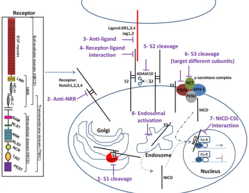
Figure 1: Notch signaling pathway and potential drug intervention sites (see text for details).
1) Furin cleavage at S1 site can be inhibited. 2) Notch antibodies targeting Notch receptors 3) or ligands would target individual receptor pathway 4) Targeting the interaction of Notch receptor with ligand after receptor maturation abrogates the pathway activity 5) Cleavage by a disintegrin and metalloproteinase Adam10 at S2 site and 6) γ-secretase complex at S3 site can be inhibited to limit Notch signaling. 7) Interfering with NICD/CSL interaction using small peptides disrupts the canonical Notch pathway signaling. 8) Inhibition of endosomal Notch trafficking could potentially reduce Notch signaling activity regardless of ligand activity. The Notch receptor is comprised of a Notch extracellular domain (NECD) and Notch intracellular domain (NICD). EGFR: epidermal growth factor repeats; HD: heterodimerization domain; NRR: negative regulatory region; LNR: cysteine-rich LNR repeats; RAM: RAM domain; NLS: nuclear localization signals; ANK: ankyrin repeat domain; NCR: cysteine response region; TAD: transactivation domain; PEST: region rich in proline (P), glutamine (E), serine (S) and threonine (T) residues.squamous cell carcinoma in cutaneous and head-and-neck
tumors [114-117] and potentially also in small cell lung
carcinoma (SCLC), which is a neuroendocrine subtype of
lung cancer [118].
This indicates that the outcome for aberrant Notch
activity is highly context-dependent. Notch inhibitors
are being investigated in clinical studies to treat these
malignancies where Notch acts as an oncogene whereas
methodologies activating the Notch pathway may have
therapeutic potential in those cancers where Notch
suppresses tumor growth, although any experimental
evidence for the latter is still lacking.
We will first provide an overview of the different
possibilities to inhibit the Notch pathway (Figure 1)
and discuss how these may interact with radiation in a
subsequent section. In canonical Notch signaling, Notch
receptors are subjected to a series of sequential proteolytic
cleavages. The site1 (S1) cleavage is controlled by a Furin
convertase and is responsible for the receptor maturation.
Therefore, it is possible to interfere with Notch maturation
in Golgi by using Furin inhibitors [119-120]. After
receptor maturation, the receptor is transported to the
cell’s surface, a process that can be blocked by using an
inhibitor of calcium transporter Atp2a3/SERCA [121].
Following surface expression, receptor activation is
mediated by binding to ligand on adjacent cells.
Notch-ligand interactions can be blocked using soluble versions
of the receptor that function as decoy [122-123] or by
blocking the ligand-induced conformational changes
in the Notch receptor [124-126]. Upon receptor-ligand
interaction, mammalian Notch receptors are cleaved by the
disintegrin metalloprotease ADAM10 at site 2 (S2)
[127-128] leading to shedding of the large Notch ectodomain
(NECD). This cleavage is followed by a S3-cleavage
caused by a γ-secretase complex and results in the release
of the cytoplasmic NICD, which subsequently translocates
to the nucleus where it binds to the DNA binding protein
CSL (CBF1/Suppressor of Hairless/Lag-1; also known
as Rbp-j) and the co-activator Mastermind-like (MAML)
to induce expression of target genes [128-129]. The S2
cleavage can be inhibited by blocking ADAM proteases
[128, 130-132] and S3 cleavage by γ-secretase inhibitors
(GSI).
A detailed overview of ways to intervene with Notch
signaling in various disease has been described elsewhere
[17]. Here, we focus on different strategies to target Notch
specifically in cancer.
1- GSIs: GSIs are pan-Notch inhibitors used in both
pre-clinical and clinical settings. They target γ-secretase
cleavage of NOTCH by presenilin, a rate-limiting step
in the Notch activation cascade. Use of GSI is however
hampered by the dose-limiting toxicity in the gut as
Notch inhibitors promote goblet cell metaplasia leading
to severe diarrhea in animals and humans [133-135].
Intermittent dosing schedules [136-138], glucocorticoid
administration or anti-estrogen therapy [139] have been
shown to mitigate the adverse effects while maintaining
GSI’s anti-tumor efficacy [140-141]. Obviously, most
drugs used in oncology, including targeted agents and
immunotherapeutics have significant acute and chronic
toxicities, and in every situation the ratio between
risks and benefits must be carefully weighted. Cyclical
(intermittent) dosing, dose adjustments and patient
stratification are therefore important to minimize the
toxicities of chemo- and radiation therapy [139, 142-146].
While GSI’s are potent inhibitors of the Notch
signaling pathway, they are not designed to be
receptor-specific and they target all four Notch isoforms that within
the same cell type can have either tumor promoting or
tumor suppressive roles. Thus, there is a need for receptor
specific antagonists.
2- Notch receptor specific targeting: Monoclonal
antibodies have been developed that target the negative
regulatory region (NRR) of Notch1, Notch2 [125] and
Notch2/3 [126, 147] and act by keeping the receptor in
an unresponsive “closed” confirmation or by blocking
receptor-ligand interactions through hindering EGF
repeats required for binding [148]. These antibodies are
able to target cancers by inhibiting simultaneously cancer
cell growth and by disrupting tumor angiogenesis that
depends on DLL4/Notch1 signaling.
3- Notch ligand targeting: To interfere with tumor
angiogenesis, Notch ligands are important targets. Along
this line, Dll4 blocking antibodies have been used to
suppress tumor vascularization and tumor growth [83,
149]. Data from a Phase1 clinical trial showed that
Demsizumab (anti-Dll4) suppressed tumor vascularization,
was well tolerated and resulted in reduced tumor size
[150-151]. In animals, the long-term use of these antibodies,
however caused marked histopathological changes in liver
endothelial cells and induced vascular tumors [152]. While
Dll4 mainly has a function in the vasculature, Jagged1 is
important in immunosuppressive T regulatory cells and
promotes the maintenance or expansion of hematopoietic
precursor cells [153] as well as tumor/stem cells
[154-155]. Targeting Jagged1 in stroma and tumor cells can thus
result in synergistic effects as demonstrated in ovarian
cancer [54, 156].
Alternatively, activating Notch signaling could be a
way to inhibit angiogenesis. While overexpression of Dll4
was shown to promote tumor growth, overexpression of
a soluble DSL domain of Dll1 resulted in reduced tumor
growth by attenuating vascularization [157]. Therefore, it
is important to further explore the differential activities of
the Notch ligands on both stroma and tumor cells.
Recent work has shown that the Notch pathway can
also be used non-canonically [158] and that Notch proteins
can become activated in a DSL-independent manner as
shown in various cancers including melanoma [159] and
T-ALL [160]. Breast cancer stem cell expansion has also
been shown to be dependent on a ligand-independent
Notch activation mechanism [161]. Taking into account
the radioresistant phenotype of breast CSC, targeting
Notch ligands in such case may be suboptimal, and other
strategies to target Notch cleavage or downstream events
may be more effective.
4- Alternatives: Cleavage of Notch proteins by
ADAM metalloproteases is a rate-limiting step preceding
γ-secretase cleavage. Specifically, metalloproteases
ADAM 10 and 17 have been implicated in ligand
dependent and independent signaling, respectively [127,
162-163]. ADAM metalloproteases bind many signaling
molecules and receptors, including TNFα and EGF
receptors and thus unless specifically targeted to tumors
are likely to yield dose-limiting toxicities in normal
tissues. Moreover, we reported Notch cleavage and
transcriptional activity of oncogenic Notch1 signaling in
cells treated with both broad-spectrum metalloprotease
inhibitors as well as specific ADAM17/10 hydroxamate
inhibitors [128], suggesting the involvement of unknown
proteases engaged in the activation of oncogenic Notch1.
While this hypothesis requires further validation, it may
open the possibility of targeting disease-specific Notch
proteases while leaving normal Notch signaling intact.
The main components of the γ-secretase complex
are presenilin, APH-1, Pen-2, and Nicastrin. Currently
Table 1: Possible sites to intervene in Notch pathway
Intervening Notch at
various sites
Mechanism of action
Pre-clinical studies
Clinical studies
S1 cleavage
- Inhibition of Furin and block
receptor maturation
- Not reported
(experimental studies
available)
- Not Reported
Receptor
-Anti Notch1,Notch2,
Notch3
-Anti-Notch4
- Unresponsive receptor to
ligand binding by targeting
NRR in case of Notch1,2 and
3
- Blocking receptor–ligand
interactions by hindering EGF
repeats required for binding
- Anti-Notch1, Notch2
[125, 202]
- Anti-Notch3 [147]
- Anti-Notch4 [203]
- Anti-Notch2, Notch3
[138]
- Anti-Notch1 [204]
Ligand
-Anti-Dll4
-Anti-Jagged1
- Non-functional vasculature
- Abrogation of angiogenesis,
targeting CSCs, targeting
EMT and inhibiting the
immunosuppressive T-
regulatory cells
- [83]
- [156, 205]
- [151]
- Not reported
Receptor-Ligand Interaction
-Notch1 receptor decoy
-Dll1 and Jag1 ligand decoy
- Ligand dependent Notch
antagonist
- [122]
- Not reported
S2 cleavage
(Adam metalloproteases)
- Targeting both Adam
metalloprotease 10/17 and
block ectodomain shedding
- [131]
- [206-207]
S3 cleavage
(γ-secretase complex)
- Inhibition of different
subunits
of
γ-secretase
complex and block NICD
release
- [208]
- [209]
NICD-CSL interaction
- Suppressing transcriptional
activation by preventing
binding of MAML1 to the
ICN–CSL complex
- [174, 210]
- Not reported
Endosomal activation
- Disrupting γ-secretase
cleavage in acidic endosome
- Inhibition of V-ATPase
- [211]
- [173]
used GSIs inhibit the catalytic activity of presenilin and
lead to off-target effects on the wide range of γ-secretase
complex substrates. [164]. Targeting Nicastrin as the key
component of the γ-secretase complex using neutralizing
antibodies reduced proliferation of cancer cells [165]
and resulted in anti-tumor and anti-metastatic effects in a
model of triple negative breast cancer [166].
γ-secretase cleavage and activity not only occurs at
the cell surface but also in the acidic environment of the
endosomes and lysosomes [167-168] where the activity
of vacuolar ATPase (V-ATPase) regulates acidification of
endocytic compartments necessary for Notch signaling
activation [23, 169]. Endocytic trafficking is also
essential for Notch ligand internalization and promoting
Notch activation [170-171]. Pharmacologic inhibition of
V-ATPase decreases Notch signaling activity [172] and
pretreatment with V-ATPase inhibitors can sensitize solid
human tumors to chemotherapy drugs and might also a be
a good strategy for radiosensitization [173].
Finally, hydrocarbon-stapled peptides that mimic a
dominant negative fragment of
Notch-CSL-mastermind-like (dnMAML) and that prevent binding of full-length
MAML to NICD/CSL have been developed. Unlike GSIs,
these peptides have shown a reduced gastrointestinal
toxicity in treated animals [174].
Treatment scheduling and personalized treatment
While numerous clinical trials using various Notch
inhibitors were ongoing, several of these trials have
stopped due to dose-limiting toxicity and lack of efficacy.
Combining Notch inhibition with (chemo) radiation can
only be successful if these hurdles can be overcome.
Appropriate treatment scheduling and patient selection
will be key to achieve this goal.
a) Treatment scheduling
One of the most important aspects that have been
understudied is the scheduling for Notch inhibitors in
conjunction with other treatments. For example, it has
been reported that Notch inhibition caused hypersprouting
of non-functional vasculature resulting in decreased
tumor growth [175-176]. Impaired angiogenesis has also
in other studies been shown to reduce tumor growth, yet
at the same time, these tumors became strongly hypoxic
[177]. Thus, administration of a Notch inhibitor in patients
before radiotherapy may induce hypoxia and contribute to
a more malignant phenotype and radio- and chemotherapy
resistance [88].
It has also been shown that irradiation can induce
Notch expression and activity and promote stem cell
like characteristics [7, 33, 178-180]. Therefore, it may
be critically important to continue Notch inhibition after
radiotherapy. Significantly enhanced radiation-mediated
tumor cytotoxicity has indeed been demonstrated
upon treatment with GSI following irradiation in lung
xenografts [178]. A similar study in glioma determined
that Notch inhibition before temozolomide administration
diminished the efficacy of chemotherapy while Notch
inhibition after chemotherapy strongly inhibited tumor
formation [181]. The reason for this could likely be due to
the induced Notch activity after chemotherapy treatment
[47]. More data are clearly needed to determine the most
appropriate treatment schedule and such results will be
invaluable for translation into the clinic with the aim to
improve outcome. In colorectal cancer patients, sequence
of the drug treatment was shown to be more important
and effective than the drug exposure itself likely due to
enhancing the subsequent treatment [182].
It will also be of utmost importance to determine
the interaction between Notch inhibition, chemotherapy
and fractionated radiation. Recovery from radiation injury
and tumor cell repopulation between fractions reduces
tumor control, while reoxygenation of hypoxic cells and
redistribution of cells into a more radiosensitive phase
increases tumor control [183-184]. In addition, the effect
on the therapeutic ratio when Notch inhibition will be
combined with alternative fractionation schedules such
as accelerated (decreasing the overall treatment time) or
hypofractionated (lower number of fractions with a higher
dose per fraction) treatment needs careful attention as
such alternative fractionation schemes have been shown
to improve tumor control [185-186].
Alternatively, increased radiotherapy effectiveness
can potentially also be achieved by properly scheduling
in combination with angiostatic drugs such as anti-Dll4
antibodies through “vascular normalization”. This concept
proposes that in order to have an effect when using
anti-angiogenesis drugs, an equilibrium between pro- and
anti-angiogenic factors in the tumor microenvironment
rather than complete angiogenesis inhibition is needed.
Dysfunctional vasculatures become then more normal,
hence tumor oxygenation and perfusion will be improved
thereby increasing the efficacy of administered drugs and/
or radiation [187]. Defining the optimal time point during
radiotherapy at which anti-angiogenesis drugs such as
Notch inhibitors should be administered will be crucial
[188].
b) Genetic profile of cancer types and signatures
The different classes of gene expression profiles,
reflecting the consequences of different sets of oncogenic
mutations, correlate with different prognoses and different
responses to therapy. Therefore, cancer cells vary widely
in their response to radiation therapy [189] as well as
Notch targeted therapies reflecting their particular genetic
profile. For example, estrogen receptor negative (ERα
-) breast cancer cells have higher Notch activity and
respond better to Notch inhibition. In ERα
+cells when
estrogen is deprived or upon anti-estrogen-treatment,
breast cancer stem cells are selectively enriched and
Notch-4 activity increased [190-191]. Combination of a
Notch inhibitor with an anti-estrogen could therefore be
a promising therapeutic strategy in ERα
+breast cancer
cells. In both ERα
-and ERα
+tumors radiosensitivity is
expected to increase, as Notch inhibition will specifically
target the more radioresistant stem cell compartment.
Yet, there are currently no valid predictive factors that
reliably identify patients who would greatly benefit
from radiation treatment. In triple-negative breast cancer
(TNBC) patients, approximately 19.5% carry BRCA
mutations [192]. These mutation carriers are defective
in DNA repair; therefore, it would be expected that these
tumors might exhibit sensitivity rather than insensitivity
to radiation therapy. One possible explanation for this
response is that these tumors might possess compensatory
DNA repair mechanisms that are more effective at
dealing with radiation-induced DNA damage. In this
regard, identification of a marker in TNBC cells would
be invaluable in identifying potential radiosensitizing
agents. Gene expression profiling analysis performed
on these tumors revealed that oncogenic PEST domain
mutations in Notch1, 2 and 3 receptors occur in ~13% of
TNBC cells conferring GSI sensitivity [104] and provides
a strong rationale for a Notch-driven personalized
medicine strategy. Notch4, but not Notch1-3 was shown to
contribute to the induction of proliferation, tumorigenesis
and invasiveness in TNBC cells and its inhibition was
shown to suppress tumorigenicity and tumor volume
[193-194]. Furthermore, mutations of Notch receptors resulting
in an active Notch pathway are frequent in TNBC
conferring GSI sensitivity [104]. Notch targeting can thus
be a potential therapeutic target for the radiosensitization
of TNBC cells.
In skin squamous cell carcinomas (SCCs),
EGFR signaling plays a significant role in suppressing
differentiation through negative regulation of Notch1
gene expression and activity [195]. Especially for large
skin SCC and at sites where surgery is not an option,
radiation is often used as first-line treatment. Notch
blockade counteracts the differentiation-inducing effects
of EGFR inhibitors, while at the same time, synergizes
with these compounds in induction of apoptosis. This
indicates an attractive combination therapy that may
enhance the potency of EGFR inhibitory agent. This study
provides a mechanistic explanation for the Notch
loss-of-function mutations found in squamous skin carcinomas
[113]. Squamous tumors without such mutations may
thus be sensitive to Notch inhibitors, and treatment
efficacy enhanced especially for indications involving
radiotherapy.
In NSCLC patients, Dll4/Notch1 signaling was
reported to negatively influence NSCLC growth via PTEN
up-regulation [97]. This indicates that the therapeutic
application of a Notch inhibitor could be adversely
affected in different categories of lung cancer [196]. Notch
inhibition could be specifically beneficial in lung cancers
with inactive PTEN [197]. In contrast, in glioma, loss of
PTEN has been reported as a critical event that leads to
Notch inhibitor resistance by transferring the “oncogene
addiction” from the Notch to the PI3K/AKT pathway
[198], supporting the regulatory link between Notch and
the PTEN/PI3K/AKT pathway. Therefore, attenuation of
cell growth using a combination of Notch inhibition and
PI3K inhibitors in PTEN mutant glioma CSCs may lead
to increased treatment efficacy [199]. As glioma stem
cells promote radioresistance by preferential activation
of the DNA damage response [8] and Notch has been
shown to enhance radiation resitance in glioma [29], the
combination of Notch inhibitors with radiation can be
expected to yield beneficial outcomes in these patients.
These data and similar other data arising from
genomic, transcriptional and proteomic analysis in glioma
[31] or breast cancer [200] exemplify how understanding
the molecular signatures that could predict the therapeutic
response allow identification of a subset of patients who
are likely to benefit from the Notch inhibition/radiotherapy
combination therapies.
Patient selection could also be performed based on
determination of activated (i.e. cleaved) Notch proteins
levels or target genes as shown in TNBC [201], indicating
their potential as prognostic biomarker to identify TNBC
patients who are most likely to respond to anti-Notch
based therapeutics. Likewise, adenoid cystic carcinoma
(ACC) tumor xenografts with activating Notch1 mutations
responded to Notch inhibition, whereas the tumors
without Notch1 mutation and low levels of NICD1 were
resistant [201]. Therefore, establishing an association
between the drug responses and molecular subclasses of
the specific cancer type may help to identify potential
cohorts of patients for targeted therapy and to be treated in
combination with radiotherapy.
Taken together, while Notch deregulation is
frequent in cancers, the failure of clinical trials using
Notch inhibitors may be explained by our incomplete
understanding of the unique and redundant functions of
the Notch receptors and our inability to select the correct
patients and lack of knowledge on the correct timing of
intervention. However, it appears that Notch signaling
plays a key role in tumor initiation, progression and
treatment response and that combining Notch therapeutics
with radiotherapy may lead to synergistic improvements.
More basic and translational research is needed to address
these issues prior to conducting clinical trials. Only
then can we expect to see therapeutic profit from Notch
inhibitors on cancer response.
ACKNOWLEDGMENTS
Supported by the EU, ERC-Consolidator Grant
(617060).
CONFLICTS OF INTEREST
None.
REFERENCES
1. Milas L and Hittelman WN. Cancer stem cells and tumor response to therapy: current problems and future prospects. Semin Radiat Oncol. 2009; 19(2):96-105.
2. Marusyk A and Polyak K. Tumor heterogeneity: causes and consequences. Biochim Biophys Acta. 2010; 1805(1):105-117.
3. Gerlinger M, Rowan AJ, Horswell S, Larkin J, Endesfelder D, Gronroos E, Martinez P, Matthews N, Stewart A, Tarpey P, Varela I, Phillimore B, Begum S, McDonald NQ, Butler A, Jones D, et al. Intratumor heterogeneity and branched evolution revealed by multiregion sequencing. N Engl J Med. 2012; 366(10):883-892.
4. Clarke MF, Dick JE, Dirks PB, Eaves CJ, Jamieson CH, Jones DL, Visvader J, Weissman IL and Wahl GM. Cancer stem cells—perspectives on current status and future directions: AACR Workshop on cancer stem cells. Cancer Res. 2006; 66(19):9339-9344.
5. Lapidot T, Sirard C, Vormoor J, Murdoch B, Hoang T, Caceres-Cortes J, Minden M, Paterson B, Caligiuri MA and Dick JE. A cell initiating human acute myeloid leukaemia after transplantation into SCID mice. Nature. 1994; 367(6464):645-648.
6. Woodward WA, Chen MS, Behbod F, Alfaro MP, Buchholz TA and Rosen JM. WNT/beta-catenin mediates radiation resistance of mouse mammary progenitor cells. Proc Natl Acad Sci U S A. 2007; 104(2):618-623.
7. Phillips TM, McBride WH and Pajonk F. The response of CD24(-/low)/CD44+ breast cancer-initiating cells to radiation. J Natl Cancer Inst. 2006; 98(24):1777-1785. 8. Bao S, Wu Q, McLendon RE, Hao Y, Shi Q, Hjelmeland
AB, Dewhirst MW, Bigner DD and Rich JN. Glioma stem cells promote radioresistance by preferential activation of the DNA damage response. Nature. 2006; 444(7120):756-760.
9. Baumann M, Krause M and Hill R. Exploring the role of cancer stem cells in radioresistance. Nat Rev Cancer. 2008; 8(7):545-554.
10. Pajonk F, Vlashi E and McBride WH. Radiation resistance of cancer stem cells: the 4 R’s of radiobiology revisited. Stem Cells. 2010; 28(4):639-648.
11. Korkaya H and Wicha MS. Selective targeting of cancer stem cells: a new concept in cancer therapeutics. BioDrugs. 2007; 21(5):299-310.
12. Koch U, Lehal R and Radtke F. Stem cells living with a Notch. Development. 2013; 140(4):689-704.
13. Ranganathan P, Weaver KL and Capobianco AJ. Notch signalling in solid tumours: a little bit of everything but not all the time. Nat Rev Cancer. 2011; 11(5):338-351.
14. Joshi I, Minter LM, Telfer J, Demarest RM, Capobianco AJ, Aster JC, Sicinski P, Fauq A, Golde TE and Osborne BA. Notch signaling mediates G1/S cell-cycle progression in T cells
via
cyclin D3 and its dependent kinases. Blood. 2009; 113(8):1689-1698.15. Ross DA, Rao PK and Kadesch T. Dual roles for the Notch target gene Hes-1 in the differentiation of 3T3-L1 preadipocytes. Mol Cell Biol. 2004; 24(8):3505-3513. 16. VanDussen KL, Carulli AJ, Keeley TM, Patel SR, Puthoff
BJ, Magness ST, Tran IT, Maillard I, Siebel C, Kolterud A, Grosse AS, Gumucio DL, Ernst SA, Tsai YH, Dempsey PJ and Samuelson LC. Notch signaling modulates proliferation and differentiation of intestinal crypt base columnar stem cells. Development. 2012; 139(3):488-497.
17. Andersson ER and Lendahl U. Therapeutic modulation of Notch signalling—are we there yet? Nat Rev Drug Discov. 2014; 13(5):357-378.
18. Vermezovic J, Adamowicz M, Santarpia L, Rustighi A, Forcato M, Lucano C, Massimiliano L, Costanzo V, Bicciato S, Del Sal G and d’Adda di Fagagna F. Notch is a direct negative regulator of the DNA-damage response. Nat Struct Mol Biol. 2015; 22(5):417-424.
19. Deng X, Michaelson D, Tchieu J, Cheng J, Rothenstein D, Feldman R, Lee SG, Fuller J, Haimovitz-Friedman A, Studer L, Powell S, Fuks Z, Hubbard EJ and Kolesnick R. Targeting Homologous Recombination in Notch-Driven C. elegans Stem Cell and Human Tumors. PLoS One. 2015; 10(6):e0127862.
20. Giambra V, Jenkins CR, Wang H, Lam SH, Shevchuk OO, Nemirovsky O, Wai C, Gusscott S, Chiang MY, Aster JC, Humphries RK, Eaves C and Weng AP. NOTCH1 promotes T cell leukemia-initiating activity by RUNX-mediated regulation of PKC-theta and reactive oxygen species. Nat Med. 2012; 18(11):1693-1698.
21. Tatarek J, Cullion K, Ashworth T, Gerstein R, Aster JC and Kelliher MA. Notch1 inhibition targets the leukemia-initiating cells in a Tal1/Lmo2 mouse model of T-ALL. Blood. 2011; 118(6):1579-1590.
22. Armstrong F, Brunet de la Grange P, Gerby B, Rouyez MC, Calvo J, Fontenay M, Boissel N, Dombret H, Baruchel A, Landman-Parker J, Romeo PH, Ballerini P and Pflumio F. NOTCH is a key regulator of human T-cell acute leukemia initiating cell activity. Blood. 2009; 113(8):1730-1740. 23. Fan X, Khaki L, Zhu TS, Soules ME, Talsma CE, Gul N,
Koh C, Zhang J, Li YM, Maciaczyk J, Nikkhah G, Dimeco F, Piccirillo S, Vescovi AL and Eberhart CG. NOTCH pathway blockade depletes CD133-positive glioblastoma cells and inhibits growth of tumor neurospheres and xenografts. Stem Cells. 2010; 28(1):5-16.
24. Fan X, Matsui W, Khaki L, Stearns D, Chun J, Li YM and Eberhart CG. Notch pathway inhibition depletes stem-like cells and blocks engraftment in embryonal brain tumors. Cancer Res. 2006; 66(15):7445-7452.
development and breast cancer: the role of stem cells. Curr Mol Med. 2011; 11(4):270-285.
26. Hassan KA, Wang L, Korkaya H, Chen G, Maillard I, Beer DG, Kalemkerian GP and Wicha MS. Notch pathway activity identifies cells with cancer stem cell-like properties and correlates with worse survival in lung adenocarcinoma. Clin Cancer Res. 2013; 19(8):1972-1980.
27. Sikandar SS, Pate KT, Anderson S, Dizon D, Edwards RA, Waterman ML and Lipkin SM. NOTCH signaling is required for formation and self-renewal of tumor-initiating cells and for repression of secretory cell differentiation in colon cancer. Cancer Res. 2010; 70(4):1469-1478. 28. Hovinga KE, Shimizu F, Wang R, Panagiotakos G, Van Der
Heijden M, Moayedpardazi H, Correia AS, Soulet D, Major T, Menon J and Tabar V. Inhibition of notch signaling in glioblastoma targets cancer stem cells
via
an endothelial cell intermediate. Stem Cells. 2010; 28(6):1019-1029. 29. Wang J, Wakeman TP, Lathia JD, Hjelmeland AB, WangXF, White RR, Rich JN and Sullenger BA. Notch promotes radioresistance of glioma stem cells. Stem Cells. 2010; 28(1):17-28.
30. Chu Q, Orr BA, Semenkow S, Bar EE and Eberhart CG. Prolonged inhibition of glioblastoma xenograft initiation and clonogenic growth following
in vivo
Notch blockade. Clin Cancer Res. 2013; 19(12):3224-3233.31. Saito N, Fu J, Zheng S, Yao J, Wang S, Liu DD, Yuan Y, Sulman EP, Lang FF, Colman H, Verhaak RG, Yung WK and Koul D. A high Notch pathway activation predicts response to gamma secretase inhibitors in proneural subtype of glioma tumor-initiating cells. Stem Cells. 2014; 32(1):301-312.
32. Harrison H, Farnie G, Howell SJ, Rock RE, Stylianou S, Brennan KR, Bundred NJ and Clarke RB. Regulation of breast cancer stem cell activity by signaling through the Notch4 receptor. Cancer research. 2010; 70(2):709-718. 33. Lagadec C, Vlashi E, Alhiyari Y, Phillips TM, Bochkur
Dratver M and Pajonk F. Radiation-induced Notch signaling in breast cancer stem cells. Int J Radiat Oncol Biol Phys. 2013; 87(3):609-618.
34. Diehn M, Cho RW, Lobo NA, Kalisky T, Dorie MJ, Kulp AN, Qian D, Lam JS, Ailles LE, Wong M, Joshua B, Kaplan MJ, Wapnir I, Dirbas FM, Somlo G, Garberoglio C, et al. Association of reactive oxygen species levels and radioresistance in cancer stem cells. Nature. 2009; 458(7239):780-783.
35. Paul MK, Bisht B, Darmawan DO, Chiou R, Ha VL, Wallace WD, Chon AT, Hegab AE, Grogan T, Elashoff DA, Alva-Ornelas JA and Gomperts BN. Dynamic changes in intracellular ROS levels regulate airway basal stem cell homeostasis through Nrf2-dependent Notch signaling. Cell Stem Cell. 2014; 15(2):199-214.
36. Weinstein IB and Joe AK. Mechanisms of disease: Oncogene addiction—a rationale for molecular targeting in cancer therapy. Nat Clin Pract Oncol. 2006; 3(8):448-457.
37. Kobayashi S, Boggon TJ, Dayaram T, Janne PA, Kocher O, Meyerson M, Johnson BE, Eck MJ, Tenen DG and Halmos B. EGFR mutation and resistance of non-small-cell lung cancer to gefitinib. N Engl J Med. 2005; 352(8):786-792. 38. Akimoto T, Hunter NR, Buchmiller L, Mason K, Ang KK
and Milas L. Inverse relationship between epidermal growth factor receptor expression and radiocurability of murine carcinomas. Clin Cancer Res. 1999; 5(10):2884-2890. 39. Schutze C, Dorfler A, Eicheler W, Zips D, Hering S, Solca
F, Baumann M and Krause M. Combination of EGFR/ HER2 tyrosine kinase inhibition by BIBW 2992 and BIBW 2669 with irradiation in FaDu human squamous cell carcinoma. Strahlenther Onkol. 2007; 183(5):256-264. 40. Arasada RR, Amann JM, Rahman MA, Huppert SS and
Carbone DP. EGFR Blockade Enriches for Lung Cancer Stem-like Cells through Notch3-Dependent Signaling. Cancer Res. 2014; 74(19):5572-5584.
41. Lundholm L, Haag P, Zong D, Juntti T, Mork B, Lewensohn R and Viktorsson K. Resistance to DNA-damaging treatment in non-small cell lung cancer tumor-initiating cells involves reduced DNA-PK/ATM activation and diminished cell cycle arrest. Cell Death Dis. 2013; 4:e478.
42. Chiang MY, Xu L, Shestova O, Histen G, L’Heureux S, Romany C, Childs ME, Gimotty PA, Aster JC and Pear WS. Leukemia-associated NOTCH1 alleles are weak tumor initiators but accelerate K-ras-initiated leukemia. J Clin Invest. 2008; 118(9):3181-3194.
43. Izrailit J, Berman HK, Datti A, Wrana JL and Reedijk M. High throughput kinase inhibitor screens reveal TRB3 and MAPK-ERK/TGFbeta pathways as fundamental Notch regulators in breast cancer. Proc Natl Acad Sci U S A. 2013; 110(5):1714-1719.
44. Liu ZJ, Xiao M, Balint K, Smalley KS, Brafford P, Qiu R, Pinnix CC, Li X and Herlyn M. Notch1 signaling promotes primary melanoma progression by activating mitogen-activated protein kinase/phosphatidylinositol 3-kinase-Akt pathways and up-regulating N-cadherin expression. Cancer Res. 2006; 66(8):4182-4190.
45. Bernhard EJ, Stanbridge EJ, Gupta S, Gupta AK, Soto D, Bakanauskas VJ, Cerniglia GJ, Muschel RJ and McKenna WG. Direct evidence for the contribution of activated N-ras and K-ras oncogenes to increased intrinsic radiation resistance in human tumor cell lines. Cancer Res. 2000; 60(23):6597-6600.
46. Weijzen S, Rizzo P, Braid M, Vaishnav R, Jonkheer SM, Zlobin A, Osborne BA, Gottipati S, Aster JC, Hahn WC, Rudolf M, Siziopikou K, Kast WM and Miele L. Activation of Notch-1 signaling maintains the neoplastic phenotype in human Ras-transformed cells. Nat Med. 2002; 8(9):979-986.
47. Baumgart A, Mazur PK, Anton M, Rudelius M, Schwamborn K, Feuchtinger A, Behnke K, Walch A, Braren R, Peschel C, Duyster J, Siveke JT and Dechow T. Opposing role of Notch1 and Notch2 in a Kras-driven
murine non-small cell lung cancer model. Oncogene. 2014. 48. Ronchini C and Capobianco AJ. Induction of cyclin D1
transcription and CDK2 activity by Notch(ic): implication for cell cycle disruption in transformation by Notch(ic). Mol Cell Biol. 2001; 21(17):5925-5934.
49. Capobianco AJ, Zagouras P, Blaumueller CM, Artavanis-Tsakonas S and Bishop JM. Neoplastic transformation by truncated alleles of human NOTCH1/TAN1 and NOTCH2. Mol Cell Biol. 1997; 17(11):6265-6273.
50. Ling H, Sylvestre JR and Jolicoeur P. Notch1-induced mammary tumor development is cyclin D1-dependent and correlates with expansion of pre-malignant multipotent duct-limited progenitors. Oncogene. 2010; 29(32):4543-4554.
51. Weng AP, Millholland JM, Yashiro-Ohtani Y, Arcangeli ML, Lau A, Wai C, Del Bianco C, Rodriguez CG, Sai H, Tobias J, Li Y, Wolfe MS, Shachaf C, Felsher D, Blacklow SC, Pear WS, et al. c-Myc is an important direct target of Notch1 in T-cell acute lymphoblastic leukemia/lymphoma. Genes Dev. 2006; 20(15):2096-2109.
52. Loosveld M, Castellano R, Gon S, Goubard A, Crouzet T, Pouyet L, Prebet T, Vey N, Nadel B, Collette Y and Payet-Bornet D. Therapeutic Targeting of c-Myc in T-Cell Acute Lymphoblastic Leukemia, T-ALL. Oncotarget. 2014; 5(10):3168-3172. doi: 10.18632/oncotarget.1873.
53. Sharma VM, Calvo JA, Draheim KM, Cunningham LA, Hermance N, Beverly L, Krishnamoorthy V, Bhasin M, Capobianco AJ and Kelliher MA. Notch1 contributes to mouse T-cell leukemia by directly inducing the expression of c-myc. Mol Cell Biol. 2006; 26(21):8022-8031.
54. Wang Z, Li Y, Kong D, Banerjee S, Ahmad A, Azmi AS, Ali S, Abbruzzese JL, Gallick GE and Sarkar FH. Acquisition of epithelial-mesenchymal transition phenotype of gemcitabine-resistant pancreatic cancer cells is linked with activation of the notch signaling pathway. Cancer Res. 2009; 69(6):2400-2407.
55. Leong KG, Niessen K, Kulic I, Raouf A, Eaves C, Pollet I and Karsan A. Jagged1-mediated Notch activation induces epithelial-to-mesenchymal transition through Slug-induced repression of E-cadherin. J Exp Med. 2007; 204(12):2935-2948.
56. Mani SA, Guo W, Liao MJ, Eaton EN, Ayyanan A, Zhou AY, Brooks M, Reinhard F, Zhang CC, Shipitsin M, Campbell LL, Polyak K, Brisken C, Yang J and Weinberg RA. The epithelial-mesenchymal transition generates cells with properties of stem cells. Cell. 2008; 133(4):704-715. 57. Singh A and Settleman J. EMT, cancer stem cells and drug
resistance: an emerging axis of evil in the war on cancer. Oncogene. 2010; 29(34):4741-4751.
58. Bhangu A, Wood G, Brown G, Darzi A, Tekkis P and Goldin R. The role of epithelial mesenchymal transition and resistance to neoadjuvant therapy in locally advanced rectal cancer. Colorectal Dis. 2014; 16(4):O133-143.
59. Yauch RL, Januario T, Eberhard DA, Cavet G, Zhu W, Fu
L, Pham TQ, Soriano R, Stinson J, Seshagiri S, Modrusan Z, Lin CY, O’Neill V and Amler LC. Epithelial versus mesenchymal phenotype determines
in vitro
sensitivity and predicts clinical activity of erlotinib in lung cancer patients. Clin Cancer Res. 2005; 11(24 Pt 1):8686-8698.60. Gort EH, Groot AJ, van der Wall E, van Diest PJ and Vooijs MA. Hypoxic regulation of metastasis
via
hypoxia-inducible factors. Curr Mol Med. 2008; 8(1):60-67. 61. Gomez-Casal R, Bhattacharya C, Ganesh N, Bailey L,Basse P, Gibson M, Epperly M and Levina V. Non-small cell lung cancer cells survived ionizing radiation treatment display cancer stem cell and epithelial-mesenchymal transition phenotypes. Mol Cancer. 2013; 12(1):94.
62. Xie M, Zhang L, He CS, Xu F, Liu JL, Hu ZH, Zhao LP and Tian Y. Activation of Notch-1 enhances epithelial-mesenchymal transition in gefitinib-acquired resistant lung cancer cells. J Cell Biochem. 2012; 113(5):1501-1513. 63. Yang Y, Ahn YH, Gibbons DL, Zang Y, Lin W,
Thilaganathan N, Alvarez CA, Moreira DC, Creighton CJ, Gregory PA, Goodall GJ and Kurie JM. The Notch ligand Jagged2 promotes lung adenocarcinoma metastasis through a miR-200-dependent pathway in mice. J Clin Invest. 2011; 121(4):1373-1385.
64. Choi SH, Yang H, Lee SH, Ki JH, Nam DH and Yoo HY. TopBP1 and Claspin contribute to the radioresistance of lung cancer brain metastases. Mol Cancer. 2014; 13:211. 65. Breindel JL, Haskins JW, Cowell EP, Zhao M, Nguyen DX
and Stern DF. EGF receptor activates MET through MAPK to enhance non-small cell lung carcinoma invasion and brain metastasis. Cancer Res. 2013; 73(16):5053-5065. 66. Wang X, Song N, Zhang Y, Cai Y, Liu Y, Qu X, Li Z, Li
D, Hou K, Kang J and Hu X. Coexpression of c-Met and Notch-1 correlates with poor prognosis in resected non-small-cell lung cancer. Tumour Biol. 2015; 36(9):7053-7059.
67. Akervall J, Nandalur S, Zhang J, Qian CN, Goldstein N, Gyllerup P, Gardinger Y, Alm J, Lorenc K, Nilsson K, Resau J, Wilson G and Teh B. A novel panel of biomarkers predicts radioresistance in patients with squamous cell carcinoma of the head and neck. Eur J Cancer. 2014; 50(3):570-581.
68. Apostolou P, Toloudi M, Ioannou E, Kourtidou E, Chatziioannou M, Kopic A, Komiotis D, Kiritsis C, Manta S and Papasotiriou I. Study of the interaction among Notch pathway receptors, correlation with stemness, as well as their interaction with CD44, dipeptidyl peptidase-IV, hepatocyte growth factor receptor and the SETMAR transferase, in colon cancer stem cells. J Recept Sig Transd. 2013; 33(6):353-358.
69. Sarkar FH, Li Y, Wang Z, Kong D and Ali S. Implication of microRNAs in drug resistance for designing novel cancer therapy. Drug Resist Updat. 2010; 13(3):57-66.
70. Shiiba M, Shinozuka K, Saito K, Fushimi K, Kasamatsu A, Ogawara K, Uzawa K, Ito H, Takiguchi Y and Tanzawa H.
MicroRNA-125b regulates proliferation and radioresistance of oral squamous cell carcinoma. Br J Cancer. 2013; 108(9):1817-1821.
71. Zhang B, Chen J, Ren Z, Chen Y, Li J, Miao X, Song Y, Zhao T, Li Y, Shi Y, Ren D and Liu J. A specific miRNA signature promotes radioresistance of human cervical cancer cells. Cancer Cell Int. 2013; 13(1):118.
72. Chun-Zhi Z, Lei H, An-Ling Z, Yan-Chao F, Xiao Y, Guang-Xiu W, Zhi-Fan J, Pei-Yu P, Qing-Yu Z and Chun-Sheng K. MicroRNA-221 and microRNA-222 regulate gastric carcinoma cell proliferation and radioresistance by targeting PTEN. BMC Cancer. 2010; 10:367.
73. Li Y, Guessous F, Zhang Y, Dipierro C, Kefas B, Johnson E, Marcinkiewicz L, Jiang J, Yang Y, Schmittgen TD, Lopes B, Schiff D, Purow B and Abounader R. MicroRNA-34a inhibits glioblastoma growth by targeting multiple oncogenes. Cancer Res. 2009; 69(19):7569-7576.
74. Bu P, Chen KY, Chen JH, Wang L, Walters J, Shin YJ, Goerger JP, Sun J, Witherspoon M, Rakhilin N, Li J, Yang H, Milsom J, Lee S, Zipfel W, Jin MM, et al. A microRNA miR-34a-regulated bimodal switch targets Notch in colon cancer stem cells. Cell Stem Cell. 2013; 12(5):602-615. 75. Bommer GT, Gerin I, Feng Y, Kaczorowski AJ, Kuick
R, Love RE, Zhai Y, Giordano TJ, Qin ZS, Moore BB, MacDougald OA, Cho KR and Fearon ER. p53-mediated activation of miRNA34 candidate tumor-suppressor genes. Curr Biol. 2007; 17(15):1298-1307.
76. Duan W, Xu Y, Dong Y, Cao L, Tong J and Zhou X. Ectopic expression of miR-34a enhances radiosensitivity of non-small cell lung cancer cells, partly by suppressing the LyGDI signaling pathway. J Radiat Res. 2013; 54(4):611-619.
77. Kang J, Kim E, Kim W, Seong KM, Youn H, Kim JW, Kim J and Youn B. Rhamnetin and cirsiliol induce radiosensitization and inhibition of epithelial-mesenchymal transition (EMT) by miR-34a-mediated suppression of Notch-1 expression in non-small cell lung cancer cell lines. J Biol Chem. 2013; 288(38):27343-27357.
78. Ji Q, Hao X, Meng Y, Zhang M, Desano J, Fan D and Xu L. Restoration of tumor suppressor miR-34 inhibits human p53-mutant gastric cancer tumorspheres. BMC Cancer. 2008; 8:266.
79. Ji Q, Hao X, Zhang M, Tang W, Yang M, Li L, Xiang D, Desano JT, Bommer GT, Fan D, Fearon ER, Lawrence TS and Xu L. MicroRNA miR-34 inhibits human pancreatic cancer tumor-initiating cells. PLoS One. 2009; 4(8):e6816. 80. Hellstrom M, Phng LK, Hofmann JJ, Wallgard E, Coultas
L, Lindblom P, Alva J, Nilsson AK, Karlsson L, Gaiano N, Yoon K, Rossant J, Iruela-Arispe ML, Kalen M, Gerhardt H and Betsholtz C. Dll4 signalling through Notch1 regulates formation of tip cells during angiogenesis. Nature. 2007; 445(7129):776-780.
81. Diez H, Fischer A, Winkler A, Hu CJ, Hatzopoulos AK, Breier G and Gessler M. Hypoxia-mediated activation of
Dll4-Notch-Hey2 signaling in endothelial progenitor cells and adoption of arterial cell fate. Exp Cell Res. 2007; 313(1):1-9.
82. Eberhard A, Kahlert S, Goede V, Hemmerlein B, Plate KH and Augustin HG. Heterogeneity of angiogenesis and blood vessel maturation in human tumors: implications for antiangiogenic tumor therapies. Cancer Res. 2000; 60(5):1388-1393.
83. Noguera-Troise I, Daly C, Papadopoulos NJ, Coetzee S, Boland P, Gale NW, Lin HC, Yancopoulos GD and Thurston G. Blockade of Dll4 inhibits tumour growth by promoting non-productive angiogenesis. Nature. 2006; 444(7122):1032-1037.
84. Ridgway J, Zhang G, Wu Y, Stawicki S, Liang WC, Chanthery Y, Kowalski J, Watts RJ, Callahan C, Kasman I, Singh M, Chien M, Tan C, Hongo JA, de Sauvage F, Plowman G, et al. Inhibition of Dll4 signalling inhibits tumour growth by deregulating angiogenesis. Nature. 2006; 444(7122):1083-1087.
85. Hoey T, Yen WC, Axelrod F, Basi J, Donigian L, Dylla S, Fitch-Bruhns M, Lazetic S, Park IK, Sato A, Satyal S, Wang X, Clarke MF, Lewicki J and Gurney A. DLL4 blockade inhibits tumor growth and reduces tumor-initiating cell frequency. Cell Stem Cell. 2009; 5(2):168-177. 86. Liu SK, Bham SA, Fokas E, Beech J, Im J, Cho S, Harris
AL and Muschel RJ. Delta-like ligand 4-notch blockade and tumor radiation response. J Natl Cancer Inst. 2011; 103(23):1778-1798.
87. Dings RP, Loren M, Heun H, McNiel E, Griffioen AW, Mayo KH and Griffin RJ. Scheduling of radiation with angiogenesis inhibitors anginex and Avastin improves therapeutic outcome
via
vessel normalization. Clin Cancer Res. 2007; 13(11):3395-3402.88. Wilson WR and Hay MP. Targeting hypoxia in cancer therapy. Nat Rev Cancer. 2011; 11(6):393-410.
89. Edward C. Halperin CAP, Luther W. Brady. (2008). Perez and Brady’s Principles and Practice of Radiation Oncology. (Philadelphia: Lippincott Williams & Wilkins).
90. Nordsmark M, Bentzen SM, Rudat V, Brizel D, Lartigau E, Stadler P, Becker A, Adam M, Molls M, Dunst J, Terris DJ and Overgaard J. Prognostic value of tumor oxygenation in 397 head and neck tumors after primary radiation therapy. An international multi-center study. Radiother Oncol. 2005; 77(1):18-24.
91. Nordsmark M, Overgaard M and Overgaard J. Pretreatment oxygenation predicts radiation response in advanced squamous cell carcinoma of the head and neck. Radiother Oncol. 1996; 41(1):31-39.
92. Theys J, Yahyanejad S, Habets R, Span P, Dubois L, Paesmans K, Kattenbeld B, Cleutjens J, Groot AJ, Schuurbiers OC, Lambin P, Bussink J and Vooijs M. High NOTCH activity induces radiation resistance in non small cell lung cancer. Radiother Oncol. 2013; 108(3):440-445. 93. Lee JH, Suk J, Park J, Kim SB, Kwak SS, Kim JW, Lee