233/
ACTA CHIRURGIAE ORTHOPAEDICAEET TRAUMATOLOGIAE ČECHOSL., 74, 2007, p. 233–246
CURRENT CONCEPTS REVIEW
SOUBORNÝ REFERÁT
Current Concepts in the Management of Distal
Radius Fractures
Současný stav v oblasti léčby zlomenin distálního radia
E. K. SHIN1, J. B. JUPITER2
1Brigham and Women’s Hospital, Hand and Upper Extremity Service, Boston, USA 2Massachusetts General Hospital, Hand and Upper Extremity Service, Boston, USA
SUMMARY
The treatment of fractures at the distal end of the radius continues to challenge orthopaedic and upper extremity sur-geons. As our understanding of the injury mechanism and local anatomy continues to improve, so too have our surgical techniques in helping patients regain functional use of the injured extremity. The purpose of this maniscript is to review the treatment methods available for distal radius fracture management.
INTRODUCTION
Distal radius fractures represent one of the most com-mon injuries treated by upper extremity surgeons (15, 16), accounting for 16% of all fractures treated in the emergency room in the United States and 75% of frac-tures of the forearm. The age distribution for injuries to the distal radius is typically bimodal with peaks in the 5–14 year age group and in elderly patients older than 60. Most distal radius fractures occur in elderly fema-les with a male–to–female ratio of 1 to 4.
The treatment of fractures at the distal end of the radi-us has certainly evolved since Abraham Colles provi-ded the first description to the English–speaking com-munity in 1814 (8). In his initial report, Colles noted that these fractures tended to do well despite conside-rable deformity. This assertion was later supported by Cassebaum (4) in 1950. Finally in 1988, McQueen and Caspers (21) demonstrated a clear correlation between malunion of the distal radius and poor functional out-comes. Since then, we have witnessed a growing body of literature devoted exclusively to the treatment of the distal radius fracture.
This review will briefly discuss methods for appro-priate evaluation of distal radius fractures, highlight salient characteristics of various classification systems, and explore current concepts in the treatment of these injuries.
EVALUATION
Since 1898, radiographic analysis has formed the foundation for evaluation of distal radius injuries. An initial set of radiographs generally include standard anteroposterior, lateral, and oblique views of the affec-ted wrist. Scaphoid views and full–length forearm views
may be obtained as needed. Guidelines for optimal posi-tioning of the distal end of the radius have evolved such that most orthopaedic and hand surgeons strive for radi-al inclination greater than 15 degrees; radiradi-al tilt betwe-en 15 degrees of dorsal angulation to 20 degrees of volar angulation; radial length with less than 5 mm of shor-tening at the distal radioulnar joint; and less than 2 mm of intraarticular step–off between fracture fragments (13) (Table 1).
Standard radiographic views, however, may be ina-dequate to characterize complex distal radius fractures. While infrequently required, computed tomography may be particularly helpful in defining the fracture lines of complex articular injuries or in defining an injury to the distal radioulnar joint. Computed tomography may also be useful for suspected die–punch, volar rim, or scaphoid facet fractures. With improving technology, three–dimensional reconstructions can provide the sur-geon detailed information for appropriate preoperative planning.
Radiographic Criterion Acceptable Measurement
Radioulnar length Radial shortening of < 5 mm at DRUJ compared with contralateral wrist Radial inclination Inclination on PA film ≥15 degrees Radial tilt Sagittal tilt on lateral projection
between 15-degree dorsal tilt and 20-degree volar tilt
Articular incongruity Incongruity of intra-articular fracture is ≤2 mm at aradiocarpal joint
Tab. 1. Radiographic Criteria for Acceptable Healing of a Distal Radial Fracture
234/
ACTA CHIRURGIAE ORTHOPAEDICAEET TRAUMATOLOGIAE ČECHOSL., 74, 2007
CURRENT CONCEPTS REVIEW
SOUBORNÝ REFERÁT
CLASSIFICATIONVarious classification systems have been proposed for fractures at the distal radius. Each distal radius frac-ture differs in the mechanism of injury, the energy of injury, the degree of articular involvement, and associ-ated injuries. Among the early attempts to define the varying patterns of articular fractures of the distal radi-us were those of Castaing (5) in 1964 and Frykman (11) in 1967. Though the latter classification has been wide-ly cited in the English language literature, it fails to give critical information about the extent and direction of articular fracture displacement. Melone (23) observed four basic components in the classification system which bears his name: the radial shaft, the radial stylo-id, the posteromedial portion of the lunate facet of the distal radius, and the palmar medial portion of the luna-te facet.
The Fernandez classification system (10) is based on the mechanism of injury and may facilitate manual reduction of a distal radius fracture. Type I fractures are simple bending fractures of the metaphysis that are low energy, typically the result of a ground level fall. The-se include extraarticular Colles’ or Smith’s fractures. Type II fractures are shearing injuries that result from obliquely directed forces through part of the articular surface. These fractures are inherently unstable and typically require open reduction internal fixation. Type III fractures are compression injuries of the joint surfa-ce with impaction of the subchondral bone. Depending on the degree of energy there may be minimal or con-siderable displacement. Type IV injuries are fractu-re–dislocations and may be deceptively simple–appea-ring. A fracture through the radial styloid, for example, may propogate through the scapholunate ligament, resulting in an intercarpal dislocation. Type V fracture are high energy injuries associated with soft tissue loss and are also referred to as combined complex injuries. The outcome of these injuries is dictated as much by bony reconstruction as it is by the extent of soft tissue injury and reconstruction (Fig. 1).
GOALS OF TREATMENT
How aggressively one pursues surgical reconstructi-on of distal radius fractures is dictated by the demands of the patient and the radiographic findings at the time of injury. Those patients with low demand activities may be best served with nonoperative techniques. High demand patients, however, may require surgical fixati-on to allow early range of motifixati-on and to prevent stiff-ness, which could be detrimental for certain activities.
The significance of articular incongruity is still deba-ted in treating patients with distal radius fractures. Arti-cular incongruity for intraartiArti-cular fractures may lead to decreased ability to remodel as the joint stepoff exce-eds the thickness of the articular cartilage. Knirk and Jupiter (20) found that articular incongruity predispo-sed patients to the development of degenerative joint disease in the radiocarpal joint. In their study of young
Fig. 1. The fracture classification of Diego Fernandez, M. D. based upon the mechanism of injury.
235/
ACTA CHIRURGIAE ORTHOPAEDICAEET TRAUMATOLOGIAE ČECHOSL., 74, 2007
CURRENT CONCEPTS REVIEW
SOUBORNÝ REFERÁT
patients, the absence of joint stepoff following treatment for an intraarticular distal radius fracture led to arthro-sis in only 11% of patients. Stepoffs of 2 mm or grea-ter, however, led to generative joint disease in 91% of patients.
Catalano (6) also found a strong association betwe-en intraarticular stepoff and degbetwe-enerative joint disease but found that all patients presented with good or excel-lent outcomes an average of 7 years following surgery, regardless of the initial deformity. Goldfarb and Cata-lano (12) followed up this study by evaluating the same cohort of patients an average of 15 years after surgery. The authors found that patients continued to function at high levels, that strength and range of motion measure-ments were unchanged, and that the joint space was reduced an additional 67% in those patients with radi-ocarpal arthrosis. No correlation was noted between the presence or degree of arthrosis and upper extremity function as measured by DASH scores and the Gartland and Werley criteria. Clearly, intraarticular stepoff may not be as significant as previously believed.
CAST IMMOBILIZATION
Multiple surgical treatments are available to upper extremity surgeons treating fractures of the distal radi-us. However, cast immobilization is an appropriate tre-atment for all non–displaced fractures and stable dis-placed fractures that have been reduced. It may also be appropriate for low demand patients who would not be able to tolerate surgery for medical reasons. Closed reduction of displaced fractures consist of longitudinal traction, palmar translation of the hand, pronation of the
hand relative to the forearm, and finally ulnar tilt. This reduction maneuver does not require wrist flexion.
Determining which fractures will heal uneventfully with cast immobilization may be difficult. An unstable distal radius fracture can be defined by several criteria, which include: comminution greater than 50% from dorsal to volar, angulation greater than 20 degrees of dorsal tilt, shortening greater than 10 mm, a shearing fracture pattern, and significant displacement with 100% loss of opposition (2). All unstable fracture pat-terns require surgical intervention.
Cast treatment typically consists of immobilization in a sugar–tong splint for three weeks immediately fol-lowing closed reduction, which is then converted to a short arm cast for an additional three weeks. Patients are usually given a removable splint for a final three weeks and instructed to perform active assist range of motion exercises to regain flexibility. Early in the tre-atment course, radiographs should be obtained weekly to ensure fracture stability. The palmar crease should be free to allow full motion about the metacarpophalange-al joints (Fig. 2).
PERCUTANEOUS PIN FIXATION
Because unstable distal radius fractures have a ten-dency to redisplace in plaster, percutaneous pinning is a relatively simple and effective method of fixation that is recommended for reducible extraarticular fractures, simple intraarticular fractures that are nondisplaced, and in patients with good bone quality.
Multiple different techniques have been described for pinning distal radius fractures. These include pins
pla-Fig. 2. A well molded short arm cast applied for a stable distal radius fracture.
236/
ACTA CHIRURGIAE ORTHOPAEDICAEET TRAUMATOLOGIAE ČECHOSL., 74, 2007
CURRENT CONCEPTS REVIEW
SOUBORNÝ REFERÁT
ced through the radial styloid, two or three crossed pins across the fracture site, or intrafocal pinning within the fracture site. Some techniques also incorporate transfi-xation wires across the distal radioulnar joint for added stability. The actual technique used is probably not sig-nificant as long as the wires confer sufficient fixation to the fractured radius.
Kapandji (18) popularized the technique of double intrafocal pinning to both reduce and maintain distal radius fractures. This procedure is probably best reser-ved for noncomminuted extraarticular injuries. Kapand-ji’s technique first requires a Kirschner wire introduced into the fracture site in a radial–to–ulnar direction. When the wire reaches the ulnar cortex, the wire is used to elevate the radial fragment and recreate the radial inc-lination. This wire is then driven through the ulnar cor-tex for stability. A second wire is introduced 90 degre-es to the first in a similar manner to rdegre-estore volar tilt. Generous skin incisions must be made about the pin sites to prevent skin tethering. Care must also be taken to avoid injury to the cutaneous nerves (Fig. 3a-d).
EXTERNAL FIXATION
External fixators are typically used as an adjunct to other forms of fixation, particularly for the treatment of highly unstable or comminuted injuries. External fixa-tors provide ligamentotaxis that can help to maintain fracture reduction, thereby preventing collapse. In addi-tion, they function by neutralizing compressive, torsio-nal, and bending forces across the fracture site. Occa-sionally, external fixators will be used for definitive reduction of fractures, but more often it will be used in conjunction with other forms of fixation.
Several biomechanical studies support the use of augmented external fixation with supplemental Kirschner wires. Wolfe (33) et al. performed a cadave-ric study comparing osteotomized distal radii stabilized with an external fixator alone or with various supple-mental Kirschner wire configurations. Fracture transfi-xation wires placed into the distal fragment and secu-red to the external fixator were superior to ex–fixation alone in reducing fracture motion. A single wire was
Fig. 3a-d. Percutaneous pin fixation of an unstable distal radius fracture: 3a. The xrays of the initial fracture. 3b. Two percu-taneous pins through the radial styloid. 3c. Fracture healing in an anatomic position. 3d. Functional result.
a b c d
237/
ACTA CHIRURGIAE ORTHOPAEDICAEET TRAUMATOLOGIAE ČECHOSL., 74, 2007
CURRENT CONCEPTS REVIEW
SOUBORNÝ REFERÁT
enough to gain appreciable stability, and additional wires did not improve stability further.
Despite its usefulness, the rate of complications with external fixation is high. Complications include stiff-ness, pin tract infections, pin loosening, radial sensory nerve injury, and reflex sympathetic dystrophy. These complications may be avoided to some degree by avoi-ding carpal overdistraction, excessive wrist flexion, and prolonged fixator treatment (Fig. 4a-c, Fig. 5a-f). ARTHROSCOPICALLY ASSISTED FIXATION
Wrist arthroscopy is a technique that provides a mini-mally invasive way of monitoring closed reduction of distal radius fractures with percutaneous pin fixation. Obviously, it allows assessment of the articular joint sur-face as well as the diagnosis of interosseous carpal liga-ment injury or triangular fibrocartilage complex injury. Finally, it facilitates the excision of osteochondral flaps and loose bodies as needed. Disadvantages of arthros-copically–assisted fixation include the steep learning curve associated with any arthroscopy procedure. To date, there are few studies that demonstrate improved functional outcomes with the use of arthroscopy.
To perform wrist arthroscopy, a small joint (2.7 mm) arthroscope may be introduced through the 3–4 portal. Instrumentation can be introduced through the 4–5 or 6–R portals. Wrist arthroscopy with fixation is
general-a b c c
Fig. 4a-c. An unstable intraarticular fracuture treated with an nonbridging external fixation:
a. The preoperative x-ray; b. The non bridging fixation with an anatomic reduction; c. Follow-up healing with excellent position.
Fig. 5a-f. A complex intrarticular fracture in a dentist treated with K-wires and a bridging external fixation: a. The original fracture; b. The CT scan; c. The fracture fixation; d. The den-tist at work with his external fixation in place; e. X-rays of the healed fracture; f. Functional follow-up.
Pokračování Fig. 5a s_233_246 27.7.2007 19:52 Stránka 237
238/
ACTA CHIRURGIAE ORTHOPAEDICAEET TRAUMATOLOGIAE ČECHOSL., 74, 2007
CURRENT CONCEPTS REVIEW
SOUBORNÝ REFERÁT
ly best achieved about four to seven days from the time of injury. Surgery performed too soon after injury may face difficulties with fracture hematoma impeding arti-cular visualization. Likewise, fractures treated after one week from the time of injury may be difficult to mani-pulate with percutaneous wires. Fracture fragments are
typically elevated using Kirschner wires as joysticks. Fractures can then be pinned transversely beneath sub-chondral bone (Fig. 6a-b).
Doi et al. (9) analyzed the usefulness of arthroscopi-cally assisted reduction of intraarticular fractures of the distal radius by comparing the results of that procedu-re with those of conventional open procedu-reduction and inter-nal fixation. The authors performed a randomized pro-spective study, treating 34 patients with arthroscopically guided reduction and pinning and 48 with conventional open reduction internal fixation. The authors found with 30 month average follow–up that patients with arthros-Fig. 5b
Fig. 5c
Pokračování s_233_246 27.7.2007 19:52 Stránka 238
239/
ACTA CHIRURGIAE ORTHOPAEDICAEET TRAUMATOLOGIAE ČECHOSL., 74, 2007
CURRENT CONCEPTS REVIEW
SOUBORNÝ REFERÁT
Fig. 5d
Fig. 5f Fig. 5e
240/
ACTA CHIRURGIAE ORTHOPAEDICAEET TRAUMATOLOGIAE ČECHOSL., 74, 2007
CURRENT CONCEPTS REVIEW
SOUBORNÝ REFERÁT
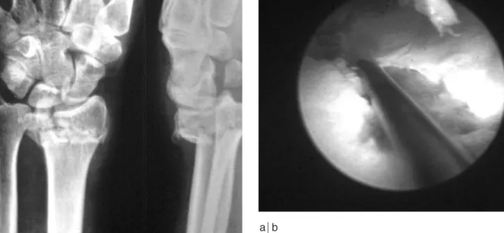
Fig. 6 a-b. Arthroscopic assisted fixation: a. A displaced intraarticular fracture; b. Arthroscopic view of the articular facture.
a b
copically guided reduction had good or excellent results 82% of the time using the Gartland and Werley criteria, better ROM and grip strength, and an improved radio-graphic appearance compared to the open reduction cohort. On the basis of this study, Doi et al. concluded that arthroscopically assisted fixation of distal radius fractures is an effective technique in patients less than 70 years of age with intraarticular injuries.
OPEN REDUCTION INTERNAL FIXATION: DORSAL AND VOLAR
Open reduction internal fixation has obvious advan-tages over the other methods discussed so far. It allows direct restoration of anatomy, stable internal fixation, a decreased period of immobilization, and an earlier return of wrist function. There are a number of diffe-rent indications for open reduction internal fixation, and these include: unstable articular fractures (such as a volar Barton’s injury), impacted articular fractures, radiocarpal fracture–dislocations, complex fractures requiring direct visualization of the fracture fragments, and failed closed reductions.
Historically, distal radius fractures were treated nonoperatively until 1929, when techniques utilizing pins and plaster were introduced. External skeletal fixation evolved in 1944 and remained popular even after the AO group designed plates specifically for the treatment of distal radius fractures in the 1970s. In 1994, Agee introduced the Wrist Jack (Hand Biome-chanics Lab Inc, Sacramento, CA), which utilized adjustable gears for multiplanar ligamentotaxis, but by this time open reduction internal fixation was beco-ming more popular, particularly when it was noted that precise reductions of the articular surface led to better outcomes (14).
The approach is generally dictated by the location of major fracture fragments and the direction of displace-ment. However, orthopaedic and upper extremity sur-geons continue to move away from dorsal plating whe-re complications can include extensor tendon ruptuwhe-re and hardware irritation. Volar plating is generally well–tolerated. Fixation of dorsally angulated and com-minuted fractures is also possible with newer fixed ang-le devices.
Although overall satisfactory outcomes have been reported with dorsal plating systems, disadvantages of dorsal plates include the need for mobilization of exten-sor tendons to achieve proper plate placement, possible tendon irritation or rupture, and the possibility of addi-tional surgery to remove the symptomatic dorsal plate, some reporting up to 30% (1, 3, 19, 27). Tendon ruptu-re has been ruptu-reported as early as 8 weeks and as late as 7 months after surgery. To prevent tendon injury, some recommend that a portion of the extensor retinaculum be interposed between the plate and the tendon sheaths, or that dorsal plates be removal routinely (7).
The advantages of a volar exposure and plating inc-lude the following: (1) Dorsally displaced fractures are simpler to reduce because the volar cortex is usually disrupted by a simple transverse line; (2) anatomic reduction of the volar cortex facilitates restoration of radial length, radial inclination, and volar tilt; (3) the avoidance of dissection dorsally helps to preserve the vascular supply to the dorsal fragments; (4) because the implant is separated from the flexor tendons by the pro-nator quadratus, the incidence of flexor tendon com-plications is lessened (17, 22); and (5) when stabilized with a fixed angle internal fixation device, shortening and secondary displacement of articular fragments is improved, and the need for bone grafting is reduced (Fig. 7a-b).
241/
ACTA CHIRURGIAE ORTHOPAEDICAEET TRAUMATOLOGIAE ČECHOSL., 74, 2007
CURRENT CONCEPTS REVIEW
SOUBORNÝ REFERÁT
Fig 7a-b. A complex intraarticular volar displaced fracture; a. The xray and CT views of the fracture; b. Volar plate fixation with angular stable Synthes plate with functional result
With the recent popularity of volar plating for treat-ment of distal radius fractures, we have witnessed a mul-titude of medical devices introduced into the market. Not surprisingly, these devices exhibit many similar charac-teristics. Volar plates are typically T–shaped to allow multiple fixation points in the distal fracture fragment with locking screws recessed into screw holes to create a lower profile distally. The angulation of these distal screws create a „scaffold“ to optimize subchondral bone
support with one or two screws devoted to fixation of the lunate facet and radial styloid. Most plating systems also feature an oblong hole for metaphyseal fixation and for plate positioning (25).
Several studies have compared outcomes of dorsal versus volar plating of distal radius fractures. Ruch and Papadonikolakis (29) performed a retrospective review of 34 patients, 20 of whom had undergone dorsal pla-ting and 14 of whom had volar plapla-ting. The authors Fig. 7a
Fig. 7b s_233_246 27.7.2007 19:52 Stránka 241
242/
ACTA CHIRURGIAE ORTHOPAEDICAEET TRAUMATOLOGIAE ČECHOSL., 74, 2007
CURRENT CONCEPTS REVIEW
SOUBORNÝ REFERÁT
Fig. 8. Complication of volar plate with screw too long resul-ting in extensor pollicis rupture requiring reconstruction.
found that both groups of patients had similar DASH scores, but the functional outcome in terms of Gartland and Werley scores was better in the volar plating group. In addition, there was a higher rate of volar collapse and late complications in the dorsal plating group compa-red with the volar plating group.
Volar plating, however, is not without its complicati-ons. Rozental and Blazar (28) retrospectively reviewed a cohort of 41 patients with dorsally displaced fractu-res treated with volar plating. Though Gartland and Wer-ley scores were overwhelmingly good and excellent, 9 complications were noted. Four of these patients actu-ally experienced loss of reduction and fracture collap-se following volar plating (Fig. 8).
OPEN REDUCTION INTERNAL FIXATION: FRAGMENT SPECIFIC
The concept of fragment specific fixation has been touted as a surgical alternative to volar plating alone. Some basic tenets of fracture fixation for fragment spe-cific systems include: (1) application of small contou-red plates on the specific components of the fracture;
(2) fixation of distal fragments is based on the strong bone proximally; (3) hardware should allow for gliding motion of tendons; (4) the exposure should cause mini-mal soft tissue disruption; and (5) the fracture should be stable to allow early range of motion. The type of implant used should match the specific fracture frag-ment being reduced via the use of limited volar and dor-sal incisions (Fig. 9a–c).
Schnall et al. (30) documented their early findings with fragment specific fixation in a retrospective study of 18 patients with intraarticular fractures. At 6 weeks, none of the patients demonstrated loss of reduction. At 6 months, patients demonstrated an average of 52 degre-es of wrist flexion and 55 degredegre-es of wrist extension. Grip strength was measured to be approximately 55 pounds. Two of the 18 patients required hardware remo-val for extensor tendon irritation.
Similarly, Rikli and Regazzoni (27) reviewed a seri-es of 20 patients with distal radius fracturseri-es fixed with two 2.0 mm titanium plates placed at 50 – 70 degrees to one another. No cases of extensor tendon problems were noted, most likely because they were able to pla-ce a flap of retinaculum over the small dorsal plates. Clearly, fragment specific fixation may provide some advantages over the traditional methods of dorsal or volar plating for fractures at the distal end of the radi-us ( Fig. 10 a-c).
INTRAMEDULLARY FIXATION
Intramedullary fixation of fractures is not a new con-cept in orthopaedic trauma surgery. Intramedullary devices increase fracture stability of the affected bone, allow load transfer across the fracture site, minimize soft tissue problems by minimizing scarring and adhe-sions, and maintain vascular blood supply to promote fracture healing. Two implants have recently been desc-s_233_246 27.7.2007 19:52 Stránka 242
243/
ACTA CHIRURGIAE ORTHOPAEDICAEET TRAUMATOLOGIAE ČECHOSL., 74, 2007
CURRENT CONCEPTS REVIEW
SOUBORNÝ REFERÁT
Fig. 9a-c. A complex articular fracture treated with fragment specific fixation with small Synthes plates (case of Daniel Rik-li MD): a. The preoperative CT scan; b. Two plate dorsal fixa-tion; c. Functional result.
Fig. 10 a-c. A complex intraarticular fracture: a. The preo-perative x-rays and CT scans; b. Opreo-perative fixation with small Synthes plates and postoperative CT scan; c. Functional fol-low up and folfol-low up x-rays
a b c c
Pokračování Fig. 10b Fig. 10a
244/
ACTA CHIRURGIAE ORTHOPAEDICAEET TRAUMATOLOGIAE ČECHOSL., 74, 2007
CURRENT CONCEPTS REVIEW
SOUBORNÝ REFERÁT
Fig. 10b
Fig. 10c
ribed for use in the distal radius, namely the Micronail (Wright Medical Technology Inc., Arlington, TN) and the Dorsal Nail Plate (Hand Innovations LLC, Miami, FL). Both are used for metaphyseal distal radius frac-tures with minimal articular involvement.
The Micronail is placed through an incision made over the radial styloid, taking care to avoid injury to branches of the radial sensory nerve. The interval bet-ween the 1st and 2nd dorsal extensor compartments is used. Kirschner wires may be used for temporary fixa-tion of large articular fragments. Difficulties that have
been described with use of this nail include possible soft tissue irritation with placement of the interlocking screws, possible screw penetration into the distal radi-oulnar joint, and difficulty observing sagittal alignment secondary to use of the jig (31).
Tan et al. (32) presented their experience with the Micronail in 23 patients with isolated unstable distal radius fractures. The minimum follow–up was 6 months. Range of motion and grip strength parameters were noted at 1 month and 6 month follow–up. The aut-hors found that all wrist and forearm range of motion s_233_246 27.7.2007 19:52 Stránka 244
245/
ACTA CHIRURGIAE ORTHOPAEDICAEET TRAUMATOLOGIAE ČECHOSL., 74, 2007
CURRENT CONCEPTS REVIEW
SOUBORNÝ REFERÁT
parameters improved significantly from 1 month to 6months. Grip strength also improved significantly from 40% to 80%. Three of the 23 patients had some loss in initial reduction, and one patient ultimately required conversion to open reduction and internal fixation.
The Dorsal Nail Plate is a hybrid plating and intra-medullary device, again intended for fractures with minimal articular involvement. The approach is perfor-med through a limited dorsal incision centered over the extensor pollicis longus. Lister’s tubercle is removed, and the exposed bone is the insertion point of this intra-medullary device. To our knowledge, outcomes with this device have not been reported. Orbay et al. (26) reported their technique and noted that complications were relatively infrequent with satisfactory functional results in over 200 patients.
BIOABSORBABLE IMPLANTS
Another emerging technology for treatment of distal radius fractures is the use of bioabsorbable implants. While popular in the use of sports medicine procedu-res, bioabsorbable implants have seen limited applica-tions in orthopaedic trauma procedures. The plates and screws in bioabsorbable constructs are typically made of polylactic acid or polyglycolic acid.
The Inion OTPS Hand System (Inion Inc, Oklaho-ma City, OK) is the only known bioabsorbable distal radius plate which is commercially available. This pla-te takes at least two years to degrade complepla-tely wit-hin the body, is contourable after placing in a hot water bath, and accepts polyaxial locking screws up to 20 degrees.
The advantages to using a bioabsorbable implant are manifold: Bioabsorbable distal radius plates obviously undergo resorption and obviate the need for hardware removal in the future, making revision surgery potenti-ally less complicated. The implants also do not incite an inflammatory response and are MRI compatible. Bioabsorbable devices are, however, relatively new and unproven technology for fracture fixation. There are valid concerns regarding the initial fixation strength, and most of these implants are slightly thicker than the-ir metal counterparts. Finally, one cannot visualize the implants on radiographs.
There is only one study, to our knowledge, which explores the possibility of using bioabsorbable implants for distal radius fracture fixation. One hundred patients with distal radius fractures were queried regarding implant preferences after they were given a brief sum-mary of advantages and disadvantages of bioabsorbab-le and metal implants. The results of the questionnaires demonstrated that 95% preferred bioabsorbable implants for their absorption feature. 91% cited ware removal as the most negative aspect of metal hard-ware. 80% of patients stated they would be interested in participating in a clinical trial comparing the two implants, setting the stage for a prospective randomized study (24).
CONCLUSION
How aggressively we pursue reconstruction of distal radius fractures is dictated by the demands of the pati-ent and radiographic findings at the time of injury. Nonoperative treatment is best reserved for low demand patients or patients too infirm to ably tolerate surgery. A multitude of different techniques can be utilized for fixation of distal radius fractures. Orthopaedic and upper extremity surgeons who manage these injuries should have all of these techniques at their disposal. As our experience with volar plating and fragment speci-fic fixation continues to deepen, newer techniques and approaches become available but are still unproven. ZÁVĚR
Léčba zlomenin distálního radia je stále výzvou pro ortopedy i traumatology. S tím, jak se zlepšují naše zna-losti anatomie a mechanismu poranění této oblasti,zdo-konalují se i chirurgické postupy přispívající k obnově funkce postižené končetiny. Tato práce podává přehled současných metod léčby zlomenin distálního radia.
References
1. AXELROD, T. S., McMURTRY, R. Y.: Open reduction and inter-nal fixation of comminuted, intraarticular fractures of the distal radius. J. Hand Surg. Amer., 15: 1–11, 1990.
2. CAPO, J. T., SWAN, K. G. Jr., TAN, V.: External fixation tech-niques for distal radius fractures. Clin. Orthop., 445: 30–41, 2006. 3. CARTER, P. R., FREDERICK, H. A., LASETER, G. F.: Open reduction and internal fixation of unstable distal radius fractures with a low–profile plate: A multicenter study of 73 fractures. J. Hand Surg. Amer., 23: 300–307, 1998.
4. CASSEBAUM, W. H.: Colles’ fracture: a study of end results. J. Amer. Med. Assoc., 143: 963–965, 1950.
5. CASTAING, J.: Les fractures récentes de l’extrémité inférieure du radius chez l’adulte. Rev. Chir. Orthop., 50: 581–696, 1964. 6. CATALANO, L. W. 3rd, COLE, R. J., GELBERMAN, R. H.,
EVANOFF, B. A., GILULA, L. A., BORRELLI, J. Jr.: Displaced intra–articular fractures of the distal aspect of the radius. Long–term results in young adults after open reduction and inter-nal fixation. J. Bone Jt Surg., 79–A: 1290–1302, 1997. 7. CHIANG, P. P., ROACH, S., BARATZ, M. E.: Failure of a
reti-nacular flap to prevent dorsal wrist pain after titanium pi plate fixation of distal radius fractures. J. Hand Surg. Amer., 27: 724–728, 2002.
8. COLLES, A.: On the fracture of the carpal extremity of the radi-us. Edinburgh Med. Surg. J., 10: 182–186, 1814.
9. DOI, K., HATTORI, Y., OTSUKA, K., ABE, Y., YAMAMOTO, H.: Intra–articular fractures of the distal aspects of the radius: arth-roscopically assisted reduction compared with open reduction and internal fixation. J. Bone Jt Surg., 81–A: 1093–1110, 1999. 10. FERNANDEZ, D. L., WOLFE, S. W.: Distal radius fractures. In:
GREEN, D. P., HOTCHKISS, R. N., PEDERSON, W. C., WOL-FE, S. W. (eds): Green’s operative hand surg. Philadelphia, Else-vier Churchill Livingstone 2005, 645–710.
11. FRYKMAN, G. K.: Fracture of the distal radius including sequ-elae: shoulder–hand–finger syndrome–disturbance in the distal radioulnar joint and impairment of nerve function: A clinical and experimental study. Acta orthop. scand., 108 (Suppl.): 1–155, 1967.
12. GOLDFARB, C. A., RUDZKI, J. R., CATALANO, L. W., HUGHES, M., BORRELLI, J. Jr.: Fifteen–year outcome of dis-placed intra–articular fractures of the distal radius. J. Hand Surg. Amer., 31: 633–639, 2006.
246/
ACTA CHIRURGIAE ORTHOPAEDICAEET TRAUMATOLOGIAE ČECHOSL., 74, 2007
CURRENT CONCEPTS REVIEW
SOUBORNÝ REFERÁT
13. GRAHAM, T. J.: Surgical correction of malunited fractures of thedistal radius. J. Amer. Acad. Orthop. Surg., 5: 270–281, 1997. 14. HARNESS, N. G., MEALS, R. A.: The history of fracture
fixati-on of the hand and wrist. Clin. Orthop., 445: 19–29, 2006. 15. JUPITER, J. B.: Fractures of the distal end of the radius. J. Bone
Jt Surg., 73–A: 461–469, 1991.
16. JUPITER, J. B.: Complex articular fractures of the distal radius: classification and management. J. Amer. Assoc. Orthop. Surg., 5: 119–129, 1997.
17. JUPITER, J. B., RING, D., WEITZEL, P. P.: Surgical treatment of redisplaced fractures of the distal radius in patients older than 60 years. J. Hand Surg. Amer., 27: 714–723, 2002.
18. KAPANDJI, A. I.: Treatment of non–articular distal radial frac-tures by intrafocal pinning with arum pins. In: SAFFER, P., COONEY, W. P. (eds): Fractures of the distal radius. Philadelp-hia, JB Lippincott 1995, 71–83.
19. KARAMBOUROGLU, G. K., AXELROD, T. S.. Complications of the AO/ASIF titanium distal radius plate system (πplate) in internal fixation of the distal radius: A brief report. J. Hand Surg. Amer., 23: 737–741, 1998.
20. KNIRK, J. L., JUPITER, J. B.: Intra–articular fractures of the distal end of the radius in young adults. J. Bone Jt Surg., 68–A: 647–659, 1986.
21. McQUEEN, M., CASPERS, J.: Colles’ fracture: does the anato-mical result affect the final function? J. Bone Jt Surg., 70–B: 649–651, 1988.
22. McQUEEN, M. M., SIMPSON, D., COURT–BROWN, C. M.: Use of the Hoffman 2 compact external fixator in the treatment of redisplaced unstable distal radial fracture. J. Orthop. Trauma, 13: 501–505, 1999.
23. MELONE, C. P. Jr.: Distal radius fractures: Patterns of articular fragmentation. Orthop. Clin. N. Amer., 24: 239–253, 1993. 24. MITTAL, R., MORLEY, J., DINOPOULOS, H.,
DRAKOU-LAKIS, E. G., VERMANI, E., GIANNOUDIS, P. V.: Use of bio–resorbable implants for stabilisation of distal radius fractu-res: The United Kingdom patients’ perspective. Injury, 36: 333–338, 2005.
25. MUDGAL, C. S., JUPITER, J. B.: Plate and screw design in frac-tures of the hand and wrist. Clin. Orthop., 445: 68–80, 2006.
26. ORBAY, J. L., TOUHAMI, A., ORBAY, C.: Fixed angle fixation of distal radius fractures through a minimally invasive approach. Tech. Hand Upper Extrem. Surg., 9: 142–148, 2005.
27. RIKLI, D. A., REGAZZONI, P.: Fractures of the distal end of the radius treated by internal fixation and early function: a prelimi-nary report of 20 cases. J. Bone Jt Surg., 78–B: 588–592, 1996. 28. ROZENTAL, T. D., BLAZAR, P. E.: Functional outcome and
complications after volar plating for dorsally displaced, unstable fractures of the distal radius. J. Hand Surg. Amer., 31: 359–65, 2006.
29. RUCH, D. S., PAPADONIKOLAKIS, A.: Volar versus dorsal pla-ting in the management of intra–articular distal radius fractures. J. Hand Surg. Amer., 31: 9–16, 2006.
30. SCHNALL, S. B., KIM, B. J., ABRAMO, A., KOPYLOV, P.: Fixation of distal radius fractures using a fragment–specific sys-tem. Clin. Orthop., 445: 51–57, 2006.
31. VIRAK, T., CAPO, J., WARBURTON, M.: Distal radius fractu-re fixation with an intramedullary nail. Tech. Hand Upper Extfractu-rem. Surg., 9: 195–201, 2005.
32. VIRAK, T., CAPO, J., WARBURTON, M.: Minimally invasive distal radius fracture fixation with an intramedullary nail. Pre-sented at the Annual Meeting of the American Society for Surge-ry of the Hand. San Antonio, Texas, September 22–24, 2005. 33. WOLFE, S. W., SWIGART, C. R., GRAUER, J., SLADE, J. F.
3rd, PANJABI, M. M.: Augmented external fixation of distal
radi-us fractures: a biomechanical analysis. J. Hand Surg., 23–A: 127–134, 1998.
Correspondence to: Jesse B. Jupiter, M.D.,
Massachusetts General Hospital Hand and Upper Extremity Service 55 Fruit Street, YAW–2–2C Boston, Massachusetts 02114 Telephone: (617) 726–8530
Práce byla přijata 10. 5. 2007. s_233_246 27.7.2007 19:52 Stránka 246