Selection of our books indexed in the Book Citation Index in Web of Science™ Core Collection (BKCI)
Interested in publishing with us?
Contact book.department@intechopen.com
Numbers displayed above are based on latest data collected.For more information visit www.intechopen.com Open access books available
Countries delivered to Contributors from top 500 universities
International authors and editors
Our authors are among the
most cited scientists
Downloads
We are IntechOpen,
the world’s leading publisher of
Open Access books
Built by scientists, for scientists
12.2%
122,000
135M
TOP 1%
154
Proliferative Diabetic Retinopathy: An Overview of
Vitreous Immune and Biomarkers
Andi Arus Victor and Ratna Sitompul
Additional information is available at the end of the chapter http://dx.doi.org/10.5772/intechopen.74366
© 2016 The Author(s). Licensee InTech. This chapter is distributed under the terms of the Creative Commons Attribution License (http://creativecommons.org/licenses/by/3.0), which permits unrestricted use, distribution, and reproduction in any medium, provided the original work is properly cited.
Andi Arus Victor and Ratna Sitompul
Additional information is available at the end of the chapter
Abstract
This chapter discusses about the effect of vitreous immune system and biomarkers on the progression of proliferative diabetic retinopathy. Immune system and biomarkers have been believed to have an important role in the progression of diabetic retinopathy (DR) severity. Hyperglycemic will influence immune cells resulting in chronic inflammation on the retina. This condition progressively disrupts the blood-retinal barrier in retina causing those inflammatory molecules and immune cells to transfer from circulation. The transfer of these molecules plays an important part in the progression of prolifera-tive diabetic retinopathy. In addition, biomarkers are indicators for some complex pro-cesses happened in our body, and are measured to determine diagnosis and prognosis of some treatment. There are several biomarkers that have been identified in DR patients including biomarkers of oxidative stress, hypoxia-inducible factors, angiogenic factors, pro-inflammatory cytokines, chemokines, cell adhesion molecules, and soluble CD200. The value of these biomarkers will tell us their possible role in the progression of DR. By improving the knowledge of molecular pathway in DR pathophysiology, the advance-ment of selective therapy approaches could be discovered and the manageadvance-ment of DR could be more efficient.
Keywords: biomarker, diabetic retinopathy, hyperglycemia, immune system, inflammation
1. Introduction
Diabetic retinopathy (DR) is the most common chronic microvascular complication of uncon-trolled diabetes mellitus leading to preventable blindness. Diabetic retinopathy is often clas-sified based on its severity into mild non-proliferative diabetic retinopathy (NPDR), moderate
© 2018 The Author(s). Licensee IntechOpen. This chapter is distributed under the terms of the Creative Commons Attribution License (http://creativecommons.org/licenses/by/3.0), which permits unrestricted use, distribution, and reproduction in any medium, provided the original work is properly cited.
NPDR, severe NPDR, and proliferative diabetic retinopathy (PDR) [1–3]. The major risk
fac-tors for developing DR are the duration of diabetes, hyperglycemia, hypertension, and dys-lipidemia [4]. Glucose concentration increases in retinal cells leading to saccular capillary
microaneurysms, pericyte deficient capillaries, and degenerate capillaries that decrease the retinal perfusion and contribute to the progression of DR [4]. Several types of evidence prove
the benefits of tight glycemic and blood pressure control in decelerating the progression of DR. Nevertheless, the numbers of DR patients and the development of DR complications are still increasing, while therapeutic approaches are limited [1, 2].
For the last several decades, many studies have been performed in order to better understand DR progression from a molecular viewpoint. The biochemical mechanisms implicated in DR progression have been shown in various animal models and patients with diabetes [1]. It is
believed that the involvement of hyperglycemia and hormonal factors in diabetic patients could disturb hemostasis in the retina and change the balance of some mediators including growth factors, cytokines, inflammatory, and adhesion molecules [5]. These changes result in
altered capillary permeability, apoptosis of capillary cells, and angiogenesis, leading to DR complications [3]. With improved clarity of molecular pathways in DR pathophysiology, the
advancement of selective therapeutic approaches could be discovered and the management of DR could be more effective [1, 5]. This chapter focuses on the inflammatory molecules and
biomarkers involved in the pathophysiology of DR.
2. The immune system in proliferative diabetic retinopathy
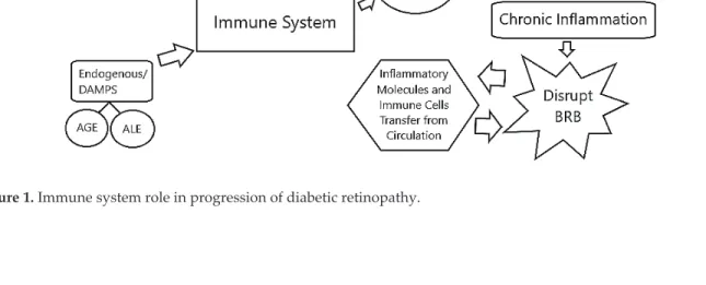
The immune system protects the body from both exogenous pathogens called pathogen-associated molecular patterns (PAMPs) and endogenous harmful molecules known as dam-age-associated molecular patterns (DAMPs). DAMPs include oxidized or glycated proteins, mislocated proteins/antigens, and intracellular contents released by necrotic cells. In normal conditions, the immune system regulates the inflammatory process and prevents uncon-trolled inflammation that damages cells. In hyperglycemic conditions, the accumulation of DAMPs induces chronic inflammation in various tissues, which in turn manifests into the various complications of diabetes, including diabetic retinopathy [6].
The retina is one of few tissues in the human body that has immune privilege. It is protected from the attack of the systemic immune system by a series of complex defense mechanisms. This protection is afforded by a physical barrier formed between endothelial cells of retinal vasculature as the inner blood-retinal barrier (BRB) and retinal pigmented epithelial cells as the outer BRB. This barrier limits the movement of cells and molecules from the systemic circulation into the retinal parenchyma. The BRB also separates retinal antigens within the intraocular compartment, avoiding activation of T cells. This phenomenon is known as immu-nological ignorance. In addition, there is no lymphatic system in the retina. This inhibits sys-temic immune cells from detecting damage-associated molecular patterns in the retina thus preventing an overt systemic inflammatory response. Retinal cells (retinal neurons and RPE cells) express immune modulators that can suppress immune cells and complement system activation. The retina is protected by the local innate immune system (microglia, perivascular macrophages, and the complement system) whose activation is tightly controlled [6].
The immune system plays an important role in the progression of DR. Under hyperglycemic conditions, over activation of the innate immune system takes place, resulting in chronic inflam-mation of the retina. A study by Urbančič et al. showed the presence of T lymphocytes in the vit-reous of patients with PDR. They found that the CD4/CD8 lymphocyte ratio in vitvit-reous is higher compared to the blood ratio in these PDR patients, demonstrating the presence of a local inflam-matory process [7]. Prolonged local inflammation in hyperglycemic conditions in the retina may develop into a chronic inflammatory response that is detrimental to the integrity of BRB [6, 8–10]. The destruction of the barrier shifts the retina from its “privileged state” when the BRB functions normally to “compromised state” when the BRB has broken down. Complement system activa-tion also increases in diabetic condiactiva-tions and this dysregulated activaactiva-tion is known to be involved in the degeneration of retinal vessels. Dysfunctional barriers permit inflammatory molecules and immune cells from systemic circulation to enter the retina and cause further deterioration of the tissue [6, 11]. Cytological examination of the vitreous samples from PDR patients were found to contain significant amounts of macrophages suggesting the infiltration of systemic immune cells into the retina [12, 13]. In addition, there was an increase in adhesion molecule expression and pro-inflammatory cytokine production, suggesting the role of defective neutrophil activity in the development of chronic inflammation in diabetic retinopathy [14, 15] (Figure 1).
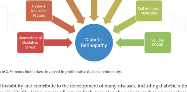
3. Vitreous biomarkers in proliferative diabetic retinopathy
A biomarker is an objective measurement that is evaluated as an indicator for some com-plex processes happening in our body [16]. Biomarkers are usually measured to determine the diagnosis and prognosis of some treatments [17]. There are several biomarkers that can be found in diabetic retinopathy patients including biomarkers of oxidative stress, hypoxia-inducible factors, angiogenic factors, pro-inflammatory cytokines, chemokines, cell adhesion molecules, and CD200. The value of these biomarkers tells us their possible role in the pro-gression of diabetic retinopathy [5, 6, 18–21] (Figure 2).
3.1. Biomarkers of oxidative stress
The presence of oxidative stress biomarkers indicate an imbalance of reactive oxygen species (ROS) and the functional capabilities of cellular antioxidants [18, 22]. This imbalance can cause
cell instability and contribute to the development of many diseases, including diabetic retinop-athy [18, 23]. Oxidative stress will remain high even after the patient reaches a normoglycemic state. This phenomenon is called “metabolic memory” and can lead to the accumulation of ROS in diabetic patients [24]. The biological markers of oxidative stress can include changes in mol-ecules of the antioxidant system and molmol-ecules modified by ROS. Antioxidant enzymes like the superoxide dismutases are an example of changes in molecules of the antioxidant system, and malondialdehyde is the best known oxidative stress marker [18].
3.1.1. Superoxide dismutases
Superoxide dismutases (SODs) are a group of enzymes found in our cells, which function as major antioxidant defense systems against ROS in the body. SODs consist of three isoforms: the cytoplasmic Cu/ZnSOD (SOD1), the mitochondrial MnSOD (SOD2), and the extracellular Cu/ZnSOD (SOD3), all of which require catalytic metal (Cu or Mn) to activate. SOD activi-ties will increase due to the presence of oxidative stress in the body. Vitreous SOD activity can also be used to measure oxidative stress levels inside the eye, allowing it to be a viable biomarker of oxidative stress in patients with PDR. Brzović-Šarić et al. state that PDR patients serum oxidative stress markers were higher than non-diabetic patients with an eye disorders (NDED) serum. Brzović-Šarić et al. found a mean activity level of SODs in the vitreous of male diabetic patients at 30.5 ± 2.5 U/mL, and 28.5 ± 3.8 U/mL in vitreous of female patients with diabetes [25]. Our previous study found a mean activity level of SODs in vitreous of patients with PDR at 0.403 + 0.50 U/mL [26].
3.1.2. Malondialdehyde
Malondialdehyde (MDA) is a highly reactive compound produced by lipid peroxidation of polyunsaturated lipid found in cell membranes. MDA exerts its oxidative stress effect inside
cells and forms molecules called advanced lipoxidation end-products (ALE). MDA levels in specific tissues can be measured to represent oxidative damage induced by physical or chemi-cal oxidative stress in the corresponding tissues [24, 25, 27]. Brzović-Šarić et al. found a signifi-cant difference between vitreous MDA values in non-diabetic patients with an eye disorder and PDR patients [25]. On the other hand, several studies found an increase in MDA serum of diabetic patients compared to control patients, but there was no significant difference in MDA serum level between non-proliferative DR and proliferative DR patients [24, 27]. Our study found a mean activity level of MDA in the vitreous of patients with PDR at 1.661 ± 1.21 nmol/mL [26]. Another study about oxidative stress levels with PDR by Mancino et al. found a mean activity level of MDA in vitreous of patients with PDR at 520 ± 210 nmol/mL [24]. What causes these differences in vitreous MDA levels still needs to be explored.
3.2. Hypoxia-inducible factors
HIF-1α is a DNA-binding protein complex that is continuously expressed and degraded by cells in the body. Under hypoxic conditions, the HIF-1α degradation rate decreases, causing increased concentration of HIF-1α which then translocates into the nucleus and dimerizes with HIF-1β. The HIF-1 complex then regulates the expression of genes responsible for the hypoxic response of the cell by binding into the hypoxia response element (HRE) [28]. The HIF-1 complex is known to cause angiogenic effects on these hypoxic tissues [29]. Previous studies by Arden et al. on patients with diabetic retinopathy shows that hypoxia is present in retinal tissues suffering from oxidative damage [30]. Accordingly, Wang and co-workers found increased levels of HIF-1α protein in vitreous samples of PDR patients compared to levels in non-diabetic subjects [28]. Furthermore, the vitreous levels of vascular endothelial growth fac-tor (VEGF) and HIF-1α were highly correlated in PDR patients. Several studies demonstrated positive immunohistochemical staining for HIF-1α and VEGF proteins in epiretinal neurovas-cular membranes. This evidence shows that HIF might play an important role in regulating the neovascularization of retina in PDR [31, 32].
3.3. Angiogenic factors
Angiogenesis is a complex multistep process that involves angiogenic factors and is induced by various cytokines and growth factors [33]. These factors have been suggested to be correlated
with the development of diabetic retinopathy [5, 33–35]. These are also known to be
hypoxia-responsive factors [5, 35]. Pro-angiogenic factors, like VEGF, angiopoientin, and erythropoietin
are well-known factors contributing to neovascularization and whose levels increase in dia-betic retinopathy patients [3, 5, 33–35]. Several therapies designed to target these factors have
been proven effective in decreasing the progression of the disease [5].
3.3.1. Vascular endothelial growth factor
Vascular endothelial growth factor (VEGF) is a signaling molecule that promotes devel-opment of new blood vessels. It is released by cells in response to hypoxic conditions. Abcouwer stated that VEGF increases vascular permeability by promoting the disassem-bly of junctions between endothelial cells. This leakage can cause diabetic macular edema
(DME) [5]. Several studies have shown marked increases of VEGF in vitreous and vitreous compared to plasma concentration in DME and PDR patients [19, 36–46]. Treatments that target VEGF have been proven highly effective in treating DR. VEGF antibodies, which were originally used for cancer treatments, such as bevacizumab and its correlate ranibi-zumab have been used effectively. These have also been tested in several small trials which showed improved vision in DR patients, demonstrating the involvement of VEGF in the pathophysiology of PDR [5, 36].
Brzović-Šarić also demonstrated a significant difference between vitreous VEGF values in non-diabetic patients with eye disorders and PDR patients [25]. Loukovaara et al. state that VEGF is a major factor in PDR development and found significant increases of VEGF levels in the vitre-ous of DR patients (465.1 ± 1470.2 pg/mL) compared to control patients (40.3 ± 165.8 pg/mL) [37]. Our study found a mean level of VEGF in vitreous of patients with PDR of 0.356 + 0.60 pg/mL [26]. Yoshimura et al. found that there was significantly elevated VEGF in PDR patients, but not in DME patients [47]. The increased levels of VEGF expression in patients with diabetic retinopathy was mainly produced by Muller glial cells. Experiments in diabetic mice, demon-strated that conditional knockout of VEGF in Muller cells effectively blocked the increase in retinal VEGF expression [48]. Lange and co-workers suggest that oxygen tension levels were positively correlated with vitreous VEGF levels, and oxygen tension levels at the posterior pole were increased in PDR patients [49]. The vitreous levels of VEGF will decrease in the most severe stage of PDR, when there is a transition from angiogenesis to fibrosis [50].
3.3.2. Angiopoietin
Angiopoietins are a group of proteins with the role of regulating vascular development and angiogenesis. Two types of angiopoietins, angiopoietin-1 and angiopoietin-2, contribute to the maintenance of retinal vasculature. The former exerts a stabilizing effect on vessels, orga-nizing and limiting the angiogenesis response, while the latter exhibits angiogenic activity if VEGF is present, but promotes endothelial cell death and vascular regression in the absence of VEGF. The ratio between these two angiopoietins represents the inflammatory process in the cell. Fiedler et al. state that hypoxia/ischemia activates endothelial cells upregu-lating angiopoientin-2 thus lowering the angiopoientin-1/angiopoientin-2 ratio [37, 51]. A recent publication by Loukovaara et al. demonstrates significant correlation between intravitreal concentrations of Ang-2 with MMP-9, VEGF, EPO and TGFb1 levels in dia-betic eyes undergoing vitrectomy, indicating its role in retinal tissue neovascularization in PDR patients. The study shows a slight increase in angiopoietin-1 from the control group (19.1 ± 25.4 pg/mL) to the study group (25.6 ± 27.1 pg/mL), and a great increase in angiopoi-etin-2 from the control group (43.0 ± 60.9 pg/mL) to the study group (317.1 ± 419.1 pg/mL), thus lowering the angiopoietin-1/angiopoietin-2 ratio in study group. The plasma value of angiopoietin-1 is similar in both groups, but the plasma value of angiopoietin-2 is increased from the control group (2623.4 ± 2142.0 pg/mL) compared to the study group (5690.4 ± 8064.7 pg/mL) [36, 37, 52]. Several studies state that angiopoietin-1 can be used for the prevention and treatment of diabetic retinopathy by its ability to suppress VEGF expression in diabetic retina [53].
3.3.3. Erythropoietin
Erythropoietin (EPO) is a glycoprotein cytokine that acts as a major regulator of erythropoi-esis. Besides erythropoiesis, several studies state that erythropoietin has a neuroprotective and angiogenic effect in brain and retina. Production of EPO in serum and vitreous is mainly caused by hypoxia [54–57]. EPO is found in many organs, including kidney, liver, brain, and
retina [55]. The angiogenic effect of EPO is a potential equivalent to VEGF, and has been
suspected as an important factor in the angiogenesis of PDR [56]. Watanabe et al. showed
that vitreous EPO levels of PDR patients are significantly higher (464.0 mlU/mL) compared to non-diabetic patients (36.5 mlU/mL). They also found that EPO levels are higher with active as compared to quiescent PDR [54]. These are consistent with Katsura et al., who also reported
increases of vitreous EPO levels in PDR patients compared to controls [55]. Cristina et al.
found that EPO levels in vitreous fluid are significantly higher (326 mU/mL) compared to serum EPO (11.2 mU/mL) in PDR patients [56]. This shows that intraocular production is
responsible for the high concentration of erythropoietin found in the vitreous fluid of retinal degeneration patients [54, 56, 57]. Garci et al. found increased vitreous EPO concentrations in
DME patients (430 mU/mL) compared to control patients (25 mU/mL) [57]. Treatment
involv-ing the erythropoietin blockade is likely to be beneficial, but may worsen the disease due to the decrease of its neuroprotective function [54].
3.3.4. Matrix metalloproteinases 9
Matrix metalloproteinases (MMPs) are a family of zinc ion-binding endopeptidases that degrade most of the extracellular matrix (ECM). MMPs regulate many cellular functions including apop-tosis, wound healing, and angiogenesis. In angiogenesis, MMPs increase VEGF production and remove physical barriers to new vessel growth [58, 59]. MMPs are produced as a response to increased oxidative stress. Diabetic patients often have increased MMP, mainly MMP-9 and MMP-2 in the retina and vitreous. These are controlled by endogenous tissue inhibitors of metalloproteinases (TIMPs). TIMP-1 regulates MMP-9 and TIMP-2 regulates MMP-2 [59]. Several studies suggest that MMPs are responsible for many diabetic complications, includ-ing cardiomyopathy, nephropathy, and retinopathy. MMPs are suspected to facilitate apop-tosis of retinal capillary cells during early stages leading to disruption of blood-retinal barrier integrity [58–60]. Kowluru et al. found an increase in MMP-9 and a decrease in TIMP-1 in the
retina of DR patients [58]. Abu et al. found significant increases in vitreous zymography levels of MMP-9 in PDR patients (392.3 ± 253.6 scanning units) compared to non-diabetic control patients (168.2 ± 65.0 scanning units). However, the levels of vitreous MMP-2 in PDR patients (540.9 ± 185.6 scanning units) did not differ significantly from non-diabetic control patients (505.4 ± 216.1 scanning units) [60]. Inhibitors of MMPs have been used to treat several diseases, however, there have been no studies using these inhibitors to treat DR patients [59].
3.3.5. Transforming growth factor β
Transforming growth factor β (TGF-β) is a polypeptide responsible for controlling cell pro-liferation and differentiation. It is usually secreted in a latent phase and must be transformed
to become a mature active form. In the human eye, there are three known TGF-β isoforms (TGF-β1, TGF-β2, and TGF-β3), where the posterior segment of the eye mainly contains TGF-β2 as the dominant form [61–63]. Hirase et al. found an increase in total vitreous TGF-β
2 levels in PDR patients (2634 ± 1652 pg/mL) compared to control patients (1305 ± 972 pg/mL) [61]. This result is also consistent with a McAuley et al. study about vitreous biomarkers in diabetic reti-nopathy [62]. The mature active form of TGF-β2 levels are also increased in PDR patients. This increase correlates with the disease severity, suggesting that TGF-β2 angiogenesis properties play a role in the progression of PDR [61].
3.4. Pro-inflammatory cytokines
Pro-inflammatory cytokines are usually secreted by inflammatory cells in response to hypoxia or hyperglycemia [64]. Well-known pro-inflammatory cytokines, such as tumor necrosis factor, inter-leukin, interferon, and receptor tyrosine kinase are found to be elevated in the vitreous of diabetic retinopathy patients, suggesting their important role in the pathogenesis of this disease [5, 64, 65]. Cytokines can induce the progression of diabetic retinopathy directly and indirectly. Direct mech-anisms include the direct engagement with target cells to induce neovascularization [64]. While indirect mechanisms induce leukocytes and endothelial cells to produce pro- angiogenic media-tors, which in turn induce neovascularization [64, 65]. Therapy targeting these cytokines may be beneficial, but we need better understanding about the cytokine roles to do so [5].
3.4.1. Tumor necrosis factor-α
Tumor necrosis factors-α (TNF-α), a pro-inflammatory cytokine, is primarily synthesized by macrophages and T cells. Its expression is regulated by NF-κβ and it has been associated with the pathogenesis of several chronic inflammatory diseases including type 2 diabetes. Its func-tion is primarily as an immune-modulator and it also plays a role in neovascularizafunc-tion and fibroplasia [3]. Costagliola et al. suggest that TNF-α is a potent mediator of leukostasis and contributes to blood-retinal barrier breakdown [3, 66]. TNF-α concentration is found elevated in the vitreous of PDR patients and the vitreous/serum ratio of TNF-α is also found higher compared to non-diabetic patients. Costagliola et al. found that TNF-α levels were lower in controls (1.9 pg/mL) than the PDR group (13.5 pg/mL) and increased with the severity of the disease [3, 66]. TNF-α has a short half-life (∼4 min), making its analyzation prone to produc-ing false negative results. Soluble TNF-α receptors (sTNF-α-Rs) have a longer half-life, mak-ing it a more reliable marker of the activation of TNF-α system [29, 31, 67–71].
3.4.2. Interleukin
Several studies have shown that there is involvement of interleukins in the development of PDR. The most common interleukins found in DR patients are IL-6 and IL-8, where their con-centrations were found increased in the vitreous of patients with PDR and prolonged hyper-glycemia [3, 42, 47, 72–82]. Their role in the pathogenesis of PDR is still under investigation but evidence suggests the possibility of a rather direct contribution. IL-6 controls immune cells responses by shifting T-helper cell populations, inhibiting the production of Th1 cells, promot-ing the differentiation of Th2 and Th17 cells, and infiltration of monocytes and T cells [9, 10, 83].
In vitro study of IL-6 reports its ability to increase endothelial cell and vascular cell permeability by rearranging actin filaments and by changing the shape of endothelial cells [3, 65]. Several
studies state that IL-6 also plays an important role in angiogenesis by activating VEGF, and reg-ulating expression of metalloproteinases [3, 64]. IL-8 is known to be a potent angiogenic factor
and also a potent chemoattractant and activator of neutrophils and T lymphocytes [64, 84, 85].
Increase of IL-8 concentrations in PDR patients, suggest that they are upregulated in response to oxygen stress and contribute to triggering inflammatory reactions. Study by Takahashi et al. shows that there is a significant increase in IL-6 and IL-8 values in PDR patients (918.0 and 2168.0 ng/mL) compared to control patient (517.0 and 343.0 ng/mL) [85]. Elner et al. also found increased levels of IL-8 in active PDR patients (24.7 ± 4.5 ng/mL) compared to control patients (7.5 ± 2.3 ng/mL), however inactive PDR patients (11.6 ± 5.2 ng/mL) did not differ significantly from controls [79]. It is most likely that VEGF expression causes an increase of IL-8 [86]. On the other hand, IL-10 concentration is not increased in the vitreous of patients with PDR. IL-10 is another important immunoregulatory cytokine that is induced by cell hypoxia. IL-10 activates nitric oxide and increases vascular permeability during the development of PDR [3, 65, 84, 85]. 3.4.3. Monokine induced by interferon-γ
Monokine induced by interferon-γ (Mig) attracts activated T cells and has potent angiostatic activity. Several studies suggest that Mig correlates with VEGF and contributes to the progres-sion of neovascularization in DR patients. The main function of Mig in the progresprogres-sion of DR might be related to its leukostasis function [88, 89]. Wakabayashi et al. found significant increases in vitreous concentration of Mig in active (148 pg/mL) and inactive (82.3 pg/mL) DR patients compared with non-diabetic patients who had macular disease (21 pg/mL). However, there was no significant difference in serum Mig concentration between DR patients (85.9 pg/mL) and control subjects (70.4 pg/mL) [87]. Takeuchi et al. also found an increase in Mig vitreous concentration in PDR patients compared to epiretinal membrane patients, idiopathic macular hole patients, and uveitis patients [88].
3.4.4. Receptor tyrosine kinase
Receptor tyrosine kinase (c-kit) is expressed by bone marrow and involved in intracellular signaling. It plays an important role in cell proliferation, cell adhesion, cell survival, and neo-vascularization [89]. Several studies have shown that C-kit plays an important role in the angiogenic process of PDR. C-kit has a soluble form called s-kit that can be generated by proteolytic cleavage [90]. Abu et al. found an increase of c-kit expression in membranes from patients with active neovascularization (697.4 ± 1528.1 pg/mL) compared to patients with inactive PDR (205.3 ± 106.4 pg/mL) and control patients (87.5 ± 91.5 pg/mL). This demonstrates that an increase of c-kit expression is correlated to the progression of PDR [90]. However, Lee et al. found a slight decrease of c-kit values in the PDR group compared to NPDR group [91]. 3.5. Chemokine
Chemokines are low molecular weight proteins that have many functions, including enhanced immune responses, regulation of homeostasis, and controlling angiogenesis [20, 92, 93].
Chemokines are often referred to as secondary pro-inflammatory mediators, whose activa-tion is induced by pro-inflammatory cytokines or primary pro-inflammatory mediators. Chemokines induce a specific leukocyte type and can bind to chemokine-receptors on target cells [20, 92]. Chemokines are usually categorized into two groups, the CXC group is che-motactic for neutrophils and the CC group is cheche-motactic for monocytes and lymphocytes [20, 92]. Several studies show an increase of chemokines in vitreous of PDR patients, suggest-ing that they have roles in mediatsuggest-ing angiogenesis and fibrosis in PDR patients [20, 93, 94]. Struyf et al. stated that chemokines have different roles based on disease progression. In the early phase, chemokines can induce leukocyte attraction and in late phase, they can induce neovascularization [93]. Das et al. introduced a new therapy targeting chemokines in patients with DME [94].
3.5.1. Monocyte chemotactic protein-1
Monocyte chemotactic protein-1 (MCP-1) is a member of the chemokine group which is responsible for regulating migration and infiltration of monocyte/macrophages to the site of inflammation, making MCP-1 a pro-inflammatory cytokine that plays a central role in CNS inflammation [2]. Hyun et al. stated that MCP-1 is a major cause of vascular complications in diabetes [95]. It is also a potent inducer of angiogenesis and fibrosis. MCP-1 levels were found elevated in the vitreous of diabetic patients and their levels are higher than serum [2, 97]. Ning et al. stated that advanced glycation end product (AGE) stimulation activates retinal neurons to release MCP-1 activating retinal microglial cells. Their study also shows a progres-sive increase of MCP-1 along with the progression of disease, indicating it may be an impor-tant link in diabetic retinopathy pathogenesis [2, 96]. Hyperglycemia also has been shown to increase MCP-1 expression from retinal vascular endothelial cells, RPE cells, and Muller glial cells [2, 97]. Reddy et al. demonstrated significantly higher levels of MCP-1 in PDR patients compared to normal glucose tolerance (NGT) patients. MCP-1 is also steadily increased along with the progression of PDR [97].
3.5.2. Interferon gamma-induced protein-10
Interferon gamma-induced protein-10 (IP-10), also known as CXCL10, is one of the CXC che-mokine members. CXC cheche-mokine has unique properties in which it can act as either an angio-genic or angiostatic factor, depending on the protein configuration of the molecule. IP-10 is inducible directly or through activation of IFN-γ, TNF-α, NFkB, viruses, or microbial products. Boulday et al. reported that VEGF induced the expression of IP-10 [1]. IP-10 binds CXCR3 receptors inducing apoptosis, angiostasis, and chemotaxis. It has been suggested that IP-10 is associated with inflammatory diseases including immune dysfunction and infectious disease. This protein has also been proposed to be involved in the pathophysiology of diabetic reti-nopathy, especially in the development of neovascularization. Elner et al. found a significant increase in the level of IP-10 in patients with PDR compared to the patients with non-diabetic eye diseases (NDED; 11.7 ± 1.1 ng/mL and 4.6 ± 0.9 ng/mL; p < 0.001, CI 95%). They also assumed that pan-retinal laser photocoagulation (PRP) might influence elevated IP-10 levels. The exact mechanism of the PRP-induced IP-10 involution of PDR remains to be elucidated [79].
3.5.3. Stromal cell-derived factor-1
Stromal cell-derived factor-1 (SDF-1) is a chemokine with a major role in the ischemic dam-age repair process. It recruits endothelial progenitor cells (EPCs) from the bone marrow to the site of repair and upregulates expression of VEGF, increasing the angiogenic process. This pro-angiogenic factor is categorized as being hypoxia-responsive and is found to be upregu-lated in PDR [98–100]. Chen et al. found that vitreous concentrations of SDF-1 and VEGF are
correlated in eyes with PDR. They also found that vitreous levels of SDF-1 are significantly higher in PDR patients (306.37 ± 134.25 pg/mL) than in patients with idiopathic macular hole (86.91 ± 55.05 pg/mL) [101]. Butler et al. demonstrate an increase of SDF-1 concentration in the vitreous of patients with PDR and this increase correlates directly with disease severity. They also demonstrated that intravitreal injection of triamcinolone dramatically decreased the con-centration of vitreal VEGF and SDF-1, suggesting it as another possible treatment for PDR [98].
3.5.4. High-mobility group box-1
High-mobility group box-1 (HMGB1) is a nonhistone DNA-binding protein that facilitates transcription. HMGB1 can be released into the extracellular space by active secretion from certain cells such as activated monocytes and macrophages, mature dendritic cells, natural killer cells, and endothelial cells. Necrotic cell death can also cause passive leakage of HMGB1 from the nucleus as the protein is no longer bound to DNA. HMGB1 can bind to the receptor for advanced glycation end products (RAGE) and toll-like receptor 2 (TLR-2), where it acts as a inflammatory cytokine, activating NF-κβ resulting in the overexpression of other pro-inflammatory molecules such as TNF-α, MCP-1, and ICAM-1 [41, 102, 103]. El-Asrar et al.
dem-onstrated a significant correlation between neovascularization levels in epiretinal membranes of patients with PDR and the expression of HMGB1 and RAGE [41]. Yao Yu et al. also found
an increase of HMGB1 concentration in the vitreous of PDR patients. This increase in vitreous happens in the later phases of DR, and differs from other inflammatory cytokines. They also found increases of RAGE protein and decreases of TLR-2 protein in DR rats, suggesting that the involvement of HMGB-1 is mainly through its binding with RAGE [102].
3.6. Cell adhesion molecules
Adhesion molecules have many roles in our body, including embryology, immunology, and malignancy [21]. Several studies show increases of these molecules in PDR patients, suggest-ing that cell to cell interaction plays a major role in the development of PDR [79, 104–106].
These molecules regulate lymphocyte recruitment to vascular endothelium. Well-known adhesion molecules found in PDR patients are intercellular adhesion molecule-1 (ICAM-1) and vascular cell adhesion molecules-1 (VCAM-1), which are required for initiation of adhe-sion-dependent immune response [21, 106].
3.6.1. Intercellular adhesion molecule-1
Intercellular adhesion molecule-1 (ICAM-1), also known as CD54, is a cell surface glycopro-tein encoded by the ICAM1 gene. ICAM-1 is usually expressed on the surface of endothelial
cells and cells of the immune system. It works as a cell adhesion molecule that recruits nearby circulating leukocytes to the inflamed location. In PDR patients, ICAM-1 is suspected as one of the deteriorating factors, promoting leukostasis and inflammation on nearby retinal tissue. Several experiments show leukostasis as a possible mechanism in diabetic retinal vasculature injury. Cells which are attached, mainly granulocytes and monocytes cause microvascular occlusion and capillary injury [106]. Leukostasis in DR is mainly caused by endothelial acti-vation and increased surface expression of intercellular adhesion molecules (ICAM-1) [107]. Hillier et al. stated that increases in ICAM-1 correlate with the severity of DR in patients [108]. Our study on ICAM-1 showed an increase of ICAM-1 expression in PDR patients with more than 10 years of diabetes history [103]. Yan et al. in their study about the effects of intravitreal ranibizumab injection on ICAM-1 levels in PDR patients, demonstrated a decrease of ICAM-1 levels a week after intravitreal injection [109].
3.6.2. Vascular cell adhesion molecule-1
Vascular cell adhesion molecule-1 (VCAM-1) is an immunoglobulin supergene family of cellular adhesion molecules that are involved in the transmigration of monocytes, eosino-phils, and lymphocytes [105, 110–112]. Oxidative stress, VEGF, and hypercholesterolemia
increase the expression of VCAM-1 in the brain and retina [111, 113, 114]. It is released
by endothelial cells and is present as an early feature of inflammatory disease [111, 113].
Several studies state that VCAM-1 promotes angiogenesis in PDR patients [105, 112–114].
Burgos et al. demonstrated increases in vitreous concentration of VCAM-1 in PDR patients (26 ng/mL) compared to non-diabetic patients in whom a vitrectomy was performed (22 ng/mL) [104]. These results are also consistent with Mroczek et al. in their study about the influence of glucose control on the activation of the intraocular molecular system [114].
There are also reports of increase VCAM-1 concentration in the retinal vessels and serum of PDR patients [111, 112].
3.7. Soluble CD200
CD200 is a novel immunosuppressive molecule found in neuronal cells. CD200 exists in a cell membrane-bound form and a soluble form. It exerts inhibitory effects on microglia/ macrophages via interaction with the CD200 receptor (CD200R) [115]. DR-related
neuro-nal degeneration also reduces CD200 concentration and further induces microglial activa-tion [6]. Recent study on CD200 revealed that levels of sCD200 in vitreous of patients with
PDR are significantly higher compared to that in the vitreous of patients without PDR. Xu et al. showed increases in mean sCD200 levels in the PDR group (182 ± 17.63 pg/mL) com-pared to non- diabetic patients with other conditions who requires pars plana vitrectomy (56.86 ± 6.573 pg/mL). This study also showed that vitreous levels of sCD200 are higher in PDR patients with DME (266.9 ± 28.82 pg/mL) or traction retinal detachment (TRD) (256.9 ± 34.50 pg/mL) compared to PDR patients without DME (136.9 ± 15.13 pg/mL) or TRD (146.9 ± 15.97 pg/mL). sCD200 level increases also have significant statistical correlations with the increase of several angiogenic and inflammatory molecules such as VEGF, IL-6, IL-8 and IL-10 [115].
Acknowledgements
This article’s publication is partially supported by the United States Agency for International Development (USAID) through the Sustainable Higher Education Research Alliance (SHERA) Program for Universitas Indonesia’s Scientific Modeling, Application, Research and Training for City-centered Innovation and Technology (SMART CITY) Project, Grant #AID-497-A- 1600004, Sub Grant #IIE-00000078-UI-1.
Conflict of interest
There are no conflicts of interest in this chapter.
Author details
Andi Arus Victor* and Ratna Sitompul
*Address all correspondence to: arvimadao@yahoo.com
Department of Ophthalmology, Faculty of Medicine, Universitas Indonesia, Cipto Mangunkusumo National General Hospital, Jakarta, Indonesia
References
[1] Simó-Servat O, Hernández C, Simó R. Usefulness of the vitreous fluid analysis in the trans-lational research of diabetic retinopathy. Mediators of Inflammation. 2012;2012:872978. DOI: 10.1155/2012/872978
[2] Dong N et al. Upregulation of retinal neuronal MCP-1 in the rodent model of diabetic ret-inopathy and its function in vitro. Investigative Ophthalmology & Visual Science. 2012; 53:7567. DOI: 10.1167/iovs.12-9446
[3] Paine SK et al. Association of tumor necrosis factor α, interleukin 6, and interleukin 10 promoter polymorphism with proliferative diabetic retinopathy in type 2 diabetic sub-jects. Retina. 2012;32:1197-1203. DOI: 10.1097/IAE.0b013e31822f55f3
[4] Lü H. Inflammation and diabetic retinopathy. Diabetic Retinopathy. 2012;1:125-136. DOI: 10.5772/28635
[5] Abcouwer SF. Angiogenic factors and cytokines in diabetic retinopathy. Journal of Clinical & Cellular Immunology. 2013;11:1. DOI: 10.4172/2155-9899
[6] Xu H, Chen M. Diabetic retinopathy and dysregulated innate immunity. Vision Research. 2017;139:39-46. DOI: 10.1016/j.visres.2017.04.013
[7] Urbančič M, Kloboves Prevodnik V, Petrovič D, Globočnik Petrovič M. A flow cytomet-ric analysis of vitreous inflammatory cells in patients with proliferative diabetic reti-nopathy. BioMed Research International. 2013;2013:1-7. DOI: 10.1155/2013/251528
[8] Khandelwal PJ, Herman AM, Moussa CEH. Inflammation in the early stages of neu-rodegenerative pathology. Journal of Neuroimmunology. 2011;238:1-11. DOI: 10.1016/j.
jneuroim.2011.07.002
[9] Holm TH, Draeby D, Owens T. Microglia are required for astroglial toll-like receptor 4 response and for optimal TLR2 and TLR3 response. Glia. 2012;60:630-638. DOI: 10.1002/
glia.22296
[10] Graeber MB, Li W, Rodriguez ML. Role of microglia in CNS inflammation. FEBS Letters. 2011;585:3798-3805. DOI: 10.1016/j.febslet.2011.08.033
[11] Bresgen M, Baum U, Esser P, Wiedemann P, Heimann K. Protein composition of the vit-reous body in proliferative diabetic retinopathy. An analysis with 2-D-electrophoresis. Der Ophthalmologe. 1994;91:758-762. PMID: 7849428
[12] Canataroglu H et al. Interleukin (IL)-6, interleukin (IL)-8 levels and cellular composi-tion of the vitreous humor in proliferative diabetic retinopathy, proliferative vitreo-retinopathy, and traumatic proliferative vitreoretinopathy. Ocular Immunology and Inflammation. 2005;13:375-381. DOI: 10.1080/09273940490518900
[13] Cutler CW, Eke P, Arnold RR, Van Dyke TE. Defective neutrophil function in an insu-lin-dependent diabetes mellitus patient. A case report. Journal of Periodontology. 1991;62:394-401. DOI: 10.1902/jop.1991.62.6.394
[14] Bouma G, Lam-Tse WK, Wierenga-Wolf AF, Drexhage HA, Versnel MA. Increased serum levels of MRP-8/14 in type 1 diabetes induce an increased expression of CD11b and an enhanced adhesion of circulating monocytes to fibronectin. Diabetes. 2004;53:1979-1986.
DOI: 10.2337/diabetes.53.8.1979
[15] Hatanaka E, Monteagudo PT, Marrocos MSM, Campa A. Neutrophils and monocytes as potentially important sources of proinflammatory cytokines in diabetes. Clinical and Experimental Immunology. 2006;146:443-447. DOI: 10.1111/j.1365-2249.2006.03229.x
[16] Zhang R et al. Association between myeloperoxidase levels and risk of coronary artery disease. JAMA: The Journal of the American Medical Association. 2001;286:2136-2142.
DOI: 10.1001/jama.286.17.2136
[17] Ho E, Karimi Galougahi K, Liu C-C, Bhindi R, Figtree GA. Biological markers of oxida-tive stress: Applications to cardiovascular research and practice. Redox Biology. 2013;
1:483-491. DOI: 10.1016/j.redox.2013.07.006
[18] Zelen I et al. Antioxidant enzymes activities and plasma levels of oxidative stress mark-ers in B-chronic lymphocytic leukemia patients. Journal of BUON. 2010;15:330-336.
PMID: 20658731
[19] Nawaz MI et al. Autocrine CCL2, CXCL4, CXCL9 and CXCL10 signal in retinal endo-thelial cells and are enhanced in diabetic retinopathy. Experimental Eye Research. 2013;109:67-76. DOI: 10.1016/j.exer.2013.01.008
[20] Abu El-Asrar AM, Struyf S, Kangave D, Geboes K, Van Damme J. Chemokines in pro-liferative diabetic retinopathy and propro-liferative vitreoretinopathy. European Cytokine Network. 2006;17:155-165. PMID: 17194635
[21] Etzioni A. Adhesion molecules-their role in health and disease. Pediatric Research. 1996; 39:191-198. DOI: 10.1203/00006450-199602000-00001
[22] Pandey KB, Rizvi SI. Markers of oxidative stress in erythrocytes and plasma during aging in humans. Oxidative Medicine and Cellular Longevity. 2010;3:2-12. DOI: 10.4161/ oxim.3.1.10476
[23] Kowluru RA, Chan PS. Oxidative stress and diabetic retinopathy. Experimental Diabetes Research. 2007;43603:1-12. DOI: 10.1155/2007/43603
[24] Mancino R et al. Lipid peroxidation and total antioxidant capacity in vitreous, aqueous humor, and blood samples from patients with diabetic retinopathy. Molecular Vision. 2011;17:1298-1304. PMID: 21633716
[25] Brzović-Šarić V et al. Levels of selected oxidative stress markers in the vitreous and serum of diabetic retinopathy patients. Molecular Vision. 2015;21:649-664. PMID: 26120270 [26] Victor AA et al. Effect of laser photocoagulation and bevacizumab intravitreal in
prolif-erative diabetic retinopathy: Review on biomarkers of oxidative stress. Medical Journal of Indonesia. 2014;23:79-86. DOI: http://dx.doi.org/10.13181/mji.v23i2.756
[27] Dave A, Kalra P, Gowda BHR, Krishnaswamy M. Association of bilirubin and malo-ndialdehyde levels with retinopathy in type 2 diabetes mellitus. Indian Journal of Endocrinology and Metabolism. 2015;19:373-377. DOI: 10.4103/2230-8210.152777
[28] Greer SN, Metcalf JL, Wang Y, Ohh M. The updated biology of hypoxia-inducible factor. EMBO Journal. 2012;31:2448-2460. DOI: 10.1038/emboj.2012.125
[29] Weidemann A, Johnson RS. Biology of HIF-1α. Cell Death and Differentiation. 2008; 15:621-627. DOI: 10.1038/cdd.2008.12
[30] Arden GB, Sivaprasad S. Hypoxia and oxidative stress in the causation of diabetic reti-nopathy. Current Diabetes Reviews. 2011;7:291-304. PMID: 21916837
[31] Han XX, Guo CM, Li Y, Hui YN. Effects of bevacizumab on the neovascular membrane of proliferative diabetic retinopathy: Reduction of endothelial cells and expressions of VEGF and HIF-1α. Molecular Vision. 2012;18:1-9. PMID: 22232563
[32] Abu El-Asrar AM, Missotten L, Geboes K. Expression of hypoxia-inducible factor-1alpha and the protein products of its target genes in diabetic fibrovascular epiretinal membranes. The British Journal of Ophthalmology. 2007;91:822-826. DOI: 10.1136/ bjo.2006.109876
[33] Noma H et al. Regulation of angiogenesis in diabetic retinopathy: Possible balance between vascular endothelial growth factor and endostatin. Archives of Ophthalmology (Chicago, Ill. 1960). 2002;120:1075-1080. PMID: 12149062
[34] Praidou A et al. Angiogenic growth factors and their inhibitors in diabetic retinopathy. Current Diabetes Reviews. 2010;6:304-312. DOI: BSP/CDR/E-Pub/00029
[35] Crawford TN, Alfaro DV, Kerrison JB, Jablon EP. Diabetic retinopathy and angiogenesis. Current Diabetes Reviews. 2009;5:8-13. DOI: 10.2174/157339909787314149
[36] Watanabe D et al. Vitreous levels of angiopoietin 2 and vascular endothelial growth factor in patients with proliferative diabetic retinopathy. American Journal of Ophthalmology. 2005;139:476-481. DOI: 10.1016/j.ajo.2004.10.004
[37] Loukovaara S et al. Ang-2 upregulation correlates with increased levels of MMP-9, VEGF, EPO and TGFβ1 in diabetic eyes undergoing vitrectomy. Acta Ophthalmologica. 2013;91:531-539. DOI: 10.1111/j.1755-3768.2012.02473.x
[38] Aiello LP et al. Vascular endothelial growth factor in ocular fluid of patients with diabetic retinopathy and other retinal disorders. New England Journal of Medicine. 1994;331:1480-1487. DOI: 10.1056/NEJM199412013312203
[39] Abu El-Asrar AM et al. Angiogenesis regulatory factors in the vitreous from patients with proliferative diabetic retinopathy. Acta Diabetologica. 2013;50:545-551. DOI: 10.1007/ s00592-011-0330-9
[40] Zhou J, Wang S, Xia X. Role of intravitreal inflammatory cytokines and angiogenic fac-tors in proliferative diabetic retinopathy. Current Eye Research. 2012;37:416-420. DOI: 10.3109/02713683.2012.661114
[41] El-Asrar AMA et al. High-mobility group box-1 and biomarkers of inflammation in the vitreous from patients with proliferative diabetic retinopathy. Molecular Vision. 2011; 17:1829-1838. PMID: 21850157
[42] Funatsu H et al. Vitreous levels of interleukin-6 and vascular endothelial growth fac-tor are related to diabetic macular edema. Ophthalmology. 2003;110:1690-1696. DOI: 10.1016/S0161-6420(03)00568-2
[43] Wakabayashi Y et al. Correlation of vascular endothelial growth factor with chemo-kines in the vitreous in diabetic retinopathy. Retina. 2010;30:339-344. DOI: 10.1097/ IAE.0b013e3181bd2f44
[44] Mohan N, Monickaraj F, Balasubramanyam M, Rema M, Mohan V. Imbalanced lev-els of angiogenic and angiostatic factors in vitreous, plasma and postmortem retinal tissue of patients with proliferative diabetic retinopathy. Journal of Diabetes and its Complications. 2012;26:435-441. DOI: 10.1016/j.jdiacomp.2012.05.005
[45] Wang X, Wang G, Wang Y. Intravitreous vascular endothelial growth factor and hypoxia-inducible factor 1a in patients with proliferative diabetic retinopathy. American Journal of Ophthalmology. 2009;148:883-889. DOI: 10.1016/j.ajo.2009.07.007
[46] Wakabayashi Y et al. Persistent overproduction of intraocular vascular endothelial growth factor as a cause of late vitreous hemorrhage after vitrectomy for proliferative diabetic retinopathy. Retina. 2017;1:2317-2325. DOI: 10.1097/IAE.0000000000001490 [47] Yoshimura T et al. Comprehensive analysis of inflammatory immune mediators in
[48] Wang J, Xu X, Elliott MH, Zhu M, Le YZ. Müller cell-derived VEGF is essential for dia-betes-induced retinal inflammation and vascular leakage. Diabetes. 2010;59:2297-2305. DOI: 10.2337/db09-1420
[49] Lange CAK et al. Intraocular oxygen distribution in advanced proliferative diabetic reti-nopathy. American Journal of Ophthalmology. 2011;152:406-412. DOI: 10.1016/j.ajo.2011. 02.014
[50] Kuiper EJ et al. The angio-fibrotic switch of VEGF and CTGF in proliferative diabetic retinopathy. PLoS One. 2008;3:2675. DOI: 10.1371/journal.pone.0002675
[51] Fiedler U, Augustin HG. Angiopoietins: A link between angiogenesis and inflammation. Trends in Immunology. 2006;27:552-558. DOI: 10.1016/j.it.2006.10.004
[52] Huber M, Wachtlin J. Vitreous levels of proteins implicated in angiogenesis are mod-ulated in patients with retinal or choroidal neovascularization. Ophthalmologica. 2012;228:188-193. DOI: 10.1159/000339952
[53] Joussen AM et al. Suppression of diabetic retinopathy with angiopoietin-1. The American Journal of Pathology. 2002;160:1683-1693. DOI: 10.1016/S0002-9440(10)61115-7
[54] Watanabe D et al. Erythropoietin as a retinal angiogenic factor in proliferative diabetic retinopathy. New England Journal of Medicine. 2005;353:782-792. DOI: 10.1056/NEJMoa 041773
[55] Katsura Y et al. Erythropoietin is highly elevated in vitreous fluid of patients with prolif-erative diabetic retinopathy. Diabetes Care. 2005;28:2252-2254. DOI: 10.2337/DIACARE. 28.9.2252
[56] Hernández C et al. Erythropoietin is expressed in the human retina and it is highly elevated in the vitreous fluid of patients with diabetic macular edema. Diabetes Care. 2006;29:2028-2033. DOI: 10.2337/dc06-0556
[57] Garcí-Arumí J et al. Vitreous levels of erythropoietin in patients with macular oedema secondary to retinal vein occlusions: A comparative study with diabetic macular oedema. Eye. 2009;23:1066-1071. DOI: 10.1038/eye.2008.230
[58] Kowluru RA. Role of matrix metalloproteinase-9 in the development of diabetic reti-nopathy and its regulation by H-Ras. Investigative Ophthalmology & Visual Science. 2010;51:4320-4326. DOI: 10.1167/iovs.09-4851
[59] Kowluru RA, Zhong Q, Santos JM. Matrix metalloproteinases in diabetic retinopathy: Potential role of MMP-9. Expert Opinion on Investigational Drugs. 2012;21:797-805. DOI: 10.1517/13543784.2012.681043
[60] Abu El-Asrar AM et al. Relationship between vitreous levels of matrix metalloprotein-ases and vascular endothelial growth factor in proliferative diabetic retinopathy. PLoS One. 2012;8:85857. DOI: 10.1371/journal.pone.0085857
[61] Hirase K et al. Transforming growth factor β2 in the vitreous in proliferative diabetic retinopathy. Archives of Ophthalmology. 1998;116:738. DOI: 10.1001/archopht.116.6.738
[62] McAuley AK et al. Vitreous biomarkers in diabetic retinopathy: A systematic review and meta-analysis. Journal of Diabetes and its Complications. 2014;28:419-425. DOI: 10.1016/j.jdiacomp.2013.09.010
[63] Gacka M, Adamiec J. The role of transforming growth factor-beta in the pathogenesis of diabetic retinopathy. Przegla̧d Lekarski. 2006;63:296-298. PMID: 17036509
[64] Semeraro F et al. Diabetic retinopathy: Vascular and inflammatory disease. Journal of Diabetes Research. 2015;2015:582060. DOI: 10.1155/2015/582060
[65] Murugeswari P, Al E. Proinflammatory cytokines and angiogenic and anti-angiogenic factors in vitreous of patients with proliferative diabetic retinopathy and eales’ disease. Retina. 2008;28:817-824. DOI: 10.1097/IAE.0b013e31816576d5
[66] Costagliola C et al. TNF-alpha levels in tears: A novel biomarker to assess the degree of diabetic retinopathy. Mediators of Inflammation. 2013;2013:629529. DOI: 10.1155/2013/629529
[67] Koleva-Georgieva DN, Sivkova NP, Terzieva D. Serum inflammatory cytokines IL-1beta, IL-6, TNF-alpha and VEGF have influence on the development of diabetic retinopathy. Folia Medica (Plovdiv). 2011;53:44-50. PMID: 21797106
[68] Myśliwiec M et al. The role of vascular endothelial growth factor, tumor necrosis factor alpha and interleukin-6 in pathogenesis of diabetic retinopathy. Diabetes Research and Clinical Practice. 2008;79:141-146. DOI: 10.1016/j.diabres.2007.07.011
[69] Doganay S et al. Comparison of serum NO, TNF-alpha, IL-1beta, sIL-2R, IL-6 and IL-8 levels with grades of retinopathy in patients with diabetes mellitus. Eye (London, England). 2002;16:163-170. DOI: 10.1038/sj/EYE/6700095
[70] Gustavsson C, Agardh E, Bengtsson B, Agardh C D. TNF-α is an independent serum marker for proliferative retinopathy in type 1 diabetic patients. Journal of Diabetes and its Complications. 2008;22(5):22309-22316. DOI: 10.1016/j.jdiacomp.2007.03.001
[71] Ben-Mahmud BM et al. Clinical validation of a link between TNF-α and the glycosyl-ation enzyme core 2 GlcNAc-T and the relglycosyl-ationship of this link to diabetic retinopathy. Diabetologia. 2006;49:2185-2191. DOI: 10.1007/s00125-006-0332-2
[72] Funatsu H et al. Aqueous humor levels of cytokines are related to vitreous levels and progression of diabetic retinopathy in diabetic patients. Graefe's Archive for Clinical and Experimental Ophthalmology. 2005;243:3-8. DOI: 10.1007/s00417-004-0950-7
[73] Gustavsson C, Agardh CD, Agardh E. Profile of intraocular tumour necrosis factor-α and interleukin-6 in diabetic subjects with different degrees of diabetic retinopathy. Acta Ophthalmologica. 2013;91:445-452. DOI: 10.1111/j.1755-3768.2012.02430.x
[74] Lee WJ, Kang MH, Seong M, Cho HY. Comparison of aqueous concentrations of angio-genic and inflammatory cytokines in diabetic macular oedema and macular oedema due to branch retinal vein occlusion. British Journal of Ophthalmology. 2012;96:1426-1430. DOI: 10.1136/bjophthalmol-2012-301913
[75] Schoenberger SD et al. Increased prostaglandin E2 (PGE2) levels in proliferative dia-betic retinopathy, and correlation with VEGF and inflammatory cytokines. Investigative Ophthalmology and Visual Science. 2012;53:5906. DOI: 10.1167/iovs.12-10410
[76] Kauffmann DJ et al. Cytokines in vitreous humor: Interleukin-6 is elevated in prolif-erative vitreoretinopathy. Investigative Ophthalmology and Visual Science. 1994;35
:900-906. DOI: 10.1167/iovs.12-10410
[77] Yuuki T et al. Inflammatory cytokines in vitreous fluid and serum of patients with dia-betic vitreoretinopathy. Journal of Diabetes and its Complications. 2001;15:257-259. DOI:
10.1016/S1056-8727(01)00155-6
[78] Adamiec-Mroczek J, Oficjalska-Młyńczak J. Assessment of selected adhesion molecule and proinflammatory cytokine levels in the vitreous body of patients with type 2 dia-betes — Role of the inflammatory–immune process in the pathogenesis of proliferative diabetic retinopathy. Graefe's Archive for Clinical and Experimental Ophthalmology. 2008;246:1665-1670. DOI: 10.1007/s00417-008-0868-6
[79] Elner SG et al. Cytokines in proliferative diabetic retinopathy and proliferative vitreo-retinopathy. Current Eye Research. 1995;14:1045-1053. PMID: 8585935
[80] Fonollosa A et al. Vitreous levels of interleukine-8 and monocyte chemoattractant pro-tein-1 in macular oedema with branch retinal vein occlusion. Eye. 2010;24:1284-1290.
DOI: 10.1038/eye.2009.340
[81] Umazume K et al. Effects of soluble CD14 and cytokine levels on diabetic macular edema and visual acuity. Retina. 2013;33:1020-1025. DOI: 10.1097/IAE.0b013e31826f0688
[82] Rincon M. Interleukin-6: From an inflammatory marker to a target for inflammatory diseases. Trends in Immunology. 2012;33:571-577. DOI: 10.1016/j.it.2012.07.003
[83] Ghasemi H, Ghazanfari T, Yaraee R, Faghihzadeh S, Hassan ZM. Roles of IL-8 in ocular inflammations: A review. Ocular Immunology and Inflammation. 2011;19:401-412. DOI:
10.3109/09273948.2011.618902
[84] Taub DD, Anver M, Oppenheim JJ, Longo DL, Murphy WJ. T lymphocyte recruitment by interleukin-8 (IL-8). IL-8-induced degranulation of neutrophils releases potent che-moattractants for human T lymphocytes both in vitro and in vivo. Journal of Clinical Investigation. 1996;97:1931-1941. DOI: 10.1172/JCI118625
[85] Takahashi S, Adachi K, Suzuki Y, Maeno A, Nakazawa M. Profiles of inflammatory cyto-kines in the vitreous fluid from patients with rhegmatogenous retinal detachment and their correlations with clinical features. BioMed Research International. 2016;2016:1-9.
DOI: 10.1155/2016/4256183
[86] Lee TH, Avraham H, Lee SH, Avraham S. Vascular endothelial growth factor modulates neutrophil transendothelial migration via up-regulation of interleukin-8 in human brain microvascular endothelial cells. Journal of Biological Chemistry. 2002;277:10445-10451.
[87] Wakabayashi Y et al. Increased levels of monokine induced by interferon-γ (Mig) in the vitreous of patients with diabetic retinopathy. Diabetic Medicine. 2008;25:875-877. DOI:
10.1111/j.1464-5491.2008.02466.x
[88] Takeuchi M et al. Elevated levels of cytokines associated with th2 and th17 cells in vit-reous fluid of proliferative diabetic retinopathy patients. PLoS One. 2015;10:e0137358.
DOI: 10.1371/journal.pone.0137358
[89] Ray P, Krishnamoorthy N, Ray A. Emerging functions of c-kit and its ligand stem cell factor in dendritic cells. Cell Cycle. 2008;7:2826-2832. DOI: 10.4161/cc.7.18.6752
[90] Abu El-Asrar AM, Nawaz MI, Kangave D, Mairaj Siddiquei M, Geboes K. Angiogenic and vasculogenic factors in the vitreous from patients with proliferative diabetic reti-nopathy. Journal of Diabetes Research. 2013;2013:539658. DOI: 10.1155/2013/539658
[91] Lee IG, Chae SL, Kim JC. Involvement of circulating endothelial progenitor cells and vasculogenic factors in the pathogenesis of diabetic retinopathy. Eye. 2006;20:546-552.
DOI: 10.1038/sj.eye.6701920
[92] Dai Y, Wu Z, Wang F, Zhang Z, Yu M. Identification of chemokines and growth factors in proliferative diabetic retinopathy vitreous. BioMed Research International. 2014;2014:
1-9. DOI: 10.1155/2014/486386
[93] Struyf S et al. Role of chemokines in diabetic retinopathy. Acta Ophthalmologica. 2015;93:1-8. DOI: 10.1111/j.1755-3768.2015.0221
[94] Das A. Inflammatory chemokines: A novel target in early diabetic retinopathy. National Institute of Health. 2015;1:1-10
[95] Jeon HJ, Choi HJ, Park BH, Lee YH, Oh T. Association of monocyte chemoattrac-tant protein-1 (MCP-1) 2518A/G polymorphism with proliferative diabetic retinopa-thy in korean type 2 diabetes. Yonsei Medical Journal. 2013;54:621. DOI: 10.3349/
ymj.2013.54.3.621
[96] Abu el-Asrar AM et al. Monocyte chemotactic protein-1 in proliferative vitreoretinal disorders. American Journal of Ophthalmology. 1997;123:599-606. PMID: 9152065
[97] Reddy S et al. Association of increased levels of MCP-1 and cathepsin-D in young onset type 2 diabetes patients (T2DM-Y) with severity of diabetic retinopathy. Journal of Diabetes and its Complications. 2017;31:804-809. DOI: 10.1016/j.jdiacomp.2017.02.017
[98] Butler JM et al. SDF-1 is both necessary and sufficient to promote proliferative retinopa-thy. Journal of Clinical Investigation. 2005;115:86-93. DOI: 10.1172/JCI22869
[99] Brooks HL et al. Vitreous levels of vascular endothelial growth factor and stromal-derivedfactor 1 in patients with diabetic retinopathy and cystoid macular edema beforeand after intraocular injection of triamcinolone. Journal of Clinical Investigation. 2004;122:1801. DOI: 10.1172/JCI22869
[100] Campochiaro PA. Ocular neovascularization. Angiogenesis: An Integrative Approach from Science to Medicine. 2013;91:311-321. DOI: 10.1007/978-0-387-71518-6_44
[101] Chen L et al. Expression of stromal cell-derived factor-1 in diabetic retinopathy. Chinese Medical Journal. 2010;123:984-988. DOI: 10.3760/cma.j.issn.0366-6999.2010.12.018
[102] Yu Y et al. The role of high mobility group box 1 (HMGB-1) in the diabetic retinopa-thy inflammation and apoptosis. International Journal of Clinical and Experimental Pathology. 2015;8:6807-6813. PMID: 26261566
[103] Victor A et al. Intercellular adhesive molecule-1 (ICAM-1) in proliferative diabetic reti-nopathy. 2016. (Unpublished data)
[104] Burgos R et al. Vitreous levels of IGF-I, IGF binding protein 1, and IGF binding protein 3 in proliferative diabetic retinopathy: A case-control study. Diabetes Care. 2000;23
:80-83. DOI: 10.2337/diacare.24.3.516
[105] Tang S, Le-Ruppert KC, Gabel VP. Expression of intercellular adhesion molecule-1 (ICAM-1) and vascular cell adhesion molecule-1 (VCAM-1) on proliferating vas-cular endothelial cells in diabetic epiretinal membranes. The British Journal of
Ophthalmology. 1994;78:370-376. PMID: 7517695
[106] Schröder S, Palinski W, Schmid-Schönbein GW. Activated monocytes and granulocytes, capillary nonperfusion, and neovascularization in diabetic retinopathy. The American Journal of Pathology. 1991;139:81-100. PMID: 1713023
[107] Patel N. Targeting leukostasis for the treatment of early diabetic retinopathy. Cardio-vascular & Hematological Disorders Drug Targets. 2009;9:222-229. PMID: 19619127
[108] Hillier RJ et al. Aqueous humor cytokine levels as biomarkers of disease severity in diabetic macular edema. Retina. 2017;37:761-769. DOI: 10.1097/IAE.0000000000001210
[109] Yan Y et al. The impact of ranibizumab on the level of intercellular adhesion molecule type 1 in the vitreous of eyes with proliferative diabetic retinopathy. Acta Ophthalmo-logica. 2016;94:358-364. DOI: 10.1111/aos.12806
[110] Gustavsson C et al. Vascular cellular adhesion molecule-1 (VCAM-1) expression in mice retinal vessels is affected by both hyperglycemia and hyperlipidemia. PLoS One. 2010;5:e12699. DOI: 10.1371/journal.pone.0012699
[111] Lekaa A, Moemen MAA, Elhamid T, El Belltagi A. Contribution of vascular cell adhe-sion molecule 1 (VCAM-1) and erythropoietin (EPO) in the pathogenesis of prolifera-tive diabetic retinopathy (PDR). Research Journal of Medicine and Medical Sciences. 2008;3:111-114
[112] Marui N et al. Vascular cell adhesion molecule-1 (VCAM-1) gene transcription and expression are regulated through an antioxidant-sensitive mechanism in human vascu-lar endothelial cells. Journal of Clinical Investigation. 1993;92:1866-1874. DOI: 10.1172/
JCI116778
[113] Semeraro F et al. Diabetic retinopathy: Vascular and inflammatory disease. Journal of Diabetes Research. 2015;2015:582060. DOI: 10.1155/2015/582060
[114] Adamiec-Mroczek J, Oficjalska-Młyńczak J, Misiuk-Hojło M. Proliferative diabetic retinopathy—The influence of diabetes control on the activation of the intraocular mol-ecule system. Diabetes Research and Clinical Practice. 2009;84:46-50. DOI: 10.1016/j. diabres.2009.01.012
[115] Xu Y et al. Increased sCD200 levels in vitreous of patients with proliferative diabetic retinopathy and its correlation with vegf and proinflammatory cytokines. Investigative Opthalmology & Visual Science. 2015;56:6565. DOI: 10.1167/iovs.15-16854