Acute Myelogenous
Leukemia
LEUKEMIA LYMPHOMA MYELOMA
Printing of this publication made possible by an education grant from:
Introduction 2
Normal Blood and Marrow 3
Leukemia 5
Acute Myelogenous Leukemia 6
Incidence, Causes and Risk Factors 7
Signs and Symptoms 8
Diagnosis 8
Subtypes of Acute Myelogenous Leukemia 9
Treatment 11
Acute Promyelocytic Leukemia Treatment 18
Acute Monocytic Leukemia Treatment 19
Treatment Side Effects and Their Management 19
Refractory Leukemia and Relapsed Leukemia 21
Long-Term and Late Effects of Treatment 21
Follow-up Care 22
Outcomes 23
Research and Clinical Trials 24
Social and Emotional Effects 29
Glossary 32
Resources 48
Advances in the treatment of acute myelogenous leukemia (AML) have resulted in improved remission and cure rates.
About 13,410 new cases of AML will be diagnosed in the United States in 2007 (source: Surveillance, Epidemiology and End Results [SEER] Program;
Cancer Statistics Review, 2000-2004; National Cancer Institute, 2007). Although AML can occur at any age, adults aged 65 years and older are more likely to develop the disease than younger people are. In 2004, AML accounted for almost 15 percent of childhood acute leukemia cases (source: Surveillance,
Epidemiology, and End Results [SEER] Program; 1969-2004 Counties, National Cancer Institute, 2007).
This booklet provides information about AML for patients and their families. A brief description of normal blood and marrow is provided for background,
followed by a detailed description of AML and its treatment. The booklet includes a glossary to help readers understand medical terms.
Some of the medical terms used throughout this booklet may be synonyms for other words or phrases used by healthcare professionals. For example, acute myelogenous leukemia may be called AML or other names, including acute myelocytic leukemia, acute myeloblastic leukemia,
acute granulocytic leukemia or acute nonlymphocytic leukemia. Another example is “neutrophil,” a type of white blood cell that may also be called a “polymorphonuclear neutrophil,” a “PMN” or a “poly” for short. Check with your physician if you have questions about how the terms used in this booklet apply to you. We hope this information is of assistance, and we welcome comments about the booklet.
This publication is designed to provide accurate and authoritative information about the subject matter covered. It is distributed as a public service by The Leukemia & Lymphoma Society, with the understanding that The Leukemia & Lymphoma Society is not engaged in rendering medical or other professional services.
A brief description of normal blood and marrow is provided to help readers understand the AML-specific information that follows.
Blood is composed of plasma and cells suspended in plasma. The plasma is largely made up of water in which many chemicals are dissolved. These chemicals include • Proteins, such as albumin; antibodies, including those developed by the body
after vaccination (such as poliovirus antibodies); and clotting factors
• Hormones, such as thyroid hormones
• Minerals, such as iron, calcium, magnesium, sodium and potassium • Vitamins, such as folate and B12.
The cells suspended in plasma include red cells, platelets and white cells (neutrophils, eosinophils, basophils, monocytes and lymphocytes).
• The red cells make up about 40 to 45 percent of the blood. They are filled with
hemoglobin, the protein that picks up oxygen in the lungs and delivers oxygen to
the cells all around the body.
• The platelets are small cell fragments, one-tenth the size of red cells, which help
stop bleeding at the site of an injury in the body. For example, when a person
gets a cut, the vessels that carry blood are torn open. Platelets stick to the torn surface of the vessel, clump together and plug up the bleeding site. Later, a firm clot forms. The vessel wall then heals at the site of the clot and returns to its normal state.
• The neutrophils (also called “polymorphonuclear leukocytes,”“PMNs” or “polys”) and monocytes are white cells. They are called “phagocytes” (eating cells) because they can ingest bacteria or fungi and kill them. Unlike the red cells and platelets, the white cells leave the blood and enter the tissues, where they can ingest invading organisms and help combat infection. Eosinophils and basophils are two additional types of white cells that respond to allergens.
• Most lymphocytes, another type of white cell, are in the lymph nodes, the spleen
and the lymphatic channels, but some enter the blood. There are three major
types of lymphocytes: T cells, B cells and natural killer cells. These cells are a key part of the immune system.
Marrow is a spongy tissue where blood cell development takes place. It occupies
the central cavity of bones. In newborns, all bones have active marrow. By the time
a person reaches young adulthood, the bones of the hands, feet, arms and legs
no longer have functioning marrow. The back bones (vertebrae), hip and shoulder
bones, ribs, breastbone and skull contain marrow that makes blood cells in adults. Blood passes through the marrow and picks up formed red and white cells and platelets for circulation.
The process of blood cell formation is called “hematopoiesis.” A small group of cells,
the hematopoietic stem cells, develop into all the blood cells in the marrow by the
process of differentiation (see Figure 1).
When the fully developed and functional cells are formed, they leave the marrow
and enter the blood. In healthy individuals, there are enough stem cells to keep
producing new blood cells continuously. Some stem cells enter the blood and circulate. They are present in such small numbers that they cannot be counted or identified in the usual type of blood cell counts. Their presence in the blood is important because they can be collected by a special technique and transplanted into a recipient if enough stem cells are harvested from a compatible donor. Stem cell circulation, from marrow to blood and back, also occurs in the fetus. After birth,
Blood Cell & Lymphocyte Development
Figure 1. This simplified diagram depicts the process in which stem cells develop into functional blood cells (hematopoiesis) and lymphatic cells.
STeM CeLLS
Differentiate & mature into six types
of blood cells
Differentiate & mature into three types of lymphocytes Red Cells Neutrophils eosinophils Basophils Monocytes Platelets Multipotential
Hematopoietic Cells Lymphocytic CellsMultipotential
T Lymphocytes B Lymphocytes Natural Killer Cells
placental and umbilical cord blood can be collected, stored and used as a source of stem cells for transplantation. (To learn more about stem cell transplantation, see
The Leukemia & Lymphoma Society’s free booklet Blood and Marrow Stem Cell
Transplantation and the fact sheet Cord Blood Stem Cell Transplantation.)
Leukemia is a cancer of the marrow and blood. The earliest observations of patients who had marked elevation of their white cells were made by European physicians in the 19th century and led to coinage of the term Weisses Blut, or “white blood,”
as a designation for the disorder. Later, the term “leukemia,” derived from the Greek words leukos, meaning “white,” and haima, meaning “blood,” was used to designate the disease.
The major forms of leukemia are divided into four categories. The terms
“myelogenous” and “lymphocytic” denote the cell type involved. Myelogenous and
lymphocytic leukemia each have an acute or chronic form. Thus, the four major
types of leukemia are acute or chronic myelogenous leukemia and acute or chronic
lymphocytic leukemia. The term “acute lymphocytic leukemia” is synonymous with
“acute lymphoblastic leukemia.” The latter term is more frequently used to denote
cases in children.
Acute leukemia is a rapidly progressing disease that affects mostly cells that are immature (not yet fully developed or differentiated). These immature cells cannot carry out their normal functions. Chronic leukemia progresses slowly and permits the
growth of greater numbers of more developed cells. In general, these more mature
cells can carry out some of their normal functions.
The ability to observe additional specific features of cells has led to further subclassification of the major categories of leukemia. The categories and subtypes allow the physician to decide what treatment works best for a given cell type and assess how quickly the disease may progress.
AML results from acquired changes (mutations) in the DNA (genetic material) of a developing cell in the marrow. Once the marrow cell undergoes the leukemic
change, it multiplies into many cells. These cells grow and survive better than normal
cells and crowd out healthy cells. The uncontrolled growth leads to an accumulation of cells called “leukemic blasts,” which 1) fail to function as normal blood cells
and 2) block production of normal marrow cells, leading to a deficiency of red cells
(anemia), of platelets (thrombocytopenia), and of normal white cells, especially neutrophils (neutropenia), in the blood.
Leukemia cells look somewhat like normal immature white cells. However, their
developmental process is incomplete. When leukemia is diagnosed, the quantity of
normal, healthy blood cells is insufficient (see Figure 2).
Acute Myelogenous Leukemia
AML Blast Cells
Figure 2. Panel A shows normal marrow cells as seen through a microscope. The darker silhouettes are the nuclei of the cells. Note the differences in their shapes. Some are circular and some are horseshoe shaped. The distinct nuclear configurations reflect the different developmental stages of the cells as well as different types of cells. Panel B shows the blast cells of acute myelogenous leukemia as seen through a microscope. The consistent appearance of these cells, which are “arrested” in an earlier stage of development, is in
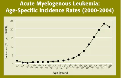
Older people are more likely to develop AML than younger adults or children.
However, almost 15 percent of acute childhood leukemias are cases of AML. The
risk for developing AML increases about 10-fold from age 30 to 34 years (about
1 case per 100,000 people) to age 65 to 69 years (about 11 cases per 100,000
people; see Figure 3).
In most cases the cause of AML is not known. Several factors have been associated with an increased risk of the disease. These include exposure to
• Very high doses of radiation, as carefully studied in the survivors of atomic bomb detonations in Japan during World War II
• The chemical benzene, above threshold levels for prolonged periods of time,
usually in an industrial setting; the stringent regulation of benzene use in the workplace has diminished the frequency of benzene as a risk factor for AML • Chemotherapy used to treat cancers such as breast cancer, ovarian cancer or
the lymphomas; the chemotherapy drug classes known as alkylating agents and
topoisomerase II inhibitors are most frequently associated with an increased risk of AML
• Therapeutic radiation, depending on the dose and duration of treatment
• Tobacco smoke.
Incidence, Causes and Risk Factors
Acute Myelogenous Leukemia:
Age-Specific Incidence Rates (2000-2004)
Figure 3. The horizontal axis shows five-year age intervals. The vertical axis shows the frequency of new cases of AML per 100,000 in a given age-group (source: Surveillance, epidemiology, and end Results [SeeR] Program; National Cancer Institute; 2007). 0 5 10 15 20 25 Incidence >85 80-84 75-79 70-74 65-69 60-64 55-59 50-54 45-49 40-44 35-39 30-34 25-29 20-24 15-19 10-14 5-9 1-4 <1 Incidence (No. per 100.000) Age (years)
AML is not contagious. Uncommon genetic disorders such as Fanconi anemia,
Shwachman-Diamond syndrome and Down syndrome are associated with an
increased risk of AML. Very rarely, an unexpectedly high number of cases of AML
may be diagnosed within the same family. It is thought that offspring in these families inherit a gene that makes them more susceptible to developing AML.
Most patients with AML feel a loss of well-being. They tire more easily and may feel short of breath in the course of normal physical activities. They may have a pale complexion from anemia. Several signs of bleeding caused by a very low platelet count may be noticed. These include black-and-blue marks or bruises occurring
for no reason or because of a minor injury, the appearance of pinhead-sized spots
under the skin, called “petechiae,” or prolonged bleeding from minor cuts. Mild fever, swollen gums, frequent minor infections such as pustules or perianal sores,
slow healing of cuts, or discomfort in bones or joints may occur. Rarely, a chloroma,
also called a “granulocytic sarcoma” or an “extramedullary myeloid tumor,” which is a collection of leukemic cells outside the marrow, occurs in patients with AML.
Blood and marrow cells are examined to diagnose AML. An accurate diagnosis is important to aid the physician in determining the most appropriate treatment. Discuss the diagnostic tests that are being done and the results, including
cytogenetic and genetic testing, with your physician. Your oncologist will work with
a hematopathologist, a physican who studies diseases of blood cells by looking at
peripheral blood smears, bone marrow aspirates and biopsies, and other tissues,
and uses his or her expertise to identify diseases such as AML and its subtypes. In addition to findings such as lower-than-expected red cell and platelet counts, a peripheral blood smear, an examination of the stained (dyed) blood cells with a microscope, usually shows the presence of leukemic blast cells. The diagnosis is
confirmed by bone marrow aspiration and biopsy, an examination of the marrow that
also shows leukemic blast cells. The blood and/or marrow cells are also used for • Studies of the number and size of chromosomes (cytogenetic analysis) • Genetic testing (which may include polymerase chain reaction [PCR])
• Immunophenotyping, a process for identifying cells based on the types of markers
Signs and Symptoms
These tests are important in determining the patient’s AML subtype. Some of the tests may be repeated during and after therapy to measure the effects of treatment.
The subclassification of AML provides important information about the expected
course of the disease. Cytogenetic analysis, a microscopic examination of a sample
of blood or marrow cells to look for changes in the chromosomes, provides
information that helps in predicting the expected outcome of both induction and postremission therapy. The cell immunophenotype and the age and general health of the patient are also important in predicting expected outcomes of treatment. Physicians are aided in identifying subtypes based on seeing different types and
patterns of cells in a patient’s blood or marrow. Most people who are diagnosed
with AML clearly have one of eight different patterns (see Table 1). Treatment is
similar for most subtypes, with the exception of acute promyelocytic leukemia (APL).
Treatment for APL (subtype M3; see Table 1) is described on page 18.
Subtypes of Acute Myelogenous
Leukemia
Table 1. Acute Myelogenous Leukemia
Cell Subtypes
Designation Cell Subtype
M0 Myeloblastic, on special analysis M1 Myeloblastic, without maturation M2 Myeloblastic, with maturation M3 Promyelocytic
M4 Myelomonocytic
M5 Monocytic
M6 erythroleukemia M7 Megakaryocytic
Myeloblasts. A myeloblast is a type of undeveloped white cell.
• If myeloblasts are the dominant leukemic cells in the marrow at the time of diagnosis,
the leukemia is referred to as a “myeloblastic” type (M0, M1 subtypes; see Table 1). • If there are many myeloblasts but some cells are developing toward fully formed blood
cells, the added designation “with maturation” is used (M2 subtype; see Table 1). • If there are cells developing features of monocytes (“monocytic” type), red cells
(“erythroleukemia” type) or platelets (“megakaryocytic” type), these designations are used (M5, M6, M7 subtypes; see Table 1).
Chromosomal Changes. Certain chromosomal changes can give important
information for patient management. For example, three chromosomal changes—
which account for between 20 and 25 percent of all cases of AML —indicate a relatively favorable prognosis, especially in younger patients. They are
• AML associated with a translocation between chromosomes 8 and 21 (t8;21),
(M2 subtype; see Table 1)
• AML associated with an inversion or translocation of chromosome 16 (t16;16)
(M4 subtype; see Table 1)
• AML associated with a translocation between chromosomes 15 and 17 (t15;17)
(M3 subtype; see Table 1). AML characterized by this translocation requires
different treatment than other types of AML (see Acute Promyelocytic Leukemia Treatment on page 18).
AML in both younger and older patients with certain leukemic cell characteristics,
including the FLT3 gene mutation, may be more difficult to treat.
Nearly all patients with AML need treatment as soon after diagnosis as possible. The principal goal of treatment is to bring about a remission, in which
• There is no evidence of leukemic blast cells in the blood or marrow, and
• Normal blood cell production is restored and blood cell counts return to normal levels.
Certain factors may help in assessing the patient’s treatment options and chances for
recovery. These include
• The patient’s age and general health
• The subtype of AML (see Table 1, page 9)
• The involvement of the central nervous system • The presence of systemic infection at diagnosis
• A history of myelodysplastic syndrome or another type of cancer • A history of AML (relapsed AML).
In most patients, intensive chemotherapy is required to achieve complete remission.
At least two drugs are combined to treat patients initially (see Chemotherapy on page 12). Variations on standard approaches to treatment are undergoing intensive
study throughout the world. Thus, a patient may receive a different number of drugs,
a different sequence of drugs, or drugs different from those described here and still be receiving appropriate and effective treatment. However, it is essential to seek treatment in a center where physicians are experienced in the care of patients with acute leukemia.
In order to prepare the patient for chemotherapy, an indwelling catheter or port
is placed surgically in a vein in the upper chest. The catheter, sometimes called a central line or a port, is tunneled under the skin of the chest so that it stays firmly
in place. The external end of the port can be used to administer medications,
fluids, or blood products or to withdraw blood samples. This is to give ready access
for the infusion of drugs or blood cells and the removal of blood samples for cell
counts and chemical tests. (Please see the Society’s free booklet Understanding
Drug Therapy and Managing Side Effects for additional information about drug administration.)
Some AML patients may build up uric acid in their blood as a result of a very high
white cell count. The use of chemotherapy may also increase uric acid, which is a
chemical in the cell. Uric acid enters the blood and is excreted in the urine. If many cells are killed simultaneously by therapy, the amount of uric acid in the urine can be so high that kidney stones can form. This may seriously interfere with the flow of urine. Drugs such as allopurinol or rasburicase can be given to minimize the buildup of uric acid in the blood.
Chemotherapy. The initial phase of chemotherapy treatment is called “induction therapy.” AML patients must be hospitalized for induction therapy, often for four to six weeks or sometimes longer.
In most cases, an anthracycline drug, such as daunorubicin, doxorubicin, or idarubicin, is combined with cytarabine (also called “cytosine arabinoside,” or
“ara-C”; see Table 2). The anthracycline and the cytarabine act in different ways
to prevent DNA synthesis in leukemia cells, stopping their growth and leading to
their death. The anthracycline is usually given in the first three days of treatment. Cytarabine is started at the same time but is given for seven to 10 days of treatment. Both drugs are dissolved in fluids and given to the patient via an indwelling catheter. The goal of induction therapy is to rid the blood and marrow of visible leukemic blast cells. If blast cells are still evident, a second course of chemotherapy may be
required to rid the marrow of blasts. Usually, the same drugs described above and
shown in Table 2 are used for each course of treatment.
When chemotherapy is effective, normal blood cells are eliminated from the marrow
along with leukemia cells. This results in a severe deficiency in the patient’s blood of • Red cells (anemia)
• Phagocytes (neutropenia and monocytopenia) • Platelets (thrombocytopenia).
Table 2. Some Drugs Used in the Treatment of and/or
in Clinical Trials for Acute Myelogenous Leukemia
Antitumor Antibiotics
These drugs interact directly with the DNA in the nucleus of cells, thus interfering with cell survival • daunorubicin (Cerubidine®) • doxorubicin (Adriamycin®) • idarubicin (Idamycin®) • mitoxantrone (Novantrone®) Antimetabolites
These chemicals are generally similar to natural building blocks of DNA, RNA or some vitamins. However, they are changed from the natural chemical. When they substitute for the DNA or RNA building blocks within a leukemic cell, the cell is unable to form normal DNA or RNA. This prevents the cell from growing.
• cytarabine (cytosine arabinoside, ara-C; Cytosar-U®)
• cladribine (2-CdA; Leustatin®)
• fludarabine (Fludara®)
• hydroxyurea (Hydrea®)
• 6-mercaptopurine (Purinethol®)
• methotrexate
• 6-thioguanine (thioguanine; Tabloid®)
• clofarabine (Clolar®)
DNA Repair Enzyme Inhibitors
These drugs act on certain proteins (enzymes) that help to repair injury to the DNA. The drugs prevent the enzymes from working and make the DNA more susceptible to injury.
• etoposide (VP-16, VePesid®, etopophos®)
• teniposide (VM-26; Vumon®)
• topotecan (Hycamtin®)
DNA Synthesis Inhibitor
This drug reacts with DNA to alter it chemically and keep it from permitting cell growth.
• carboplatin (Paraplatin®)
Cell-Maturing Agents
• all-trans retinoic acid (ATRA, tretinoin, Vesanoid®)
• arsenic trioxide (Trisenox®)
Monoclonal Antibody
• gemtuzumab ozogamicin (Mylotarg®)
Hypomethylating Agents
• azacitidine (Vidaza®)
• decitabine (Dacogen®)
Transfusion of red cells, and often platelets, may be required. During this time, the deficiency of phagocytes (microbe-eating cells) permits bacteria and fungi normally present on the skin or in the nose, mouth or large bowel (colon), or present in the environment, to cause infection. As a result, antibiotic therapy is frequently needed to treat infection. Growth factors are sometimes given to increase white cells. G-CSF
(Granulocyte-Colony Stimulating Factor or filgrastim; Neupogen®) and GM-CSF
(Granulocyte-Macrophage Colony Stimulating Factor or sargramostim; Leukine®) are
drugs that may be used in certain circumstances to increase a patient’s white cell
counts.
Normal blood cell production will return in most patients several weeks after
treatment is completed; transfusion of cells and antibiotics will no longer be needed.
Blood cell counts gradually approach normal, well-being returns and leukemia cells
cannot be identified in blood or marrow. This is called a remission. In this state,
residual leukemic cells cannot be detected. They do not interfere with normal blood cell development but have the potential to regrow and cause a relapse of the leukemia.
Radiation Therapy. Occasionally, radiation therapy may be used to treat a large,
localized accumulation of leukemia cells, called a “chloroma.” A chloroma is an uncommon occurrence in AML.
Consolidation Therapy. Consolidation therapy, also called “postremission therapy,”
is needed to rid the patient of remaining leukemia cells in order to prevent relapse. Postremission treatment of AML consists of additional intensive chemotherapy after
remission has been achieved, with or without autologous stem cell infusion or
allogeneic stem cell transplantation. There is no consensus on the best approach. Some of the main factors that influence the approach used are
• The age of the patient
• The patient’s ability to tolerate intensive treatment • Cytogenetic findings
• The availability of a stem cell donor.
Patients are hospitalized for consolidation, or postremission, therapy. The length of stay will vary depending on the treatment and other factors. If chemotherapy is to be used, the best results occur if intensive treatment is applied. Intensive chemotherapy can be given with high dosages of cytarabine or other drugs.
Some patients may not benefit from intensive chemotherapy alone but may benefit from an allogeneic stem cell transplant or an autologous stem cell infusion. These treatment options are described below. Cytogenetic test results, the availability of
an HLA-related or -unrelated matched donor and the patient’s age are some of the
factors that aid the physician in determining the appropriate postremission therapy for a given patient.
Intensive Chemotherapy and Autologous Stem Cell Infusion. Therapy can be further intensified for AML patients who do not have an HLA-matched stem cell
donor by giving very intensive chemotherapy and reinfusing the patient’s own
marrow or blood stem cells. An autologous stem cell infusion involves harvesting
the patient’s own stem cells from blood or marrow after remission is achieved,
freezing the cells for later use, then thawing them and infusing them into the patient after intensive chemotherapy. Reinfusion will restore blood cell production that would otherwise be profoundly impaired by this amount of chemotherapy. Special techniques are required to keep marrow cells from being damaged during the freezing and thawing process. (For more information about autologous stem cell infusion, see the Society’s free booklet Blood and Marrow Stem Cell Transplantation.)
Allogeneic Stem Cell Transplantation. Patients between the ages of approximately one and 55 years who are in remission and have an HLA-matched stem cell donor may be candidates for allogeneic stem cell transplantation. Allogeneic stem cell transplantation is a high-risk procedure and the decision to perform a transplant depends on the features of the patient’s leukemia, the age of the patient and the patient’s (or his or her family’s) understanding of the potential benefits and risks. For example, a younger patient with cytogenetic findings that are associated with a higher probability of relapse would be a candidate for allogeneic stem cell transplantation early in treatment. (See the Society’s free materials Blood and Marrow Stem Cell Transplantation and Cord Blood Stem Cell Transplantation for comprehensive information about allogeneic stem cell transplantation.)
See Nonmyeloablative Allogeneic Stem Cell Transplantation on page 26 for information about reduced-intensity or “mini” transplantation.
AML Treatment in Children.Acute myelogenous leukemia accounts for about 15 percent of cases of acute childhood leukemias. Most cases of acute leukemia in children are acute lymphocytic (lymphoblastic) leukemia.
Children are treated with a similar remission-induction regimen as adults with AML:
cytarabine and an anthracycline antibiotic, such as doxorubicin or daunomycin, and
often a third drug, such as mitoxantrone. This regimen is followed by a complex
multidrug program that results in about an 80 percent remission rate and a nearly 50 percent five-year, relapse-free remission rate. Slightly more than half of those children in relapse-free remission are considered cured. Infants and older children are usually treated with the same regimens.
Central nervous system (CNS) therapy may be given during the induction phase
of therapy because AML cells may collect in the lining of the spinal cord and brain,
called the “meninges.” If not treated, the meninges can harbor leukemia cells,
and relapse can occur in this site (meningeal leukemia). The treatment involves injecting drugs, such as methotrexate, into the spinal column (intrathecal therapy), or irradiating the covering of the CNS using x-rays. Sometimes both forms of treatment are used. These areas of the body that are less accessible to chemotherapy when
given by mouth or injected into the vein are sometimes referred to as “sanctuary
sites.”
Very young children with AML (less than two years of age) also have a decreased
rate of remission and cure. In addition, acute monocytic leukemia, a subtype of AML,
and a very high blast count leukemia referred to as “hyperleukocytic leukemia” are variants of AML that are much more difficult to treat, with resultant lower remission and cure rates than the average results noted above.
Certain gene abnormalities (for example,FLT3 mutations) and various chromosomal
abnormalities (such as those involving chromosome 5 or chromosome 7) are markers that suggest a poor outcome. Allogeneic stem cell transplantation may be
used in children who have a poor prognosis, who relapse after intensive multidrug
therapy or who have primary induction failure. Multi-institution clinical trials are needed to determine the best treatments for high-risk patients. When chromosomal
abnormalities occur in children with AML, their prognoses may be different from
those of adults who have the same chromosomal abnormalities. Many of the chromosomal abnormalities that occur in childhood AML are under study. Other factors also influence prognosis in children.
See the Society’s free booklet Learning & Living with Cancer: Advocating for your child’s educational needs for information about planning for the child’s entry or return to school following diagnosis and treatment.
AML Treatment in Older Adults. Acute myelogenous leukemia occurs more
frequently with advancing age. At least half of patients are more than 65 years of age when the disease is diagnosed. Today there are curative options available for older patients, including those who may have other significant health issues.
Patient age alone is a limited predictor of tolerance to chemotherapy. Standardized measures of strength, reaction time, balance, and other indicating factors, developed by experts in geriatrics, are being applied to determine the patient’s physiological age. The latter measurement rather than chronological age is a better indicator of tolerance to therapy. Such determinations may permit some older patients to receive
more intensive therapy, when appropriate and desired by the patient. However,
older patients are more difficult to treat and may have a poorer response to therapy for several reasons:
• The principal reason is that the leukemic cells of older patients with AML are more
resistant to treatment with chemotherapy. Older patients’ leukemia cells have a
much higher occurrence of unfavorable cytogenetics (chromosome abnormalities).
Mutated genes may also be more common. For example, the FLT3 gene is more
likely to be mutated in older AML patients than in younger ones. The leukemic cells of older patients more frequently overexpress drug resistance genes as
compared to cells of younger patients. Thus, the response to therapy is usually
inadequate to produce a remission or to lead to sustained remission.
• Older patients may have other medical problems, including heart, lung or kidney
disease or diabetes mellitus. The treating physician often has to select less toxic but less effective drugs or decrease the dosage and frequency of treatment to avoid further compromising the patient’s general health.
• Patients of advanced age, even in the absence of other medical disorders, tend to be more intolerant than younger patients to optimal dosages of chemotherapy. The drugs, dosages, and frequency of treatment are often individualized to take into account the features of the leukemia, the health of the patient and the patient’s anticipated tolerance of therapy.
See Research and Clinical Trials on page 24 for treatment studies of interest to older patients. Patients should discuss these treatment options with their physicians.
The treatment of the acute promyelocytic leukemia (APL) subtype of AML (M3
subtype; see Table 1 on page 9) differs from the treatment for other AML subtypes
described in the previous section. With APL, the cells that accumulate in the marrow
can be identified as promyelocytes, the step in blood cell formation that follows the development of myeloblasts. These cells also have a specific chromosome
abnormality involving chromosome 15, usually in conjunction with chromosome 17.
A derivative of vitamin A called all-trans retinoic acid, often abbreviated as ATRA,
is administered with chemotherapy. ATRA is also known as tretinoin (Vesanoid®).
Retinoic acid is capable of inducing the leukemic promyelocytes to develop into mature cells (neutrophils). It causes a marked decrease in the concentration of leukemic blast cells in the marrow, and a remission frequently follows.
Treatment with ATRA must be followed by or given with chemotherapy in order for the remission to be long-lasting. ATRA often minimizes the side effects of chemotherapy because blood cell counts may be improved and the number of leukemic cells may be decreased at the time that chemotherapy is started.
Arsenic trioxide (Trisenox®) has been approved by the Food and Drug Administration
(FDA) to treat patients who have relapsed or are resistant to treatment with chemotherapy and ATRA.
The remission rate of patients with APL treated with ATRA and an anthracycline,
such as idarubicin, is about 70 to 80 percent. Patients with this subtype of AML
are among the most frequently cured. Nevertheless, problems with hemorrhage
during the initial phases of treatment, resistance to treatment and relapse occur in a proportion of patients, as they do in some patients with other types of AML. Therefore, long-term follow-up of patients in remission is required to identify those who are cured and those who may require further therapy.
A small number of APL patients have persistent minimal residual disease (MRD) at the end of consolidation therapy. These patients may benefit from treatment with
arsenic trioxide with or without gemtuzumab ozogamicin (Mylotarg®), followed by
allogeneic stem cell transplantation, if an HLA-matched donor is available. Patients who do not have a donor or cannot have an allogeneic stem cell transplant for other reasons may be candidates for an autologous stem cell infusion.
Acute Promyelocytic Leukemia
Treatment
In some types of leukemia, including the AML subtype of monocytic leukemia (M5;
see Table 1, page 9), the leukemic blast cells may invade the lining of the spinal cord or brain. This does not usually occur with other types of AML. When the lining of the spinal cord or brain is involved, chemotherapy is injected into the spinal fluid.
A spinal tap (lumbar puncture) is a commonly used medical procedure, performed
under local anesthesia or with heavy sedation. During a spinal tap, a needle is placed
into the spinal canal and the spinal fluid is removed and examined for leukemia cells. The extracted fluid volume is then replaced with fluid containing appropriate drugs, usually cytarabine or methotrexate.
Most side effects of treatment for AML, although severe, are temporary and subside
once the body adjusts to therapy or when therapy is completed. Severe side effects
are treated on an in-patient basis. During and following the completion of therapy,
healthy new cells begin to grow and develop each day. Less commonly, a drug or
drug combination used to treat blood cancer has side effects that continue for a
period of time after treatment ends. Some effects may be permanent. (See
Long-Term and Late Effects of Treatment on page 21.) Physicians and patients should
discuss the possible side effects of treatment so that proper planning, evaluation and
follow-up can take place.
AML decreases the production of normal blood cells, and the blood cell counts
are further decreased by the added effects of chemotherapy. The intensity of chemotherapy required to destroy sufficient numbers of leukemia cells to permit
a remission leads to even more severe decreases in the numbers of red cells,
phagocytes and platelets. Severe anemia, risk of bleeding due to a low platelet count
and a high likelihood of infection result. Red cell and platelet transfusions are usually effective replacements until the beneficial effects of treatment occur several weeks later and blood cell counts return toward normal. Practical methods for transfusion of phagocytes are not currently available, except occasionally in infants and very small children. Therefore, when the white count is low and infection risk is increased,
antibiotic prophylaxis is used.
Treatment Side Effects and Their
Management
A severe or prolonged low white cell count may occur, especially after intensive drug therapy, and may increase the patient’s risk of developing an infection. Medical staff and visitors need to take precautions—such as frequent and vigorous hand washing— to avoid exposing patients to bacteria, viruses and other infection-causing agents.
They may also wear masks, gowns and gloves in some circumstances. Caregivers for
patients with ports need to be meticulous in the cleaning of the catheter to reduce the risk of bacteria infecting the body through this device.
A rise in temperature or the onset of chills may be the only sign of infection in a patient with a very low white cell concentration. In such patients, other signs of infection may include persistent coughing; tenderness at a site prone to infection,
such as the area surrounding the anus or the facial sinuses; sore throat; pain on urination; or frequent loose stools.
Blood cell growth factors may be used to stimulate the production of phagocytes and can shorten the time during which the white cell count is low. The growth factors used most frequently are colony stimulating factor (G-CSF) and granulocyte-macrophage colony stimulating factor (GM-CSF). These agents are used in children only in special circumstances. The identification of pediatric AML patients most likely to benefit from treatment to prevent infection with growth factors is under study.
Chemotherapy affects tissues that normally have a high rate of cell turnover (also called “cell division” or “mitosis”). Thus, the lining of the mouth, the lining of the intestines, the skin and the hair follicles may be affected. As a result, mouth ulcers, diarrhea, and hair loss are common after chemotherapy. Hair loss from
chemotherapy is temporary, and hair growth resumes when treatment is completed.
Rashes may also occur. Treatment for these side effects can make patients more comfortable and may prevent serious problems from developing. Nausea and vomiting may also be side effects of chemotherapy. These side effects result from actions both on the intestines and on centers of the brain that, when triggered, lead
to vomiting. Fortunately, in most cases drugs can be given to prevent nausea and
vomiting. Diarrhea can be managed with treatment. Cancer-treatment-related fatigue affects many individuals. It is an important issue that can have a major impact on quality of life. (For more information see the Society’s free materials Fatigue [fact
Some patients have residual leukemic cells in their marrow even after intensive treatment. This is referred to as “refractory leukemia.” There are other patients who have a return of leukemia cells in the marrow and a decrease in normal blood cells after achieving a remission of leukemia following therapy. This situation is referred to as “relapse.”
With refractory leukemia, approaches such as drugs not used in the first course of
treatment or stem cell transplantation may be used in an effort to induce remission. In patients who relapse, the duration of the remission, the patient’s age and the cytogenetic findings in the leukemia cells influence the approach to therapy. Drugs similar to those administered initially, different drugs or stem cell transplantation may
be used to treat the leukemia. Gemtuzumab ozogamicin (Mylotarg®), a monoclonal
antibody that is coupled with a potent cell-killing agent that targets myelogenous
leukemia blast cells, has been approved for treatment of older patients who have
relapsed AML (see Table 2 on page 13). This agent is also being studied in clinical trials in combination with other drugs to treat relapsed AML. For more information on clinical trials for relapsed and refractory leukemia see page 28.
Several drugs and drug combinations that can be used to treat AML are being studied in clinical trials. Among these are clofarabine (Clolar®), either alone and with other
drugs; azacitidine (Vidaza®); several FLT3 inhibitors; farnesyl transferase inhibitors
such as tipifarnib; and the alkylating agent VNP40101M (Cloretazine®).
Treatment for individuals with AML may cause complications that persist long after treatment ends (long-term effects) or develop much later in life (late effects). Not everyone who is treated for AML will develop long-term or late effects. Various factors can influence the risk of developing long-term or late effects, including the type and duration of treatment, age at the time of treatment, gender and the patient’s overall health.
With induction therapy for AML, most patients are treated with an anthracycline, such as daunorubicin. Anthracyclines have been associated with increased risk for heart muscle injury or chronic heart failure. Heart disease may not become apparent until
Refractory Leukemia and Relapsed
Leukemia
Stem cell transplantation may be used to treat patients with AML. Stem cell transplantation has been associated with a variety of long-term or late effects,
including infertility, thyroid dysfunction, chronic fatigue, and risk for developing a
second cancer (lymphoma, melanoma of the skin, or cancer of the tongue and
salivary glands, brain, CNS, bone, soft tissue, and thyroid gland). The number of patients who develop secondary cancers is small.
Various options exist to manage long-term and late effects. It is important that patients be aware of the potential for long-term and late effects of treatment, and that some of these effects may not appear until years after treatment ends.
For more information on long-term and late effects, see the Society’s free fact sheets
Long-Term and Late Effects of Treatment for Childhood Leukemia or Lymphoma
and Lymphoma and Long-Term and Late Effects of Treatment in Adults.
Patients who are in remission continue to be examined regularly by their physicians.
After the induction of remission and the completion of postremission therapy,
careful periodic assessment of the patient’s state of health, blood cell counts and, if
necessary, marrow is required. As time progresses, the interval between assessments
may be lengthened, but assessments should continue indefinitely.
Children and young adults who have been treated for AML may be at increased
risk for heart damage, other cancers and neurologic or cognitive problems. Patients
should be seen by a primary care physician for general health examinations at least once a year. They should also be examined regularly by an oncologist.
Sensitive molecular techniques permit the identification of small amounts of cells (minimal residual disease [MRD]) that cannot be detected by standard tests of the
patient’s blood and marrow. This approach can be used if the leukemia cells have a
detectable molecular abnormality. This feature can permit more sensitive follow-up of patients who are in remission and can help determine whether additional treatment is necessary. It is worth noting that, after treatment, a finding that 1 to 5 percent of the white cells in a patient’s marrow are blast cells is not an indication of MRD. This percentage of blast cells may be found in persons who do not have leukemia.
Follow-up Care
Patients with AML have a difficult disease to cure. However, a few decades ago
almost no adults with AML were cured. Today, advances in AML treatment have
resulted in improved remission and cure rates.
Age is the most important determinant of cure rate. Children with the disease have a cure rate just below 50 percent. Younger patients with certain cytogenetic patterns and with certain subtypes, such as APL, have a greater possibility of cure. The application of allogeneic stem cell transplantation can also cure some patients. Relative survival compares the survival rate of a person diagnosed with a disease to that of a person without the disease. The relative survival rates for AML differ by age of the patient at diagnosis, as well as by gender, race and subtype of AML. Patients diagnosed with AML before age 65 have an overall five-year relative survival rate of 34.9 percent. Children under 15 years of age have an overall five-year survival rate of 54.1 percent. Patients diagnosed at age 65 and older have an overall five-year survival rate of 4.1 percent. Table 3 shows additional five-year relative survival by age
data. Note that these numbers do not take into account significant individual factors,
such as the patient’s cytogenetics.
Outcomes
Table 3. Acute Myelogenous Leukemia:
Five-Year Survival Rates (1996-2003)
Source: Surveillance, epidemiology, and end Results [SeeR] Program, 1996-2003; National Cancer Institute, 2007.
The proportion of patients with AML who enter remission, stay in remission for years or are cured has increased during the last 30 years. Research in several areas has contributed to this progress. In children from birth to 14 years, the expectation of a cure is now just below 50 percent; with each decade of life, the probability of cure
decreases. However, AML is still one of the most difficult cancers of the blood and
marrow to treat. The challenge remains to develop treatment programs that cure all younger and older patients.
Society Research Program. The Leukemia & Lymphoma Society invests research funds in both basic and applied research programs to improve the cure rate for AML patients. Society-supported research under way includes studies to
• Identify new drug targets
• Find methods to overcome drug resistance
• Explore ways to attack leukemic stem cells thought to give rise to and sustain the disease
• Develop new immune therapies, such as vaccines
• Improve techniques of stem cell transplantation.
The following strategies, along with other new approaches, hold the promise of
increasing the rate of remission and cure of patients with AML.
Clinical Trials. New approaches to therapy are under study in clinical trials. These trials, conducted under rigorous guidelines, help physicians to determine the beneficial effects of new treatments and what, if any, adverse effects they have.
New drugs, new combinations of drugs, new types of immunotherapy, and new
approaches to stem cell transplantation are being explored to bring new and better treatments to patients. Clinical trials are conducted at many hospitals throughout the United States and also at hospitals worldwide.
The Society’s Information Resource Center, at (800) 955-4572, offers guidance to help patients work with their physicians to find out if a specific clinical trial is an appropriate treatment option. Information specialists will conduct individualized clinical trial searches for patients, family members and healthcare professionals. This service is also available on the Society’s Web site, at www.LLS.org. Information about clinical trials that is maintained by the U.S. National Institutes of Health can be
Research and Clinical Trials
Research Approaches. To find better treatments, researchers are studying the • Causes of AML
• Cell changes that make AML cells resistant to treatment
• Criteria for identifying disease subtypes, such as chromosome abnormalities
• Approaches that will permit patients to get the least toxic therapies without compromising treatment goals
• Better ways to manage side effects of therapy.
Oncogenes and Mutations. Understanding the ways that certain DNA changes cause a normal cell to become an AML cell may lead to the development of new therapies. These therapies would work by blocking instructions from
cancer-causing genes (called “oncogenes”). A new therapy might target the oncogene.
For example, a new drug might block the making of a protein that carries
cancer-causing instructions, or it might block the cancer-causing instructions from being executed. Most likely, in order for AML to arise, several interacting gene mutations are necessary. This means that any of the interacting gene mutations may also be
therapeutic targets. FLT3 inhibitors are an example of a new class of drugs under
study to target mutations of the gene FLT3, which is found in the AML cells of
about 30 percent of patients. Several FLT3-inhibitor drugs are being studied in AML
treatment.
Overcoming Drug Resistance. Among patients with AML, a subset has leukemia cells that are resistant to drug therapy; current treatment options may not cure these
patients’ disease or bring them into remission. Research is uncovering mechanisms
in the leukemia cell that protect it from the effects of chemotherapy, and ways to
reverse drug resistance are being studied.
Immunotherapy and Cytokines. Extensive testing is being conducted to synthesize new drugs or find them from natural (botanical) sources. Researchers are also investigating new combinations of existing drugs for their usefulness in AML treatment (see examples of specific agents under study in Table 4 on page 27).
Research is being conducted to develop approaches that may enhance the body’s
• An antibody that targets specific AML cells and carries a potent cell toxin,
gemtuzumab ozogamicin (Mylotarg®), has been approved for use by the Food
and Drug Administration (FDA) in patients in first relapse who are 60 years of age or older and who are not considered candidates for cytotoxic chemotherapy. Mylotarg® specifically targets leukemic cells that have the CD33 receptor.
The safety and efficacy of Mylotarg® are now being studied in other patient
populations, including children.
• Antibodies that target leukemic cells and carry a radioactive element, such as
isotopes of iodine or yttrium (radioimmunotherapy), have been developed and
are being tested.
• Another approach uses vaccines made of immune cells that have been primed to attack leukemia cells.
• Cytokines are naturally occurring chemicals that can be made commercially using the techniques of biotechnology. Some cytokines are used to help restore normal blood cells during treatment. Others are being studied for their effectiveness in enhancing the immune system to better attack leukemic cells.
Nonmyeloablative Allogeneic Stem Cell Transplantation. This type of stem cell transplantation therapy may be useful for older patients with AML. The conditioning therapy used for a nonmyeloablative transplant (also called a “mini” transplant or
“reduced-intensity” transplant) is of much lower intensity than a standard stem cell transplant; it does not completely inactivate the patient’s immune system or treat the AML as intensively. A nonmyeloablative transplant is based on two considerations: 1) Much-improved immunosuppressive therapy prevents the patient from rejecting
the donor’s stem cells, even though the patient’s immune system has not been
fully suppressed by the lower intensity conditioning therapy, and 2) the anticipated
attack of the donor’s immune cells successfully suppresses the leukemia cells of the
patient. This attack is referred to as “graft versus leukemia” or “GVL.” Over time, if successful, the donor’s stem cells result in the replacement of the patient’s immune system. The donor’s immune cells, which are now engrafted into the patient,
recognize minor tissue antigens on the patient’s leukemia cells and continue to
suppress their growth.
Nonmyeloablative transplantation is relatively new, and its risks and benefits have not yet been clearly established. It has benefited some patients. Thus, in patients
with a matched-related donor, it may be an appropriate option for carefully
selected older individuals. As is the case with allogeneic stem cell transplantation in middle-aged individuals, the risk of graft versus host disease (GVHD) is an
important consideration and a potentially disabling side effect of nonmyeloablative stem cell transplantation. A patient who is interested in exploring the option of nonmyeloablative transplantation should talk with his or her physician. If appropriate,
the physician can help the patient locate a transplant center that is investigating the procedure through a clinical trial.
Cord Blood Stem Cell Transplantation. Umbilical cord blood, like bone marrow and peripheral blood, is a rich source of stem cells for transplantation. A cord blood stem cell transplant may be a consideration for a patient who could benefit from allogeneic stem cell transplantation but does not have a related or unrelated
HLA-matched donor. Research studies of cord blood transplantation outcomes,
including transplants with two or more cord blood units, show promising results.
This topic is presented in detail in the Society’s free fact sheet Cord Blood Stem Cell Transplantation.
Table 4 describes some of the drugs under study for AML treatment.
Table 4. Some Drugs Under Study
for AML Treatment
• Farnesyl transferase inhibitors (tipifarnib [Zarnestra®], lonafarnib
[Sarasar®])
• FLT3 inhibitors (CeP-701, lestaurtinib for children; sorafenib [Nexavar®] in people 60 years of age and older)
• Proteasome inhibitor (bortezomib [Velcade®]) )
• Multidrug resistance modulators (cyclosporine A, PSC-833
[valspodar])
• Antisense molecules (G3139, oblimersen sodium, [Genasense®],
GTI-2040)
• Hypomethylating agents (azacitidine [Vidaza®], decitabine
[Dacogen®])
• Histone deacetylase inhibitors (depsipeptide)
• Histamine dichloride (Ceplene®) and IL-2*
• Alkylating agents (VNP40101M [Cloretazine®])
• Monoclonal antibodies (gemtuzumab ozogamicin [Mylotarg®])
• Immunosuppressive agents_mTOR inhibitors (sirolimus, rapamycin
[Rapamune®], tacrolimus [Prograf®])
*These drugs are not used as first-line treatment for AML. They are being studied for maintenance of remission after induction therapy.
Examples of specific agents under study in clinical trials for adults with AML include:
• Gemtuzumab ozogamicin (Mylotarg®), an anti-CD33 antibody, in combination
with other drugs such as daunorubicin and cytarabine, is being studied to find out
if it is better than standard treatment for adult patients with newly diagnosed AML. • FLT3 inhibitors, such as CEP-701, sorafenib, and others are being studied in
older, newly diagnosed AML patients, along with standard primary therapy to
see if FLT3 inhibitors produce better outcomes than chemotherapy alone. FLT3
inhibitors are also being studied in patients with relapsed or refractory AML. • Farnesyl transferase inhibitors, such as tipifarnib (Zarnestra®), are being
studied in older patients in second or subsequent remission to determine how well the drug can keep the AML in remission and to see what side effects the drug may cause. Tipifarnib is also being studied for older adults in combination
with bortezomib (Velcade®).
• Clofarabine (Clolar®) is being studied as a single agent in newly diagnosed older
adults with AML for whom standard induction chemotherapy is unlikely to be of benefit. The drug is also being studied in older adults with relapsed or refractory AML in combination with intermediate-dose cytarabine.
Examples of specific therapies under study in clinical trials for adults with APL include:
• ATRA and arsenic trioxide used together are being studied in APL patients who
have a good prognosis. This approach does not include chemotherapy.
• ATRA and arsenic trioxide, combined with gemtuzumab ozogamicin
(Mylotarg®) is being studied in APL patients who have a poorer prognosis in
comparison with ATRA and an anthracycline drug.
Clinical Trials for Childhood AML. AML is one of the most challenging childhood cancers to treat. Chemotherapy has been used in different combinations and
dosages over the past several decades, leading to improved childhood AML cure
rates, but more research is needed to further improve cure rates and decrease the
side effects and long-term and late effects of chemotherapy.
Researchers have identified cell targets that appear to be the key to treatment with the new generation of chemotherapy agents. These new targeted agents are being studied in conjunction with chemotherapy to examine their impact upon cure rates and their effect on toxic complications associated with traditional chemotherapy.
Gemtuzumab ozogamicin (Mylotarg®) is currently being tested in children with
newly diagnosed AML.
Researchers are also studying how to group patients by their risk for relapsed AML.
Patients with higher risks may benefit from more intensive treatments, including
stem cell transplantation, while patients at a lower risk may benefit from less intensive treatment.
Researchers are studying risk factors and treatments for AML chemotherapy complications, especially infections, to make AML therapy safer for children.
A diagnosis of AML often brings a strong emotional response in patients, family members and friends. Denial, depression, hopelessness and fear are some of the reactions or emotions people may experience. No one response is either universal or unexpected. Most people with AML are able to cope with what at first may seem too hard to accept. This adjustment usually takes a while. However, with information and time, many people shift their focus to the therapy process ahead and the prospect of recovery.
Patients may initially want to focus on learning about their disease and its treatment. Knowing more about the disease and its treatment helps many individuals to cope. Patients and caregivers are advised to discuss the disease and its treatment, to ask questions and convey fears or concerns to the patient’s physicians, nurses, social workers and other members of the oncology team. They are available to spend time with the patient, answer questions, lend emotional support and provide referrals to other useful resources.
During and after treatment, patients may want to have friends, family members or
caregivers help them obtain and process information from the physician and other members of the oncology team. The presence of another individual may help ease the patient’s stress. This person can also help the patient ask questions and record and retain information. While it is not always possible to have this type of support,
patients can reach out in other ways—for example, local or Internet support groups
can provide a forum for discussion. Often, patients with cancer become acquainted
with one another, and these friendships provide support. Over time, some patients
form supportive relationships with members of their healthcare team.
Social and Emotional Effects
Treatment for AML will mean changes in daily life, at least for a time. Hospitalizations,
disease and treatment side effects, and concerns about survival, finances, work or family life may cause a person to question his or her self-worth or identity. These issues may affect relationships, including intimate relationships. Recognition that these feelings are normal and knowing that many side effects are temporary may
be reassuring. Open, honest communication regarding fears and concerns can be
very helpful.
Finances. Cancer treatment can be financially difficult for many families due to loss
of income and the high cost of many medications and procedures. The Society’s
Patient Financial Aid Program offers financial reimbursement for some medications,
transportation and procedures for those in need. The Society’s Co-Pay Assistance
Program offers patients assistance with private health insurance premiums,
private insurance co-pay obligations, Medicare Part B, Medicare Plan D, Medicare Supplementary Health Insurance and Medicare Advantage premium or co-pay obligations. Prescription drugs covered under this program include those supplied to the patient by a pharmacy or administered in an office or hospital by a healthcare provider. Public or private prescription drug coverage is required to qualify for this program.
Depression. It is important to seek medical advice if a patient’s mood does not
improve over time—for example, if a patient is feeling depressed every day for a
two-week period. Depression is an illness that should be treated even when a person is undergoing treatment for AML. Treatment for depression has proven benefits for people living with cancer. There are many sources of help available to patients and
caregivers. Aspects of care such as making treatment choices, finding the time and
money for medical care and communicating with family members and friends can be stressful. Contact the Society or ask the healthcare team for guidance and referrals
to other sources of help such as support groups, counseling services or community
programs. The National Institute of Mental Health (NIMH) has several publications about depression that may be helpful. For more information go to www.nimh.nih.gov
and enter “depression” in the search box at the top of the Web page or call NIMH at
(866) 615-6464.
Children’s Concerns. Children with AML may face long periods of treatment,
including hospitalizations. However, many can expect to enter or return to school,
attend college, enter the workforce, marry and become parents. Still, each family
living with a childhood AML diagnosis is thrown into an unfamiliar world. The child,
to ask for assistance for your child, yourself or other family members, even if you are already working with a psychologist, social worker or child life specialist. Many families will benefit from extra support.
A child with AML is usually admitted to the hospital as soon as the diagnosis is known. For some children this is the first time they have stayed away from home for an extended period of time. Providing age-appropriate information to your child about the illness and treatment will help him or her build trust in both you and the treatment team and feel comfortable talking about fears and concerns. For practical
guidance on how to support your child and other family members, deal with your
own concerns, share the news with extended family and friends and make the
transition to life after treatment ends, see the Society’s free booklet Coping With Childhood Leukemia and Lymphoma.
We Can Help. The Leukemia & Lymphoma Society also offers financial assistance and support programs through its national office and local chapters to help ease the economic and emotional pressure and that comes with a cancer diagnosis. Visit the Society’s Web site at www.LLS.org or contact the Society’s Information Resource
Center at (800) 955-4572 to locate a chapter in your area, order free publications
or speak directly to an Information Specialist.
For more information, see the Society’s free booklets.
Acute Myelogenous Leukemia: A Guide for Patients and Caregivers; 2007 (in press).
Coping: Support for People Living with Leukemia, Lymphoma or Myeloma; 2007 (in press).
Coping With Childhood Leukemia and Lymphoma; 2007.
Each New Day: Ideas for Coping with Leukemia, Lymphoma or Myeloma; 2006.
Learning & Living with Cancer: Advocating for your child’s educational needs; 2006.
Touching Lives: A Directory of Patient Services Programs; 2007 (in press). For children:
Pictures of My Journey: Activities for kids with cancer; 2007.
Absolute Neutrophil Count (ANC)
The number of neutrophils (a type of white cell) that a person has to fight infection. It is calculated by multiplying the total number of white blood cells by the percentage of neutrophils.
Allogeneic Stem Cell Transplantation
A treatment that uses donor stem cells to restore a patient’s marrow and blood cells.
First, the patient is given “conditioning therapy” (dose chemotherapy or
high-dose chemotherapy with total body radiation) to treat the leukemia and to “turn
off” the patient’s immune system so that the donor stem cells will not be rejected. A type of transplant called a “nonmyeloablative” transplant (or “mini” transplant) is
under study. It uses lower doses of conditioning therapy and may be safer, especially
for older patients. (For more information, see the Society’s free booklet Blood and Marrow Stem Cell Transplantation.)
Anemia
A decrease in the number of red cells and, therefore, the hemoglobin concentration
of the blood. This results in a decreased ability of the blood to carry oxygen. If
severe, anemia can cause a pale complexion, weakness, fatigue and shortness of
breath on exertion. Antibodies
Proteins released by plasma cells (derived from B lymphocytes) that recognize and bind to the specific foreign substances called antigens. Antibodies coat, mark for destruction or inactivate foreign particles like bacteria and viruses or harmful toxins. Antibodies can also be made in the laboratory in two ways. If one injects material from one species into another, the latter will recognize the material as foreign and make antibodies to it. These antibodies are usually polyclonal antibodies, that is,
they react to multiple targets (antigens). A laboratory technique is used to produce a specific antibody known as a monoclonal antibody. Monoclonal antibodies react to only one target (antigen) and can be used in several important ways. They can be used to identify and classify human leukemias and lymphomas or can be altered to make them useful in antibody-mediated immunotherapy.
Antigen
A foreign substance, usually a protein, that stimulates an immune response when
it is ingested, inhaled or comes into contact with the skin or mucous membranes.
Examples of antigens are bacteria, viruses, or allergens. Antigens stimulate plasma cells to produce antibodies.
Antioncogene See Tumor Suppressor Gene. Apheresis
The process of removing components of a donor’s blood and returning the
unneeded parts to the donor. The process, also called hemapheresis, uses
continuous circulation of blood from a donor through an apparatus and then back to the donor. This process makes it possible to remove desired elements from large volumes of blood. Platelets, red cells, white cells and plasma can be removed separately. For example, this technique permits the harvest of enough platelets for
transfusion from one donor (rather than six to eight separate donors). In so doing,
the recipient of the platelets is exposed to fewer donors or can be given HLA-matched platelets from a single related donor. This technique is also used to remove circulating blood stem cells, which can be frozen, stored and later used, instead of marrow stem cells, for transplantation.
Autologous Stem Cell Infusion
A technique, often referred to as “autologous stem cell transplantation,” involving 1) harvesting the patient’s stem cells from blood or marrow, 2) freezing them for later use and 3) thawing and infusing them via an indwelling catheter after the patient has been given intensive chemotherapy or radiation therapy. The blood or
marrow may be obtained from a patient with a disease of the marrow, such as acute
myelogenous leukemia, when in remission or when the marrow and blood are not
overtly abnormal (for example, in lymphoma). Technically, this procedure is not
transplantation, which implies taking tissue from one person (donor) and giving it
to another person (recipient). The purpose of this procedure is to restore blood cell production from the preserved and reinfused stem cells after intensive therapy has
severely damaged the patient’s remaining marrow. This procedure can be performed
using marrow or blood stem cells. The latter can be harvested by hemapheresis.
(For more information, see the Society’s free booklet Blood and Marrow Stem Cell