INTRODUCTION
E
ruption is a process of biological maturation, which comprises the axial movement of a tooth from the developmental position within the jaw towards the functional position in the occlusal plane. The eruption of the first and second permanent molars is especially important for the co-ordination of facial growth, and for providing sufficient occlusal support for undisturbed mastication. Eruption is a multifactorial process, whose biological mechanism remains unknown. Among the various hypotheses that have been proposed, the root growth and peri-odontal ligament theories are largely disregarded today, because eruption also occurs in the absence of root formation and PDL. In more recent studies the dental follicle theory has gained popularity. This organ is considered an essential requisite for bone resorp-tion in the erupresorp-tion path as well as for the formaresorp-tion of bone below the roots.1,2There is a close relationship between the moment of eruption of a tooth and its stage of root development. Just after emergence, three quarters of the roots of the tooth have normally been formed. In the case of lower first permanent molars and central incisors, half the roots have been formed by this time.3
The eruption of some molars may be delayed, and sometimes may not occur at all. This failure of
eruption is associated with a range of medical con-ditions.3 Nevertheless, on occasion the failure of
eruption of first and second permanent molars is not associated with any systemic conditions or genetic alterations.5
Failure of eruption of first and second permanent molars is rare;6the prevalence in the normal population
is 0.01% in the case of the first permanent molar, and 0.06% in the case of the second.7 Failure of eruption
may occur due to an impaction, primary retention, or secondary retention.
Impaction is the cessation of the eruption of a tooth caused by a clinically or radiographically detectable physical barrier in the eruption path or due to an abnormal direction of the tooth.2Lack of space in the
arches is a common factor in the etiology of impacted teeth,8especially in second permanent molars.9In the
case of upper first permanent molars impaction is usually associated with an ectopic eruption path. Other factors that cause impaction are supernumerary teeth, odontogenic tumors or cysts and idiopathic factors. If impaction occurs before emergence, the radiograph shows an abnormal orientation of the molar in its eruption path.2
Primary retention refers to the cessation of eruption before emergence, without a physical barrier in the eruption path and not due to an abnormal position.2
Primary retention is probably caused by a disturbance in the dental follicle, which fails to initiate the meta-bolic events responsible for bone resorption in the eruption path.10Radiography shows normal orientation
of the molar in its eruption path. When eruption of a permanent tooth is at least two years behind schedule, primary retention should be suspected.2
Secondary retention refers to the cessation of erup-tion of a tooth after emergence without a physical barrier in the eruption path and not due to an abnormal position.11 Ankylosis is probably the main etiological
factor2 and infraocclusion is the most reliable clinical
finding.11
Failure of eruption of first and second permanent molars
Camila Palma* /Ana Coelho* / Yndira González* / Abel Cahuana**
Failure of eruption of permanent molars is an uncommon condition with a range of possible causative factors. This retrospective study of 35 pediatric subjects assesses the influence of these factors in the prognosis. The study aims to broaden our understanding of an abnormality, which has a considerable clinical impact, and proposes a treatment protocol for the management of these patients.
J Clin Pediatr Dent 27(3): 239-246, 2003
* Master of Pediatric Dentistry, Universitat de Barcelona, Barcelona, Spain.
** Department of Pediatric Dentistry and Orthodontics, Hospital Sant Joan de Déu, Barcelona, Spain.
Send all correspondence to: Dr. Abel Cahuana Cárdenas, Hospital Sant Joan de Déu, Passeig Sant Joan de Déu, 2; 08950 Esplugues de Llobregat, Barcelona, Spain.
Voice: 0034 93 253 2117 Fax: 0034 93 203 3959
The common feature of these conditions is the failure of eruption, which impedes functional occlusion, causing alterations of neighboring and opposing teeth and eventually malocclusions. Multiple local factors are involved in the failure of eruption and influence its prognosis and treatment. Among them are lack of space in the arch, dental anatomy, inclination axis, the developmental stage of the root and the depth of the molar, although their exact roles have not been established. The age of the patient is probably a key factor in the evolution of the cases.
The aims of the present study were to:
1. Analyse the hospital’s records of children with failure of eruption of first and second permanent molars.
2. Classify the entities related to the condition and to study the influence of the following variables: age, stage of root formation, degree of non-eruption, dental inclination, posterior dento-alveolar discrepancy and root deflections of the unerupted molar.
3. Determine the influence of these variables on the evolution of the affected molar and to propose a treatment protocol for the condition.
MATERIAL AND METHODS
Patients referred to the Dental Department of the Hospital Universitari Sant Joan de Déu of Barcelona between 1995 and 2001, who presented failure of eruption of the first or second permanent molar were included.
Failure of eruption was considered as the inability of the tooth to emerge in the oral cavity, thus remaining totally covered by bone or soft tissue. Cases of secondary retention were not included in the study.
All patients underwent complete clinical and radio-graphic examinations, during which the following data were recorded (Table 1):
1. Dental anomalies
2. Associated local pathology
3. Characteristics of the unerupted molar 4. Treatment options
5. Evolution
Characteristics of the unerupted molar
The stage of root formation was classified using Nolla’s Stages of Dental Development.12 The degree of
non-eruption was measured radiographically in millimetres of bone, from the alveolar ridge to the central fossa of the unerupted molar (Table 2). The inclination axis was evaluated according to Fonseca’s Classification of Angulations of Impacted Teeth.13
Evolution
The evolution of the case was considered:
1. A success, when the molar erupted, was asympto-matic and functional in the arch.
2. A failure, when the molar did not erupt in spite of treatment, or when it was extracted because the probability of eruption was considered to be low. The SPSS 9.0 for Windows program was used for the statistical analysis and the Chi-Square Tests and Fisher’s Exact Test were used for the comparison of variables. A p value < 0.05 was considered statistically significant.
RESULTS
The study population consisted of 26 patients (19 males, 7 females, age range 7 to 17 years). The mean age (±SD)
Table 1. Record of the patient’s oral condition
1. DENTAL ANOMALIES
Macrodontia, supernumerary teeth, delay of eruption, rotations, ectopia, agenesis, microdontia
Alterations of neighboring and opposing teeth: ectopia, extrusion, impaction, inclination
2. ASSOCIATED LOCAL PATHOLOGY
Local pathology (cysts, tumors) Posterior dento-alveolar discrepancy
3. CHARACTERISTICS OF THE UNERUPTED MOLAR
Stage of root formation Degree of non eruption Inclination axis
Root anatomy (normal / dilacerated)
4. TREATMENT OPTION
Observation, exposure, luxation, extraction
5. EVOLUTION
Success, failure
Table 2. Characteristics of the unerupted molar
Stage of root formation
A Crown completed, no root formation B One third of root completed C Two thirds of root completed D Root almost completed, open apex E Apical end of root completed
Degree of non eruption
0 0-2 mm or covered by gingiva 1 2-4 mm 2 4-6 mm 3 6-8 mm 4 8-10 mm 5 More than 10 mm
was 13.3 years ± 2.7 years. Non-eruption of more than one permanent molar (excluding third molars) was present in 8 patients. There were 35 unerupted molars, of which 16 were impactions (46%) and 19 were pri-mary retentions (54%) (Table 3). Failure of eruption was most common in the second lower molars (15 cases, 42.9% of the total) (Table 4).
Other dental anomalies: an association with other dental anomalies was observed in 80% of cases, delay of eruption of other teeth being the most common (31.4%). Alterations of neighboring or opposing teeth were seen in 83% of cases; ectopic position of neigh-boring teeth (40%) and extrusion of opposing teeth (28.6%) were the most common anomalies.
Associated local pathology: dento-alveolar posterior discrepancy was recorded in 8 cases (23%) and local tumors or cysts in 4 cases (11%) (Figure 1).
Characteristics of the unerupted molar: the most common stages of root formation were D and E (80%). The most frequent degrees of non-eruption were 0 and 1 (80%). The most common inclination axis was vertical (74%), followed by mesioangular (14%) and distoangu-lar (12%). Nine modistoangu-lars (26%) had root dilacerations.
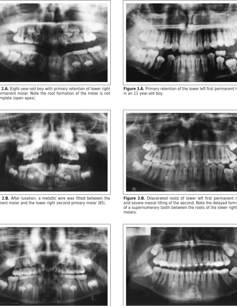
Treatment options: 26% of molars were luxated (9/35). Of these 9 luxations, 8 evolved favorably (89%) (Figure 2) and in one the molar had to be extracted. Overall, the extraction of the unerupted molar was the most frequent treatment (68%) and the only option in the cases of poor prognosis (Figure 3). Of the four cases with associated dental pathology, one underwent an extraction and the obstacle was removed in the other three.
Evolution: evolution was considered a success in 10 cases (29%) and a failure in 25 cases (71%). The results of the analysis of the influence of each factor in the evolution of the case are shown in Table 5.
There was a statistically significant relationship between poor evolution of the unerupted molar and the following factors: age over 14, and root formation of the unerupted molar in its last stages.
Moreover, there was a significant correlation between posterior dento-alveolar discrepancy and the impaction of lower and upper second molars.
The following factors did not have a significant effect on evolution: degree of non-eruption, inclination
axis, root dilaceration and posterior dento-alveolar discrepancy.
DISCUSSION
We observed a high percentage of loss of permanent molars in cases of failure of eruption, reflecting the fact that the prognosis of this abnormality is still unfavora-ble. This may be because failure of eruption is an asymptomatic pathology, which means that it is usually
Figure 1.A. Panoramic radiograph of a 9 year-old girl showing
impaction of upper left first molar and ectopic position of upper left second molar, caused by an Ameloblastic Fibrodontoma.
Figure 1.B. Final radiograph after removal of the obstacle and
lux-ation of upper left first permanent molar.
CASE # 1
Table 3. Distribution of impacted and primarily retained cases.
Failure of eruption 1st. Molars 2nd. Molars (35 cases)
Impaction
16 cases - 46 % 5 11*
Primary retention
19 cases - 54 % 5 14
* 7/11 were associated with an ectopy of the third molar
Table 4. Distribution of unerupted molars.
Frequency of unerupted molars
Molar Nº cases %
Upper first 5 14,3%
Lower first 5 14,3%
Upper second 10 28,6%
Figure 3.A. Primary retention of the lower left first permanent molar
in an 11 year-old boy.
Figure 3.B. Dilacerated roots of lower left first permanent molar
and severe mesial tilting of the second. Note the delayed formation of a supernumerary tooth between the roots of the lower right pre-molars.
Figure 3.C. Final radiograph showing orthodontic treatment after
the extraction of the lower left first permanent molar.
CASE # 3
Figure 2.A. Eight-year-old boy with primary retention of lower right
first permanent molar. Note the root formation of the molar is not yet complete (open apex).
Figure 2.B. After luxation, a metallic wire was fitted between the
permanent molar and the lower right second primary molar (85).
Figure 2.C. Lower right permanent molar erupted completely and
is functional in the arch; the apical end of the root is completed.
a casual discovery and its diagnosis is made late.14When
this is so, repercussions on the permanent dentition may be already present. These include: malocclusions, shortening of the facial height,15 incomplete
develop-ment of the alveolar process and risk of root resorption of neighboring teeth.8
Previous studies14have discussed the failure of
erup-tion of permanent molars in adults in whom significant occlusal disturbances have already taken place. This study of an exclusively pediatric population shows that anomalies in neighboring and opposing teeth and the resulting malocclusions are frequent and start at very early ages. Therefore, the age of the patient is a key fac-tor in the prognosis of a case.
Like Valmaseda,14we found lower second molars to
be the most commonly affected, followed by upper sec-ond molars. We also found a marked predominance of this abnormality in males, as Varpio reported.16A third
of the population described had more than one unerupted molar, and indeed the delay of eruption of other teeth was a common finding. These results sug-gest the probability of a shared genetic background underlying the onset of the eruption disturbance.6,14,16,17
Most of the molars involved were in the last stages of root development, a fact which, confirms the assump-tion that root formaassump-tion is independent of the eruptive process.14 In addition, we found that when roots of the
unerupted tooth are completely formed, the chances of successful treatment decrease.9The depth of the molar
in the maxilla (degree of non-eruption), contrary to our expectations, was a less decisive factor in the evolution of the case than the stage of root formation.
The position of the molar in our series (that is, incli-nation axis) had no influence on evolution, though Wellfelt18 reported that the mesioangular inclination
was the most successfully treated.
Wellfelt maintains that ankylosis is often suspected in vertically positioned teeth.18 Nevertheless, the
radi-ographic diagnosis of ankylosis in multi-rooted teeth is very difficult because of overlapping structures,11
because labial and lingual ankylotic surfaces are not visible in the radiograph19 and because ankylosis may
be located in a minute area.20 Because of these
limita-tions, it is impossible to specify the diagnosis of this dis-order, although several orthopantomographs suggested its presence.
Root dilacerations were observed in a quarter of the molars in our series. Other studies have reported an association between root anomalies and eruptive disor-ders in permanent molars,17but the finding has not been
quantified before. Kaban5 states that the follicle of
unerupted molars near the inferior border begins to curve, resulting in “hooked” roots. In our group we observed dilacerated roots not only in the lower molars, but in upper molars too, and this anomaly was not related to a particularly deep position of the molar in the bone. We think that root deflection may be due to the local disturbance leading to failure of eruption, rather than to the depth of the molar in the maxilla. In the pre-sent study, the relationship between failure of eruption and root deflection was not statistically significant, prob-ably due to the reduced number of cases seen (9/35). Nonetheless, 8 of the 9 cases with deflected roots failed. In agreement with Kaban,5we think that the presence of
dilacerated roots determines a poor prognosis.
Another factor analyzed was posterior discrepancy. Insufficient space in the dental arches has been com-monly considered an etiological factor for impaction of lower second molars.18,21 Our population study also
showed the major influence of the lack of space in impactions of permanent second molars, both lower and upper.
Table 5. Determining factors in the evolution of unerupted molars
Determining factors in Success Failure Total Statistically evolution cases Significant
Age Group Younger
than14 10 16 26 Yes Older than 14 0 9 9 Stage of Root Fromation A, B, C 5 2 7 Yes D, E 5 23 28 Degree of non eruption 0, 1 7 21 28 No 2, 3, 4 3 4 7 Inclination axis Vertical 9 17 26 No Mesial /Distal 1 8 9 Posterior Discrepancy Yes 2 6 8 No No 8 19 27 Root Dilaceration Yes 1 8 9 No No 9 17 26
The relationship between impaction of lower second molars and ectopic third molars is a contro-versial subject. Levy22reported a case of impaction of
second molars, which was due to malposition of the tooth germs of the third molars caused by dento-alveolar disproportion. Pogrel23 suggests the
extrac-tion of third molar as a prophylactic measure when uprighting second molars. However, like Valmaseda,14
we believe that third molars cannot be considered the primary cause of impaction of a second molar because formation and eruption take place at different times. Moreover, in our view dento-alveolar disproportion is a key factor in the ectopia of the third molar, and has an indirect influence on the eruption of the second molar.
The treatment of failure of eruption of permanent molars depends on several factors, the most important being age. Treatment options include observation, extraction of the obstacle, surgical exposure, luxation and extraction of the unerupted molar.
When failure of eruption is due to an obstacle, the early removal of the barrier usually allows the molar to erupt spontaneously.24
The usual treatment in cases with a favorable prognosis was surgical exposure and luxation. Molars luxated prior to complete root formation erupted spontaneously and continued their normal root development.5
Luxation is an effective technique with minimal morbidity and good long-term prognosis.23,25The
prog-nosis is better than that of dental transplant because the tooth is not removed from its socket and the apical blood vessels are not damaged. It has even been successfully used in ankylosed permanent molars.20The
potential risks of luxation include pulpal devitalization and root fracture,20although a prophylactic endodontic
treatment of the luxated molar is not recommended.23
Some luxated cases require orthodontics.25,26 In our
study group, the four cases treated with luxation and orthodontics evolved favorably.
Extraction of the permanent molar is indicated when exposure, luxation and orthodontics treatment fail, in the presence of a pathological process,2or when
prognosis is poor because of the factors described above. If the second molar is extracted at a time when the third molar is at a low Nolla stage (5 to 8), it can erupt in a position in which it replaces the lost second molar.14 If the second permanent molar is removed at
an earlier stage, the third molar will take years to erupt, and control of the eruption of the opposing teeth must be considered.14,18
We believe that prompt diagnosis is essential in order to improve prognosis and to palliate the consequences of the failure of eruption of permanent molars. We pro-pose a treatment protocol for the management of these patients based on the type of eruptive abnormality and the age of the patient (Table 6).
CONCLUSIONS
1. In this series, the failure of eruption of permanent molars was the consequence of impactions and primary retentions.
2. Unfavorable prognosis is associated with advanced age and with molars in the last stages of root formation. 3. Root dilaceration is a major factor limiting eruption
and an indicator of poor prognosis.
4. The degree of non-eruption and the inclination axis are not key factors in prognosis.
5. Posterior dento-alveolar discrepancy is associated with impaction of second molars.
REFERENCES
1. Ten Cate AR. Oral histology: development, structure, and function. 3rd ed. St Louis, CV Mosby, pp 275 – 98, 1989. 2. Raghoebar GM, Boering G, Vissink A, Stegenga B. Eruption
disturbances of permanent molar: a review. J Oral Pathol Med 20:159-66, 1991.
3. Canut Brusola JA. Ortodoncia clínica y terapéutica. 2nd ed. Barcelona: Masson, pp 25-42, 2000.
4. Cahill DR, Marks SC. Tooth eruption: evidence for the central role of the dental follicle. J Oral Pathol 9: 189-200, 1980. 5. Kaban LB, Needleman HL, Hertzberg J. Idiopathic failure of
eruption of permanent molar teeth. Oral Surg 42: 155-63, 1976. 6. Baccetti T. Tooth anomalies associated with failure of eruption of
first and second permanent molars. Am J Orthod Dentofac Orthop 118: 608-10, 2000.
7. Grover PS, Lorton L. The incidence of unerupted permanent teeth and related clinical cases. Oral Surg Oral Med Oral Pathol 59: 420-25, 1985.
8. Shafer WG, Hine MK, Levy BM. A textbook of Oral Pathology. 4th ed. Philadelphia, WB Saunders pp 66-9, 1983.
9. Johnson JV, Quirk GP. Surgical repositioning of impacted mandibular second molar teeth. Am J Orthod Dentofac Orthop 91: 242-51, 1987.
10. Oliver RG, Richmond S, Hunter B. Submerged permanent molars: four case reports. Br Dent J 160: 128-30, 1986.
11. Raghoebar GM, Boering G, Jansen HWB, Vissink A. Secondary retention of permanent molar: a histologic study. J Oral Pathol Med 18: 427-31, 1989.
12. Nolla CM. The development of the permanent teeth. J Dent Child 27: 254-66, 1960.
13. Fonseca RJ. Oral and Maxillofacial Surgery: Volume 1. 1st ed. Philadelphia, WB Saunders, pp 257-8, 2000.
14. Valmaseda-Castellón E, De-la-Rosa-Gay C, Gay-Escoda C. Eruption disturbances of the first and second permanent molars: Results of treatment in 43 cases. Am J Orthod Dentofac Orthop 116: 651-8, 1999.
15. Proffit WR. Equilibrium theory revisited: factors influencing position of the teeth. Angle Orthod 48: 175-86, 1978.
16. Varpio M, Wellfelt B. Disturbed eruption of the lower second molar: clinical appearance, prevalence, and etiology. J Dent Child 55: 114-8, 1988.
17. Vedtofte H, Andreasen JO, Kjaer I. Arrested eruption of the permanent lower second molar. European J Orthod 21: 31-40, 1999.
18. Wellfelt B, Varpio M. Disturbed eruption of the permanent lower second molar: treatment and results. J Dent Child 55: 183-9, 1988. 19. Andersson L, Blomlöf, Lindskog S, Feiglin B, Hammarström L. Tooth ankylosis; clinical, radiographic and histological assess-ments. Int J Oral Surg 13: 423-31, 1984.
20. Geiger A.M, Bronsky MJ. Orthodontic management of ankylosed permanent posterior teeth: A clinical report of three cases. Am J Orthod Dentofac Orthop 106: 543-8, 1994.
21. Evans R. Incidence of lower second permanent molar impaction. Br J Orthod 15: 199-203, 1998.
22. Levy I, Regan D. Impaction of maxillary permanent second molars by the third molars. J Paed Dent 5: 31-4, 1989.
23. Pogrel M.A. The surgical uprighting of mandibular second molars. Am J Orthod Dentofac Orthop 108: 180-3, 1995.
24. Jacobs SG. The Surgical exposure of teeth: simplest, safest and best? Austr Orthod J 10: 5-11, 1987.
25. Owen A. Early surgical management of impacted mandibular second molars. J Clin Orthod 32: 446-50, 1998.
26. Dibiase A.T, Leggat TG. Primary failure of eruption in the permanent dentition of siblings. Int J Paed Dent 10: 153-7, 2000.