Spontaneous Thoracic Spinal Subarachnoid Hemorrhage 139 Tohoku J. Exp. Med., 2013, 231, 139-144
139
Received July 23, 2013; revised and accepted September 26, 2013. Published online October 17, 2013; doi: 10.1620/tjem.231.139. Correspondence: Tatsuro Sasaji, Department of Orthopedic Surgery, Iwate Prefectural Central Hospital, 1-4-1, Ueda, Morioka, Iwate 020-0066, Japan.
e-mail: [email protected]
Spontaneous Thoracic Spinal Subarachnoid Hemorrhage
Diagnosed with Brain Computed Tomography
Tatsuro Sasaji,
1Kiyotsugu Shinagawa
1and Shigetsune Matsuya
1 1Department of Orthopedic Surgery, Iwate Prefectural Central Hospital, Morioka, Iwate, JapanSpontaneous thoracic spinal subarachnoid hemorrhage is rare, and thus no useful radiological findings for preoperative diagnosis have been reported. We experienced a patient with spontaneous thoracic spinal subarachnoid hemorrhage. A 37-year-old female presented with sudden-onset paraplegia and numbness in the trunk and bilateral lower extremities. The patient had no past history of trauma, lumbar puncture and bleeding disorder. T2-weighted sagittal magnetic resonance imaging (MRI) of the cervical and thoracic spines showed a mass occupied in the ventral space of spinal cord that was dorsally shifted. The mass extended from C6 to Th6 levels, with its largest size at Th2 level. Thoracic spine T2-weighted sagittal and axial MRI showed that the mass compressed spinal cord and was located in the intradural space. There was no spinal cord tumor and no spinal vascular malformation around the mass. Brain computed tomography (CT) showed a high-density area in the subarachnoid space, indicating the possibility of subarachnoid hemorrhage. Brain MRI showed no ruptured aneurysm. The patient was diagnosed as a spontaneous thoracic spinal subarachnoid hemorrhage and emergency surgery was selected. We performed right-side hemilaminectomy at Th1-Th6 and opened dura mater and arachnoid membrane. Hematoma was found in the ventral space of spinal cord and was removed. One year after surgery, numbness in the trunk and bilateral lower extremities had disappeared but paraplegia remained unchanged. Thoracic spine T2-weighted MRI confirmed no hematoma but showed a newly formed intradural cyst. Preoperative combination of brain CT and thoracic MRI is useful to diagnose thoracic spinal subarachnoid hemorrhage.
Keywords: paraplegia; spinal canal; spinal cord; subarachnoid hemorrhage; thoracic vertebrae Tohoku J. Exp. Med., 2013 October, 231(2), 139-144. © 2013 Tohoku University Medical Press
Introduction
Intraspinal hematoma causing neurological symptom is an emergency disease and sometimes encountered. In case of intraspinal hematoma, early diagnosis is necessary. Intraspinal hematoma is divided into extradural hematoma, subdural hematoma, subarachnoid hemorrhage and hemato-myelia, depending on the location (Hoshimaru 2005). Spinal extradural hematoma is the most frequent entity of spinal bleeding (Sarubbo et al. 2009). However, spinal sub-arachnoid hemorrhage is rare.
For useful radiological findings of spinal subarachnoid hemorrhage, Domenicucci et al. (2005) reported one patient whose hematoma was surrounded by cerebrospinal fluid. Their patient underwent a conservative treatment, and thus the diagnosis was not confirmed as subarachnoid hemor-rhage. To the best of our knowledge, there have been no studies for specific radiological findings of spinal subarach-noid hemorrhage. Accordingly, preoperative diagnosis of spinal subarachnoid hemorrhage is difficult.
We experienced a patient with spontaneous thoracic spinal subarachnoid hemorrhage and present specific radio-logical findings and the treatment course. The patient pro-vided consent for her data to be published.
Clinical Report
A 37-year-old female presented to our hospital with sudden-onset back pain, numbness in the trunk and bilateral lower extremities, and paraplegia. There was no history of trauma, lumbar puncture, blood disorders, or anticoagulant therapy. The patient complained of headache for several months ago; however, no neurological symptoms or back pain were observed. On brain magnetic resonance images (MRI), there were no tumor and hematoma. And so, to investigate spinal cord, cervical and thoracic spine MRI was obtained at another department one month before the onset of paraplegia. On cervical and thoracic spine T2-weighted MRI, there were no spinal tumor and no ves-sel malformation (Fig. 1A and B).
T. Sasaji et al. 140
Neurological Examination
On neurological examination at the initial visit to our department, muscle power was zero in bilateral lower extremities and the patellar tendon, Achilles tendon, and bulbocavernous reflexes could not be elicited. Sensory dis-turbance was detected in the trunk and bilateral lower extremities. The patient had no upper extremity complaints and was diagnosed as having a thoracic spinal cord injury. The patient was considered at Grade A (Complete) of the Frankel scale (Frankel et al. 1969). All laboratory findings were within normal limits.
Radiological Findings
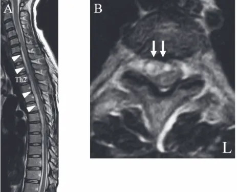
Preoperative cervical and thoracic spine T2-weighted sagittal MRI showed that the mass was located in the dura mater and extended from C6 to Th6, and the spinal cord had been dorsally shifted (Fig. 2A). Preoperative T2-weighted axial MRI showed a mass of high-intensity signal, which was located in the right ventral space of the spinal cord, and this mass maximally compressed spinal cord at Th2 level (Fig. 2B). The dura mater was deformed into an oval shape from the inner side by the intradural mass. The reported signs of intradural-extra-arachnoid hematoma on MRI included a “black line,” which is a line in the dura mater on T2-weighted axial image, but it could not be observed in the present case (Shimada et al. 1996). Brain computed tomography (CT) showed a high-density area in the subarachnoid space that appeared to be a sub-arachnoid hemorrhage (Fig. 3). Brain MRI showed no
rup-tured aneurysm (Fig. 4). On the basis of the history of no bleeding tendency and radiological findings of no tumor or no vessel malformation, the patient was diagnosed with spontaneous thoracic spinal subarachnoid hemorrhage. Therefore, we performed the emergency surgery.
Operation
The right-side thoracic spine was explored through a straight posterior midline approach. Right-side hemilami-nectomy at Th1-Th6 revealed no hematoma in the extradu-ral space. The dura mater and arachnoid membrane were exposed and the spinal cord was centrally retracted reveal to a hematoma in the ventral space of the spinal cord at Th1-Th6. We scraped out and removed the hematoma (Fig. 5A). After removal of the hematoma, the spinal cord was decompressed (Fig. 5B). The bleeding point was unclear, but bleeding spontaneously stopped.
Postoperative Course
Numbness in the patient’s trunk and lower extremities disappeared after surgery. One year later, the patient had no complaints of numbness. However, paraplegia and neuro-genic bladder did not resolve. The patient was considered at Grade B (Sensory only) of the Frankel scale (Frankel et al. 1969). The patient has returned to a daily life and uses a wheel chair.
Follow-up cervical and thoracic spine T2-weighted MRI on postoperative day 10 showed no indications of the hematoma, and the spinal cord was decompressed (Fig. 6A Fig. 1. Magnetic resonance images of the cervical and thoracic spine (one month before onset of symptom).
A: Sagittal plane on T2-weighted image. B: Axial plane at Th2 level on T2-weighted image.
Sagittal plane (A) and axial plane at the Th2-Th3 level (B) T2-weighted images showing no spinal tumor and no vessel malformation.
Spontaneous Thoracic Spinal Subarachnoid Hemorrhage 141
and B). Follow-up thoracic spine T2-weighted MRI 1 year after surgery showed that a new intradural cyst in the ven-tral left side had appeared and was compressing the spinal
cord (Fig. 7A and B). However, because the patient’s symptoms had not worsened, we thought that the cyst had not evoked any new spinal cord symptoms. We did not per-form revision surgery, but we continued the patient follow-up.
Discussion
Spinal subarachnoid hemorrhage accounts for < 1% of all cases of subarachnoid hemorrhage (Walton 1953). In Fig. 2. Preoperative magnetic resonance images of the cervical and thoracic spine.
A: Sagittal plane on T2-weighted image. B: Axial plane at Th2 level on T2-weighted image.
A: Sagittal plane on T2-weighted image showing a mass in the right ventral space of the spinal cord. The mass extend-ed from C6 to Th6 level (arrow heads). Compression of the spinal cord was maximal at Th2 level. B: Axial plane at the Th2 -Th3 level on T2-weighted image showing that the spinal cord was compressed and deformed by the intradural mass (arrows).
Fig. 3. Preoperative computed tomographic image of the brain.
There was a high-density area in the subarachnoid space (arrows).
Fig. 4. Preoperative magnetic resonance image of the brain. There was no ruptured aneurysm.
T. Sasaji et al. 142
addition, according to a review of spinal subarachnoid hem-orrhage, 12 of the 69 cases (17.3%) were spontaneous cases (Domenicucci et al. 2005). The rate of secondary cases was
higher than that of spontaneous cases. The reported causes were coagulopathy, hematological pathology, alcoholic hepatitis, lumbar puncture, spinal trauma, previous spinal Fig. 5. Intraoperative photographs.
A: Opening of the dura mater and arachnoid membrane. B: After removal of the hematoma.
A: Opening of the dura mater and arachnoid membrane revealed that the hematoma surrounded the spinal cord. B: The spinal cord was decompressed after removal of the hematoma.
Fig. 6. Postoperative magnetic resonance images of the cervical and thoracic spine (10 days after surgery). A: Sagittal plane on T2-weighted image. B: Axial plane at Th2 level on T2-weighted image.
Sagittal plane (A) and axial plane at the Th2-Th3 level (B) T2-weighted images showing decompressed spinal cord and intraspinal high-intensity area.
Spontaneous Thoracic Spinal Subarachnoid Hemorrhage 143
surgery and arteriovenous malformation (Russell and Benoit 1983; Domenicucci et al. 2005). About spontaneous spinal subarachnoid hemorrhage, Plotkin et al. firstly reported in 1966 and there have been some subsequent reports (Plotkin et al. 1966; Owaki et al. 1975; Russell and Benoit 1983; Swann et al. 1984; Hiyama et al. 1990; Langmayr et al. 1995; Sunada et al. 1995; Komiyama et al. 1997; Ruelle et al. 2001; Domenicucci et al. 2005; Kim and Lee 2009). Spontaneous spinal subarachnoid hemorrhage is a rare disease.
The reported preoperative diagnosis of spinal sub-arachnoid hemorrhage was unclear by myelography (Plotkin et al. 1966), intraspinal tumor by myelography (Owaki et al. 1975), subarachnoid hematoma by lumbar puncture (Swann et al. 1984), intradural hematoma by MRI (Hiyama et al. 1990), and spinal hematoma by MRI (Sunada et al. 1995). Swann et al. (1984) reported that xanthochro-mic cerebrospinal fluid was useful for diagnosis. Except in one study (Swann et al. 1984), the diagnosis was intraoper-atively confirmed. Preoperative diagnosis is difficult because there have been no specific radiological findings regarding spinal subarachnoid hemorrhage. Domenicucci et al. (2005) reported that MRI and CT are not usually diag-nostic. In the present patient, the hematoma was located in the dura mater based on MRI. However, we could not determine whether hematoma was located in the intradural-extra-arachnoid space or subarachnoid space. Reported specific radiological findings of intradural-extra-arachnoid hematoma have included the “black line” (Shimada et al.
1996). In the present patient, a “black line” was not observed on MRI. We thought that the hematoma may have been located in the subarachnoid space. However, the extra-arachnoid and subarachnoid spaces were so close that we could not confirm the diagnosis only by the MRI.
There have been a few reports regarding radiological findings of brain CT in spinal subarachnoid hemorrhage case (Hiyama et al. 1990; Ruelle et al. 2001; Kim and Lee 2009). Subarachnoid hemorrhage on brain CT could not be found in cases of lumbar spine subarachnoid hemorrhage (Ruelle et al. 2001; Kim and Lee 2009). It was identified in case of thoracic spine subarachnoid hemorrhage (Hiyama et al. 1990). However, the authors did not describe the inter-pretation of brain CT. In the present case, the radiological findings of subarachnoid hemorrhage on brain CT were useful for diagnosis. Because the upper thoracic spine was so close to the brain, blood spread from the upper thoracic spine to the brain through the subarachnoid space. To the best of our knowledge, this is the first report to describe the usefulness of brain CT for spinal subarachnoid hemorrhage, and we believe that brain CT may be useful for diagnosis in patients with upper thoracic subarachnoid hemorrhage.
The reported treatments for spinal subarachnoid hem-orrhage included surgery, needle aspiration, and conserva-tive treatment (Plotkin et al. 1966; Owaki et al. 1975; Russell and Benoit 1983; Swann et al. 1984; Hiyama et al. 1990; Langmayr et al. 1995; Sunada et al. 1995; Komiyama et al. 1997; Ruelle et al. 2001; Domenicucci et al. 2005; Kim and Lee 2009). In paralysis cases, surgery has been Fig. 7. Postoperative magnetic resonance images of the thoracic spine (1 year after surgery).
A: Sagittal plane on T2-weighted image. B: Axial plane at Th2 level on T2-weighted image.
Sagittal plane (A) and axial plane at the Th2-Th3 level (B) T2-weighted images showing newly formed intradural cyst (arrow heads) in the left ventral space and the spreading intraspinal high-intensity area (arrows).
T. Sasaji et al. 144
performed (Owaki et al. 1975; Russell and Benoit 1983; Hiyama et al. 1990; Langmayr et al. 1995; Domenicucci et al. 2005). Prognosis has been influenced by preoperative neurological status, duration between the onset of symp-toms and operation, and rapidity of symptom progression (Sunada et al. 1995). We thought that the poor preoperative neurological function (Frankel A) and acute-onset paraple-gia were the causes of poor neurological recovery in the present case.
No reports have described newly formed intradural cyst following spinal subarachnoid hemorrhage. In the present patient, we thought that the hematoma had damaged the arachnoid membrane and caused cyst formation.
Conflict of Interest The authors declare no conflict of interest.
References
Domenicucci, M., Ramieri, A., Paolini, S., Russo, N., Occhiogrosso, G., Di Biasi, C. & Delfini, R. (2005) Spinal subarachnoid hematomas: our experience and literature review. Acta Neuro-chirurgica (Wien), 147, 741-750.
Frankel, H.L., Hancock, D.O., Hyslop, G., Melzak, J., Michaelis, L.S., Ungar, G.H., Vernon, J.D.S. & Walsh, J.J. (1969) The value of postural reduction in the initial management of closed injuries of the spine with paraplegia and tetraplegia. Para-plegia, 7, 179-192.
Hiyama, H., Shimizu, T., Yato, S., Kobayashi, N., Ono, Y. & Kakinoki, Y. (1990) Wide-spread spontaneous spinal subarachnoid hematoma. Case report. Neurol. Med. Chir (Tokyo),30, 842-847 (in Japanese).
Hoshimaru, M. (2005) Diagnosis and treatment of spinal hemor-rhage due to lesions other than spinal AVM. Spine & Spinal
Cord, 18, 979-986 (in Japanese).
Kim, J.S. & Lee, S.H. (2009) Spontaneous spinal subarachnoid hemorrhage with spontaneous resolution. J. Korean Neuro-surg. Soc., 45, 253-255.
Komiyama, M., Yasui, T., Sumimoto, T. & Fu, Y. (1997) Sponta-neous spinal subarachnoid hematoma of unknown pathofen-esis: case reports. Neurosurgery, 41, 691-693.
Langmayr, J.J., Ortler, M., Dessl, A., Twerdy, K., Aichner, F. & Felber, S. (1995) Management of spontaneous extramedullary spinal haematomas: results in eight patients after MRI diag-nosis and surgical decompression. J. Neurol. Neurosurg. Psychiatry, 59, 442-447.
Owaki, K., Nakazawa, S., Yajima, K., Sugiura, K. & Yabe, Y. (1975) Spontaneous spinal subarachnoidal hematoma—case report. No Shinkei Geka, 3, 593-597 (in Japanese).
Plotkin, R., Ronthal, M. & Froman, C. (1966) Spontaneous subarachnoid haemorrhage. Report of 3 cases. J. Neurosur-gery, 25, 443-446.
Ruelle, A., Zerbi, D. & Andrioli, G. (2001) Spinal subarachnoid bleeding of unknown etiology. Case reports. J. Neurosurg. Sci., 45, 53-57.
Russell, N.A. & Benoit, B.G. (1983) Spinal subdural hematoma. A review. Surg. Neurol., 20, 133-137.
Sarubbo, S., Garofano, F., Maida, G., Fainardi, E., Granieri, E. & Cavallo, M.A. (2009) Spontaneous and idiopathic chronic spinal epidural hematoma: two case reports and review of the literature. Eur. Spine J., 18, 1555-1561.
Shimada, Y., Sato, K., Abe, E., Miyakoshi, N. & Tsutsumi, Y. (1996) Spinal subdural hematoma. Skeletal Radiol., 25, 477-480.
Sunada, I., Akano, Y., Kidosaki, Y., Shimokawa, N. & Yamamoto, S. (1995) Spontaneous spinal subarachnoid hematoma—case report. Surg. Neurol., 44, 133-136.
Swann, K.W., Ropper, A.H., New, P.F. & Poletti, C.E. (1984) Spontaneous spinal subarachnoid hemorrhage and subdural hematoma. Report of two cases. J. Neurosurg., 61, 975-980. Walton, J.N. (1953) Subarachnoid haemorrhage of unusual