Sodium nitroprusside enhanced cardiopulmonary resuscitation
improves survival with good neurological function in a porcine
model of prolonged cardiac arrest*
Demetris Yannopoulos, MD; Timothy Matsuura, BS; Jason Schultz, MD; Kyle Rudser, PhD;
Henry R. Halperin, MD; Keith G. Lurie, MD
T
he vast majority of patients
who suffer an out-of-hospital
cardiac arrest die despite
ceiving cardiopulmonary
re-suscitation (CPR) for 20 –30 mins by
emergency medical personnel at the
scene (1– 4). Such patients, when treated
with the standard of care as
recom-mended by the American Heart
Associa-tion guidelines, typically receive
epineph-rine every 3–5 mins (5). While the
standard of care for decades, this practice
has recently been challenged by animal
and clinical trials showing that use of
epinephrine may actually have more
det-rimental than beneficial effects (6 –9).
In stark pharmacologic contrast to
epinephrine, sodium nitroprusside (SNP)
is a potent vasodilator. SNP is used to
treat hypertensive emergencies and heart
failure and its primary mode of action is
the release of nitric oxide (10, 11). Use of
SNP would typically be considered an
anathema for the treatment of the
pro-found hypotension associated with
car-diac arrest and manual CPR.
Contrary to current thinking, large
doses of SNP during CPR do not cause a
significant decrease in central aortic
pressure (12). This can be explained by
the way aortic pressure is generated
dur-ing CPR. With chest compression, the
resultant increase in intrathoracic
pres-sure results in an instantaneous increase
in the pressure of all of the intrathoracic
vascular structures (13). Therefore, when
arterial vascular resistance is decreased
by SNP, forward blood flow can be
signif-icantly increased with each chest
com-pression. The new approach, SNPeCPR or
SNP-“enhanced” CPR (pronounced
“snappy CPR”), incorporates three
com-ponents: 1) active
compression-decom-pression CPR with the use of an
imped-ance threshold device (ACD CPR
⫹
ITD),
which by actively decreasing
intratho-racic pressure during the decompression
phase decreases right atrial and
intracra-nial pressures and improves vital organ
perfusion (14, 15); 2) a potent
vasodila-tor, SNP, which by decreasing vascular
resistance promotes forward blood flow;
and 3) lower abdominal binding (AB) that
limits descending aortic blood flow and
redirects the augmented cardiac output
to the heart and brain, where it is needed
the most. For simplicity, the combination
of ACD CPR
⫹
ITD
⫹
AB without SNP will
be called from here thereafter as eCPR.
SNPeCPR consists of easily deployable
components that can be readily applied
during CPR. ACD CPR
⫹
ITD is currently
clinically practiced. AB can be performed
easily either manually or with a belt/
pneumatic bladder/compression clamp.
*See also p. 1548.From the Departments of Medicine and Emergency Medicine (DY, TM, JS, KGL) and Biostatistics (KR), University of Minnesota, Minneapolis-St. Paul, MN; and Department of Medicine (HRH), Johns Hopkins University, Baltimore, MD.
Supported, in part, by an Institutional Division of Cardiology grant at the University of Minnesota to Dr. Yannopoulos.
Presented, in part, as preliminary data at the Re-suscitation Science Symposium in Chicago, IL, Novem-ber 14, 2010; published in abstract form: Yannopoulos D, Matsuura T, McKnite S, et al: Abstract 164: Sodium nitroprusside improves carotid blood flow and 24-hour neurological intact survival in a porcine model of pro-longed CPR.Circulation. 2010; 122:A164.
Dr. Lurie is the founder of Advance Circulatory Sys-tems Incorporated (ACSI), and coinventor of the inspira-tory impedance threshold device and ACD CPR technique used in this study. Dr. Yannopoulos received funding from the Division of Cardiology at the University of Minnesota. Dr. Lurie received a patent and holds stock ownership with ACD and ITD CPR (ACSI). The remaining authors have not disclosed any potential conflicts of interest.
For information regarding this article, E-mail: yanno001@umn.edu
Copyright © 2011 by the Society of Critical Care Medicine and Lippincott Williams & Wilkins
DOI: 10.1097/CCM.0b013e31820ed8a6
Objective:
To assess the effectiveness of sodium nitroprusside
(SNP)-“enhanced” cardiopulmonary resuscitation (SNPeCPR) on
24-hr survival rates compared to standard CPR in animals after
cardiac arrest. SNPeCPR consists of large intravenous SNP bolus
doses during CPR enhanced by active compression-decompression
CPR, an inspiratory impedance threshold device (ITD), and abdominal
binding (AB). The combination of active compression-decompression
CPR
ⴙ
ITD
ⴙ
AB
without SNP
will be called “enhanced” or eCPR.
Design:
Randomized, blinded, animal study.
Setting:
Preclinical animal laboratory.
Subjects:
Twenty-four female farm pigs (30
ⴞ
1 kg).
Interventions:
Isoflurane anesthetized and intubated pigs were
randomized after 8 mins of untreated ventricular fibrillation to
receive either standard CPR (n
ⴝ
8), SNPeCPR (n
ⴝ
8), or eCPR
(n
ⴝ
8) for 25 mins followed by defibrillation.
Measurements and Main Results:
The primary end point was
carotid blood flow during CPR and 24-hr survival with good neurologic
function defined as an overall performance category score of
<
2 (1
ⴝ
normal, 5
ⴝ
brain dead or dead). Secondary end points included
hemo-dynamics and end-tidal CO
2. SNPeCPR significantly improved carotid
blood flow and 24-hr survival rates with good neurologic function
compared to standard CPR or eCPR (six of eight vs. zero of eight vs. one
of eight,
p
<
.05). The improved survival rates were associated with
higher coronary perfusion pressure and ET
CO2during CPR.
Conclusion:
In pigs, SNPeCPR significantly improved
hemody-namics, resuscitation rates, and 24-hr survival rates with good
neurologic function after cardiac arrest when compared with
standard CPR or eCPR alone. (Crit Care Med 2011; 39:1269 –1274)
K
EYW
ORDS: vasodilators; cardiopulmonary resuscitation;
neu-rological function; resuscitation rates; carotid blood flow
SNP can be given intravenously or
in-traosseously as any other drug that is
currently delivered during CPR without
further complexity.
We hypothesize that SNPeCPR will
im-prove carotid blood flow, resuscitation
rates, and 24-hr survival rates compared to
standard CPR (S-CPR) plus epinephrine.
MATERIALS AND METHODS
The study was approved by the Institu-tional Animal Care Committee of the Minne-apolis Medical Research Foundation of Hen-nepin County Medical Center. All animal care was compliant with the National Research Council’s 1996 Guidelines for the Care and Use of Laboratory Animals. All studies were performed by a qualified, experienced research team in Yorkshire female farm pigs weighing 30⫾1.5 kg. A certified and licensed veteri-narian provided a blinded neurologic assess-ment in the 24-hr survival experiassess-ments.
Preparatory Phase
The anesthesia, surgical preparation, data monitoring, and recording procedures used in this study have been described previously by our group (16). Briefly, we employed aseptic surgical conditions, using initial sedation with intramuscular ketamine (7 mL of 100 mg/mL, Ketaset, Fort Dodge Animal Health, Fort Dodge, IA) followed by inhaled isoflurane at a dose of 0.8% to 1.2%. Pigs were intubated with a size 7.0 endotracheal tube. The animal’s temperature was maintained at 37.5⫾0.5°C, with a warming blanket (Bair Hugger, Augus-tine Medical, Eden Prairie, MN). Central aortic blood pressure was recorded continuously with a micromanometer-tipped (Mikro-Tip Transducer, Millar Instruments, Houston, TX) catheter placed at the beginning of the de-scending thoracic aorta. A second Millar cath-eter was inserted in the right atrium via the right external jugular vein. All animals re-ceived an intravenous heparin bolus (100 units/kg). A ultrasound flow probe (Transonic 420 series multichannel, Transonic Systems, Ithaca, NY) was placed to the left internal carotid to record blood flow (mL/min). The animals were then ventilated with room air, using a volume-control ventilator (Narcomed, Telford, PA), with a tidal volume of 10 mL/kg and a respiratory rate adjusted to continually maintain a PaCO2of 40 Torr and PaO2of 80 Torr (blood oxygen saturation⬎95%), as mea-sured from arterial blood (Gem 3000, Instru-mentation Laboratory, Lexington, MA) to ad-just the ventilator as needed. Surface electrocardiographic tracings were continu-ously recorded. All data were recorded with a digital recording system (Superscope II ver-sion 1.295, GW Instruments, Somerville, MA).
End-tidal CO2 (ETCO2), tidal volume, minute ventilation, and blood oxygen saturation were continuously measured with a respiratory monitor (CO2SMO Plus, Novametrix Medical Systems, Wallingford, CT). Right atrial pres-sure was adjusted between 2 and 5 mm Hg with saline infusion as needed before induc-tion of ventricular fibrillainduc-tion.
Measurements and Recording
Aortic pressure, right atrial pressure, ETCO2, and carotid blood flow were
continu-ously recorded. Coronary perfusion pressure during CPR was calculated from the mean arithmetic difference between right atrial pressure and aortic pressure during the de-compression phase. Doppler derived common carotid blood flow was reported in mL/sec.
Experimental Protocol
After the surgical preparation was com-plete, oxygen saturation on room air was
⬎95% and ETCO2was stable between 35 and 42 mm Hg for 5 mins. Ventricular fibrillation was induced by delivering direct current via a temporary pacing wire (Daig Division, St Jude Medical, Minnetonka, MN) positioned in the right ventricle. The ventilator was discon-nected from the endotracheal tube. S-CPR and ACD CPR were performed with a pneumati-cally driven automatic piston device (Pneu-matic Compression Controller, Ambu Interna-tional, Glostrup, Denmark) as previously described (17). Uninterrupted chest compres-sions at a rate of 100 comprescompres-sions/min, with a 50% duty cycle and a compression depth of 25% of the anteroposterior chest diameter, were provided. During CPR, asynchronous positive-pressure ventilations were delivered to simulate advanced life support with a man-ual resuscitator bag. The fraction of inspired
oxygen was 1.0, the tidal volume was main-tained at⬃10 mL/kg, and the respiratory rate was 10 breaths/min.
Protocol
Following 8 mins of untreated ventricular fibrillation, 24 pigs were randomized to 25 mins of S-CPR (n⫽8), ACD CPR⫹ITD⫹AB (n⫽8), or SNP (eCPR) (n⫽8). Epinephrine was administered to the S-CPR control group in 0.5-mg boluses every 5 mins, whereas SNP was delivered in the SNPeCPR group in 1-mg bolus every 5 mins (timetable is shown in Fig. 1). SNPeCPR and eCPR groups did not receive epinephrine during CPR. Defibrillation was delivered after 25 mins of CPR. Earlier defi-brillations were not attempted because the study was designed to assess the effectiveness of SNPeCPR after 25 mins of CPR compared to S-CPR (since most of the patients require sup-port for that period of time clinically). Animals that had a return of spontaneous circulation (ROSC) were then observed under general an-esthesia with isoflurane until hemodynamics were stable. Hemodynamic stability was de-fined as a mean aortic pressure of⬎55 mm Hg without epinephrine for 10 mins, carotid blood flow equal or higher than baseline, and normalization of ETCO2 and acidosis. At that point, vascular repair of the internal jugular and the left common femoral artery was then performed.
Animals that had a stable post-ROSC rhythm but were hypotensive (mean arterial pressure of ⬍50 mm Hg) received 250 –500 mL of intravenous normal saline bolus. If mean arterial pressure was still⬍50 mm Hg, they received increments of 0.1– 0.2 mg of epinephrine every 5 mins until mean arterial pressure rose above 50 mm Hg. Survivors were given intramuscular analgesic injections of nonsteroidal anti-inflammatory drugs as
Figure 1.Protocol timelines.ROSC, return of spontaneous circulation;S-CPR, standard cardiopul-monary resuscitation,ACD⫹ITD, active compression-decompression CPR with an inspiratory imped-ance threshold device;AB, abdominal binding;SNP, sodium nitroprusside;VF, ventricular fibrillation; epi., epinephrine. (The combination of ACD CPR⫹ITD⫹AB is called “enhanced” eCPR.)Electric bolt sign, DC cardioversion.
previously described (16) and had free access to water and food. There was no other post-ROSC medical care provided after the vascular repair. Animals were not treated with hypo-thermia. All surviving pigs with stable circu-lation for at least 30 mins received intrave-nous antibiotics with a third-generation cephalosporin and a repeat dose at 12 hrs. At 24 hrs, a certified veterinarian blinded to the intervention assessed the pig’s neurologic function based on an overall performance cat-egory (OPC) scoring system. The veterinarian used clinical signs such as response to open-ing the cage door, response to noxious stimuli if unresponsive, response to trying to lift the pig, whether the animal could stand, walk, eat, urinate, and defecate, and appropriate re-sponse to the presence of a person walking around the cage. The following scoring system was used: 1⫽normal; 2⫽slightly disabled; 3⫽severely disabled but conscious; 4⫽ veg-etative state; or 5⫽dead (16).
Postresuscitation care was not blinded, since the same team performed CPR and pro-vided post-ROSC care.
Echocardiographic Evaluation of Left Ven-tricular Function.After 1 hr, all survivors had a transthoracic echocardiogram to evaluate their left ventricular ejection fraction. Images were obtained from the right parasternal win-dow, which provides similar views as the long and short parasternal windows for humans (18). The ejection fraction was assessed with trace planimetry and was reported by a blinded independent echocardiographer both for base-line and 1 hr post-ROSC.
Statistical Analysis
Values were expressed as mean⫾SD.
Base-line data were compared using attest.
Hemo-dynamics and blood gases during CPR were analyzed with two-way ANOVA. A two-tailed Fisher’s exact test was used to compare ROSC rates and 24-hr survival as well as OPC pro-portions, which were dichotomized to OPC 1 and OPC 2 (good outcome) and OPC 3, 4, and 5 (bad outcome). A p value of ⬍0.05 was considered statistically significant.
RESULTS
There were no baseline differences
be-tween the three treatment groups in any
hemodynamic or respiratory parameters.
S-CPR vs. SNPeCPR or eCPR
Pigs treated with S-CPR with
epineph-rine demonstrated significantly low
aor-tic and coronary perfusion pressures
compared with SNPeCPR or eCPR. There
was profound progressive acidosis and
lower ET
CO2with progressive decrease in
carotid blood flow despite repetitive
epi-nephrine doses (Table 1 and Fig. 2).
SNPeCPR vs. eCPR
With the exception of carotid blood
flow and ET
CO2, the other measured
he-modynamic parameters did not differ
sig-nificantly between the two groups. This
was not the case for arterial blood gases:
there was a progressive metabolic
acido-sis in the eCPR group but not the
SNPeCPR group (Tables 1 and 2 and Fig.
2). It is noteworthy that carotid blood
flows after 20 and 25 mins of SNPeCPR
were similar to baseline pre-ventricular
fibrillation values.
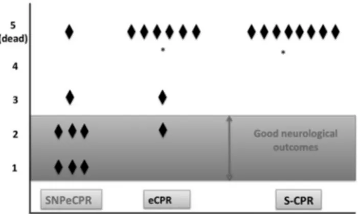
ROSC and 24-hr Survival
No animal treated with S-CPR with
epinephrine group had successful ROSC
and therefore could not be assessed for
24-hr survival. Eight of eight animals
that received SNPeCPR had a ROSC, and
seven of eight survived for both 1 and 24
hrs. A total of five of eight animals with
eCPR had an initial ROSC; two of eight
survived to 1 hr and two of eight survived
24 hrs (
p
⫽
.04 for 1-hr and 24-hr
sur-vival compared with SNPeCPR). Good
outcomes at 24 hrs (OPC 1 or 2) were
significantly better in the SNPeCPR
group than in the eCPR (six of eight vs.
one of eight) (
p
⫽
.04) (Fig. 3).
Left Ventricular Function.
There was
no baseline difference between groups in
the left ventricular ejection fraction
(SNPeCPR: 64%
⫾
7%, S-CPR: 66%
⫾
8%, eCPR: 64%
⫾
9%). An
echocardio-graphic evaluation performed 1 hr after
cardiac arrest revealed that the SNPeCPR
animals had an ejection fraction of
58%
⫾
12%. The two animals in the
eCPR group had ejection fractions of 38%
and 44%, respectively.
In addition, it was observed that six of
eight animals in the eCPR and six of eight
animals in the S-CPR group developed
severe pulmonary edema that started
af-ter 15 mins of CPR. Only one of eight
animals developed mild pulmonary
edema in the SNPeCPR group, which
self-terminated once the animals was
connected to the ventilator (six of eight
vs. one of eight,
p
⬍
.05). The one animal
that died in the SNPeCPR group
devel-oped intractable ventricular tachycardia/
fibrillation 30 mins after successful
ROSC. No antiarrhythmic drugs were
used.
Postresuscitation Care.
SNPeCPR
an-imals received an average of 0.2
⫾
0.1 mg
and 358
⫾
128 mL of epinephrine and
saline, respectively. The two survivors in
the eCPR group received 0.4 and 0.3 mg
of epinephrine and 500 mL of saline,
respectively.
DISCUSSION
Results from this first series of
exper-iments with SNPeCPR demonstrate the
feasibility of using SNP in the treatment
of cardiac arrest to enhance brain flow
and increase 24-hr survival with favorable
neurologic outcomes. When SNP was
Table 1.Hemodynamics and resuscitation rates Resuscitation
Type Baseline CPR 10 min CPR 20 min
1-hr Return of Spontaneous Circulation 24-hr Survival SNPeCPR SBP 100⫾12 72⫾16 73⫾13 7 of 8 7 of 8 DBP 72⫾12 38.6⫾6 40⫾5 RA 1.8⫾1 7⫾6 10⫾7 CPP 70⫾12 31⫾7 30⫾5 eCPR SBP 93⫾15 77⫾13 81⫾5 2 of 8a 2 of 8a DBP 68⫾11 35⫾5 36⫾3 RA 1.5⫾1 9⫾7 11⫾5 CPP 66⫾13 26⫾7 25⫾8 S-CPR SBP 98⫾11 58⫾8a 51⫾5a 0 of 8a 0 of 8a DBP 71⫾9 22⫾5a 18⫾3a RA 1.5⫾2 5⫾3 4⫾3a CPP 69⫾8 17⫾5a 14⫾8a
SBP, systolic blood pressure; DBP, diastolic blood pressure; RAP, right atrial pressure; CPP, coronary perfusion pressure.
Values are shown as mean⫾SD. Cardiopulmonary resuscitation (CPR) for 25 mins with SNPeCPR,
eCPR, and S-CPR (see text for definitions). All pressures are in mm Hg and carotid blood flow is in ml/min.
combined with the “mechanical
plat-form” of eCPR, a high level of cardiac and
cerebral perfusion pressure as well as
ca-rotid blood flow was maintained for 25
mins, and 24 hrs later most of the
ani-mals were alive with good neurologic
function.
By contrast, none of the pigs treated
with traditional means of S-CPR and
epi-nephrine survived for 24 hrs, and only
two of eight treated with eCPR survived
for 24 hrs. Thus, SNP was an essential
element of this novel therapeutic
ap-proach. The combination of
hemody-namic findings, pH and ET
CO2measure-ments, and 24-hr survival outcomes
supports the hypothesis that SNPeCPR
provided superior hemodynamics and
es-pecially carotid blood flow than either
S-CPR or eCPR.
In this study, S-CPR could not sustain
forward blood flow for the duration of the
protocol and carotid blood flow, and
ET
CO2deteriorated over time. This
con-tributed to the inability to successfully
defibrillate the animals. Coronary
perfu-sion pressure also significantly decreased
after the first 5 mins of S-CPR. In the
clinical setting, where most patients
re-ceive CPR for up to 30 mins and few are
resuscitated, the pathophysiology is likely
to be similar to what was observed in this
study, The temporal deterioration of
S-CPR efficiency could explain the grim
prognosis of patients with out-of-hospital
cardiac arrest (1). The significant
deteri-oration of ET
CO2in the S-CPR treated
animals could also be due to the
develop-ment of pulmonary edema that was
com-monly observed in this group.
By contrast, the addition of SNP to
eCPR significantly increased carotid
blood flow and resulted in improved
sur-vival rates with more favorable
neuro-logic outcomes. Many clinical
resuscita-tion efforts last even longer than the
protocol used in these pig studies. As
such, the findings underscore the
criti-cality of using a CPR method that can
have a sustainable effect on vital organ
perfusion until ROSC is finally achieved.
Superior blood flow appears to be
essen-tial for an improved neurologic function.
In addition, the absence of hypotension
during SNPeCPR and the immediate
post-ROSC period, as well as the minimal
need for fluid or vasopressor
administra-tion during the recovery phase, suggests
that intra-CPR SNP administration is also
safe.
The mechanisms underlying the
ben-eficial effects of SNP during CPR
ob-served in this study remain speculative
but are likely due to the significant
in-crease in blood flow secondary to nitric
oxide, generated by the metabolism of
SNP, and also by endothelial donation, as
it has been recently described in a smaller
animal model (19). Exogenous nitric
ox-ide donation offered by SNP has been
shown to alleviate ischemia reperfusion
injury in a number of organs such as the
heart, kidney, and liver (20).
Animal studies have also documented
the protective effect of nitric oxide in the
early stages of cerebral ischemia and
point to the therapeutic potential of SNP
in the management of brain ischemic
damage (21). We speculate that the global
ischemia of cardiac arrest promotes a
sys-temic microcirculatory dysfunction and
possibly a “low-flow” or “no-reflow”
phe-nomenon similar to coronary flow after
percutaneous revascularization for ST
el-evation myocardial infarction, where
in-tracoronary infusion of SNP has been
shown to be beneficial (22). In addition,
the presence of pulmonary edema during
S-CPR and eCPR suggests that the left
ventricular compliance is significantly
al-tered in a favorable manner with SNP.
This has been previously shown in
pa-tients when global intracoronary infusion
of SNP significantly improved left
ven-tricular diastolic distensibility and
signif-icantly increased compliance (23).
Our study has several limitations.
First, we did not investigate the
biochem-ical mechanism responsible for the
im-proved outcomes with SNP. While we
presume the observed physiologic benefit
was largely secondary to the vasodilatory
effects of this drug, it could also be due in
part to theoretically protective effects of
nitric oxide donation. Levels of cyanide, a
metabolic byproduct of SNP, and its
po-tentially protective or detrimental effect
on mitochondrial metabolism were not
studied. There was no evidence of cyanide
toxicity, since none of the animals
showed evidence of persistent metabolic
acidosis. All SNPeCPR animals had
al-most normal arterial pH and acid base
balance within 30 mins. Regardless of the
exact mechanism that SNP offers its
ben-efits in prolonged CPR, when
coadminis-tered with a mechanical means to
main-tain adequate coronary perfusion and
aortic pressures, it appears appropriate
for use in the setting of CPR. Second, we
did not rigorously study the combination
of SNP and S-CPR. However, we did
per-form preliminary studies, not reported
herein, that demonstrated that the
hemo-dynamics of S-CPR and SNP were equal
to or less than S-CPR with epinephrine,
described above for the control group
(12). Those findings are the subject of a
separate report. Finally, we continued to
treat animals in ventricular fibrillation
with CPR but without defibrillation for a
total of 25 mins in an effort to simulate
the duration of CPR reported in most
clin-ical trials and effectively assess the ability of
Figure 2.Carotid blood flow and end-tidal CO2. Carotid blood flow (mL/min) (A), end-tidal CO2(Torr) (B),
and coronary perfusion pressure (mm Hg) (C) with sodium nitroprusside-enhanced cardiopulmonary resuscitation (SNPeCPR), CPR enhanced by active compression-decompression CPR, an inspiratory im-pedance threshold device, and abdominal bindingwithoutSNP (eCPR), and standard CPR (S-CPR) over 25 mins of CPR. *Statistically significant difference with apvalue of⬍.05 compared to SNPeCPR. †Significant difference with apvalue of⬍.05 between eCPR and S-CPR.epi., epinephrine.
Figure 3.Overall performance category score and 24-hr survival. Overall performance category score at 24 hrs after 25 mins with sodium nitroprusside-enhanced cardiopulmonary resuscitation (SNPeCPR), CPR enhanced by active compression-decompression CPR, an inspiratory impedance threshold device, and abdominal bindingwithoutSNP (eCPR), and standard CPR (S-CPR). *p⬍.05 compared to SNPeCPR.
Table 2.Arterial blood gases during cardiopulmonary resuscitation and after return of spontaneous circulation Resuscitation Type Baseline CPR Return of Spontaneous Circulation (30 min) 5 min 10 min 20 min
SNPeCPR pH 7.38⫾0.2 7.35⫾0.1 7.28⫾0.06 7.24⫾0.07 7.38⫾0.06 PCO2 44⫾3 42⫾7 40⫾46 43⫾7 36⫾46 PO2 90⫾13 172⫾32 188⫾63 145⫾45 102⫾13 HCO3 24⫾2 24⫾2 19.8⫾2 16⫾4 21⫾2a SaO2 96⫾2 100 100 100 100 eCPR pH 7.42⫾0.2 7.33⫾0.1 7.28⫾0.07 7.15⫾0.04a 7.21⫾0.05a PCO2 44⫾4 42⫾6 42⫾7 47⫾5 40⫾5 PO2 92⫾8 195⫾65 203⫾77 131⫾59 108⫾25 HCO3 22⫾2 22⫾4 18⫾5 11⫾3a 16⫾3 SaO2 95⫾3 100 100 100 100 S-CPR pH 7.39⫾0.1 7.23⫾0.1a 7.21⫾0.07a 7.11⫾0.04a N/A PCO2 42⫾3 44⫾6 40⫾7 44⫾5 N/A PO2 89⫾9 195⫾51 176⫾59 121⫾45 N/A HCO3 22⫾3 14⫾3a 13⫾5a 9⫾3a N/A SaO2 94⫾5 100 100 100 N/A
See text for definitions of SNPeCPR, eCPR, and S-CPR.
Mean⫾SD. Arterial blood gas measurements during cardiopulmonary resuscitation (CPR) and 30
mins after return of spontaneous circulation. Partial pressures in Torr. SaO2, % arterial oxygen
saturation. HCO3, bicarbonate (mEq/L).
the CPR method to sustain heart and brain
viability for that duration.
CONCLUSION
Use of SNPeCPR significantly
im-proved carotid blood flow,
hemodynam-ics, acid-base status, ROSC, and 24-hr
survival rates with good neurologic
out-comes compared with either S-CPR with
epinephrine or eCPR. SNPeCPR can be
delivered with currently available drugs
and devices.
REFERENCES
1. Nichol G, Aufderheide TP, Eigel B, et al: Regional systems of care for out-of-hospital cardiac arrest: A policy statement from the American Heart Association. Circulation 2010; 121:709 –729
2. Nichol G, Thomas E, Callaway CW, et al: Regional variation in out-of-hospital cardiac arrest incidence and outcome.JAMA 2008; 300:1423–1431
3. Aufderheide TP, Pirrallo RG, Provo TA, et al: Clinical evaluation of an inspiratory imped-ance threshold device during standard car-diopulmonary resuscitation in patients with out-of-hospital cardiac arrest.Crit Care Med 2005; 33:734 –740
4. Thayne RC, Thomas DC, Neville JD, et al: Use of an impedance threshold device improves short-term outcomes following out-of-hospital cardiac arrest. Resuscitation2005; 67:103–108
5. 2005 American Heart Association Guidelines for Cardiopulmonary Resuscitation and Emergency Cardiovascular Care.Circulation 2005; 112(Suppl 24):IV1–IV203
6. Voelckel WG, Lurie KG, McKnite S, et al: Effects of epinephrine and vasopressin in a piglet model of prolonged ventricular
fibril-lation and cardiopulmonary resuscitation. Crit Care Med2002; 30:957–962
7. Ristagno G, Sun S, Tang W, et al: Effects of epinephrine and vasopressin on cerebral mi-crocirculatory flows during and after cardio-pulmonary resuscitation. Crit Care Med 2007; 35:2145–2149
8. Berg RA, Otto CW, Kern KB, et al: High-dose epinephrine results in greater early mortality after resuscitation from prolonged cardiac arrest in pigs: A prospective, randomized study.Crit Care Med1994; 22:282–290 9. Wenzel V, Krismer AC, Arntz HR, et al: A
comparison of vasopressin and epinephrine for out-of-hospital cardiopulmonary resusci-tation.N Engl J Med2004; 350:105–113 10. Cohn JN, Burke LP: Nitroprusside.Ann
In-tern Med1979; 91:752–757
11. Guiha NH, Cohn JN, Mikulic E, et al: Treat-ment of refractory heart failure with infusion of nitroprusside. N Engl J Med 1974; 291: 587–592
12. Yannopoulos D, Matsuura T, Kotsifas K, et al: Use of sodium nitroprusside in combination with active enhancement of venous return improves cerebral blood flow during pro-longed CPR.Circulation2009; 120:S1450 13. Halperin HR, Tsitlik JE, Guerci AD, et al:
Determinants of blood flow to vital organs during cardiopulmonary resuscitation in dogs.Circulation1986; 73:539 –550 14. Lurie KG, Coffeen P, Shultz J, et al:
Improv-ing active compression-decompression car-diopulmonary resuscitation with an inspira-tory impedance valve.Circulation1995; 91: 1629 –1632
15. Yannopoulos D, Nadkarni VM, McKnite SH, et al: Intrathoracic pressure regulator during continuous-chest-compression advanced cardiac resuscitation improves vital organ perfusion pressures in a porcine model of cardiac arrest. Circulation 2005; 112: 803– 811
16. Yannopoulos D, Matsuura T, McKnite S, et
al: No assisted ventilation cardiopulmonary resuscitation and 24-hour neurological out-comes in a porcine model of cardiac arrest. Crit Care Med2010; 38:254 –260
17. Shultz JJ, Coffeen P, Sweeney M, et al: Eval-uation of standard and active compression-decompression CPR in an acute human model of ventricular fibrillation.Circulation 1994; 89:684 – 693
18. Marino BS, Yannopoulos D, Sigurdsson G, et al: Spontaneous breathing through an in-spiratory impedance threshold device aug-ments cardiac index and stroke volume index in a pediatric porcine model of hemorrhagic hypovolemia.Crit Care Med2004; 32(Suppl 9): S398 –S405
19. Dezfulian C, Shiva S, Alekseyenko A, et al: Nitrite therapy after cardiac arrest reduces reactive oxygen species generation, improves cardiac and neurological function, and en-hances survival via reversible inhibition of mitochondrial complex I.Circulation2009; 120:897–905
20. Phillips L, Toledo AH, Lopez-Neblina F, et al: Nitric oxide mechanism of protection in ischemia and reperfusion injury. J Invest Surg2009; 22:46 –55
21. Salom JB, Ortí M, Centeno JM, et al: Reduc-tion of infarct size by the NO donors sodium nitroprusside and spermine/NO after tran-sient focal cerebral ischemia in rats.Brain Res2000; 865:149 –156
22. Tesic MB, Stankovic G, Vukcevic V, et al: The use of intracoronary sodium nitroprusside to treat no-reflow after primary percutaneous coronary intervention in acute myocardial infarction.Herz2010; 35:114 –118 23. Paulus WJ, Vantrimpont PJ, Shah AM: Acute
effects of nitric oxide on left ventricular re-laxation and diastolic distensibility in hu-mans. Assessment by bicoronary sodium ni-troprusside infusion. Circulation1994; 89: 2070 –2078