Determinants of clinical outcome
in
MLL
-rearranged infant acute
lymphoblastic leukemia
Emma Driessen
Det
er
minants of clinical outcome
in
MLL
-r
ear
rang
ed inf
ant acut
e lymphoblastic leuk
emia
|
Emma Driessen
Determinants of clinical outcome in
MLL-
rearranged
infant acute lymphoblastic leukemia
Determinants of clinical outcome in MLL-rearranged infant acute lymphoblastic leukemia Copyright © 2019, E.M.C. Driessen, Rotterdam, Th e Netherlands
ISBN: 978-94-6361-291-3
Printed by: Optima Grafi sche Communicatie, Rotterdam
Th e studies described in this thesis were performed at the Department of Pediatric Oncology/ Hematology of the Erasmus MC-Sophia Children’s Hospital Rotterdam, Th e Netherlands, and were fi nancially supported by grants from the KiKa (Stichting KinderenKankervrij; project nr. 75).Th e publication of this thesis was fi nancially supported by Centrum voor Jeugd en Gezin Rijnmond.
Determinants of clinical outcome in
MLL-
rearranged infant acute
lymphoblastic leukemia
Determinanten van klinische uitkomst bij zuigelingen met acute lymfatische leukemie met een MLL-herschikking
PROEFSCHRIFT
ter verkrijging van de graad van doctor aan de Erasmus Universiteit Rotterdam
op gezag van de rector magnificus Prof.dr. R.C.M.E. Engels
en volgens besluit van het College voor Promoties. De openbare verdediging zal plaatsvinden op
dinsdag 1 oktober 2019 om 13:30 uur
door
Emma Margriet Cécile Driessen
Promotiecommissie
Promotor: Prof.dr. R. Pieters Overige leden: Prof.dr. M.L. den Boer
Prof.dr. M.M. van den Heuvel-Eibrink Prof.dr. H.G.P. Raaijmakers
ContEnts
Chapter 1 General introduction 7
Chapter 2 Outcome of relapsed infant acute lymphoblastic leukemia treated on the Interfant-99 protocol
15 Chapter 3 Relation between in vitro drugs responses and
prognostic markers in MLL-rearranged infant acute lymphoblastic leukemia
25
Chapter 4 Frequencies and prognostic impact of RAS mutations in MLL-rearranged acute lymphoblastic leukemia in infants
41 Chapter 5 MEK inhibition is a promising therapeutic strategy for
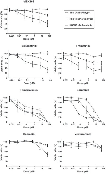
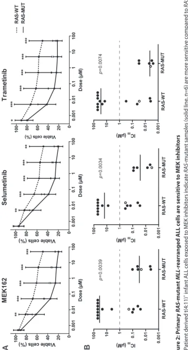
MLL-rearranged infant acute lymphoblastic leukemia patients carrying RAS mutations
63
Chapter 6 CBL mutations do not frequently occur in pediatric acute myeloid leukemia
87 Chapter 7 Versican expression is an adverse prognostic factor in
MLL-rearranged infant acute lymphoblastic leukemia
105 Chapter 8 Minimal mesenchymal stromal cell contaminations
compromise mRNA profiling in acute lymphoblastic leukemia cells derived from co-culture experiments
115
Chapter 9 Summary and general discussion 135
Chapter 10 Nederlandse samenvatting 147
Lijst van publicaties 153
Curriculum Vitae 155
PhD portfolio 157
Chapter 1
9
General introduction
LEukEMia
In the bone marrow the normal hematopoeisis (formation of blood cells) takes place. The main function of the bone marrow is to maintain the number of mature blood cells in the peripheral blood at a constant level throughout life. Leukemia is a type of cancer, character-ized by abnormal growth of immature white blood cells in the bone marrow. This overgrowth of non-functional leukemic cells compromises the formation of normal mature blood cells, leading to anemia (due to the loss of functional red blood cells), infections (due to the loss of functional white blood cells), bleedings or bruising (due to the loss of functional white blood cells). Once the bone marrow is overgrown, leukemic cells are capable of infiltrating other organs, like the spleen, liver, skin, testes and central nervous system. Leukemia can be classified in several subtypes, based on cell-growth and immunophenotype. Fast-growing leukemias are characterized as “acute” and slow-growing as “chronic”. Based on immuno-phenotype leukmias are classified into “lymphoblastic” or “myeloid”, depending on the type of with blood cells the leukemic cells rises from.
infant aCutE LyMPhobLastiC LEukEMia
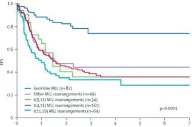
Acute lymphoblastic leukemia (ALL) in infants (<1 year of age) is a rare and aggressive malignancy with early onset, high frequencies of early relapse during treatment, and a poor clinical outcome1, 2. Infant ALL accounts for approximately 4% of all pediatric ALL cases. Infants have a distinctive biology compared to older children with ALL. Translocation of the Mixed Lineage Leukemia (MLL) gene are found in ~6% of childhood ALL cases, of which 80% is associated with infant ALL3. The presence of an MLL-rearrangement in their leukemic cells, is a strong independent predictor of poor outcome in infant ALL patients1-3. More than 70 different MLL partner genes have been identified4. The most common MLL translocation in infant ALL is t(4;11), in which the N-terminus of MLL (chromosome 11q23) fuses to the C-terminus of AF4 (chromosome 4q23). Other recurrent in-frame MLL-rearrangements found among infant ALL patients are t(11;19) and t(9;11), giving rise to the fusion proteins MLL-ENL and MLL-AF9 respectively2, 5. MLL germline infant ALL patients are patients, who lack an MLL-translocation in their leukemic cells.
The past decades survival rates for pediatric ALL patients have improved dramatically, with event-free survival (EFS) of 80% in older children with ALL. In contrast with the ~50% EFS for infant ALL patients (Figure 1), regardless of the more aggressive chemotherapy treatment they usually recieve2, 6. Although most infant ALL patients (~90-95%) achieve morphological complete remission (CR) after initial induction therapy, their prognosis is hampered by high relapse-rates. Most relapses occur very early, i.e. within the first year
10 Chapter 1
after diagnosis2, 7-9. The poor outcome of infant ALL patients in general is caused by the very poor outcome of infants which harbor an MLL-translocation in their leukemic cells (EFS of ~30-40%). This is in contrast to the outcome of infants lacking MLL-rearrangement in their leukemic cells, which nearly reaches the survival rates of older children with ALL (Figure 2)2. Other prognostic factors in infant ALL, associated with poor outcome are, high white blood cell (WBC) counts at diagnosis, very immature pro-B ALL phenotype, and glucocorticoid resistance2, 3, 10-12. More insights in the prognostic factors, molecular biol-ogy and clinical outcome of infant ALL, and in particular MLL-rearranged infant ALL, are needed in order to improve risk stratification and therapeutic strategies.
Insights into the biology and pathogenesis of MLL-rearranged ALL are hampered by the lack of genuine animal models accurately recapitulating this severe malignancy. Various attempts have been made to develop mouse models mimicking leukemogenesis of human t(4;11)-positive ALL, these mice displayed propensities towards developing lymphomas or leukemia with phenotypes that significantly differ from those found in humans13-15. Recent studies suggest involvement of the RAS-pathway, additional to the MLL-fusion, in the pathogenesis of MLL-rearranged leukemia 16-19. The RAS-pathway regulates diverse cellular functions including cell proliferation, survival, differentiation, angiogenesis, and migration 20, 21. Studies on the incidence of RAS mutations (NRAS and KRAS) in MLL-rearranged ALL demonstrate various frequencies 10-34% in MLL-rearranged and 26-63.9% in t(4;11)-positive childhood leukemia 16, 17, 22-24. Deregulation of the RAS-pathway is a common event in childhood ALL and may guide new therapy strategies for MLL-rearranged infant ALL21.
11
General introduction
outLinE of this thEsis
In Chapter 2 we detail the clinical outcome and prognostic factors of infant ALL patients, who relapsed on or after receiving the Interfant-99 treatment protocol. Relapsed infant ALL is assumed to be inevitably fatal. However, published data on outcome of relapsed infant ALL is limited. In Chapter 3 we report on the correlation of in vitro drug sensitivity with clinical outcome in a relatively large cohort of primary MLL-rearranged infant ALL cases. To further identify prognostic factors in infant ALL patients we investigated the frequencies and prognostic value of RAS mutations in our cohort of primary infant ALL cases. As we re-port in Chapter 4 we found that the presences of a RAS mutation in MLL-rearranged infant ALL cases is an independent prognostic factor for outcome. Therefore we hypothesized that inhibiting the RAS-pathway could be beneficial for infant ALL patients. In Chapter 5 we report the effect of MEK inhibitors targeting primary RAS-mutated MLL-rearranged infant ALL cells in vitro and the relation with prednisolone sensitization. Chapter 6 describes the role of Casitas B lineage lymphoma (CBL), a protein involved in the RAS-pathway, in acute myeloid leukemia and MLL-rearranged infant ALL. In Chapter 7 we address our study of the relation of high Versican expression and clinical outcome in MLL-rearranged infant ALL patients. In Chapter 8 we report on the effect of minimal contaminating stromal cells in in vitro co-culture experiments. Chapter 9 summarizes this thesis and comprises a general discussion and Chapter 10 covers a layman’s summary in Dutch.
figure 2: Clinical outcome of infant aLL patients, dissected by type ofMLL-translocation
12 Chapter 1
REfEREnCEs
1. Greaves MF. Infant leukaemia biology, aetiology and treatment. Leukemia. 1996;10(2):372-7. 2. Pieters R, Schrappe M, De Lorenzo P, Hann I, De Rossi G, Felice M, et al. A treatment protocol for
infants younger than 1 year with acute lymphoblastic leukaemia (Interfant-99): an observational study and a multicentre randomised trial. Lancet. 2007;370(9583):240-50.
3. Biondi A, Cimino G, Pieters R, Pui CH. Biological and therapeutic aspects of infant leukemia. Blood. 2000;96(1):24-33.
4. Meyer C, Hofmann J, Burmeister T, Groger D, Park TS, Emerenciano M, et al. The MLL recombi-nome of acute leukemias in 2013. Leukemia. 2013;27(11):2165-76.
5. Jansen MW, Corral L, van der Velden VH, Panzer-Grumayer R, Schrappe M, Schrauder A, et al. Immunobiological diversity in infant acute lymphoblastic leukemia is related to the occurrence and type of MLL gene rearrangement. Leukemia. 2007;21(4):633-41.
6. Pui CH, Robison LL, Look AT. Acute lymphoblastic leukaemia. Lancet. 2008;371(9617):1030-43. 7. Frankel LS, Ochs J, Shuster JJ, Dubowy R, Bowman WP, Hockenberry-Eaton M, et al. Therapeutic
trial for infant acute lymphoblastic leukemia: the Pediatric Oncology Group experience (POG 8493). J Pediatr Hematol Oncol. 1997;19(1):35-42.
8. Reaman GH, Sposto R, Sensel MG, Lange BJ, Feusner JH, Heerema NA, et al. Treatment outcome and prognostic factors for infants with acute lymphoblastic leukemia treated on two consecutive trials of the Children’s Cancer Group. J Clin Oncol. 1999;17(2):445-55.
9. Stam RW, den Boer ML, Pieters R. Towards targeted therapy for infant acute lymphoblastic leukae-mia. Br J Haematol. 2006;132(5):539-51.
10. Dordelmann M, Reiter A, Borkhardt A, Ludwig WD, Gotz N, Viehmann S, et al. Prednisone response is the strongest predictor of treatment outcome in infant acute lymphoblastic leukemia. Blood. 1999;94(4):1209-17.
11. Kaspers GJ, Pieters R, Van Zantwijk CH, Van Wering ER, Van Der Does-Van Den Berg A, Veerman AJ. Prednisolone resistance in childhood acute lymphoblastic leukemia: vitro-vivo correlations and cross-resistance to other drugs. Blood. 1998;92(1):259-66.
12. Pieters R, den Boer ML, Durian M, Janka G, Schmiegelow K, Kaspers GJ, et al. Relation between age, immunophenotype and in vitro drug resistance in 395 children with acute lymphoblastic leukemia--implications for treatment of infants. Leukemia. 1998;12(9):1344-8.
13. Chen W, Li Q, Hudson WA, Kumar A, Kirchhof N, Kersey JH. A murine Mll-AF4 knock-in model results in lymphoid and myeloid deregulation and hematologic malignancy. Blood. 2006;108 (2):669-77.
14. Krivtsov AV, Feng Z, Lemieux ME, Faber J, Vempati S, Sinha AU, et al. H3K79 methylation profiles define murine and human MLL-AF4 leukemias. Cancer Cell. 2008;14(5):355-68.
15. Metzler M, Forster A, Pannell R, Arends MJ, Daser A, Lobato MN, et al. A conditional model of MLL-AF4 B-cell tumourigenesis using invertor technology. Oncogene. 2006;25(22):3093-103. 16. Liang DC, Shih LY, Fu JF, Li HY, Wang HI, Hung IJ, et al. K-Ras mutations and N-Ras mutations in
childhood acute leukemias with or without mixed-lineage leukemia gene rearrangements. Cancer. 2006;106(4):950-6.
17. Mahgoub N, Parker RI, Hosler MR, Close P, Winick NJ, Masterson M, et al. RAS mutations in pedi-atric leukemias with MLL gene rearrangements. Genes Chromosomes Cancer. 1998;21(3):270-5. 18. Ng MH, Ng RK, Kong CT, Jin DY, Chan LC. Activation of Ras-dependent Elk-1 activity by MLL-AF4
13
General introduction 19. Tamai H, Miyake K, Takatori M, Miyake N, Yamaguchi H, Dan K, et al. Activated K-Ras protein
accelerates human MLL/AF4-induced leukemo-lymphomogenicity in a transgenic mouse model. Leukemia. 2011;25(5):888-91.
20. Chung E, Kondo M. Role of Ras/Raf/MEK/ERK signaling in physiological hematopoiesis and leuke-mia development. Immunol Res. 2011;49(1-3):248-68.
21. Knight T, Irving JA. Ras/Raf/MEK/ERK Pathway Activation in Childhood Acute Lymphoblastic Leukemia and Its Therapeutic Targeting. Front Oncol. 2014;4:160.
22. Prelle C, Bursen A, Dingermann T, Marschalek R. Secondary mutations in t(4;11) leukemia patients. Leukemia. 2013;27(6):1425-7.
23. Andersson AK, Ma J, Wang J, Chen X, Gedman AL, Dang J, et al. The landscape of somatic mutations in infant MLL-rearranged acute lymphoblastic leukemias. Nat Genet. 2015;47(4):330-7.
24. Trentin L, Bresolin S, Giarin E, Bardini M, Serafin V, Accordi B, et al. Deciphering KRAS and NRAS mutated clone dynamics in MLL-AF4 paediatric leukaemia by ultra deep sequencing analysis. Sci Rep. 2016;6:34449.
Chapter 2
Outcome of relapsed infant acute
lymphoblastic leukemia treated on
the Interfant-99 protocol
E.M.C. Driessen1, P. de Lorenzo2, M. Campbell2, M. Felice2,
A. Ferster2, I. Hann2, A.Vora2, L. Hovi2, G. Escherich2, C.K. Li2,
G. Mann2, T. Leblanc2, F. Locatelli2 ,A. Biondi2, J. Rubnitz2,
M. Schrappe2, L. Silverman2, J.Stary2 R. Suppiah2, T.
Szczepanski2, M. Valsecchi2 *, R. Pieters1,2,3*
*Shared last co-authorship
1 Pediatric Oncology/Hematology, Erasmus MC-Sophia Children’s Hospital,
Rotterdam, The Netherlands;
2 Interfant-99 Collaborative Study Group
3 Princess Máxima Center for Pediatric Oncology, Utrecht, the Netherlands
17
Outcome of relapsed infant ALL
intRoDuCtion
Acute lymphoblastic leukemia (ALL) in infants (<1 year of age) is an aggressive disease. About 80% of infant ALL patients harbor a Mixed Lineage Leukemia (MLL) translocation, which is a strong independent predictor of poor outcome1. Furthermore, infant ALL is as-sociated with higher white blood cell (WBC) counts at diagnosis, very immature pro-B ALL phenotype, and glucocorticoid resistance1, 2. Although most infant ALL patients (~95%) achieve morphological complete remission (CR) after induction therapy, due to high relapse rates their outcome is poor1. Most relapses occur early, within the first year after diagnosis. The largest series of infant ALL, described by the Interfant-99 study group, reported a re-lapse rate of 34%. Rere-lapsed infant ALL is generally assumed to be inevitably fatal. Published data on outcome of relapsed infant ALL is very limited, and available ones report on small cohorts.3 Here, we describe the clinical outcome of 202 infant ALL patients, who relapsed on or after receiving the Interfant-99 treatment protocol1.
MEthoDs
Design and inclusion criteria of the Interfant-99 trial have been described previously1. Therapy after relapse differed in participating countries according to local policy and in-dividual choice. We retrospectively asked the physicians whether the therapy was based on curative or palliative intent.
Outcome measure was overall survival (OS), defined as time from relapse until death from any cause. Curves were estimated with the Kaplan-Meier method and compared by sub-groups with the log-rank test. Follow-up was updated at December 31 2009 and median follow-up (range) was 5.2 years (1 month–10.1 years). Survival analyses regarding hemato-poietic stem cell transplantation (HSCT) were corrected for waiting time to transplantation. Association between prognostic characteristics at first diagnosis of ALL and at relapse with risk group and with assignment to curative or palliative care was evaluated with the Fisher exact test. To account for imbalances in characteristics between patients, a propensity score was derived, reflecting the probability that a patient would undergo curative treatment, by performing a multivariable logistic regression analysis. Any residual association between the covariates and treatment allocation was assessed by the Cochran–Mantel–Haenszel test. Cox model was used to evaluate outcome of curative and palliative treatment and of HSCT vs chemotherapy alone as curative treatment after relapse. Hazard ratios were tested accord-ing to Wald. SAS version 9.2 statistical software was used for data analysis.
18 Chapter 2
REsuLts
Out of 478 patients, 448 patients achieved first CR, 70.1% patients were allocated in the standard risk (SR, i.e. good prednisone response) and 29.9% in high risk (HR, i.e. poor prednisone response) group. Two-hundred and two patients (45.1%) relapsed, 37.6% and 62.7% in the SR and HR group, respectively. 56.9% (115/202) of the relapses occurred in the first year after diagnosis, with a median time to relapse of 10.0 months (range: 1.7-50.7 months). Most relapses occurred in the bone marrow (BM, 71.8%), while others were isolated extramedullary (n=23) or combined relapses (n=32). Patients in the HR group relapsed earlier than patients in the SR group (median: 9 vs. 12 months, p=0.042).
The 3-year OS after relapse was 20.9% (SE 3.5%). Infants with germline-MLL ALL relapsed later (>24 months) than infants with MLL-rearrangements (p=0.03). There was no differ-ence in site of relapse (BM involvement vs. extramedullary) between germline-MLL and MLL-rearranged infant ALLs.
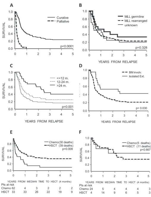
Out of 202 patients, 159 (78.7%) received relapse treatment with curative intent and 32 (15.8%) received supportive care only. For 11 patients (5.5%) treatment after relapse was not reported and these were excluded from further analyses. The 3-year OS after relapse of patients treated with curative intent was 24.9% (SE 4.0%), of whom 76.1% (121/159) died due to progressive ALL (n=93), HSCT-related causes (n=21) and other, mainly infec-tious, causes (n=7). All patients who received palliative care (n=32) died within 1 year after relapse, due to disease progression (Figure 1A, p<0.0001). As expected, this straightforward outcome comparison is biased by imbalances in the characteristics of the two groups. Cura-tive treatment was more likely to be adopted for patients with a long duration of first CR (p=0.0005).
All patients (n=28), who relapsed after 24 months received curative treatment. 84.4% of pa-tients receiving palliative care relapsed in the first year, compared to 50.3% of curative treated patients. Almost all patients carrying germline-MLL underwent curative treatment, whereas 87.5% of patients with palliative care had an MLL-rearrangement. Similarly, patients with higher WBC counts (>300x109/L) at first diagnosis received palliative care more frequently. In order to account for these imbalances, we derived a propensity score which predicted whether a patient would undergo curative treatment as a function of clinical characteristics likely af-fecting treatment choice. We used the propensity score in a stratified Cox model in order to perform a fair comparison of outcome in the two groups. The model which was also adjusted by relevant covariates, revealed that curative treatment was associated with a significantly superior OS (hazard ratio=0.17, 95% CI (0.10–0.27), p<0.0001). Interestingly, the time of
19
Outcome of relapsed infant ALL
SU R VI VAL 0.0 0.2 0.4 0.6 0.8 1.0
YEARS FROM RELAPSE
0 1 2 3 4 5 BM involv. Isolated Ext. p= 0.030 SU R VI VAL 0.0 0.2 0.4 0.6 0.8 1.0
YEARS FROM RELAPSE
0 1 2 3 4 5 MLL germline MLL rearranged unknown p=0.328 SU R VI VAL 0.0 0.2 0.4 0.6 0.8 1.0
YEARS FROM RELAPSE
0 1 2 3 4 5 Curative Palliative p<0.0001 SU R VI VAL 0.0 0.2 0.4 0.6 0.8 1.0
YEARS FROM MEDIAN TIME TO HSCT (4 months)
0 1 2 3 4 5 Chemo(30 deaths) HSCT (39 deaths) Pts at risk Chemo 62 4 3 2 2 2 HSCT 33 33 26 22 19 9 p=0.009 SU R VI VAL 0.0 0.2 0.4 0.6 0.8 1.0
YEARS FROM RELAPSE
0 1 2 3 4 5 <=12 m. 12-24 m. >24 m. p=0.001 SU R VI VAL 0.0 0.2 0.4 0.6 0.8 1.0
YEARS FROM MEDIAN TIME TO HSCT (4 months)
0 1 2 3 4 5 Chemo(5 deaths) HSCT (11 deaths) Pts at risk Chemo 24 5 4 4 4 3 HSCT 4 14 9 6 5 3 p=0.667
A
B
C
D
E
F
figure 1: overall survival of relapsed infant aLL patients
Overall survival, defined as time from relapse until death, (A) by treatment given after relapse, (B) of patients treated with curative intents by MLL status at initial diagnosis, (C) of patients treated with curative intents by time at relapse after initial diagnosis, (D) of patients treated with curative intents by site at relapse, (E) of patients treated with HSCT vs. chemotherapy only of early relapses (within 24 months from initial diag-nosis), (F) of patients treated with HSCT vs. chemotherapy only of late relapses (beyond 24 months from initial diagnosis). Curves were estimated using the Kaplan-Meier method and analyzed by Log-rank tests. Follow-up was updated at December 31 2009 and median follow-up(range) was 5.2 years(1 month–10.1 years). Survival analyses regarding HSCT were corrected for waiting time to transplantation.
20 Chapter 2
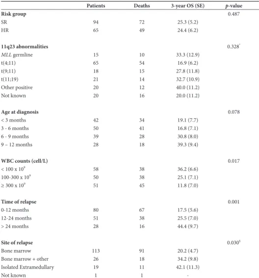
relapse retained prognostic relevance. Patients who relapsed within 24 months after diagnosis had a 1.5-fold increase in the risk of death (hazard ratio=1.40, 95% CI (0.97–2.01), p=0.016). We then restricted our attention to the outcome of 159 patients treated with curative intent (Table 1 and Figure 1B-F). We did not observe a significant difference in OS after relapse between infants with and without 11q23 abnormalities (p=0.328). Infants with a t(4;11)-re-table 1: outcome of relapsed infant aLL patients treated with curative intent
Patients Deaths 3-year OS (SE) p-value
Risk group 0.487 SR 94 72 25.3 (5.2) HR 65 49 24.4 (6.2) 11q23 abnormalities 0.328* MLL germline 15 10 33.3 (12.9) t(4;11) 65 54 16.9 (6.2) t(9;11) 18 15 27.8 (11.8) t(11;19) 21 14 32.7 (10.9) Other positive 20 12 40.0 (11.2) Not known 20 16 20.0 (11.2) Age at diagnosis 0.078 < 3 months 42 34 19.1 (7.7) 3 - 6 months 50 41 16.8 (7.1) 6 - 9 months 39 28 30.8 (8.0) 9 – 12 months 28 18 39.3 (9.4) WBC counts (cell/L) 0.017 < 100 x 109 58 38 36.2 (6.6) 100-300 x 109 50 38 25.1 (7.1) ≥ 300 x 109 51 45 11.8 (7.0) Time of relapse 0.001 0-12 months 80 67 17.5 (5.6) 12-24 months 51 38 25.5 (7.0) > 24 months 28 16 44.4 (9.7) Site of relapse 0.030§ Bone marrow 113 91 20.2 (4.7)
Bone marrow + other 26 18 34.2 (9.8)
Isolated Extramedullary 19 11 42.1 (11.3)
Not known 1 1
-Overall survival, defined as time from relapse until death, of curative treatments after relapse by relevant prognostic factors at initial diagnosis and at relapse, including risk group (based on prednisone response), 11q23-abnormalities, age at initial diagnosis, WBC counts at initial diagnosis, time of relapse, defined as time from initial diagnosis until relapse, and site of relapse. Subgroups were analyzed by using the Log-rank
21
Outcome of relapsed infant ALL
arrangement showed a worse 3-year OS compared to those with other 11q23-abnormalities (p=0.037). Age at diagnosis had a significant impact on outcome after first relapse, with a 3-year OS of 17.9% (SE 5.2%) in patients below 6 months of age and 34.3% (SE 6.1%) in older patients (p=0.012). Higher WBC count at diagnosis was associated with inferior OS after relapse (p=0.017), while risk group stratification by initial prednisone response showed no impact (p=0.487). In addition, we found that infants relapsing within one year after diagnosis had a 3-year OS of 17.5% (SE 5.6%) compared to 25.5% (SE 7.0%) and 44.4% (SE 9.7%) of those who relapsed between 12-24 and beyond 24 months after diagnosis, respectively (p=0.001). Patients with relapse involving BM, either isolated or combined, had a worse OS compared to patients with isolated extramedullary relapse (p=0.030).
Among patients treated for relapse, 87 (54.7%) underwent HSCT, the majority in second CR (n=66, 75.9%) and from an HLA-matched unrelated donor (n=41, 47.1%). The median time from relapse to HSCT was 4.2 months (range: 1.1-9.1). According to the results of the Cox model corrected for waiting time to transplantation, patients receiving HSCT after relapse had a better OS compared to patients receiving chemotherapy alone (p=0.021); similar results were found after adjusting for known prognostic factors at initial diagnosis. The advantage of HSCT over chemotherapy did not change significantly over time of follow-up, contrary to what was observed for HSCT in CR1 in the Interfant-99 study4. Interestingly, there was a significant interaction between treatment and time at relapse (p=0.010), while the interaction between treatment and site of relapse was not significant. The analysis in subgroups defined by the time at relapse showed that HSCT significantly reduced the risk of death compared to chemotherapy alone in 131 patients who relapsed within 24 months from initial diagnosis (hazard ratio=0.52, 95% CI(0.32–0.85), p=0.009), while no advantage over chemotherapy was observed for patients who relapsed later (hazard ratio=1.28, 95% CI(0.420–3.89), p=0.667). After fitting the Cox model to subgroups of early relapses, site of relapse retained its significant impact over survival (hazard ratio for BM and combined vs. extramedullary relapses was 2.00, 95% CI(1.04-3.85), p=0.038).
DisCussion
Here, we demonstrate in a large cohort that relapsed infant ALL is not inevitable fatal. We found a 3-year OS of 20.9% for all relapsed infant ALL patients and 24.9% for those treated with curative intent. This is in concordance with the previous reported 5-year OS of 25.6% of Tomizawa et al.3. Even after adjusting for imbalances of prognostic factors in the treat-ment groups, patients treated with curative intent had a significant better outcome. Young age and a high WBC count at initial diagnosis were associated with inferior outcome after first relapse, but response to prednisone prophase during first line treatment did not.
Fur-22 Chapter 2
thermore, patients who relapsed earlier and with patients BM involvement (either isolated or combined) had a worse outcome. These findings are consistent with previous studies in other ALL subtypes5-7.
The use of HSCT in infant ALL is a matter of debate. Previous studies demonstrated that routine use of HSCT does not improve outcome for infants1, 8-10. However, data from the Interfant-99 study group suggested that a small subset with high risk features (young age combined with poor response to prednisone prophase or high WBC) may benefit from HSCT in first CR4. The present study showed that HSCT improved outcome of infants who relapsed ‘early’ (i.e. within 2 years after initial diagnosis), a subset which in our cohort accounts for 82% of all relapses.
In conclusion, we demonstrated that relapsed infant ALL was not invariably lethal and underline the relevance of offering treatments with curative intents. New therapeutic strategies, such as FLT3 inhibitors, epigenetic drugs, glucocorticoid sensitization and RAS -pathway inhibition, could be beneficial for infant ALL patients11-15.
23
Outcome of relapsed infant ALL
REfEREnCEs
1. Pieters R, Schrappe M, De Lorenzo P, Hann I, De Rossi G, Felice M, et al. A treatment protocol for infants younger than 1 year with acute lymphoblastic leukaemia (Interfant-99): an observational study and a multicentre randomised trial. Lancet. 2007;370(9583):240-50.
2. Dordelmann M, Reiter A, Borkhardt A, Ludwig WD, Gotz N, Viehmann S, et al. Prednisone response is the strongest predictor of treatment outcome in infant acute lymphoblastic leukemia. Blood. 1999;94(4):1209-17.
3. Tomizawa D, Koh K, Hirayama M, Miyamura T, Hatanaka M, Saikawa Y, et al. Outcome of recurrent or refractory acute lymphoblastic leukemia in infants with MLL gene rearrangements: A report from the Japan Infant Leukemia Study Group. Pediatric blood & cancer. 2009;52(7):808-13.
4. Mann G, Attarbaschi A, Schrappe M, De Lorenzo P, Peters C, Hann I, et al. Improved outcome with hematopoietic stem cell transplantation in a poor prognostic subgroup of infants with mixed-lineage-leukemia (MLL)-rearranged acute lymphoblastic leukemia: results from the Interfant-99 Study. Blood. 2010;116(15):2644-50.
5. Gaynon PS, Qu RP, Chappell RJ, Willoughby ML, Tubergen DG, Steinherz PG, et al. Survival after relapse in childhood acute lymphoblastic leukemia: impact of site and time to first relapse--the Children’s Cancer Group Experience. Cancer. 1998;82(7):1387-95.
6. Nguyen K, Devidas M, Cheng SC, La M, Raetz EA, Carroll WL, et al. Factors influencing survival after relapse from acute lymphoblastic leukemia: a Children’s Oncology Group study. Leukemia. 2008;22(12):2142-50.
7. Tallen G, Ratei R, Mann G, Kaspers G, Niggli F, Karachunsky A, et al. Long-term outcome in children with relapsed acute lymphoblastic leukemia after time-point and site-of-relapse stratification and intensified short-course multidrug chemotherapy: results of trial ALL-REZ BFM 90. J Clin Oncol. 2010;28(14):2339-47.
8. Dreyer ZE, Dinndorf PA, Camitta B, Sather H, La MK, Devidas M, et al. Analysis of the role of hematopoietic stem-cell transplantation in infants with acute lymphoblastic leukemia in first remis-sion and MLL gene rearrangements: a report from the Children’s Oncology Group. J Clin Oncol. 2011;29(2):214-22.
9. Kosaka Y, Koh K, Kinukawa N, Wakazono Y, Isoyama K, Oda T, et al. Infant acute lymphoblastic leukemia with MLL gene rearrangements: outcome following intensive chemotherapy and hemato-poietic stem cell transplantation. Blood. 2004;104(12):3527-34.
10. Koh K, Tomizawa D, Moriya Saito A, Watanabe T, Miyamura T, Hirayama M, et al. Early use of al-logeneic hematopoietic stem cell transplantation for infants with MLL gene-rearrangement-positive acute lymphoblastic leukemia. Leukemia. 2014.
11. Brown P, Levis M, McIntyre E, Griesemer M, Small D. Combinations of the FLT3 inhibitor CEP-701 and chemotherapy synergistically kill infant and childhood MLL-rearranged ALL cells in a sequence-dependent manner. Leukemia. 2006;20(8):1368-76.
12. Daigle SR, Olhava EJ, Therkelsen CA, Majer CR, Sneeringer CJ, Song J, et al. Selective killing of mixed lineage leukemia cells by a potent small-molecule DOT1L inhibitor. Cancer cell. 2011;20(1):53-65. 13. Driessen EM, van Roon EH, Spijkers-Hagelstein JA, Schneider P, de Lorenzo P, Valsecchi MG, et
al. Frequencies and prognostic impact of RAS mutations in MLL-rearranged acute lymphoblastic leukemia in infants. Haematologica. 2013;98(6):937-44.
14. Spijkers-Hagelstein JA, Pinhancos SS, Schneider P, Pieters R, Stam RW. Chemical genomic screen-ing identifies LY294002 as a modulator of glucocorticoid resistance in MLL-rearranged infant ALL. Leukemia. 2014;28(4):761-9.
24 Chapter 2
15. Stumpel DJ, Schneider P, Seslija L, Osaki H, Williams O, Pieters R, et al. Connectivity mapping identifies HDAC inhibitors for the treatment of t(4;11)-positive infant acute lymphoblastic leukemia. Leukemia. 2012;26(4):682-92.
Chapter 3
Relation between
in vitro
drugs
responses and prognostic markers
in
MLL-
rearranged infant acute
lymphoblastic leukemia
J.A.P. Spijkers-Hagelstein1, E.M.C. Driessen1, P. Schneider1,
P. Garrido Castro1, M. L. den Boer1, R. Pieters1,2, R. W. Stam1
1 Pediatric Oncology/Hematology, Erasmus MC-Sophia Children’s Hospital,
Rotterdam, The Netherlands
2 Princess Máxima Center for Pediatric Oncology, Utrecht, the Netherlands
26 Chapter 3
abstRaCt
Despite the current successful treatment results in pediatric acute lymphoblastic leukemia (ALL), the prognosis for MLL-rearranged infant ALL patients remains poor, and novel strategies are needed to identify and predict patients at high risk of therapy failure. We explored to what extent in vitro drug resistance is associated with clinical outcome and established prognostic markers, such as age <6 months, MLL rearrangement and white blood cell count (WBC) at diagnosis.
We found that in vitro response to none of the drugs tested by itself is capable of predicting clinical outcome, although an association between in vitro glucocorticoid (i.e. prednisolone and dexamethasone) resistance and a poor outcome was observed. Also, in vitro resistance to glucocorticoids was associated with age <6 months and high WBC. The combined in vitro response to prednisolone, vincristine and L-asparaginase (PVA) was found to be predictive for an adverse outcome in MLL-rearranged infant ALL. PVA-resistant MLL-rearranged infant ALL patients are at high risk of therapy failure. PVA-resistant MLL-rearranged infant ALL patients showed cross-resistance towards dexamethasone but not to other drugs. Taken together, we conclude that in vitro PVA-sensitivity testing in MLL-rearranged ALL may be useful to stratify patients at high risk of therapy failure, and that this approach allows identification of patients urgently requiring effective novel treatment options.
27
In vitro drugs responses in MLL-rearranged infant ALL
intRoDuCtion
Over the last decades, the outcome for childhood acute lymphoblastic leukemia (ALL) in general has improved tremendously. Yet, the clinical outcome for infants (< 1 year of age) diagnosed with ALL remained dismal1-5. Infant ALL is characterized by a high incidence of balanced chromosomal translocations involving the Mixed Lineage Leukemia (MLL) gene located on chromosome 11, which are detected in ~80% of the cases6-8. As a result of such translocations, the N-terminal region of MLL fuses to the C-terminal region of one of its many translocation partner genes. Among infant ALL patients, the most recurrent MLL translocations are t(4;11), t(11;19) and t(9;11), which generate the chimeric fusion proteins MLL-AF4, MLL-ENL and MLL-AF9, respectively. The presence of leukemia-specific MLL translocations on its own represents a strong predictor of an unfavorable clinical out-come2, 3, 5, 9, 10. Additional parameters predicting a poor outcome include age at diagnosis (<6 months of age and white blood cell count (WBC) at disease presentation5. Moreover, infant ALL cells are resistant to glucocorticoids and L-asparaginase11, 12. The in vitro responses of prednisolone, vincristine and L-asparaginase combined (known as the PVA score), appeared highly predictive for clinical outcome in childhood ALL13, 14. To date it remains unclear whether in vitro drug resistance patterns obtained in primary infant ALL cells influence prognosis, or to what extent cellular resistance is associated with abovementioned prognostic factors. Therefore we generated in vitro cytotoxicity profiles in a cohort of infant ALL patients enrolled in the Interfant treatment protocols, and investigated the relation between in vitro drug responses, clinical outcome, and known prognostic factors, in order to attain evidence-based progression towards personalized medicine.
MEthoDs Patient samples
In the present study we used a total of 67 samples obtained from infant ALL patients (<1 year of age) enrolled in either the Interfant-99 or the Interfant-06 study, for whom a com-plete overview of clinical parameters and follow up data was available, as well as sufficient material for in vitro cytotoxicity testing. This cohort of infant ALL patients consists of MLL -rearranged infant ALL cases (n=55) as well as patients carrying germline (or wild-type) MLL genes (i.e. no translocation (n=12). The MLL-rearranged infant ALL patient group consists of patients carrying t(4;11) (n=28), t(11;19) (n=18) and other 11q23 translocations (n=9), as determined by fluorescence in situ hybridization (FISH) and RT-PCR analyses. Bone marrow aspirates or peripheral blood was collected at the Erasmus MC-Sophia Children’s Hospital, and within 24 hours leukemic cells were isolated by density gradation centrifuga-tion (Lymphoprep; density 1.077 g/ml, Nycomed Pharma, Oslo, Norway). Samples were
28 Chapter 3
enriched for leukemic blasts by removal of contaminating normal cells using monoclonal antibodies linked to magnetic beads as described by Kaspers et al.15. Consequently, all samples used for in vitro drug testing contained >90% leukemic cells, as determined mor-phologically on May-Grunwald Giemsa (Merck, Darmstadt, Germany) stained cytospins.
In vitro drug response and in vivo prednisone response
For all samples, in vitro drug cytotoxicity was assessed by MTT assays as described previ-ously16. Briefly, leukemic cells were incubated for 4 days in a humidified incubator at 37oC and 5% CO2, both in the absence and presence of a range of drug concentrations. Then, 10 µl of yellow 3-(4,5-dimethylthiazol-2-yl)-2,5-diphenyl tetrazoliumbromide (MTT) was added to the cells, following an additional 6-hour incubation under the same conditions, during which the yellow MTT is reduced to blue formazan crystals by viable cells only. Next, the blue formazan crystals were dissolved using HCL-isopropyl alcohol, allowing spectropho-tometrical measurement of the optical density. Culture medium without cells nor drugs served as blanks (i.e. 0% survival), and leukemic cells cultured in the absence of drugs (i.e. controls) were adjusted to 100% survival and used to calculate the LC50-value. Results were considered evaluable only if the mean control OD, after correction for the background (as determined by the blanks), at day 4 exceeded 0.05 arbitrary units. The following drugs and concentration ranges (6 dosages per drug) were used: prednisolone, 0.08-250 ug/mL (PRED; Bufa, Uitgeest, The Netherlands); Vincristine, 0.05-50 ug/mL (VCR; Pharmacy Erasmus Medical Center, Rotterdam, The Netherlands); L-Asparaginase, 0.003-10 IU/mL (L-ASP; Nycomed BV, Hoofddorp, The Netherlands); Daunorubicin, 0.002-2.0 ug/mL (DNR; Sanofi Aventis, Gouda, The Netherlands); Cytarabine, 0.04-2.5 ug/mL (Ara-C; Hospira Benelux BVBA, Brussel, Belgium) and Dexamethasone, 0.0002-6.0 ug/mL (DEX; Pharmacie Eras-mus Medical Center, Rotterdam, The Netherlands). The PVA score was based on the in vitro drug responses (i.e. LC50-values) of prednisolone, Vincristine and L-Asparaginase, in which sensitivity towards each drug individually was scored as 1, intermediate responses as 2, and resistance was scored as 3. For this, the cut-off LC50-values were: prednisolone sensitivity: <0.1 µg/mL and resistance: >150 µg/mL; vincristine sensitivity: <0.3906 µg/mL and resistance: >1.7578 ug/mL; L-asparaginase sensitivity: <0.033 IU/mL and resistance: >0.912 IU/mL. Hence, the PVA scores vary between 3 (sensitive to all three drugs) and 9 (resistant to all three drugs).
The in vivo response to prednisone was determined after one intrathecal dose of methotrex-ate and a 7-day window of prednisone mono-therapy (before the initiation of combination chemotherapy). Patients were defined as prednisone poor responders (PPRs) when >1000 leukemic cells/µL remained present in the peripheral blood17. When the amount of leukemic cells dropped below 1000/µL patients were defined as prednisone good responders (PGRs).
29
In vitro drugs responses in MLL-rearranged infant ALL
statistical analysis
Differences in the distribution of variables between patient groups were analyzed using the Mann-Whitney U-test or the Kruskall-Wallis test. Cross–resistance patterns were studied by correlating the LC50 values for different drugs using the Kruskall-Wallis test and the Spearman’s Rho-test. The probability of event-free survival (EFS) was calculated using the Kaplan-Meier method and the Log-rank (Mantel-Cox) test was performed to analyze dif-ferences in outcome between patient groups. Furthermore, a correlation between in vitro prednisolone response and age at diagnosis was analyzed using the Spearman’s Rho-test. The EFS rate is defined as time from diagnosis to death in induction, failure to achieve complete remission after induction, disease relapse, the emergence of secondary malignan-cies, or death in complete remission. Patients who did not achieve complete remission were assigned an event at time-point zero in the EFS analyses. All analyses were two-tailed and differences with p-value <0.05 were considered statistically significant.
REsuLts
Prognostic relevance of in vitro PVa scoring in infant aLL
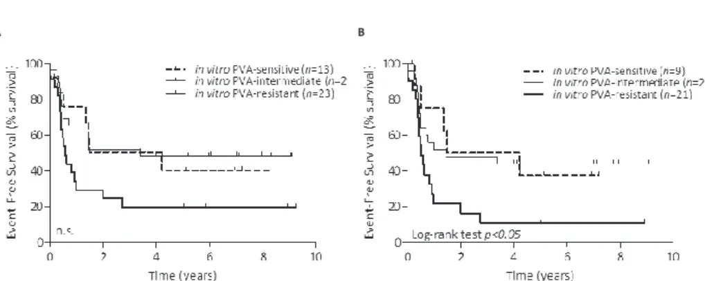
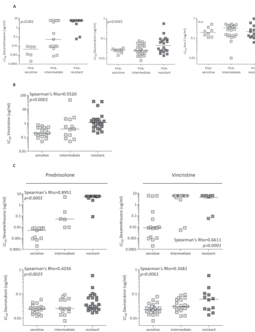
The combined in vitro responses to prednisolone, vincristine and L-asparaginase (i.e. PVA score) has been shown to be predictive for clinical outcome in pediatric ALL (i.e. children >1 year of age)13, 14, 17. We analyzed whether this also accounts for infants with ALL (<1 year of age). Patients were divided into a sensitive (PVA scores of 3-4), a intermediate (PVA scores of 5-6), or a resistant (PVA scores of 7-9) group. PVA-resistant infant ALL cases (including both patients with and without MLL translocations) tended to have a worse outcome when compared to patients with sensitive and intermediate PVA scores (Figure 1A); although this did not reach statistical significance (p=0.0686). As the presence of MLL translocations represents a strong and independent prognostic factor5, we also analyzed the influence of the PVA score on clinical outcome excluding patients with wild-type MLL genes. PVA-resistant MLL-rearranged infant ALL patients showed worse EFS compared with both PVA-sensitive and PVA-intermediate MLL-rearranged infant ALL cases (Log-rank; p<0.05, Figure 1B). The 5-year EFS for PVA-resistant MLL-rearranged infant ALL patients is with ~10% very poor. Furthermore, we also analyzed whether in vitro sensitivity towards single chemotherapeutics (prednisolone, vincristine, L-asparaginase, daunorubicin, cytarabine or dexamethasone) is predictive for EFS. MLL-rearranged infant ALL patients in vitro resistant to these single chemotherapeutics have worse outcome when compared to their sensitive counterpart. However, these differences were not significant (prednisolone, p=0.1756, L-asparaginase, p=0.2970, daunorubicin, p=0.1396).
30 Chapter 3
Next, we analyzed whether PVA resistance showed cross-resistance to other drugs. In MLL-rearranged infant ALL patients, PVA resistance was correlated to dexamethasone resistance, but not to resistance to daunorubicin or Ara-C (Figure 2A). Interestingly, when analyzing cross-resistance between prednisolone, vincristine and L-asparaginase, only a correlation was found between prednisolone and vincristine (Spearman’s Rho-test=0.5520, p<0.005) (Figure 2B); no correlation was found between prednisolone and L-asparaginase (Spearman’s Rho-test=0.044, p=0.7494) nor between vincristine and L-asparaginase (Spearman’s Rho-test=0.247, p=0.0688). Furthermore, MLL-rearranged infant ALL patient samples which were in vitro sensitive to prednisolone or vincristine only displayed in vitro sensitivity to the glucocorticoid dexamethasone (Spearman´s Rho-test prednisolone-dexa-methasone=0.8951, p<0.0001; Spearman´s Rho-test vincristine-dexamethasone=0.6611, p<0.0001) and the antracycline daunorubicin (Spearman´s Rho-test prednisolone-daunorubicin=0.4036, p=0.0025; Spearman´s Rho-test vincristine-daunorubicin=0.3681, p=0.0061) (Figure 2C). No cross-resistance or cross-sensitivity was observed for neither L-asparaginase nor Ara-C with other drugs (data not shown).
Both the in vitro prednisolone response, as well as the in vivo prednisone response have been identified as prognostic makers in pediatric ALL17 and infant ALL2, 5. Here, we investi-gated to what degree the clinical prednisone response correlates with in vitro prednisolone response as determined by MTT assays. As shown in Figure 3, PGRs are more frequent sensitive to prednisolone in vitro as compared with PPRs. Similar results are obtained when analyzing infant ALL in general (Figure 3A), or when analyzing only MLL-rearranged infant ALL cases (Figure 3B). Although the observed differences are statistically significant
A B
figure 1: Prognostic relevance of prednisolone, vincristine and L-asparaginase (PVa) in infant acute lymphoblastic leukemia
EFS analysis of (A) total infant ALL patients (with and without MLL-rearranged infant ALL) and (B) MLL
-rearranged infant ALL cases only. Infant ALL patient samples were divided based on their in vitro response
to prednisolone, vincristine and L-asparaginase (PVA): PVA-sensitive (score 3 or 4), PVA-intermediate (score 5 or 6) and PVA-resistant (score 7-9). Differences in outcome were statistically analyzed using the Log-rank test.
31
In vitro drugs responses in MLL-rearranged infant ALL
A
B
Prednisolone Vincristine
C
figure 2: In vitrocross-resistance of PVa to other induction-therapeutic drugs
(A) LC50 values of dexamethasone, daunorubicin or cytarabine (Ara-C) of MLL-rearranged patient samples
divided into in vitro sensitive, intermediate or resistant to PVA. P-values were determined by the Kruskall
Wallis-test. (B) LC50 values of vincristine of MLL-rearranged patient samples divided into in vitro sensitive,
intermediate or resistant to prednisolone. The indicated Spearman´s Rho index was determined by com-paring LC50 values of prednisolone with LC50 values of vincristine. (C) LC50 values of dexamethasone,
dauno-rubicin or cytarabine (Ara-C) of MLL-rearranged patient samples divided into in vitro sensitive, intermediate
or resistant to either prednisolone or vincristine. Indicated p-value was determined by Kruskall Wallis test.
Correlation index was determined by Spearman’s Rho-test, in which LC50 values of both indicated drugs
32 Chapter 3 A B C D figur e 3: C orr ela tion of in viv o and in vitro pr ednisolone r esistanc e LC50 v alues of pr ednisolone of (A ). infan t ALL or (B). MLL -r ear ranged infan t ALL tha t ar e in viv o pr ednisone good responders (PGR, n =39) or in viv o pr ednisone poor responders (PPR, n =29). In viv o pr ednisone response is based on the WBC coun t af ter one -w eek of pr ednisone monother ap y. PPRs demonstr at e to be in vitr o pr edniso -lone resistan t wher eas PGRs ar e in vitr o sensitiv e to pr ednisolone . P -v alues w er e det er mined by the M ann– W hitney U -t est . EFS analy sis of pr ednisolone -sensitiv e and pr ednisolone -r esistan t within (C ) PGRs and (D ) PPRs . T he median in vitr o pr ednisolone response w as used as a cut -off . In vitr o pr ednisolone response is non-sig nifican tly pr edic tiv e in PGRs , and not in PPRs .
33
In vitro drugs responses in MLL-rearranged infant ALL
A B C D E F figur e 4. In vitro dr ug r esp
onse and the t
yp e of MLL rearr angemen t LC50 v alues of (A ) pr ednisolone , (B) vincr istine , ( C) L-aspar ag inase , (D ) de xamethasone , (E) cytar abine (A ra-C) and (F) daunorubicin in infan t ALL pa tien t samples with ( MLL -R; in black) or without an MLL rear rangemen t (wild-t ype MLL , denot ed as wt MLL ; in whit e). P -v alues w er e det er mined by the M ann– W hitney U -t est , when compar ing MLL -R and wt MLL pa tien t samples . In vitr o drug response w as also depic ted in MLL -AF4 -r ear ranged (“ t(4;11)”), MLL -ENL -r ear ranged (“ t(11:19)”) and other MLL -r ear ranged (“11q23”) infan t ALL (in g rey). P -v alues w er e det er mined b y the K rusk all-W allis t est , when c ompar ing L C50 v alues of the g roups “wt MLL ”, “ t(4;11)” , “ t(11;19)” and “11q23” .
34 Chapter 3 A B figur e 6: C orr ela tion of W bC and in vitro dr ug r esp onse in MLL -r earr anged infan t a LL In vitr o drug response of MLL -r ear ranged infan t ALL with WBCs <100*10 6 c ells/mL w er e compar ed with their MLL -r ear ranged infan t ALL with WBCs 100-300*10 6 or >300*10 6 c ells/mL. A ll LC50 v alues ar e depic ted in the figur es for (A ) pr ednisolone and (B) L-aspar ag inase . T he median is sho wn as a hor iz on tal bar . P -v alues w er e det er -mined b y the K rusk all-W allis t est . A B C figur e 5: Rela tionship b et w
een age and
in vitro dr ug r esistanc e in MLL -r earr anged infan t a LL In vitr o drug response of MLL -r ear ranged infan t ALL aged <6 mon ths w er e compar ed with their coun ter par t, MLL -r ear ranged infan t ALL with aged >6 mon ths . A ll LC50 values ar e depic ted in the figur es f or ( A ) pr ednisolone , (B) de xamethasone and ( C) daunorubicin.
The median is sho
wn as a hor iz on tal bar . P -v alues w er e det er mined b y the M ann– W hitney U test .
35
In vitro drugs responses in MLL-rearranged infant ALL
(p<0.005) and the median LC50 values of the patient groups show that PPRs are >1000-fold more resistant to prednisolone in vitro, a discrepancy was found in 8/39 of the PGRs that appeared to be in vitro resistant to prednisolone, whereas 3/21 of the PPRs appeared to be sensitive to prednisolone in vitro. We found a trend of a better outcome in MLL-rearranged infant ALL patients that were PGR and in vitro sensitive for prednisolone (not significant, Figure 3C). Among PPR patients no differences in outcome between in vitro prednisolone respons was found (Figure 3D).
In vitro drug responses and MLL translocations
MLL-rearranged infant ALL patients have an inferior outcome when compared with infant ALL patients who do not carry translocations of the MLL gene5. We compared in vitro drug responses between MLL-rearranged and wild-type MLL infant ALL samples. MLL -rearranged infant ALL appeared to be more resistant to vincristine, and a trend was found for increased resistance to glucocorticoids (i.e. prednisolone and dexamethasone) and to cytarabine (Ara-C) (Figure 4). Analysis by type of MLL rearrangement showed that t(4;11)-positive infant ALL samples were significantly more resistant to prednisolone, dexametha-sone and vincristine (Figure 4). Samples of t(11;19)-positive infant ALL patients displayed a remarkable sensitivity towards the glucocorticoids prednisolone and dexamethasone, with median LC50 values comparable to that of wild-type MLL infant ALL samples.
Relation between in vitro drug response and prognostic factors in MLL -rearranged infant aLL
Among MLL-rearranged infant ALL patients, predictors of an adverse outcome identified in the Interfant-99 study, include young age, high WBC and poor prednisone response5. Central nervous system (CNS) involvement and a highly immature (CD10 negative) pro-B cell immunophenotype have been identified as factors adversely influencing outcome3, 18, but were not significantly contributing to prognosis in the Interfant-99 study5. Infant ALL patients <6 months of age have a worse outcome than infant ALL patients >6 month of age2-5 and therefore we analyzed in vitro drug sensitivity in MLL-rearranged infant ALL samples in both age groups. Significant differences in in vitro drug response were only observed for the glucocorticoids prednisolone and dexamethasone and for the anthracycline daunoru-bicin (Figure 5).
MLL-rearranged infant patients with high WBC (>300x106 cells/mL), had a worse outcome when compared with to patients with low WBC numbers (<100x106 cells/mL) or intermedi-ate WBC numbers (100-300x106 cells/mL)3-5. Here we show that samples from MLL -rear-ranged patients with high WBC numbers are in vitro more resistant to L-asparaginase when compared to samples from patients with low WBC numbers (Kruskall-Wallis Test p<0.05), and tend to be more resistant to prednisolone (Figure 6; Kruskall-Wallis Test p=0.1446).
36 Chapter 3
DisCussion
In vitro cytotoxicity testing (MTT assays) has been very informative in childhood ALL, providing an indication of therapy effectiveness before the initiation of actual treatment. Prospective analyses showed that patients who are in vitro sensitive to several drugs have a superior prognosis over patients displaying in vitro resistance13, 14.
Here we provide evidence of a marked association between clinical parameters (e.g. WBC, age at diagnosis, in vivo prednisone response) and in vitro cytotoxicity for a variety of drugs used in the treatment of MLL-rearranged infant ALL patients.
We show that the in vitro sensitivity to none of the drugs tested individually is capable of predicting clinical outcome. However, MLL-rearranged infant ALL patients displaying combined in vitro resistance to prednisolone, vincristine and L-asparaginase (i.e. PVA score), had inferior EFS rates. This is in concordance with studies in pediatric ALL13, 14. Moreover, the in vitro sensitivity to prednisolone appeared to correlate with the clinical response to prednisone. Interestingly, both in vitro prednisolone resistance as well as in vivo prednisone resistance have been identified as predictors of an adverse prognosis in pediatric ALL14, 17. For infant ALL patients the in vitro prednisolone is indicative for clinical outcome, but not significantly (data not shown). Additionally, within the PGR patient group, in vitro prednisolone sensitivity showed to be predictive for a better prognosis in childhood ALL17 and in MLL-rearranged infant ALL. Hence, the strongest indicator for an adverse outcome in infant ALL patients in terms of drug resistance is the combined in vitro resistance to prednisolone, vincristine, and L-asparaginase (PVA), especially for MLL-rearranged infant ALL cases.
Furthermore, infant ALL patients samples carrying high risk features such as a t(4;11) translocation or age <6 months, or a WBC >100x106 cells/mL demonstrated to be in vitro resistant to the glucocorticoids prednisolone and dexamethasone. In contrast, infant ALL patients carrying translocation t(11;19) appeared to be more sensitive to these glucocor-ticoids. Although, t(4;11)-positive and t(11;19)-positive infant ALL patients, as well as patients carrying other types of MLL translocations (designated 11q23-rearranged) show a comparable poor clinical outcome5, no similarity was observed based in terms of in vitro drug response profiles. Ramakers-van Woerden et al. observed no differences in in vitro drug responses in infant ALL patients by age12. Here we investigated age at presentation within only MLL-rearranged infant ALL patients; and demonstrated that MLL-rearranged infant ALL patients <6 months of age at diagnosis were in vitro more resistant to the glu-cocorticoids prednisolone and dexamethasone, when compared to MLL-rearranged infant ALL patients >6 months of age at diagnosis.
37
In vitro drugs responses in MLL-rearranged infant ALL
In summary, we show that for MLL-rearranged infant ALL patients the in vitro drug cyto-toxicity profile can predict clinical outcome, but only by combining the responses of pred-nisolone, vincristine, and L-asparaginase (PVA). Moreover, established prognostic markers such as high WBC and young age at diagnosis are associated with resistance to prednisolone in vitro. We therefore conclude that in vitro PVA testing identifies MLL-rearranged infant ALL patients at very high risk of therapy failure, and that PVA-scoring may be used to stratify these patients more accurately if confirmed in an independent patient cohort. Clearly, PVA-resistant MLL-rearranged infant ALL cases form a patient group that needs alternative, more effective therapeutic strategies.
38 Chapter 3
REfEREnCEs
1. Frankel LS, Ochs J, Shuster JJ, Dubowy R, Bowman WP, Hockenberry-Eaton M, et al. Therapeutic trial for infant acute lymphoblastic leukemia: the Pediatric Oncology Group experience (POG 8493). J Pediatr Hematol Oncol. 1997;19(1):35-42.
2. Dordelmann M, Reiter A, Borkhardt A, Ludwig WD, Gotz N, Viehmann S, et al. Prednisone response is the strongest predictor of treatment outcome in infant acute lymphoblastic leukemia. Blood. 1999;94(4):1209-17.
3. Reaman GH, Sposto R, Sensel MG, Lange BJ, Feusner JH, Heerema NA, et al. Treatment outcome and prognostic factors for infants with acute lymphoblastic leukemia treated on two consecutive trials of the Children’s Cancer Group. J Clin Oncol. 1999;17(2):445-55.
4. Chessells JM, Harrison CJ, Watson SL, Vora AJ, Richards SM, Medical Research Council Working Party on Childhood L. Treatment of infants with lymphoblastic leukaemia: results of the UK Infant Protocols 1987-1999. Br J Haematol. 2002;117(2):306-14.
5. Pieters R, Schrappe M, De Lorenzo P, Hann I, De Rossi G, Felice M, et al. A treatment protocol for infants younger than 1 year with acute lymphoblastic leukaemia (Interfant-99): an observational study and a multicentre randomised trial. Lancet. 2007;370(9583):240-50.
6. Pui CH, Kane JR, Crist WM. Biology and treatment of infant leukemias. Leukemia. 1995;9(5):762-9. 7. Felix CA, Lange BJ. Leukemia in infants. Oncologist. 1999;4(3):225-40.
8. Biondi A, Cimino G, Pieters R, Pui CH. Biological and therapeutic aspects of infant leukemia. Blood. 2000;96(1):24-33.
9. Pui CH, Behm FG, Downing JR, Hancock ML, Shurtleff SA, Ribeiro RC, et al. 11q23/MLL rear-rangement confers a poor prognosis in infants with acute lymphoblastic leukemia. J Clin Oncol. 1994;12(5):909-15.
10. Heerema NA, Sather HN, Ge J, Arthur DC, Hilden JM, Trigg ME, et al. Cytogenetic studies of infant acute lymphoblastic leukemia: poor prognosis of infants with t(4;11) - a report of the Children’s Cancer Group. Leukemia. 1999;13(5):679-86.
11. Pieters R, den Boer ML, Durian M, Janka G, Schmiegelow K, Kaspers GJ, et al. Relation between age, immunophenotype and in vitro drug resistance in 395 children with acute lymphoblastic leukemia--implications for treatment of infants. Leukemia. 1998;12(9):1344-8.
12. Ramakers-van Woerden NL, Beverloo HB, Veerman AJ, Camitta BM, Loonen AH, van Wering ER, et al. In vitro drug-resistance profile in infant acute lymphoblastic leukemia in relation to age, MLL rearrangements and immunophenotype. Leukemia. 2004;18(3):521-9.
13. Kaspers GJ, Veerman AJ, Pieters R, Van Zantwijk CH, Smets LA, Van Wering ER, et al. In vitro cellular drug resistance and prognosis in newly diagnosed childhood acute lymphoblastic leukemia. Blood. 1997;90(7):2723-9.
14. Den Boer ML, Harms DO, Pieters R, Kazemier KM, Gobel U, Korholz D, et al. Patient stratification based on prednisolone-vincristine-asparaginase resistance profiles in children with acute lympho-blastic leukemia. J Clin Oncol. 2003;21(17):3262-8.
15. Kaspers GJ, Veerman AJ, Pieters R, Broekema GJ, Huismans DR, Kazemier KM, et al. Mononuclear cells contaminating acute lymphoblastic leukaemic samples tested for cellular drug resistance using the methyl-thiazol-tetrazolium assay. Br J Cancer. 1994;70(6):1047-52.
16. Pieters R, Loonen AH, Huismans DR, Broekema GJ, Dirven MW, Heyenbrok MW, et al. In vitro drug sensitivity of cells from children with leukemia using the MTT assay with improved culture conditions. Blood. 1990;76(11):2327-36.
39
In vitro drugs responses in MLL-rearranged infant ALL
17. Kaspers GJ, Pieters R, Van Zantwijk CH, Van Wering ER, Van Der Does-Van Den Berg A, Veerman AJ. Prednisolone resistance in childhood acute lymphoblastic leukemia: vitro-vivo correlations and cross-resistance to other drugs. Blood. 1998;92(1):259-66.
18. Stam RW, den Boer ML, Pieters R. Towards targeted therapy for infant acute lymphoblastic leukae-mia. Br J Haematol. 2006;132(5):539-51.
Chapter 4
Frequencies and prognostic
impact of
RAS
mutations
in
MLL
-rearranged acute
lymphoblastic leukemia in infants
E.M.C. Driessen1, E.H.J. van Roon1,J.A.P. Spijkers-Hagelstein1, P. Schneider1, P. de Lorenzo2,
M.G. Valsecchi2, R. Pieters1,3, R. W. Stam1
1 Pediatric Oncology/Hematology, Erasmus MC-Sophia Children’s Hospital,
Rotterdam, The Netherlands
2 Interfant-99 Trial Data Center, Department of Clinical Medicine and
Prevention, University of Milano-Bicocca,Monza, Italy
3 Princess Máxima Center for Pediatric Oncology, Utrecht, the Netherlands
42 Chapter 4
abstRaCt
Acute lymphoblastic leukemia (ALL) in infants represents an aggressive malignancy as-sociated with a high incidence(~80%) of translocations involving the Mixed Lineage Leukemia(MLL) gene. Attempts to mimic MLL fusion driven leukemogenesis in mice raised the question whether these fusion proteins require secondary hits. RAS mutations are suggested as candidates. Earlier results on the incidence of RAS mutations in MLL -rearranged acute lymphoblastic leukemia are inconclusive. Therefore, we studied frequen-cies and relation with clinical parameters of RAS mutations in a large cohort of infant acute lymphoblastic leukemia patients.
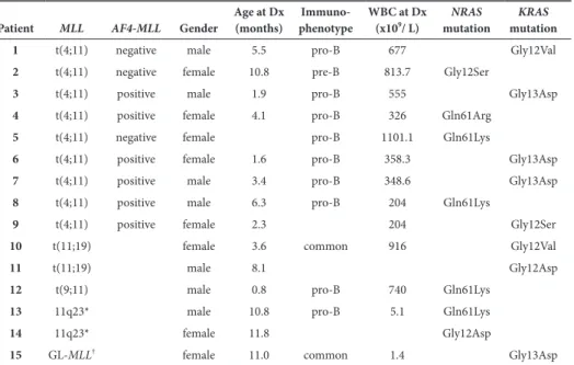
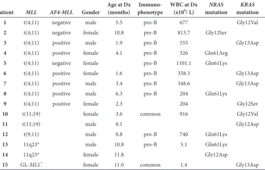
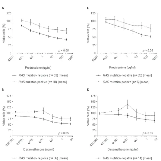
Using conventional sequencing analysis, we screened neuroblastoma RAS viral (v-ras) onco-gene homolog onco-gene(NRAS), v-Ki-ras Kirsten rat sarcoma viral oncogene homolog gene(KRAS), and v-raf murine sarcoma viral oncogene homolog B1 gene(BRAF) for mutations in a large cohort(n=109) of infant ALL patients and studied the mutations in relation to several clini-cal parameters, and in relation to Homeobox gene A9 expression and the presence of ALL1 fused gene 4-Mixed Lineage Leukemia(AF4-MLL).
Mutations were detected in ~14% of all cases, with a higher frequency of ~24% in t(4;11)-positive patients(p=0.04). Furthermore, we identified RAS mutations as an independent predictor(p=0.019) for poor outcome in MLL-rearranged infant acute lymphoblastic leuke-mia, with a hazard ratio of 3.194(95% confidence interval:1.211-8.429). Also, RAS-mutated infants have higher white blood cell counts at diagnosis(p=0.013), and are more resistant to glucocorticoids in vitro(p<0.05). Finally, we demonstrate that RAS mutations, and not the lack of Homeobox gene A9 expression nor the expression of AF4-MLL are associated with poor outcome in t(4;11)-rearranged infants.
We conclude that the presence of a RAS mutations in MLL-rearranged infant acute lym-phoblastic leukemia is an independent predictor for a poor outcome. Therefore, future risk-stratification based on abnormal RAS-pathway activation and RAS-pathway inhibition could be beneficial in RAS-mutated infant acute lymphoblastic leukemia patients.
43 RAS mutations in MLL-rearranged infant ALL
intRoDuCtion
Acute lymphoblastic leukemia (ALL) in infants (<1 year of age) represents an aggressive, early onset type of leukemia characterized by high relapse rates during treatment, and an unfavorable clinical outcome1. This poor prognosis is associated with a high incidence of balanced chromosomal translocations involving the Mixed Lineage Leukemia (MLL) gene, which occur in ~80% of the infant ALL cases1. The most common MLL translocation in infant ALL is t(4;11), in which the N-terminus of MLL (chromosome 11q23) fuses to the C-terminus of AF4 (chromosome 4q23). As the joining of MLL and AF4 occurs in-frame, the t(4;11) translocation generates a unique fusion gene encoding the chimeric, and suppos-edly oncogenic MLL-AF4 fusion protein. Other recurrent in-frame MLL-rearrangements found among infant ALL patients are t(11;19) and t(9;11), giving rise to the fusion proteins MLL-ENL and MLL-AF9 respectively. The presence of an MLL translocation is the strongest independent predictor of an adverse outcome in infant ALL patients2.
Over the past decades numerous studies provided important insights into the biology and pathogenesis of MLL-rearranged ALL, but so far in vivo validation of these achievements are hampered by the lack of genuine animal models accurately recapitulating this severe malignancy. Although various attempts have been made to develop mouse models mimicking leukemogenesis of human t(4;11)-positive ALL, these mice displayed propensities towards de-veloping lymphomas or le