A structural narrative
Arnab Bhattacharjee
University of Helsinki
Complement Factor H C-terminus and its significance: A structural narrative
Arnab Bhattacharjee
Department of Bacteriology and Immunology Haartman Institute
University of Helsinki Finland
and
Research Programs Unit, Immunobiology Faculty of Medicine
University of Helsinki Finland
To be publicly discussed with the permission of the Medical Faculty, University of Helsinki, in auditorium 1 of Haartman Institute,
Department of Bacteriology and Immunology, Haartman Institute
and
Research Programs Unit, Immunobiology, University of Helsinki, Finland
Reviewed by:
Adj. Prof. Arno Hänninen, MD, PhD
Department of Medical Microbiology and Immunology, University of Turku,
Finland
Adj. Prof. Veli-Pekka Jaakola, PhD Department of Biochemistry, University of Oulu,
Finland
Opponent:
Professor Péter Gál, PhD
Institute of Enzymology, Biological Research Center, Hungarian Academy of Sciences, Budapest,
Hungary
Illustrations by the author
ISBN 978-952-10-9854-3 (paperback) ISBN 978-952-10-9855-0 (PDF) Printed at Oasis media Finland Oy Finland 2014
‘Cogito ergo sum’,: I think, therefore I am
~René Descartes
Dedicated to
the honourable memory of my first biology teacher in school ,
‘Ajitda’
who seeded the interest in me to look through biological systems, but couldn’t live to cherish the day
CONTENTS
ORIGINAL PUBLICATIONS ... 5
CONTRIBUTION TO PUBLIC DATABASES ... 6
ABBREVIATIONS ... 7
ABSTRACT ... 8
1 INTRODUCTION ... 11
2 REVIEW OF LITERATURE ... 13
2.1 The complement system: an overview ... 13
2.2 Activation of the complement system ... 14
2.3 Regulation of the complement system ... 16
2.4 The CFH family: focus on structure and functions ... 19
2.5 CFH Interactions with Host Ligands ... 25
2.6 Diseases associated with CFH family of proteins ... 29
2.7 The microbial complement evasion: role of CFH ... 32
2.8 Anti-FH autoantibodies and aHUS ... 33
3 OBJECTIVES ... 36
4 MATERIAL AND METHODS ... 37
4.1 Materials ... 37
4.2 Methods ... 38
5 RESULTS ... 48
5.1 Both domains (19 and 20) of CFH are involved in C3b binding ... 48
5.2 A bigamic interaction of CFH revealed ... 50
5.3 Borrelia burgdorferi binds CFH20 to evade complement ... 52
5.4 Molecular basis for autoimmunity against CFH in aHUS ... 55
6 DISCUSSION ... 58
6.1 Molecular basis for the self non-self discrimination by AP ... 58
6.2 The microbial complement evasion: a molecular perspective ... 61
6.3 Link between the CFHR1 deficiency and the AI-aHUS puzzle ... 62
6.4 Structural analyses of various structures of CFH19-20 ... 68
6.5 Crystal artifacts, the pinch of salt ... 69
6.6 Conclusions and future prospects ... 70
7 ACKNOWLEDGEMENTS ... 71
ORIGINAL PUBLICATIONS
This thesis is based on the following publications:
I) Bhattacharjee A., Lehtinen M. J., Kajander T., Goldman A. and Jokiranta T. S. (2010) Both domain 19 and domain 20 of factor H are involved in
binding to complement C3b and C3d. Mol Immunol 47, 1686-91.
II) Kajander T., Lehtinen M. J.*, Hyvärinen S.*, Bhattacharjee A.*, Leung
E., Isenman D. E., Meri S., Goldman A. and Jokiranta T. S. (2010) Dual interaction of factor H with C3d and glycosaminoglycans in host-nonhost
discrimination by complement. Proc Natl Acad Sci USA, 108(7),
2897-902.
III) Bhattacharjee A., Oeemig J.S., Kolodziejczyk R., Meri T., Kajander T., Iwai H., Jokiranta T.S.*, Goldman A.* (2013) Structural basis for
complement evasion by Lyme disease pathogen Borrelia burgdorferi. J
Biol Chem 288,18685-695.
IV) Bhattacharjee A., Reuter S., Kolodziejczyk R., Seeberger H., Hyvarinen S., Prohászka Z., Szilágyi Á., Goldman A., Józsi M, Jokiranta T.S. (2013) The major AA epitope on CFH and the structure of its homologous site on CFHR1 explain why CFHR1 deficiency leads to aHUS. (Submitted)
*authors contributed equally to the article
The publications are referred to the text by their roman numerals. The articles have been reprinted with permission of the copyright holders.
CONTRIBUTION TO PUBLIC DATABASES
I) Bhattacharjee A., Lehtinen M. J., Kajander T., Goldman A. and Jokiranta
T. S. (2010): 3KXV; Structure of complement Factor H variant R1203A,
three-dimensional structural data deposited in the RCSB protein data bank (www.rcsb.org/pdb).
II) Bhattacharjee A., Lehtinen M. J., Kajander T., Goldman A. and Jokiranta
T. S. (2010): 3KZJ; Structure of complement Factor H variant Q1139A,
three-dimensional structural data deposited in the RCSB protein data bank (www.rcsb.org/pdb).
III) Kajander, T., Lehtinen, M. J., Hyvarinen, S., Bhattacharjee, A., Leung,
E., Isenman, D. E., Meri, S., Jokiranta, T. S., Goldman, A. (2010): 2XQW;
Structure of complement Factor H domains 19-20 in complex with complement C3d, three-dimensional structural data deposited in the RCSB protein data bank (www.rcsb.org/pdb).
IV) Bhattacharjee, A., Oeemig, J.S., Kolodziejczyk, R., Meri, T., Kajander,
T., Iwai, H., Jokiranta, T., Goldman, A. (2013): 2M4F; Solution structure
of Outer surface protein E, three-dimensional structural data deposited in the RCSB protein data bank (www.rcsb.org/pdb).
V) Bhattacharjee, A., Kolodziejczyk, R., Kajander, T., Goldman, A.,
Jokiranta, T.S. (2013): 4J38; Structure of Borrelia burgdorferi Outer
surface protein E in complex with Factor H domains 19-20, three-dimensional structural data deposited in the RCSB protein data bank (www.rcsb.org/pdb).
VI) Bhattacharjee, A., Kolodziejczyk, R., Goldman, A., Jokiranta, T.S.
(2013): 4MUC; Structure of the C-terminus of Factor H related protein I,
three-dimensional structural data deposited in the RCSB protein data bank (www.rcsb.org/pdb).
ABBREVIATIONS
AA Autoantibody
AI-aHUS Autoimmune Atypical haemolytic uremic syndrome
aHUS Atypical haemolytic uremic syndrome
AMD Age related macular degeneration
AP Alternative pathway
CCP Complement control protein
CFH Complement Factor H
CFH19-20 Domain 19 and domain 20 of Complement Factor H
CFHL-1 Complement Factor H like protein-1
CFHR Complement Factor H related protein
CFHR1 Complement Factor H related protein 1
CFHR14-5 Domain 4 and domain 5 of Complement Factor H related protein 1
CFH-AA anti Complement Factor H autoantibody
CP Classical pathway
CR Complement receptor
CRP C-reactive protein
CS-A Chondroitin sulfate A
DAF Decay accelerating factor (CD55)
DDD Dense deposit disease
FI Factor I GAG Glycosaminoglycan HS Heparan sulfate IC Immune complex iC3b Inactive C3b Ig Immunoglobulin LP Lectin pathway
mAb Monoclonal antibody
MAC Membrane attack complex
MASP MBL-associated serine protease
MBL Mannan binding lectin
PBS Phosphate buffered saline
RCA Regulators of complement activation
RLA Radioligand assay
RMSD Root mean square deviation
SA Sialic acid
SDS-PAGE Sodium dodecyl sulfate – polyacrylamide gel electrophoresis
SPR Surface plasmon resonance
ABSTRACT
Complement is comprised of a cascade of proteins that recognizes and attacks the invading microbes and thus is the first line of defense for the human body against invading pathogens. It is initiated via different activation pathways that lead to C3b deposition on the target and sequentially to the formation of lytic membrane attack complexes (MAC). One of the complement activation pathways – the alternative pathway (AP) – can be activated on any surface, self or non-self, and is therefore tightly regulated. Complement Factor H (CFH) is the most important complement down-regulator and it mediates target discrimination between self and non-self cells. Several point mutations in CFH and/or autoantibodies (AA) against it are found to be directly associated with atypical haemolytic uremic syndrome (aHUS), a severe and often fatal disease triggered by the impaired regulation of AP on self surfaces leading to complement attack.
CFH is composed of 20 homologous complement control protein domains (CCP). The N-terminal domains 1 to 4 (CFH1-4) mediate inactivation of C3b on the self-surfaces and the C-terminal domains 19 and 20 (CFH19-20) are critical for the ability of CFH to discriminate between self and non-self structures. Self-surfaces are rich in anionic sialic acids (SA) and glycosaminoglycans (GAGs) that are not present on pathogenic microbes. CFH19-20 contains binding sites for both the C3d part of C3b and self-surface polyanions that enhance avidity of CFH to C3b on self surfaces and thus enhance C3b inactivation. CFH mutations that have been found in aHUS patients are mostly located in CFH19-20.
The previously solved X-ray crystal structure of CFH19-20 illuminated the location of aHUS related mutations. The aims of this thesis work were to study the functionality of CFH on a molecular level by studying the molecular structure of CFH C-terminus and its mutants along with structures of CFH in complex with its different interacting partners, as well as the structure of the domains of CFH-related protein-1
highly homologous to CFH19-20. The structures solved and the their relevance are
described in the four articles attached to this thesis.
In the first article, stability of the CFH C-terminus fold by aHUS mutation(s)
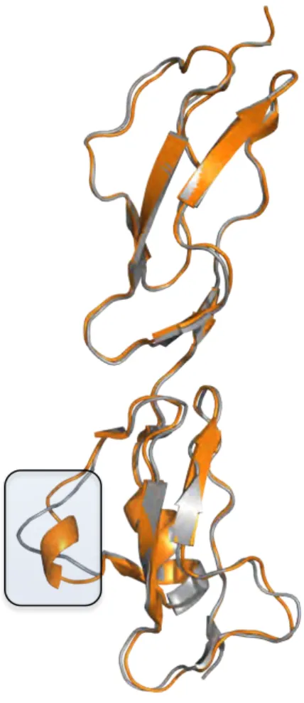
using radioligand assays and affinity chromatography. The X-ray crystal structures of CFH19-20 with two different point mutations (of residues indicated to be involved in binding C3d/C3b) were solved. It was shown that these mutations did not result in the disruption of the basic fold of CFH19-20, but maintained the same fold with a prominent difference in the surface charge distribution in the zone of the residues. The results clearly indicated that the aHUS mutations on CFH do not disrupt the basic structural fold, but induce anomaly in the charge distribution of the molecule, explaining the effects of the mutations to its C3b/heparin binding abilities suggested to be critical for the pathogenesis of the disease.
In the second article, we revealed the rationale of the molecular mechanism of CFH19-20 mediated self–nonself discrimination and showed why point mutations in CFH19-20 lead to aHUS. The CFH19-20 :C3d structure reported in this article revealed
two independent binding interfaces between CFH19-20 and C3d, namely the ‘CFH19
site’ and the ‘CFH20 site’. The results of deeper analysis of this structure also showed
that the simultaneous binding of the CFH19-20 via the CFH19 site to C3b and via CFH20
site to C3d was possible.
In the third article, the X-ray crystal structure of CFH19-20 was solved in
complex with the outer surface protein E (OspE) from Borrelia burgdorferi in order
to understand the molecular mechanism of sequestering CFH by microbes which results in complement evasion. The nucleomagnetic resonance (NMR) structure of the
OspE protein from Borrelia burgdorferi reported in this paper was required for
solving the structure of OspE in complex with CFH19-20. Chemical shift perturbations studies using NMR also confirmed the physiological viability of the complex structure. This complex structure was the first structure of CFH19-20 in complex with any microbial protein and thus answered the puzzle of the molecular mimicry used by microbes involving CFH C-terminus in order to evade complement.
In the fourth article the structure of the CFHR1 domains 4 and 5 (CFHR14-5) was solved and used to explain why AA bind to a common epitope on CFH domain 20, which is highly homologous to domain 5 of CFHR1. The CFHR14-5 structure revealed an important structural bigamy of CFH and its related proteins that can be used to understand why CFHR-1 deficiency and formation of AA against CFH (CFH-AA) lead to autoimmune aHUS (AI-aHUS). We extensively studied CFH-AA from more than a dozen of patients and their binding behavior to CFH and CFHR1. The results suggest a novel hypothesis on the pathogenesis of AI-aHUS.
In conclusion, the results have revealed the molecular mechanisms beneath different functionalities of the C-terminus of CFH. It is not only the most important molecule to facilitate the target discrimination by the AP, but is also a prominent tool used by the microbes in order to evade complement attack. Furthermore, the CFH C-terminus also houses the AA binding epitope and thus also plays a role in the pathogenesis of AI-aHUS. We were also quite surprised to find out that the molecular structure of CFH C-terminus is extremely stable, and hardly undergoes any changes in its conformation in the presence of other ligands. Cumulatively, the structures of the CFH in presence of its different partners of interactions contributed greatly to the knowledge pool of the understanding of the molecular mechanisms associated with CFH in complement activation and regulation in health and disease.
1
INTRODUCTION
The human body needs to protect itself both from invading pathogens as well as from the side effects of the immune processes going on inside the body. The continuous challenge thrown towards our body is both internal and external in nature. The security system our body puts up in order to handle these challenges is called the
immune (coming from the latin word immunitas) system, which with evolution has
developed into an organized, sensitive and a robust system.
Our immune system can be classified into two parts, namely the innate and adaptive immunity. The concept of innate immunity shaped through evolution to discriminate self from non-self cells has been expanded, during the last decades, owing to the description of additional functions of the innate defenses. This part of immunity not only recognizes non-self molecules or surfaces, but also utilizes more sophisticated mechanisms of recognition. It is the type of immunity that provides the early line of defense against invading microbes and is comprised of the cellular and biochemical defense. Innate immunity is pre-poised to respond to an external attack even before they take place, but responds to repeated infections in a non-specific manner. On the contrary, the adaptive immune responses come into action by an exposure to external invading agents and increases its defense capabilities with each successive exposure to the particular microbe.
The most important components of the innate immune system are the epithelial barriers, phagocytic cells, dendritic cells, natural killer cells, cytokines and the complement system. The complement system was described in 1889 when Hans Büchner noticed a heat labile substance in blood serum which was able to kill bacteria, thus ‘complementing’ the action of adaptive humoral immunity. Paul Ehrlich first used the term “complement” by 1899 (1) for the phenomenon mentioned below in more detail.
The mammalian complement proteins provide a first degree immune response against pathogens in blood and interstitial fluids (2). The primary aim of the complement system is to identify surfaces of bacteria, viruses and fungi, and also apoptotic host cells in order to be treated for cell lysis, clearance by phagocytosis and stimulation of the adaptive immune response.
Since 1960s, there has been a remarkable transformation in our understanding of the immune system and its functions. Advances in cell culture techniques and monoclonal antibody (mAb) production, immunochemistry, recombinant DNA technology, studies of molecule structures at atomic resolution employing different methods, and the generation of transgenic animals have changed the nature of immunological studies. Most of the immunological phenomena are now explained in structural and biochemical terms silently leading to revolutionary advances in the field.
This thesis deals with different aspects of complement system and its occasional failure in case of the onset of infections or autoimmunity using X-ray crystallography as a tool to study it at atomic (or close to atomic) resolution.
2
REVIEW OF LITERATURE
2.1
The complement system: an overview
Innate immunity is the first-line of the immune defense system, essential for cellular integrity, tissue homeostasis and modifying the adaptive immune response. The innate immune system comprises of several components and uses several receptors for the recognition of microorganisms. A major humoral component of innate immunity is the complement system, which was established early in evolution and is the primary immune system in invertebrates. Complement was initially identified more than 100 years ago as a serum component that ‘complements’ (thus contributing to the nomenclature) the antibody response towards pathogens (1,3). Now, more than a hundred years after these initial reports, the central role of this system in immune defense is much better known. It is now established as a system of a cascade of proteins and many of the biological effects of complement are understood in molecular terms and a role for complement in tissue homeostasis is emerging (2,4,5). Complement activation activates pro-inflammatory mediators, generates anaphylactic peptides, cytolytic compounds and antimicrobial compounds, recruits effector cells and induces effector responses. These proteins form a complex network of various recognition, effector, regulatory and receptor molecules that act in a finely tuned fashion, allowing complement to safely exert its functions (6,7). The activated complement system is beneficial for the protection of the host against invading pathogens (8). Other than protecting the host from infections by destroying and eliminating pathogens, complement maintains the integrity of the body by discriminating between healthy and injured tissue, in participating in the disposal of immune complexes and other cellular debris (9,10), also in inflammatory processes, angiogenesis and tissue regeneration (11,12). The individual complement reactions develop in a sequential manner, allowing regulation that modulates the intensity of the response and adjusts the effector functions for the specific immune response. It has also been established that complement is involved in the induction and regulation of both innate and adaptive cellular immune responses (2,13).
2.2
Activation of the complement system
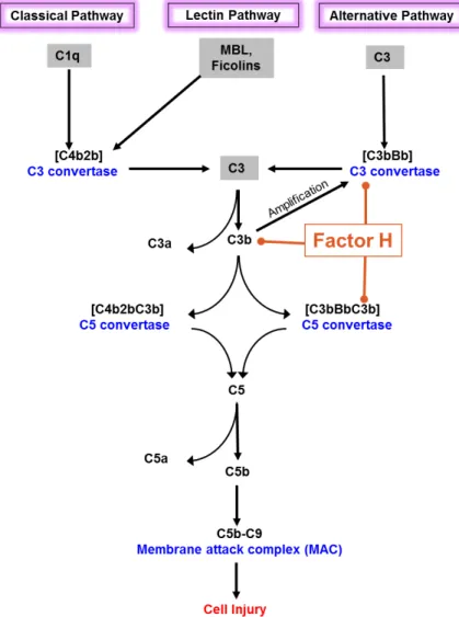
Complement can be activated via three primary pathways, the classical pathway (CP), the lectin pathway (LP), and the AP. All pathways converge at the central component C3 (Figure 1), which eventually initiates the terminal pathway forming the membrane attack complex (MAC)
The CP is initiated by C1q binding to surface-bound immunoglobulins (Ig, mainly IgG1, IgG3 or IgM) or apoptotic cells (14-18). C1q is reported to bind to the Fc regions of the antibodies in order to initiate the CP (19). It has been suggested that binding of C1q to multiple, or even multimerized Ig, on a surface results in changes of the conformation of the C1-complex; this would prevent fluid phase antibodies from activating the cascade (20). Bacterial lipopolysaccharides, DNA, C-reactive protein (CRP), serum amyloid P component (SAP), and pentraxin 3 (PTX3) have been also found to activate the CP (21-25), but the physiological importance of these phenomena remains mainly elusive.
The LP is activated by binding of mannose-binding lectin (MBL) to repetitive carbohydrates, or by binding of ficolins to carbohydrate or acetylated groups on target cell surfaces (26-28). MBL binds mannose and N-acetyl-glucosamine residues present in bacterial cell walls. The LP activation starts with association of two C1r- and C1s-like MBL-associated serine proteases (MASP-1 and MASP-2) with MBL. Complement components C4 and C2 are both cleaved by MASP-2 leading to complement activation (29-31). MBL and C1q are structurally related but MBL possesses carbohydrate-binding lectin domains which is not present in C1q (32). LP can be also be activated by M-, L-, and H-ficolins (also called 1-, 2- and 3-ficolins), respectively. All of these ficolins function as pattern recognition molecules that recognize carbohydrates on microbial surfaces (28).
The AP is spontaneously activated by the hydrolysis of the internal thioester group of C3 eventually forming the C3 convertase (classical/lectin pathway C3 convertase C4b2aand the AP C3 convertase C3bBb). (17,33). This complex cleaves fluid phase C3 to C3a (a small 9 kDa inflammatory mediator) and C3b, an opsonin that deposits on the surface of target cells and particles and promotes phagocytosis. (34-38). This spontaneous complement activation in fluid phase, a unique feature of AP, is constantly targeting all surfaces that are devoid of sufficient down-regulators.
The larger fragment C3b formed as a result of C3 cleavage can also form additional C3 convertases with Bb and amplify complement activation. The cleaved C3b exposes a thioester group that can bind covalently to a target surface similarly to C4b in CP. At this stage the interaction between C3b and either factor B or CFH forms the basis of the self vs. non-self discrimination of AP (39,40). The amplification of AP occurs at this stage when C3 convertase activates more fluid phase C3 by cleavage to C3b (41). This amplification step causes effective C3b opsonization of the target (42,43). C3b can also bind to an already existing C3 convertase, to form the C5 convertase which cleaves C5 and activates the terminal complement pathway. This activation of C5 potentially leads to inflammation (via C5a generation) and results in the lysis of the target cell through the formation of membrane pores by the MAC (44). Whereas these activation processes are needed on microbial surfaces, they are destructive to host (self) cells.
2.3
Regulation of the complement system
The complement activity is important in our body for the clearance of foreign and altered host cells, whereas on the other hand, this activity must be well controlled in order to avoid self tissue damage. The spontaneous activation of AP as well as its secondary activation via the ‘amplification loop’ of classical/lectin pathway makes its regulation extremely important. Consequently, the process of complement activation is strictly regulated by membrane-bound and blood-plasma regulatory molecules that protect against complement activation by down-regulating the central proteolytic activity of the amplification and opsonisation steps. Deficiencies or mutations in complement regulators may predispose individuals to infectious and immune-related diseases or may lead to excessive complement activation and tissue injury.
The complement system with its casacade of regulatory proteins may act during activation, amplification and effector steps of the cascade that enables fine-tuning of the activation in different physiological circumstances. These proteins compete with other complement components for the binding sites.
The regulators of complement activation (RCA) gene cluster in chromosome
1 encode several complement regulatory proteins (45-50). All the RCA proteins are composed of several homologous domains that have probably arisen by gene duplication events (51). These domains are approximately composed of 60 amino acids each and these protein domains are arranged in a bead-like structure. These short consensus repeat domains (presently known as CCP domains) are generally arranged in proteins in a head-to-tail fashion resembling beads-in-string (52). The binding sites in CCP-bearing proteins may span over its domains and therefore the orientation of the CCPs is critical for the proteins to act. These RCA proteins include C4b binding protein (C4bp), CFH, CFH-like protein-1 (CFHL-1), CFH-related (CFHR) proteins and membrane bound regulators complement receptor 1 (CR1/CD35), membrane cofactor protein (MCP/CD46), and decay accelerating factor (DAF/CD55)(46-48).
2.3.1 Soluble complement regulators
C1 inhibitor (C1inh): C1inh is a 105 kDa serine protease inhibitor (serpin) with a heavily glycosylated single chain protein with a two domain architecture (53,54) which regulates the complement activation (via CP) by binding to C1r and C1s
causing their dissociation from C1q. It also influences the C activation in fluid phase by acting on C1 that otherwise could be spontaneously autoactivated by non-immune activators at low levels (55). Deficiency in C1inh results in hereditary angioedema (HAE), a disease characterized by recurrent acute edema of skin or mucosa which is associated with the lack of regulation of kallikrein by C1inh (56).
C4b-binding protein (C4bp). C4bp is a 540-590 kDa protein consisting of several disulphide bonded subunits (57, 58) which interferes with the assembly of surface bound CP C3-convertase C4b2a by accelerating its decay. It also functions as a cofactor for factor I in cleavage of both surface bound and fluid phase C4b to C4c and C4d (59).
CFH and its family. CFH is an elongated, approximately 155 kDa single-chain glycoprotein (60) consisting of 20 CCP domains (61)(Figure 2). When bound on the surface, CFH regulates AP indirectly by acting as a cofactor for the inactivation of C3b by factor I (62). It also directly inhibits the formation of C3-convertases by interfering with the binding of FB to C3b (39) and furthermore increases the pace of the natural decay of already formed AP convertases (63,64). Mutations in CFH have been associated with aHUS (65,66), dense deposit disease (DDD, also known as membranoproliferative glomerulonephritis type II or MPGN II ) (67). CFH has been found to have some of its polymorphisms (68) associated with aHUS as well (69). One of the CFH polymorphisms also has been associated with age related macular degeneration (AMD) (70,71).
Several other proteins from the RCA gene cluster have been found to have strikingly similar structural and functional features like CFH. These are all categorized in the family of CFH that will be discussed later.
Properdin. Properdin exists in plasma as oligomers of two to four monomers linked together in a cyclic fashion (72). Each 52 kDa monomer consists of six thrombospondin repeat-domains (73). Properdin is the only positive regulator of complement activation that stabilises the nascent C3/C5 convertases of the AP and thus subsequently slowing down their rate of decay. (74).
Vitronectin. Also known as S-protein, vitronectin exists in at least two forms in plasma, a single chain molecule (84 kDa) or a double chain molecule where the subunits (69 kDa and 15 kDa) are linked together with disulfide bonds. It is also expected to form dimers and higher molecular weight aggregates in plasma (75). Vitronectin binds fluid phase C5b-7 in order to prevent its membrane-binding and thus prevent MAC insertion (76).
Factor I. Factor I is a 90 kDa enzyme consisting of a 50 kDa and a 38 kDa chain linked together by a disulphide bond. (77,78). Factor I regulates the complement system by inactivating C4b and C3b molecules (with either CFH, C4bp, CR1 or MCP working as cofactors) (79-81).
2.3.2 Membrane bound regulators
CR1/CD35. This is a 205 kDa polymorphic protein and was first isolated from erythrocyte membranes. It regulates complement activation on cell surfaces and facilitates the decay of fluid phase immune-complexes (82,83). CR1 gene is located in the RCA cluster.
Complement receptor of the Immunoglobulin family (CRIg). CRIg is a complement receptor expressed on tissue macrophages. It mediates opsonophagocytosis by binding C3b and inactive C3b (iC3b) on opsonized particles (84). CRIg regulates AP activation by binding to C3b and inhibiting the interaction of C3 and C5 with AP convertases (85).
Decay accelerating factor (DAF or CD55). DAF regulates the AP and CP by accelerating the decay of C3-convertases (86). DAF is a 70 kDa membrane protein composed of four CCP domains and a serine/threonine/proline (STP)-rich linker (87).
Membrane cofactor protein (MCP/CD46). It is 50-85 kDa heavily glycosylated membrane protein widely expressed on cells other than erythrocytes. It acts as a cofactor for C4b and C3b cleavage by factor I as mentioned before (81). MCP has four CCP domains and a heavily glycosylated STP region along with transmembrane and cytoplasmic domains as well (46).
CD59 (protectin). CD59 is a 18-20 kDa single chain protein expressed by RBC and other cells (88). This has five intra-chain disulphide bonds (89), heavily glycosylated and binds to the membrane associated C5b-8 complex preventing the unfolding of C9 (90) and thus the membrane spanning by C9.
2.4
The CFH family: focus on structure and functions
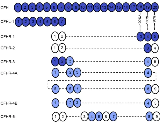
CFH belongs to a protein family that also includes CFHL-1 (an alternative splicing product of the CFH gene) and five CFH-related proteins (CFHR) (91) (Figure 2). These proteins are composed of four to nine CCP domains, and share various degrees of sequence identity with each other and with CFH (Figure 2).
2.4.1 Complement Factor H (CFH) and target discrimination
CFH is the main soluble regulator of the AP (92). The CFH coding gene (CFH) is
located in chromosome 1q32 within the RCA gene cluster (as mentioned before),
adjacent to the five genes that code for the CFHRs. CFH is constitutively expressed in Figure 2. The schematic structure of factor H and its family members. For sequence identities of 32-49% the domains are displayed in white, for 57-84% identity in light blue, and for 85-100% identity in dark blue. The identity between the domains 3-5 of CFHR1 and domains 18-20 of CFH is indicated with percentages; *, sequence identity of the domain 3 of the basic isoform of CFHR1 is 100%, whereas that of the acidic isoform is 95% (216). CFH-like molecule-1 (CFHL-1) is an alternatively spliced transcript from the CFH gene having four unique residues after domain 7.
the liver and is distributed systemically in body fluids. Reported CFH plasma concentrations vary, depending on the study population, age, genetic and environmental factors, as well as on the method used for quantification (93). Previous studies overestimated CFH concentration, which was a result of measuring both CFH and CFHRs. So, while early reports suggested that the plasma CFH concentration ranges 265–684 µg/ml (94) and 116–562 µg/ml (68), current studies using monoclonal antibodies have established mean CFH concentrations of 233 µg/ml (in young adults), 269 µg/ml (in elderly individuals) (95) and 263 µg/ml (93), in different healthy control populations with the age group between 25-66 (mean 42) years. Thus, normal CFH concentrations in human plasma correspond to approximately 1–2 µM. In addition, CFH is also produced outside the liver by different cell types such as monocytes (96), fibroblasts (97), endothelial cells (98), keratinocytes (99), thrombocytes (100) and retinal pigment epithelial cells (101). Locally released CFH in tissues may help to limit complement activation and maintain an anti-inflammatory environment.
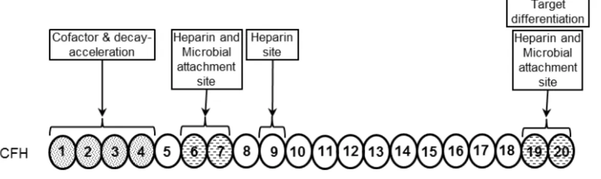
Each of the autonomously folding globular domains (see 2.3.1) is composed of about 60 amino acids and is stabilized by two internal disulfide-bonds (61). As mentioned earlier, CFH regulates complement activation by inhibiting the assembly of the AP C3 and C5 convertase enzymes via competition with FB for C3b binding; thus by facilitating the dissociation of the convertases by displacing bound factor Bb; and by acting as a cofactor for the serine protease factor I in the cleavage and inactivation of C3b (64,102). These regulatory activities are mediated by the four N-terminal domains 1–4 of CFH (103,104) (Figure 3), whereas the C-N-terminal domains 19-20 are responsible for target recognition (Figure 3) (105-107). One of the important targets for CFH binding in the vicinity of C3b on host cells are polyanionic surface molecules, such as GAGs and SA, which increase the affinity of CFH for C3b (40). Thus, CFH is also able to control complement activation on self-surfaces (Figure 2, also see section 2.5) (108,109). The target discrimination by AP is mediated by CFH and it has been established that the negatively charged polyanions or SA on the self cells contributes to higher CFH:C3b (surface) interaction and thus leads to high cofactor activity of CFH (39,40,64,110). In contrast, host-like polyanionic molecules are normally not present on the surface of pathogens, rendering them susceptible to complement attack.
In addition to C3b and polyanionic molecules, CFH interacts with several other endogenous ligands. These interactions allow CFH to regulate complement on certain host surfaces (such as the glomerular basement membrane, the extracellular matrix, and late apoptotic cells), which are otherwise not properly protected due to a reduced expression or the lack of membrane-anchored complement regulators (i.e., membrane cofactor protein, decay accelerating factor and CD59). Several of these ligands and structures activate complement via interactions with C1q, MBL, or ficolins. The simultaneous binding of both complement activating molecules (i.e., C1q, MBL) and molecules inhibiting the activation of complement (i.e., CFH, C4b-binding protein) on such ligands and cells may facilitate their opsonisation and safe removal, but at the same time prevents an exaggerated complement activation that could lead to inflammation, unnecessary cell lysis, and subsequent tissue damage (111).
Figure 3. Complement activation pathways and the regulatory role of factor H. The primary function associated with FH domains are also indicated. Sparsely dotted regions indicate (domains 6-7, domains 19-20) heparin and microbial binding sites while the densely dotted region (Domains 1-4) is associated with cofactor and decay acceleration activities.
CFH also binds to nonhost ligands, such as certain surface proteins of microbes, which hijack host CFH in order to protect themselves from complement attack (112). AP evasion by microbes is considered to the primary reason for the sequestering of CFH on their surface. Streptococcus pyogenes was the first microbe to
be identified with this phenomenon to evade complement-mediated
opsonophagocytosis (113-115).
Most microbes acquire CFH via two regions of this control protein, domains 5-7 and 18-20 (Figure 4). Domains 6-7 of CFH have been evidently functional in
binding with Streptococcus and Neisseria surface proteins(114-119). Domain 18-20
of CFH has been suggested to mediate Neisseria gonorrhoeae complement evasion
via Por1A and Por1B (119-121). Borrelia burgdorferi also uses two different surface
proteins for CFH binding, viz. OspE for binding domains 18-20 of CFH (122,123) and
BbCRASPs for binding to domains 6-7 (124,125). Some microbes such as Yersinia
enterocolitica (126,127) and Streptococcus pneumoniae (128,129) have been shown to bind to non-conventional microbial binding domains in CFH. Interestingly, there are also microbes which have been found to bind CFH via two different domains
using the same ligand on their surface, such as Pseudomonous aeruginosa (130) and
Candida albicans (131,132).
Sbi protein of Staphylococcus aureus and shiga toxin (Stx2) of
enterohemorrhagic Escherichia coli have been reported to bind CFH in a
non-conventional manner. Sbi binds domains 19-20 of CFH by forming a tripartite complex with C3b (133), whereas Stx2 binds both CFH domains 6-7 and 18-20 in fluid phase (134).
Several microbial ligands involved in CFH interactions have not been
identified. For example, Haemophilus influenzae (135), Fusobacterium necrophorum
(136), Aspergillus fumigatus (137), Bordetella pertussis, Bordetella parapertussis
(138) and Onchocerca volvulus (139) have been reported to bind to CFH, but the
bacterial ligands remain unknown. All of these microbes interact at least with CFH domains 6-7 and/or 18-20.
2.4.2 The CFH like protein-1
CFHL1 is a 43-kDa protein deriving from an alternative splice product of the CFH gene. It shares the seven domains (1-7) with CFH and has four additional C-terminal amino acids. As expected owing to high sequence similarity, CFHL1 possesses the
same complement regulatory activities as mediated by the N-terminus of CFH. CFHL1 may also play a role in age-related macular degeneration as the domain7 harbors the site for Y402H polymorphism (see 2.7). Consequently, the CFHL1 402H variant has impaired ligand-binding capacity, similar to that exhibited by the CFH 402H variant (140,141). However, an expression pattern differing from CFH and a distinct role in mediating cell adhesion have been reported for CFHL1 as well (142,143). CFHL1 has been observed to bind microbes which do not bind to CFH as well (144).
2.4.3 The CFH related proteins
The five CFHR proteins (CFHR1-CFHR5, Figure. 2) are from separate genes located adjacent to the CFH gene. They have domains homologous to the different domains of CFH, which are responsible for directing the complement regulatory activity of CFH to cell and tissue surfaces (91). These proteins have both similar and non-redundant ligands and functions with CFH. CFHR1, CFHR3, CFHR4 and CFHR5 bind to C3b, CFHR1, CFHR3 and CFHR5 to heparin, and CFHR4 and CFHR5 to CRP (145-151). In spite of similar cell and ligand binding properties, however, CFHR1 was reported to regulate the terminal complement pathway (145). In addition, it has been shown that both CFH and CFHR1 enhance neutrophil adhesion and activation during host cell-pathogen contact (152). Similar to CFH, CFHR4 binds to late apoptotic and necrotic cells, but in contrast to CFH, CFHR4 binds to the native pentameric form of CRP (153).
2.4.4 Discrimination of self and non-self by CFH
Identification of cell surfaces by the complement system is achieved by covalent binding of C3b molecules to the target surface through an exposed thioester in C3b that reacts with hydroxyl or amine groups. After initial binding of C3b or its homolog C4b by the initiation of complement cascade, the response (other than C4b2a) is amplified by enzyme complexes, or convertases, that form on the surface-bound C3b or C4b molecules.
Host cells require soluble complement inhibitors, particularly CFH, which provide effective protection from unwanted complement-mediated damage, especially under conditions with strong complement activation. It is critical to protect noncellular surfaces, which do not carry surface-bound regulators. Several mutations
in the two C-terminal domains of CFH have been linked to aHUS (109). The same mutations also exhibited reduced functioning of CFH (154). Self-nonself discrimination by CFH is mediated by domains 19-20 that bind to surface-bound C3b/C3d and host surface GAGs or SA (155). Critical for target discrimination is the formation of a ternary complex of CFH domains 19–20, surface-bound C3b and GAGs or SA or C3d from the self-surface.
Host cells, so-called non-activators of complement, possess cell surface polyanionic molecules that promote CFH binding (40). In contrast, complement activators such as microbes or rabbit erythrocytes that lack SA and host-like GAGs do not allow significant CFH binding and thus activates complement. Heparin is generally used in studies as a representative of host GAGs, and the major heparin binding sites are located in domains 7 and 20 (154-157). Polymorphisms or mutations in domains 7 and 19-20 may affect interactions of CFH with host cells and basement membranes, and are implicated in the diseases AMD and aHUS (106).
2.5
CFH interactions with host ligands
Other than its role as a complement regulator, CFH has been shown to mediate cellular interactions by binding to receptors on various cells as well.
2.5.1 CFH interaction with C3b
The central complement protein C3b is the main host ligand of CFH. The C3b-CFH interaction is of particular importance for the functions of CFH, namely complement regulation and host surface recognition. In CFH, four binding sites have been reported for C3b and its fragments, each with a different binding preference, affinity and functional relevance (155,158,159). These binding sites are located in the domains 1-4, 6-8, 12-14 or elsewhere in domains 8-18, and 19-20. Solid evidence supports the two primary C3b binding sites in domains 1-4 and 19-20, whereas evidence for additional binding sites remains elusive.
The N-terminal domains 1-4 mediate CFH binding to intact C3b, domains in the middle of CFH (domains 8-18) to the C3c part of C3b (i.e., binds both C3b and the C3c fragment), and domains 19-20 to the C3d part of C3b (i.e., binds both C3b and the C3d fragment) (159). Surface plasmon resonance (SPR) analyses indicate that the main binding sites are in domains 1-4 and domains 19-20, with the latter having
higher affinity (155), whereas additional domains may contribute to C3b binding. The crystal structure of the complex of C3b and CFH domains 1-4 showed that all four N-terminal domains of CFH are involved in this interaction (160), and that these domains are necessary and sufficient for both cofactor and decay accelerating activities (160).
Thus, the deposited C3b and the surface GAGs (the latter absent on microbes) together allow CFH domains 19-20 to recognize the host cells.
2.5.2 CFH attachment to host Cells
In addition to membrane-bound regulators, host cells require soluble complement inhibitors, particularly CFH, which provide effective protection from unwanted complement-mediated damage, especially under conditions with strong complement activation (161). This is exemplified by the attachment of CFH to endothelial cells via cell surface GAG and C3b, as discussed above, which is impaired in aHUS and is associated with endothelial damage and acute renal failure (109,162). Also, CFH can bind to cell surface polyanionic molecules in the absence of C3b, although this binding is weak and not readily detectable in physiological buffers (109). Notably, this interaction is distinct from the binding to specific cellular receptors.
Nonactivators of complement possess cell surface polyanionic molecules that allow for CFH binding (40). In contrast, complement activators such as microbes that lack SA and host-like GAG do not allow significant CFH binding and thus complement activation can proceed unchecked.
Heparin is the highly sulfated form of heparan sulfate (HS) which contributes primarily to the negative charge of the endothelial cell surface glycocalyx and extracellular matrices, like the glomerular basement membrane (163). Soluble heparin is synthesized only in mast cells, but HS in the glycocalyx of the cells has heparin-like domains where CFH is thought to bind.
Chondroitin sulfate A (CS-A) is the main GAG on thrombocytes. Upon thrombocyte activation, the fully sulfated form of CS-A is exposed on the surface and
is also released from α-granules (164). CFH also protects thrombocytes from
complement attack (100).
The prominent negative overall charge of self surfaces are mediated by the SA or neuraminic acids other than the GAGs. SA are a diverse group of nine-carbon saccharides with a negative charge that are present as terminal monosaccharide in
glycans of lipids and proteins (165). Sialic acid biology and specificity of the interactions with complement proteins are still poorly understood.
The major heparin binding sites of CFH are located in domains 7 and 19-20, (155,166-169). Polymorphisms or mutations in domains 7 and 19-20 which affect their interactions with host cells and basement membranes lead to the pathogenesis of diseases AMD and aHUS.
However, target surfaces bearing SA, especially erythrocytes, are recognized by CFH, which subsequently restricts complement activation on these surfaces (39,110).
2.5.3 CFH binding to apoptotic and necrotic Cells
To maintain tissue homeostasis, old and damaged cells must be removed and replaced by new ones, which is facilitated by apoptosis. Apoptosis is a programmed mechanism of cell death, involving changes like nuclear and cellular fragmentation, chromatin condensation and cell shrinkage. Changes in the cell membranes facilitate an efficient recognition and safe clearance of apoptotic cells by phagocytes. Complement proteins and pentraxins can bind to apoptotic cells and thus enhance their uptake by phagocytes via specific receptors (170). It has been shown that complement activation does not proceed to the terminal pathway on apoptotic cells, which is partly due to the binding of CFH (171). The loss of membrane-bound regulators on apoptotic cells is in part compensated by the acquisition of the soluble complement regulators CFH and C4b-binding protein (C4bp), protecting against complement attack that would otherwise lead to the release of potential autoantigens from the cells (172). The CFH binding site for apoptotic/necrotic cells is located within domains 6-20, which is outside the complement regulatory region (173). Thus surface-bound CFH is able to regulate complement activation.
2.5.4 CFH interactions with Pentraxins
Pentraxins are recognition molecules of the innate immune system. The classical short pentraxins CRP and serum amyloid P component circulate as pentamers in human plasma. The long pentraxins are comprised of the PTX3, PTX4 and neuronal pentraxins, which display a more complex structure (21).
Human CRP is an acute-phase protein, whose synthesis by hepatocytes is upregulated in response to inflammatory stimuli. The main effector functions of CRP
are the activation of the complement system and the stimulation of phagocytosis (174). A direct binding of CFH to CRP has been described (175,176), suggesting down-regulation of CRP-mediated complement activation on self-surfaces by CFH. Later it has been shown that CFH mainly interacts with the monomeric or denatured form of CRP (153,177,178), although an interaction of CFH with native pentameric CRP at acute phase concentrations has recently also been demonstrated (179). The interaction of CFH with monomeric CRP leads to CFH recruitment, which limits complement activation but increases phagocytosis of apoptotic cells and reduces the release of inflammatory cytokines by macrophages (180).
Aside from the short pentraxin CRP, CFH and C4bp were also shown to bind to the long pentraxin PTX3 (181,182). PTX3 has a pentraxin domain, homologous to that of the short pentraxins CRP and serum amyloid P, and has an additional unique N-terminal domain. In contrast to the short pentraxins that are mainly produced in the liver and thus act systemically in the body, PTX3 is produced locally by various cell types, such as vascular endothelial cells, fibroblasts, monocytes, macrophages, myeloid dendritic cells and neutrophil granulocytes (21). The binding of CFH to PTX3 requires the presence of calcium and is mediated by at least two binding sites in CFH. The primary binding site located within domains 19-20 of CFH interacts with the N-terminal domain of PTX3, whereas a secondary binding site on domain 7 binds to the C-terminal pentraxin domain (181).
Cumulatively, the above-mentioned in vitro studies suggest that CFH binding to pentraxins is important to limit local complement activation and inflammation.
2.5.5 CFH binding to extracellular matrix (ECM)
It has been shown that the ECM proteins fibromodulin, chondroadherin, and osteoadherin can bind C1q and the regulators CFH and C4bp, which together maintain a balance between complement activation and inhibition (111). Exaggerated complement activation in turn may lead to inflammatory disease. Complement activation and the roles of CFH and C4b-binding protein on endothelial cell-derived ECM has been analysed in vitro. ECM-bound CFH and C4b-binding protein acted as cofactors for the inactivation of C3b and C4b, respectively. Furthermore, their binding and thus cofactor activity were enhanced by PTX3 (182).
2.6
Diseases associated with CFH family of proteins
Any mishap in this complex enzyme cascade of complement system caused by complement gene mutations, AA or exogenous triggers may affect the balance between complement activation and inhibition, resulting in an attack on self tissue (13). Complement deficiencies and malfunctions in the complement system are associated with various infectious, inflammatory and (auto)immune diseases. CFH gene mutations and polymorphisms, as well as CFH-AA, are associated with several diseases that are characterized by severe complement dysregulation, e.g., the eye disorder AMD, the rare systemic diseases aHUS and DDD (91,183). Several studies in recent years (discussed below) clarified the roles of CFH and complement in the pathological conditions mentioned above.
2.6.1 Age-related macular degeneration
AMD is a leading cause of visual impairment in elderly populations. In recent years, complement gene mutations and polymorphisms have been found to be associated with AMD, pointing to a role of the AP activation in the pathogenesis of the disease (184). Although the underlying pathomechanism is not yet fully known, a role of complement-mediated inflammation in the eye is postulated. Correspondingly, several therapeutic compounds targeting the complement system are currently being evaluated in clinical trials (184).
The common CFH polymorphism 402H has been identified as a major genetic risk factor for developing AMD (70,71,185,186). Functional analyses of the CFH 402Y and 402H variants revealed a reduced binding of the AMD-associated 402H variant to murine CRP (157,187-190). Since residue 402 in domain7 is involved in the GAG-binding site of CFH, there are also subtle differences between the variants in their interaction with heparin and GAG-analogs (191,192). In contrast, the 402H variant has a higher affinity for DNA and necrotic cells compared to the 402Y variant (190). No difference in binding to retinal pigment epithelial cells was found (189), but the disease-associated variant binds less efficiently to both the extracellular matrix protein fibromodulin (190) and the Bruch’s membrane in the retina (191). In addition, malondialdehyde, a lipid peroxidation product, has been described as a novel ligand of CFH on apoptotic/necrotic cells, and shown to bind the 402H variant less strongly, thus adversely affecting the anti-inflammatory role of CFH (193,194). Very recently,
the rare CFH variant R1210C, previously described in aHUS patients, has been linked to AMD as well (195). This variant was shown to affect CFH interactions with C3b and cell surfaces (196). Altogether, these data suggest a CFH-associated defect in the proper, non-inflammatory handling of cellular waste and in the control of complement activation leads to damage retinal pigment epithelial cells. It is still unknown how these CFH defective functions cause or contribute to the late-onset disease AMD in affected individuals, and which other factors (genetic, environmental, lifestyle) influence the role of CFH (197).
2.6.2 Dense deposit disease
DDD, also termed membranoproliferative glomerulonephritis type II (MPGN II), is a rare renal disease that progresses to fatal renal failure in about 50% of patients. It is associated with uncontrolled AP activation in plasma that generates C3 activation fragments depositing in the glomeruli (198). In the majority of the DDD patients, C3-nephritic factor, i.e. an autoantibody against the C3 convertase, is detected in serum (199). This factor causes enhanced complement activation by stabilizing the convertase or limiting down regulation of the convertase. In some patients, CFH mutations have been identified in these patients (183,200). These mutations may lead to CFH deficiency and thus insufficient plasma complement control (201). Mutations in cysteine residues that are important for forming the disulfide bonds within the single CCP domains can result in a defective protein folding and a CFH secretion defect (202). Moreover, a mutation in CCP4 was described, where the mutant CFH protein was inefficient in its cofactor activity, while cell-binding functions remained unaffected (67). All these cases led to a defective C3 activation control in plasma, either due to a quantitative CFH deficiency or dysfunctional CFH.
CFH-AA have been described in DDD (203). So far, only one case has been published where the AA was characterized in detail. The isolated CFH ‘mini-AA’
consisted of lambda light-chain dimers that bound to CCP3 of CFH, i.e., within the
complement regulatory region of the molecule (204). Functional assays demonstrated that the AA inhibited the CFH-C3b interaction and caused an increased C3 turnover due to a blockade of the complement inhibitory activity of CFH (203). Due to the lack of systematic screening for such AA in DDD patients, at present the prevalence and the characteristics of DDD-associated CFH-AA are not known.
2.6.3 Atypical haemolytic uremic syndrome (aHUS)
The rare systemic disease aHUS is characterized by haemolytic anemia, thrombocytopenia (low platelet count) and impaired renal function (205). Its pathology is related to dysregulation of the AP, caused by polymorphisms, mutations and deletions in complement genes, or due to CFH-AA (206).
CFH mutations affect approximately 30% of aHUS patients. More than 100 CFH mutations have been described in aHUS patients and can be searched in an online database (http://www.fh-hus.org) (207). In most cases, these are heterozygous mutations affecting various domains of CFH. However, most of the mutations affect the C-terminal CCP domains 19 and 20. Functional analyses of several of these mutants showed an altered interaction with C3b, heparin and endothelial cells (196,208,209). Furthermore, gene conversion and gene deletions leading to hybrid CFH proteins with functionally affected C-terminal domains have been reported (210-212). These data show a disturbance in the physiological interaction of CFH with host endothelial cells. Certain mutations in domain 20 were also found to reduce the binding of CFH to CRP (180). For many of these mutations, however, there is no functional effect known to date.
CFH-AA of the IgG class are detected in approximately 10% of aHUS patients (213,214). This form of aHUS affects primarily juvenile patients. A further characteristic of this patient group is that >90% of the affected individuals lack the CFHR1 gene, indicating that this genetic defect predisposes to the development of
CFH-AA (214-217). These AA can also occur together with mutations in the CFH,
CFI, C3 or MCP genes (217). The antibody binding sites in several patients were determined using recombinant CFH fragments and it was observed that practically all patients have AA that bind to domains 19-20 of CFH, although in some cases reactivity with other domains was also seen, such as to domains 8-11 (214,217-219). In three patients anti-CFH IgA AA were found that similarly recognized domains 19-20 (219-20). Functional studies indicated that the AA interfered with the recognition functions of CFH, namely, impairing its interaction with surface-bound C3b and inhibiting the CFH complement regulatory activity on host surfaces (218,220,221). This reduced protection from complement-mediated damage is likely to be involved in the endothelial injury associated with aHUS. Owing to the similarity in the C-terminal domains of CFH and CFHR1, most of the studied AA recognise both the proteins. CFHR1 in fact can hijack AA and rescue host cells when added to
CFH-AA-positive plasma (220). CFH-AA from healthy people have not been studied for obvious reasons, so the presence of CFHR1 and CFHR3 in these patients are not known.
Altogether, these genetic and acquired abnormalities affecting CFH allow a normal fluid-phase regulation, but result in an impaired cell binding and cell surface protection from complement attack, which apparently contribute to the endothelial damage and microvascular thrombus formation in aHUS. However, further studies are needed to understand the role and relevance of mutations affecting other domains of CFH in aHUS. A recent study showed that some of the mutations do not lead to any known functional effect on CFH, thus care should be taken when interpreting genetic data and advising patients (222).
2.7
The microbial complement evasion: role of CFH
Since complement plays an important role in protection against infections, numerous viruses, bacteria, fungi and parasites have acquired the ability to sequester host complement regulators like CFH (112). Other than bacterial complement evasion (which is discussed here), viral complement evasion has been reported as well in
different families of viruses. Herpesviridae and Coronaviridae are reported to
interfere with CP, poxviruses and herpesviruses encode and express proteins with functional similarities to complement regulators (223). Viruses belonging to the
families Poxviridae, Herpesviridae, Retroviridae and Togaviridae can incorporate
host complement control proteins in their viral envelope and/or upregulate expression of these proteins in infected cells (224). It must be mentioned here that CFH has not yet been reported to have a direct involvement in any viral complement evasion. One of the vaccinia virus complement control protein has a striking similarity with domain 16 of CFH (225).
As a common theme, it appears that many of the microbial CFH binding proteins interact with the domains 6-7 and 19-20, the primary CFH domains relevant for host cell recognition (Figure 4) (226). It has been suggested that the CFH Y402H polymorphism may be associated with enhanced resistance to certain bacterial infections. The AMD-associated CFH 402H variant has a lower binding affinity to various group A streptococci compared to the 402Y variant, resulting in more efficient opsonization and phagocytosis (227,228).
However, studies indicate that microbes may use CFH for purposes other than complement inhibition, such as mediating entry into host cells as well. The interaction of CFH with neutrophils in the context of host-pathogen interactions is being
characterized (152). CFH, when bound on the human pathogenic yeast Candida
albicans, served as a bridging molecule to enhance the adherence and antimicrobial
activity of neutrophils. The CFH-CR3 interaction could enhance Streptococcus
pneumoniae adherence and uptake by epithelial cells and neutrophils (229). Similarly,
CFH facilitated the adherence of Neisseria gonorrhoeae to human CR3-transfected
cells (230). These data indicate a role for CFH in cellular adhesion by interacting with CR3, even beyond pathogen-host cell interactions.
Binding of CFH downregulates opsonization and prevents further amplification of the C cascade and formation of cytolytic MAC. While prevention of opsonization and subsequent phagocytosis is beneficial for practically all microbes, evasion of MAC formation is especially important for Gram-negative bacteria and spirochetes that have an outer membrane exposed to plasma and therefore vulnerable to MAC attack.
The molecular mechanism of CFH sequestration by Neisseria meningitidis has been demonstrated by the FHBp:FH complex structure (118). It has been observed that some microbes utilize the other microbial binding site on domains 19-20 of CFH to evade complement attack. Some microbes have also been shown to use two microbial evasion sites of CFH (domains 6-7 and 19-20). For instance, B. burgdorferi sensu stricto, which causes Lyme disease, binds CFH via domain 7 using protein CRASP-1 and potentially other CRASPs (231) and via domains 19–20 using OspE and its paralogs (122). Borrelia hermsii is known to bind CFH using FhbA in order to
evade complement attack (232). Leptospira interrogans is known to bind both CFH
and CFHR1 via LfhA (233). Recent reports have confirmed the binding of Salmonella enterica to CFH via Rck protein (234).
2.8
Anti-FH autoantibodies and aHUS
AA to CFH have been reported in aHUS and DDD patients and are associated with the diseases (see 2.6.2 and 2.6.3). However, a clear understanding of the mechanisms of how AA contribute to endothelial cell damage along with the pathophysiology of aHUS is still elusive. AA to CFH in HUS patients were first reported in 2005 for three
children of the French aHUS cohort. The AA-positive plasma inhibited binding of CFH to the C3bBb convertase, but did not affect the complement regulatory functions of CFH in fluid phase mediated by the N-terminal domains 1–4 of CFH (213).
CFH-AA were later on reported in five additional patients, and their binding epitopes were localized to the C-terminal cell surface attachment region of CFH, to domains 19–20 (218). These AA do not inhibit the complement regulatory activity of CFH in fluid phase but block cell binding of CFH. Purified IgG AA of the patients reduced C3b binding of CFH, blocked binding of CFH to cell surfaces and inhibited haemolytic activity (218).
The frequency of CFH-AA has been analyzed in different cohorts (213,214,216-218). This suggests a similar mechanism of AA and of C-terminal mutations in CFH. Consequently, it was hypothesized that these AA inhibit surface attachment and the complement regulatory functions of CFH on cellular surfaces (235).
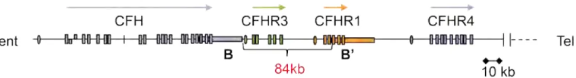
Protein analyses of the CFH-AA-positive plasma samples demonstrated the complete absence of CFHR1 and CFHR3 (214). Further detailed analyses showed for 14 patients the complete absence of both plasma proteins and for two patients rather low, barely detectable protein levels (235). Genetic analyses showed for the 14 deficient patients the complete absence of CFHR1 and CFHR3 due to a homozygous deletion of a large 84 kb genomic fragment which includes the CFHR1 and CFHR3 genes (Figure 5). The AA of practically all patients with AI-aHUS recognize the C-terminus of CFH, and inhibit the protective activity of CFH against complement attack to host cells (214,218,221).
The reason for the association between CFH-AA and the CFHR1 deficiency is not yet known. An infection often precedes emergence of CFH-AA (236), and it is known that a wide variety of microbes recruit host CFH onto their surface via domain
Figure 5. Schematic diagram of the chromosome 1, region q32. The 20 domains of CFH, the five domains of CFHR3, the five domains of CFHR1, and the nine domains of CFHR4 are indicated by vertical bars. The position of the two duplicated homologous segments (B and B’) is shown in purple and orange boxes. Failure of DNA amplification downstream reveals a deletion of a genomic ~84kb fragment of CFH in 99% of the AA-HUS patients.
20 of CFH, known as the common microbial binding site, to prevent complement attack (226). The association of CFHAA with any physiological or microbial ligand binding to CFH19-20 and the location of the binding sites of the AA within CFH19-20 have not, however, been shown.
3
OBJECTIVES
To characterize the structure-function relationship of the C-teminus of CFH via three distinct objectives:
I. To identify the molecular mechanism of the self non-self discriminatory
process by CFH, and its implications on aHUS mutations.
II. Structural basis of complement evasion by microbes using CFH20:
Borrelia as a case study.
4
MATERIAL AND METHODS
4.1
Materials
4.1.1 Proteins
Generation of the recombinant proteins generated specifically for the studies of this thesis by the author are described below in section 4.2.
Wild-type CFH19-20 was cloned without expression tags by Maria Pärepalo.
The mutants R1203A and Q1139A used in study I were cloned and expressed by
Markus J. Lehtinen. The CFH19Del-20 (Q1137A/ Q1139A/ Y1142A) and CFH19-20Del
(T1184G/ K1202A/ R1203A/Y1205A) used in study II were cloned, expressed and purified by Markus Lehtinen and Satu Hyvärinen. The C3d with its residue C17 (the thioester forming residue) mutated to alanine and the C3dg (along with the mutants D36A, E37A, E117A, D122A, E160A, D163A, and K291A) used in the study I and II respectively were cloned and expressed in E. coli and were procured from Prof. David Isenman’s lab (Toronto, Canada) (237,238).
Rabbit polyclonal antibodies against CFH19-20 and CFH, and monoclonal anti-CFH19-20 antibody VIG8 (239) were received from Dr. Jens Hellwage (Jena, Germany). The mAb C18 (107) was purchased from Enzo Life Sciences (Lörrach, Germany).
For NMR, OspE (residues 21-171) was cloned, expressed and purified with a His-tag in Dr. Hideo Iwaï’s lab. The GST-tagged OspE used for crystallization was cloned by Mia Eholuoto (122).
4.1.2 Microbial strains used
Cloning of the CFHR14-5 constructs was done in E. coli XL10-Gold® cells
(Stratagene) and the expression of CFH19-20 proteins in Pichia pastoris X-33 strain
(Invitrogen).
The construct of OspE used for NMR was cloned in XL10-Gold cells and was expressed in E. coli ER2566 strain grown in M9-medium.
4.1.3 Bioinformatical resources used
The following web resources were used to obtain sequence and structure information:
European Bioinformatics Institute (www.ebi.ac.uk), National Center for
Biotechnology Information (www.ncbi.nlm.nih.gov), Protein Data Bank
(www.rcsb.org/pdb), and “Proteins, Interfaces, Structures and Assemblies (PISA) server” (http://www.ebi.ac.uk/msd-srv/prot_int/) of the European Bioinformatics Institute.
4.1.4 Patient material used
Sera were collected from aHUS patients with CFH-AA in Germany and Hungary. The studies have been approved by the Research Ethics Committee of the Medical Faculty of Friedrich Schiller University and were performed in accordance with the declaration of Helsinki.
4.2
Methods
4.2.1 General laboratory methods for proteins
The proteins were resolved based on their size using standard sodium dodecyl sulfate – polyacrylamide gel electrophoresis (SDS-PAGE) techniques and visualized using staining with Coomassie Brilliant blue, silver staining, or by performing Western blotting where detection was done with primary antibodies and horseradish peroxidase-conjugated secondary antibodies (Jackson Immunoresearch). Protein concentrations were measured using absorbance at 280 nm wavelength, BCA assay (Thermo Scientific), and comparing intensity of the protein bands with a standard in the gel using Image J program (http://rsb.info.nih.gov/ij/). Buffer exchanges were performed using dialysis membranes or PD-10 buffer exchange columns (GE Healthcare).
4.2.2 Protein iodination
For radio immune assays (RIA) or radio ligand assays (RLA) proteins were iodinated using the Iodogen method in a hood of a class B radioactive work area (240). Iodogen tubes were prepared by mixing Iodogen to chloroform and dried. Iodination was performed by incubating 20-100 µg of protein with 0.2-1.0 mCi of 125-I for 10-20 minutes in PBS. After the reaction the mixture was transferred to a glass tube for