ACEMBL Expression System
User Manual
Vers. 09.11
Yan Nie, Christoph Bieniossek, Imre Berger
ACEMBL was developed at the
European Molecular Biology Laboratory EMBL Grenoble Outstation
38042 Grenoble CEDEX 9, France
Table of Contents
A. Synopsis: 4
B. ACEMBL System 5
B.1. ACEMBL vectors 5
B.2. The multiple integration element (MIE) 6
B.3. Tags, promoters, terminators 7
B.4. Complex Expression 8
C. Procedures 9
C.1. Cloning into ACEMBL vectors 9
C1.1. Single gene insertion into the MIE by SLIC 9
C1.2. Polycistron assembly in MIE by SLIC 13
C.1.3. Gene insertion by restriction/ligation 18
C.1.4. Multiplication by using the HE and BstXI sites 21
C.2. Cre-LoxP reaction of Acceptors and Donors 24
C.2.1. Cre-LoxP fusion of Acceptors and Donors 25
C.2.2. Deconstruction of fusion vectors by Cre 28
C.3. Coexpression by Cotransformation 30
D. ACEMBL multigene combination: Examples 31 D.1. SLIC cloning into ACEMBL vectors: human TFIIF 31 D.2. Polycistron by SLIC: human VHL/ElonginB/ElonginC complex. 32 D.3. The Homing endonuclease/BstXI module: yeast RES complex 33 D.4. Coexpression by cotransformation: human NYB/NYC 34
D.5. Coexpression from Acceptor-Donor fusions 34
E. The ACEMBL System Kit 35
F. Appendix 38
F.1. DNA sequence of MIE 38
F.2. DNA sequences of ACEMBL vectors 39
F.2.1. pACE 39
F.2.2. pACE2 40
F.2.3. pDC 41
F.2.4. pDK 42
F.2.5. pDS 43
F.2.6. pACKS tetrafusion (ACEMBL kit component) 44
Protocols
Protocol 1: Single gene insertion by SLIC 10
Protocol 2: Polycistron assembly by SLIC 13
Protocol 3: Restriction/ligation cloning into the MIE. 17 Protocol 4: Multiplication by using homing endonuclease/BstXI 21
Table:
Table I: Adaptor DNA sequences. 15
Illustrations:
ACEMBL system for multiprotein complex production 4
The multiple integration element, schematic view 5
Single gene insertion by SLIC 9
Generating a polycistron by SLIC 12
LoxP imperfect inverted repeat 23
Cre and De-Cre reaction pyramid 24
96well analysis of Cre assembly 26
ACEMBLing TFIIF 30
Multifragment SLIC of pACE-VHLbc (tricistron) 31 The HE/BstXI multiplication module 32 ACEMBL System Kit: Generating single vectors from pACKS 35 96well microtiter analysis of pACKS De-Cre reaction 36
A. Synopsis:
ACEMBL is a 3rd generation multigene expression system for complex production in E. coli, created at the European Molecular Biology Laboratory EMBL, at Grenoble.
ACEMBL can be applied both manually and also in an automated setup by using a liquid handling workstation. ACEMBL applies tandem recombination steps for rapidly assembling many genes into multigene expression cassettes. These can be single or polycistronic expression modules, or a combination of these elements. ACEMBL also offers the option to employ conventional approaches involving restriction enzymes and ligases if desired, which may be the methods of choice in laboratories not familiar with recombination approaches.
The following strategies for multigene assembly and expression are provided for in the ACEMBL system and detailed in Sections B and C:
(1) Single gene insertions into vectors (recombination or restriction/ligation) (2) Multigene assembly into a polycistron (recombination or restriction/ligation) (3) Multigene assembly using homing endonucleases
(4) Multigene plasmid fusion by Cre-LoxP reaction (5) Multigene expression by cotransformation
These strategies can be used individually or in conjunction, depending on the project and user.
In Section C, step-by-step protocols are provided for each of the methods for multigene cassette assembly that can be applied in the ACEMBL system. Each procedure is illustrated by corresponding complex expression experiments in Section D of this Supplement.
DNA sequences of ACEMBL vectors are provided in the Appendix and can be copied from there for further use.
Requests for ACEMBL system kit components can be addressed to Imre Berger ([email protected]).
B. ACEMBL System B.1. ACEMBL vectors
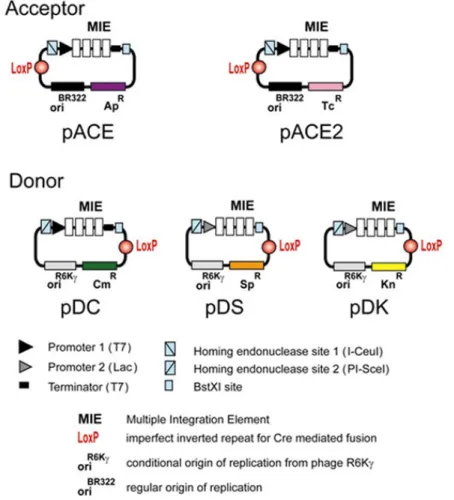
At the core of the technology are five small de novo designed vectors which are called “Acceptor” and “Donor” vectors (Illustration 1). Acceptor vectors (pACE, pACE2) contain origins of replication derived from ColE1 and resistance markers (ampicillin or tetracycline). Donor vectors contain conditional origins of replication (derived from R6Kγ), which make their propagation dependent on hosts expressing the pir gene. Donor vectors contain resistance markers kanamycin, chloramphenicol, spectinomycin. Up to three Donor vectors can be used in conjunction with one Acceptor vector
Illustration 1: ACEMBL system for multiprotein complex production.
All Donor and Acceptor vectors contain a loxP imperfect inverted repeat and in addition, a multiple integration element (MIE). This MIE consists of an expression cassette with a promoter of choice (prokaryotic, mammalian, insect cell specific or a combination thereof) and a terminator (prokaryotic, mammalian, insect cell specific
or a combination thereof). In between is a DNA segment which contains a number of restriction sites that can be used for conventional cloning approaches or also for generating double-strand breaks for the integration of expression elements of choice (further promoters, ribosomal binding sites, terminators and genes). The MIE is completed by a homing endonuclease site and a specifically designed restriction enzyme site (BstXI) flanking the promoter and the terminator (see B.2.) Vector DNA sequences are provided in the Appendix. Maps of all vectors are shown at the end of this manual.
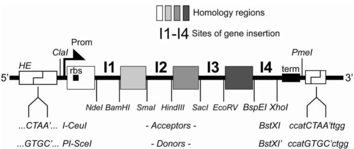
B.2. The multiple integration element (MIE)
Illustration 2: The multiple integration element, schematic view.
The MIE was derived from a polylinker1 and allows for several approaches for
multigene assembly (Section C). Multiple genes can be inserted into the MIE of any one of the vectors by a variety of methods, for example BD-In-Fusion recombination2 or SLIC (sequence and ligation independent cloning3. For this, the vector needs to be linearized, which can also be carried out efficiently by PCR reaction with appropriate primers, since the vectors are all small (2-3.0 kb). Use of ultrahigh-fidelity polymerases such as Phusion4 is recommended. Alternatively, if more conventional approaches are preferred i.e. in a regular wet lab setting without robotics, the vectors can also be linearized by restriction digestion, and a gene of interest can be integrated by restriction / ligation (Section C). The DNA sequence of the MIE is shown in the Appendix.
1 Tan, S. et al. Protein Expr. Purif. 40, 385 (2005) 2 ClonTech TaKaRa Bio Europe, www.clontech.com 3 Li, M. and Elledge, S., Nat. Methods 4, 251 (2007) 4 Finnzymes/New England BioLabs, www.neb.com
B.3. Tags, promoters, terminators
Current vectors of the ACEMBL system contain per default promoters T7 and Lac, as well as the T7 terminator element (Illustr.1, 10). The T7 system is most commonly used currently; it requires bacterial strains which contain a T7 polymerase gene in the
E. coli genome. The Lac promoter is a strong endogenous promoter which can be
utilized in most strains. All ACEMBL vectors contain the lac operator element for repression of heterologous expression.
Evidently, all promoters and terminators present in ACEMBL Donor and Acceptor vectors, and in fact the entire multiple integration element (MIE) can be exchanged with a favored expression cassette of choice by using restriction/ligation cloning with appropriate enzymes (for example ClaI/PmeI, Illustration 2) or insertion into linearized ACEMBL vectors where the MIE was removed by sequence and ligation independent approaches such as SLIC. We have substituted the T7 promoter in pDC with a trc promoter (pDCtrc), and the T7 promoter in pACE with an arabinose promoter (pACEara) and used the resulting vectors successfully in coexpression experiments by inducing with arabinose and IPTG.
Currently, the ACEMBL system vectors do not contain DNA sequences encoding for affinity tags to facilitate purification or solubilization of the protein(s) of interest. We typically use C- or N-terminal oligohistidine tags, with or without protease sites for tag removal. We introduce these by means of the respective PCR primers used for amplification of the genes of interest prior to SLIC mediated insertion. We recommend to outfit Donors or Acceptors of choice by the array of custom tags that are favored in individual user laboratories prior to inserting recombinant genes of interest. This is best done by a design which will, after tag insertion, still be compatible with the recombination based principles of ACEMBL system usage.
B.4. Complex Expression
For expression in E.coli, the ACEMBL multigene expression vector fusions with appropriate promoters or terminators are transformed into the appropriate expression host of choice. In the current version (T7 and lac promoter elements), most of the wide array of currently available expression strains can be utilized. If particular expression strains already contain helper plasmids with DNA encoding for chaperones, lysozyme or else, the design of the multigene fusion should ideally be such that the ACEMBL vector containing the resistance marker that is also present on the helper plasmid is not included in multigene vector construction (although this is probably not essential).
Alternatively, the issue can be resolved by creating new versions of the ACEMBL vectors containing resistance markers that circumvent the conflict. This can be easily performed by PCR amplifying the vectors minus the resistance marker, and combine the resulting fragments with a PCR amplified resistance marker by recombination (SLIC) or blunt-end ligation (using 5’phosphorylated primers). Note that resistance markers can also be exchanged in between ACEMBL vectors by restriction digestion with AlwNI and ClaI (for Donors) and AlwNI and PmeI (for Acceptors).
Donor vectors depend on the pir gene product expressed by the host, due to the R6K conditional origin of replication. In regular expression strains, they rely on fusion with an Acceptor for productive replication. Donors or Donor-Donor fusions can nonetheless be used even for expression when not fused with an Acceptor, by using expression strains carrying a genomic insertion of the pir gene. Such strains have more recently become available (Novagen Inc., Madison WI, USA).
Cotransformation of two plasmids can also lead to successful protein complex expression. The ACEMBL system contains two Acceptor vectors, pACE and pACE2, which are identical except for the resistance marker (Illustration 1). Therefore, genes present on pACE or pACE2, respectively, can be expressed by cotransformation of the two plasmids and subsequent exposure to both tetracyclin and ampicillin simultaneously. In fact, entire Acceptor-Donor fusions containing several genes, based on pACE or pACE2 as Acceptors, can in principle be cotransformed for mutli-expression, if needed.
C. Procedures
C.1. Cloning into ACEMBL vectors
All Donors and Acceptors contain an identical MIE with exception of the homing endonuclease site / BstXI tandem encompassing the MIE (Illustrations 1 and 12). The MIE is tailored for sequence and ligation independent gene insertion methods. In addition, the MIE also contains a series of unique restriction sites, and therefore can be used as a classical polylinker for conventional gene insertion by restriction/ligation. We suggest to choose the methods a user lab is most proficient with. For automated applications, restriction/ligation is essentially ruled out. In this case, recombination approaches can be used efficiently for gene insertion (SLIC). C1.1. Single gene insertion into the MIE by SLIC
Several procedures for restriction/ligation independent insertion of genes into vectors have been published or commercialized (Novagen LIC, Becton-Dickinson BD In-Fusion and others), each with its own merit. These systems share in common that they rely on the exonuclease activity of DNA polymerases. In the absence of dNTPs, 5’ extensions are created from blunt ends or overhangs by digestion from the 3’ end. If two DNA fragments contain the same ~20 bp sequence at their termini at opposite ends, this results in overhangs that share complementary sequences capable of annealing. This can be exploited for ligation independent combination of two or several DNA fragments containing homologous sequences.
If T4 DNA polymerase is used, this can be carried out in a manner that is independent of the sequences of the homology regions (Sequence and Ligation Independent Cloning, SLIC) and detailed protocols became available. In the context of multiprotein expression, this is particularly useful, as the presence of unique restriction sites, or their creation by mutagenesis, in the ensemble of encoding DNAs ceases to be an issue.
We adapted SLIC for inserting encoding DNAs amplified by Phusion polymerase into the ACEMBL Acceptor and Donor vectors according to the published protocols. In this way, not only seamless integration of genes into the expression cassettes, but also concatamerization of expression cassettes to multigene constructs can be achieved by applying the same, simple routine that can be readily automated.
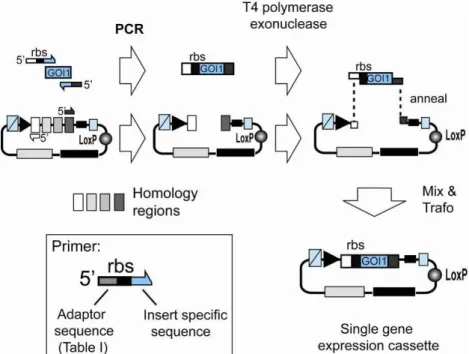
Illustration 3: Single gene insertion by SLIC. A gene of interest (GOI 1) is PCR amplified with specific primers and integrated into a vector (Acceptor, Donor) linearized by PCR with complementary primers (complementary regions are shaded in light gray or dark grey, respectively). Resulting PCR fragments contain homology regions at the ends. T4 DNA polymerase acts as an exonuclease in the absence of dNTP and produces long sticky overhangs. Mixing (optionally annealing) of T4DNA polymerase exonuclease treated insert and vector is followed by transformation, yielding a single gene expression cassette.
We use an improved protocol for SLIC which was modified from the original publication5. This protocol as applied manually is detailed below (Protocol 1). If other systems are used (BD-InFusion etc.), follow manufacturers’ recommendations. For robotics applications, modifications of the protocol may be necessary and will be detailed elsewhere6.
Protocol 1: Single gene insertion by SLIC. Reagents required:
Phusion Polymerase
5x HF Buffer for Phusion Polymerase dNTP mix (10 mM)
T4 DNA polymerase (and10x Buffer) DpnI enzyme
E. coli competent cells
100mM DTT, 2M Urea, 500 mM EDTA Antibiotics
5 Li, M. and Elledge, S., Nat. Methods 4, 251 (2007)
Step 1: Primer design
Primers for the SLIC procedure are designed to provide the regions of homology which result in the long sticky ends upon treatment with T4 DNA polymerase in the absence of dNTP:
Primers for the insert contain a DNA sequence corresponding to this region of homology (“ Adaptor sequence” in Illustration 3, inset), followed by sequence which specifically anneals to the insert to be amplified Illustration 3, inset). Useful adaptor sequences for SLIC are listed below (Table I).
If the gene of interest (GOI) is amplified from a vector already containing expression elements (e.g. the pET vector series), this “ insert specific sequence” can be located upstream of a ribosome binding site (rbs). Otherwise, the forward primer needs to be designed such that a ribosome binding site is also provided in the final construct (Illustration 3, inset).
Primers for PCR linearization of the vector backbone are simply complementary to the two adaptor sequences present in the primer pair chosen for insert amplification (Illustration 3).
Step 2: PCR amplification of insert and vector
Identical reactions are prepared in 100-µl volume for DNA insert to be cloned and vector to be linearized by PCR:
ddH2O 75 µl
5× Phusion HF Reaction buffer 20 µl
dNTPs (10 mM stock) 2 µl
Template DNA (100 ng/µl) 1 µl 5′ SLICprimer (100 µM stock) 1 µl 3′ SLICprimer (100 µM stock) 1 µl Phusion polymerase (2 U/µl) 0.5 µl
PCR reactions are then carried out with a standard PCR program (unless very long DNAs are amplified, then double extension time):
1 x 98° C for 2 min
30 x [98° C for 20 sec. -> 50°C for 30 sec. -> 72°C for 3 min] Hold at 10°C
Analysis of the PCR reactions by agarose gel electrophoresis and ethidium bromide staining is recommended.
Step 3: DpnI treatment of PCR products (optional)
PCR reactions are then supplied with 1 µl DpnI enzyme which cleaves parental plasmids (that are methylated). For insert PCR reactions, DpnI treatment is not required if the resistance marker of the template plasmid differs from the destination vector.
Reactions are then carried out as follows: Incubation: 37°C for 1-4h
Inactivation: 80°C for 20 min Step 4: Purification of PCR products
! PCR products must be cleaned of residual dNTPs !
Otherwise, the T4 DNA polymerase reaction (Step 5) is compromised.
Product purification is best performed by using commercial PCR Purification Kits or NulceoSpin Kits (Qiagen, MacheryNagel or others). It is recommended to perform elution in the minimal possible volume indicated by the manufacturer. Step 5: T4 DNA polymerase exonuclease treatment
Identical reactions are prepared in 20-µl volume for insert and for vector (eluted in Step 4):
10x T4 DNA polymerase buffer 2 µl
100mM DTT 1 µl
2M Urea 2 µl
DNA eluate from Step 3 (vector or insert) 14 µl
T4 DNA polymerase 1 µl
Reactions are then carried out as follows: Incubation: 23°C for 20 min
Arrest: Addition of 1 µl 500 mM EDTA Inactivation: 75°C for 20 min
Step 6: Mixing and Annealing
T4 DNA polymerase exonuclease treated insert and vector are then mixed, followed by an (optional) annealing step which was found to enhance efficiency7:
T4 DNA pol treated insert: 10 µl T4 DNA pol treated vector: 10 µl
Annealing: 65°C for 10 min
Cooling: Slowly (in heat block) to RT Step 7: Transformation
Mixtures are next transformed into competent cells following standard transformation procedures.
Reactions for pACE and pACE2 derivatives are transformed into standard E. coli cells for cloning (such as TOP10, DH5α, HB101) and after recovery (204h)
plated on agar containing ampicillin (100 µg/ml) or tetracycline (25 µg/ml), respectively.
Reactions for Donor derivatives are transformed into E. coli cells expressing the
pir gene (such as BW23473, BW23474, or PIR1 and PIR2, Invitrogen) and plated
on agar containing chloramphenicol (25 µg/ml, pDC), kanamycin (50 µg/ml, pDK), and spectinomycin (50 µg/ml, pDS).
Step 8: Plasmid analysis
Plasmids are cultured in small-scale in media containing the corresponding antibiotic, and analyzed by sequencing and (optionally) restriction mapping with an appropriate restriction enzyme.
C1.2. Polycistron assembly in MIE by SLIC
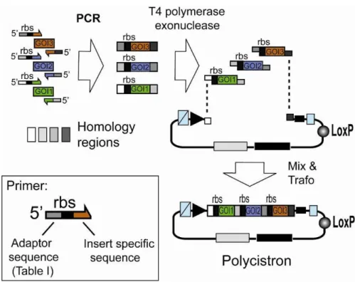
The multiple integration element can also be used to integrate genes of interest by using multi-fragment SLIC recombination as shown in Illustration 4. Genes preceded by ribosome binding sites (rbs) can be assembled in this way into polycistrons.
Illustration 4: Generating a polycistron by SLIC. Genes of interest (GOI 1,2,3) are PCR amplified with specific primers and integrated into a vector (Acceptor, Donor) linearized by PCR with primers complementary to the ends of the forward primer of the first (GOI 1) and the reverse primer of the last (GOI 3) gene to be assembled in the polycistron (complementary regions are shaded in light gray or dark grey, respectively). Resulting PCR fragments contain homology regions at the ends. T4 DNA polymerase acts as an exonuclease in the absence of dNTP and produces long sticky overhangs. Mixing (optionally annealing) of T4DNA polymerase exonuclease treated insert and vector is followed by transformation, yielding a polycistronic expression cassette.
Protocol 2. Polycistron assembly by SLIC. Reagents required:
Phusion Polymerase
5x HF Buffer for Phusion Polymerase dNTP mix (10 mM)
T4 DNA polymerase (and 10x Buffer)
E. coli competent cells
100mM DTT, 2M Urea, 500 mM EDTA Antibiotics
Step 1: Primer design
The MIE element is composed of tried-and-tested primer sequences. These constitute the “ Adaptor” sequences that can be used for inserting single genes or multigene constructs. Recommended adaptor sequences are listed below (Table I).
Adaptor sequences form the 5’ segments of the primers used to amplify DNA fragments to be inserted into the MIE. Insert specific sequences are added at 3’, DNE encoding for a ribosome binding sites can be inserted optionally if not already present on the PCR template
Step 2: PCR amplification of insert and primer
Identical reactions are prepared in 100-µl volume for all DNA insert (GOI 1,2,3) to be cloned and the vector to be linearized by PCR:
ddH2O 75 µl
5× Phusion HF Reaction buffer 20 µl
dNTPs (10 mM stock) 2 µl
Template DNA (100 ng/µl) 1 µl 5′ SLICprimer (100 µM stock) 1 µl 3′ SLICprimer (100 µM stock) 1 µl Phusion polymerase (2 U/µl) 0.5 µl
PCR reactions are then carried out with a standard PCR program (unless very long DNAs are amplified, then double extension time):
1 x 98° C for 2 min
30 x [98° C for 20 sec. -> 50°C for 30 sec. -> 72°C for 3 min] Hold at 10°C
Analysis of the PCR reactions by agarose gel electrophoresis and ethidium bromide staining is recommended.
Step 3: DpnI treatment of PCR products (optional)
PCR reactions are then supplied with 1 µl DpnI enzyme which cleaves parental plasmids (that are methylated). For insert PCR reactions, DpnI treatment is not required if the resistance marker of the template plasmids differs from the destination vector.
Reactions are then carried out as follows: Incubation: 37°C for 1-4h
Inactivation: 80°C for 20 min Step 4: Purification of PCR products
! PCR products must be cleaned of residual dNTPs !
Otherwise, the T4 DNA polymerase reaction (Step 5) is compromised.
Product purification is best performed by using commercial PCR Purification Kits or NulceoSpin Kits (Qiagen, MacheryNagel or others). It is recommended to perform elution in the minimal possible volume indicated by the manufacturer. Step 5: T4 DNA polymerase exonuclease treatment
Identical reactions are prepared in 20-µl volume for each insert (GOI 1,2,3) and for the vector (eluted in Step 4):
10x T4 DNA polymerase buffer 2 µl
100mM DTT 1 µl
2M Urea 2 µl
DNA eluate from Step 3 (vector or insert) 14 µl
T4 DNA polymerase 1 µl
Reactions are then carried out as follows: Incubation: 23°C for 20 min
Arrest: Addition of 1 µl 500 mM EDTA Inactivation: 75°C for 20 min
Step 6: Mixing and Annealing
T4 DNA polymerase exonuclease treated insert and vector are then mixed, followed by an (optional) annealing step which was found to enhance efficiency8:
T4 DNA pol treated insert 1 (GOI 1): 5 µl T4 DNA pol treated insert 2 (GOI 2): 5 µl T4 DNA pol treated insert 3 (GOI 3): 5 µl T4 DNA pol treated vector: 5 µl
Annealing: 65°C for 10 min
Cooling: Slowly (in heat block) to RT
Step 7: Transformation
Mixtures are next transformed into competent cells following standard transformation procedures.
Reactions for pACE and pACE2 derivatives are transformed into standard E. coli cells for cloning (such as TOP10, DH5α, HB101) and after recovery plated on agar containing ampicillin (100 µg/ml) or tetracycline (25 µg/ml), respectively. Reactions for Donor derivatives are transformed into E. coli cells expressing the
pir gene (such as BW23473, BW23474, or PIR1 and PIR2, Invitrogen) and plated
on agar containing chloramphenicol (25 µg/ml, pDC), kanamycin (50 µg/ml, pDK), and spectinomycin (50 µg/ml, pDS).
Step 8: Plasmid analysis
Plasmids are cultured and correct clones are selected based on specific restriction digestion and DNA sequencing of the inserts.
Table I. Adaptor sequences ! updated to commercially available kit, ATG-biosynthetics ! For single gene or multigene insertions into ACEMBL vectors by SLIC.
Adaptor1 Sequence Description
T7InsFor TAGGTATCGATAATACGACT CACTATAGGG
Forward primer for insert amplification, if gene of interest (GOI) is present in a T7 system vector (i.e. pET series).
No further extension (rbs, insert specific overlap) required.
T7InsRev CCTCAAGACCCGTTTAGAGG CCCCAAGGGGTTATGCTAG
Reverse primer for insert amplification, if GOI is present in a T7 system vector (i.e. pET series). No further extension (stop codon, insert specific overlap) required.
T7VecFor CTAGCATAACCCCTTGGGGC CTCTAAACGGGTCTTGAGG
Forward primer for vector amplification, reverse complement of T7InsRev.
No further extension required. T7VecRev CCCTATAGTGAGTCGTATTA
TCGATACCTA
Reverse primer for vector amplification, reverse complement of T7InsFor.
No further extension required. NdeInsFor GTTTAACTTTAAGAAGGAGA
TATACATATG
Forward primer for insert amplification for insertion into MIE site I1 (Illustration 2).
Further extension at 3’ (insert specific overlap) required.
Can be used with adaptor XhoInsRev in case of single fragment SLIC (Illustr. 3).
XhoInsRev pACE,pACE2,pDC (T7): CTTTGTTAGCAGCCGGATCT CTCGAG pDK,pDS (lac): GGGTTTAAACGGAACTAGTC TCGAG
Reverse primer for insert amplification for insertion into MIE site I4 (Illustr. 2).
Further extension at 3’ (stop codon, insert specific overlap) required.
Can be used with adaptor NdeInsFor in case of single fragment SLIC (Illustr. 3).
XhoVecFor pACE,pACE2,pDC (T7): CTCGAGAGATCCGGCTGCTA ACAAAG pDK,pDS (lac): CTCGAGACTAGTTCCGTTTAA ACCC
Forward primer for vector amplification, reverse complement of XhoInsRev
No further extension required.
NdeVecRev CATATGTATATCTCCTTCTT
AAAGTTAAAC Reverse primer for vector amplification, reverse complement of NdeInsFor. No further extension required.
SmaBam GAATTCACTGGCCGTCGTTT
TACAGGATCC Reverse primer for insert amplification (GOI1) for insertion into MIE site I1 (Illustr. 2). Further extension at 3’ (stop codon, insert specific overlap) required.
Use with adaptor NdeInsFor. BamSma GGATCCTGTAAAACGACGGC
CAGTGAATTC Forward primer for insert amplification (GOI2) for insertion into site I2 (Illustr. 2,4). Further extension at 3’ (rbs, insert specific overlap) required.
Use with adaptor SacHind (multifragment SLIC, Illustr. 4)
SacHind GCTCGACTGGGAAAACCC
TGGCGAAGCTT Reverse primer for insert amplification (GOI2) insertion into MIE site I2 (Illustr. 2, 4).
Further extension at 3’ (stop codon, insert specific overlap) required.
Use with adaptor BamSma (multifragment SLIC, Illustr. 4)
HindSac AAGCTTCGCCAGGGTTTT
CCCAGTCGAGC Forward primer for insert amplification (GOI3) for insertion into site I3 (Illustr. 2,4).
Further extension at 3’ (rbs, insert specific overlap) required.
Use with adaptor BspEco (multifragment SLIC, Illustr. 4)
BspEco5 GATCCGGATGTGAAATTG
TTATCCGCTGGTACC Reverse primer for insert amplification (GOI3) insertion into MIE site I3 (Illustr. 2, 4).
Further extension at 3’ (stop codon, insert specific overlap) required.
Use with adaptor HindSac.(multifragment SLIC, Illustr. 4)
Eco5Bsp GGTACCAGCGGATAACAA
TTTCACATCCGGATC Forward primer for insert amplification (GOI4) for insertion into site I4 (Illustr. 2,4).
Further extension at 3’ (rbs, insert specific overlap) required.
Use with adaptor XhoInsRev (multifragment SLIC, Illustr. 4)
1 All Adaptor primers (without extension) can be used as sequencing primers for genes of interest that were inserted into the MIE.
C.1.3. Gene insertion by restriction/ligation
The MIE can also be interpreted as a simple multiple cloning site with a series of unique restriction sites. The MIE is preceded by a promoter and a ribosome binding site, and followed by a terminator, therefore, cloning into the MIE by classical restriction/ligation also yields functional expression cassettes.
Genes of interest can be subcloned by using standard cloning procedures into the multiple integration element (MIE) (see Appendix) of ACEMBL vectors (the MIE is identical in all vectors).
Protocol 3. Restriction/ligation cloning into the MIE. Reagents required:
Phusion Polymerase
5x HF Buffer for Phusion Polymerase dNTP mix (10 mM)
10 mM BSA
Restriction endonucleases (and 10x Buffer) T4 DNA ligase (and 10x Buffer)
Calf or Shrimp intestinal alkaline phosphatase
E. coli competent cells
Antibiotics Step 1: Primer design
For conventional cloning, PCR primers are designed containing chosen restriction sites, preceded by appropriate overhangs for efficient cutting (c.f. New England Biolabs catalogue), and followed by 20 nucleotides overlapping with the gene of interest that is to be inserted.
All MIEs are identical in the ACEMBL vectors. They contain a ribosome binding preceding the NdeI site. For single gene insertions, therefore, a rbs need not be included in the primer.
If multigene insertions are planned (for example in insertion sites I1-I4 of the MIE), primers need to be designed such that a rbs preceding the gene and a stop codon at its end are provided.
In particular for polycistron cloning by restriction/ligation, is recommended to construct templates by custom gene synthesis. In the process, the restriction sites present in the MIE can be eliminated from the encoding DNAs.
Step 2: Insert preparation PCR of insert(s):
Identical PCR reactions are prepared in 100 µl volume for genes of interest to be inserted into the MIE:
ddH2O 75 µl
5× Phusion HF Reaction buffer 20 µl
dNTPs (10 mM stock) 2 µl
Template DNA (100 ng/µl) 1 µl 5′ primer (100 µM stock) 1 µl 3′ primer (100 µM stock) 1 µl Phusion polymerase (2 U/µl) 0.5 µl
PCR reactions are then carried out with a standard PCR program (unless very long DNAs are amplified, then double extension time):
1 x 98° C for 2 min
30 x [98° C for 20 sec. -> 50°C for 30 sec. -> 72°C for 3 min] Hold at 10°C
Analysis of the PCR reactions by agarose gel electrophoresis and ethidium bromide staining is recommended.
Product purification is best performed by using commercial PCR Purification Kits or NulceoSpin Kits (Qiagen, MacheryNagel or others). It is recommended to perform elution in the minimal possible volume indicated by the manufacturer. Restriction digestion of insert(s):
Restriction reactions are carried out in 40 µl reaction volumes, using specific restriction enzymes as specified by manufacturer’s recommendations (c.f. New England Biolabs catalogue and others).
PCR Kit eluate ( 1 µg) 30 µl 10x Restriction enzyme buffer 4 µl
10 mM BSA 2 µl
Restriction enzyme for 5’ 2 µl
Restriction enzyme for 3’ 2 µl (in case of double digestion, otherwise ddH2O)
Restriction digestions are performed in a single reaction with both enzymes (double digestion) or sequentially (two single digestions) if the buffer conditions required are incompatible.
Gel extraction of insert(s):
Processed insert is then purified by agarose gel extraction using commercial kits (Qiagen, MachereyNagel etc). It is recommended to elute the extracted DNA in the minimal volume defined by the manufacturer.
Step 3: Vector preparation
Restriction digestion of ACEMBL plasmid(s):
Restriction reactions are carried out in 40 µl reaction volumes, using specific restriction enzymes as specified by manufacturer’s recommendations (c.f. New England Biolabs catalogue and others).
ACEMBL plasmid ( 0.5 µg) in ddH2O 30 µl
10x Restriction enzyme buffer 4 µl
10 mM BSA 2 µl
Restriction enzyme for 5’ 2 µl
Restriction enzyme for 3’ 2 µl (in case of double digestion, otherwise ddH2O)
Restriction digestions are performed in a single reaction with both enzymes (double digestion) or sequentially (two single digestions) if the buffer conditions required are incompatible.
Gel extraction of vector(s):
Processed vector is then purified by agarose gel extraction using commercial kits (Qiagen, MachereyNagel etc). It is recommended to elute the extracted DNA in the minimal volume defined by the manufacturer.
Step 4: Ligation
Ligation reactions are carried out in 20 µl reaction volumes according to the recommendations of the supplier of T4 DNA ligase:
ACEMBL plasmid (gel extracted) 8 µl Insert (gel extracted) 10 µl 10x T4 DNA Ligase buffer 2 µl
T4 DNA Ligase 0.5 µl
Ligation reactions are performed at 25ºC (sticky end) for 1h or at 16ºC (blunt end) overnight.
Step 5: Transformation
Mixtures are next transformed into competent cells following standard transformation procedures.
Reactions for pACE and pACE2 derivatives are transformed into standard E. coli cells for cloning (such as TOP10, DH5α, HB101) and after recovery plated on agar containing ampicillin (100 µg/ml) or tetracycline (25 µg/ml), respectively. Reactions for Donor derivatives are transformed into E. coli cells expressing the
pir gene (such as BW23473, BW23474, or PIR1 and PIR2, Invitrogen) and plated
on agar containing chloramphenicol (25 µg/ml, pDC), kanamycin (50 µg/ml, pDK), and spectinomycin (50 µg/ml, pDS).
Step 6: Plasmid analysis
Plasmids are cultured and correct clones are selected based on specific restriction digestion and DNA sequencing of the inserts.
C.1.4. Multiplication by using the HE and BstXI sites
All ACEMBL system vectors contain a homing endonuclease (HE) site and a designed BstXI site that envelop the multiple integration element (MIE). The homing endonuclease site can be used to insert entire expression cassettes, containing single genes or polycistrons, into a vector already containing one gene or several genes of interest. Homing endonucleases have long recognition sites (20-30 base pairs or more). Although not all equally stringent, homing endonuclease sites are most probably unique in the context of even large plasmids, or, in fact, entire genomes.
In the ACEMBL system, Donor vectors contain a recognition site for homing endonuclease PI-SceI (Illustr. 2). This HE site yields upon cleavage a 3’ overhang with the sequence -GTGC. Acceptor vectors contain the homing endonuclease site I-CeuI, which upon cleavage will result in a 3’ overhang of -CTAA. On Acceptors and Donors, the respective HE site is preceding the MIE. The 3’ end of the MIE contains a specifically designed BstXI site, which upon cleavage will generate a matching overhang. The basis of this is the specificity of cleavage by BstXI. The recognition sequence of BstXI is defined as CCANNNNN’NTGG (apostrophe marks position of phosphodiester link cleavage). The residues denoted as N can be chosen freely. Donor vectors thus contain a BstXI recognition site of the sequence CCATGTGC’CTGG, and Acceptor vectors contain CCATCTAA’TTGG. The overhangs generated by BstXI cleavage in each case will match the overhangs generated by HE cleavage. Note that Acceptors and Donors have different HE sites.
The recognition sites are not symmetric. Therefore, ligation of a HE/BstXI digested fragment into a HE site of an ACEMBL vector will be (1) directional and (2) result in a hybrid DNA sequence where a HE halfsite is combined with a BstXI halfsite. This site will be cut by neither HE nor BstXI. Therefore, in a construct that had been digested with a HE, insertion by ligation of HE/BstXI digested DNA fragment containing an expression cassette with one or several genes will result in a construct which contains all heterologous genes of interest, enveloped by an intact HE site in front, and a BstXI site at the end. Therefore, the process of integrating entire expression cassettes by means of HE/BstXI digestion and ligation into a HE site can be repeated iteratively.
Protocol 4. Multiplication by using homing endonuclease/BstXI. Reagents required:
Homing endonucleases PI-SceI, I-CeuI 10x Buffers for homing endonucleases Restriction enzyme BstXI (and 10x Buffer) T4 DNA ligase (and 10x Buffer)
E. coli competent cells
Antibiotics
Step 2: Insert preparation
Restriction reactions are carried out in 40 µl reaction volumes, using homing endonucleases PI-SceI (Donors) or I-CeuI (Acceptors) as recommended by the supplier (c.f. New England Biolabs catalogue and others).
ACEMBL plasmid ( 0.5 µg) in ddH2O 32 µl
10x Restriction enzyme buffer 4 µl
10 mM BSA 2 µl
PI-SceI (Donors) or I-CeuI (acceptors) 2 µl
Reactions are then purified by PCR extraction kit or acidic ethanol precipitation, and next digested by BstXI according to the recommendations of the supplier.
HE digested DNA in ddH2O 32 µl
10x Restriction enzyme buffer 4 µl
10 mM BSA 2 µl
BstXI 2 µl
Gel extraction of insert(s):
Processed insert is then purified by agarose gel extraction using commercial kits (Qiagen, MachereyNagel etc). It is recommended to elute the extracted DNA in the minimal volume defined by the manufacturer.
Step 3: Vector preparation
Restriction reactions are carried out in 40 µl reaction volumes, using homing endonucleases PI-SceI (Donors) or I-CeuI (Acceptors) as recommended by the supplier (c.f. New England Biolabs catalogue and others).
ACEMBL plasmid ( 0.5 µg) in ddH2O 33 µl
10x Restriction enzyme buffer 4 µl
10 mM BSA 2 µl
Reactions are then purified by PCR extraction kit or acidic ethanol precipitation, and next treated with intestinal alkaline phosphatase according to the recommendations of the supplier.
HE digested DNA in ddH2O 17 µl
10x Alkaline phosphatase buffer 2 µl
Alkaline phosphatase 1 µl
Gel extraction of vector:
Processed vector is then purified by agarose gel extraction using commercial kits (Qiagen, MachereyNagel etc). It is recommended to elute the extracted DNA in the minimal volume defined by the manufacturer.
Step 4: Ligation
Ligation reactions are carried out in 20 µl reaction volumes: HE/Phosphatase treated vector (gel extracted) 4 µl HE/BstXI treated insert (gel extracted) 14 µl 10x T4 DNA Ligase buffer 2 µl
T4 DNA Ligase 0.5 µl
Ligation reactions are performed at 25ºC for 1h or at 16ºC overnight. Step 5: Transformation
Mixtures are next transformed into competent cells following standard transformation procedures.
Reactions for pACE and pACE2 derivatives are transformed into standard E. coli cells for cloning (such as TOP10, DH5α, HB101) and after recovery plated on agar containing ampicillin (100 µg/ml) or tetracycline (25 µg/ml), respectively. Reactions for Donor derivatives are transformed into E. coli cells expressing the
pir gene (such as BW23473, BW23474, or PIR1 and PIR2, Invitrogen) and plated
on agar containing chloramphenicol (25 µg/ml, pDC), kanamycin (50 µg/ml, pDK), and spectinomycin (50 µg/ml, pDS).
Step 6: Plasmid analysis
Plasmids are cultured and correct clones selected based on specific restriction digestion and DNA sequencing of the inserts.
Note: Integration can likewise be performed by sequence and ligation independent cloning. It is recommended to carry out linearization of the vector by digestion with HE, if heterologous genes are already present, to avoid PCR amplifications over encoding regions. The fragment to be inserted is generated by PCR amplification resulting in a PCR fragment containing a 20-25 base pair stretch at its 5’ end that is identical to the corresponding DNA sequence present at the HE site counted from the site of cleavage towards 5’ (site of cleavage is position -4). At the 3’ end of the PCR fragment, the homology region is 20-25 base pairs counted from the site of cleavage towards 3’.
C.2. Cre-LoxP reaction of Acceptors and Donors
Cre recombinase is a member of the integrase family (Type I topoisomerase from bacteriophage P1). It recombines a 34 bp loxP site in the absence of accessory protein or auxiliary DNA sequence. The loxP site is comprised of two 13 bp recombinase-binding elements arranged as inverted repeats which flank an 8 bp central region where cleavage and ligation reaction occur.
The site-specific recombination mediated by Cre recombinase involves the formation of a Holliday junction (HJ). The recombination events catalyzed by Cre recombinase are dependent on the location and relative orientation of the loxP sites. Two DNA molecules, for example an Acceptor and a Donor plasmid, containing single loxP sites will be fused. Furthermore, the Cre recombination is an equilibrium reaction with 20-30% efficiency in recombination. This provides useful options for multigene combinations for multiprotein complex expression.
Illustration 5: LoxP imperfect inverted repeat
13bp 8bp 13bp
5’…ATAACTTCGTATA GCATACAT TATACGAAGTTAT…3’
3’…TATTGAAGCATAT CGTATGTA ATATGCTTCAATA…5’
inverted repeat spacer inverted repeat
In a reaction where several DNA molecules such as Donors and Acceptors are incubated with Cre recombinase, the fusion/excision activity of the enzyme will result in an equilibrium state where single vectors (educt vectors) and all possible fusions coexist. Donor vectors can be used with Acceptors and/or Donors, likewise for Acceptor vectors. Higher order fusions are also generated where more than two vectors are fused. This is shown schematically in Illustration 6.
The fact that Donors contain a conditional origin of replication that depends on a pir+ (pir positive) background now allows for selecting out from this reaction mix all desired Acceptor-Donor(s) combinations. For this, the reaction mix is used to transform to pir negative strains (TOP10, DH5α, HB101 or other common laboratory cloning strains). Then, Donor vectors will act as suicide vectors when plated out on agar containing the antibiotic corresponding to the Donor encoded resistance marker, unless fused with an Acceptor. By using agar with the appropriate combinations of antibiotics, all desired Acceptor-Donor fusions can be selected for.
We have generated fusion vectors of 25 kb and larger. In stability tests (serial passaging for more than 60 generations), even such large plasmids proved to be stable as checked by restriction mapping, even if only one of the antibiotics corresponding to the encoded resistance markers was provided in the growth medium.
Illustration 6: Cre and De-Cre reaction pyramid
Cre-mediated assembly and disassembly of pACE, pDK, and pDS vectors are shown in a schematic representation (left). LoxP sites are shown as red circles, resistance markers and origins are labelled. White arrows stand for the entire expression cassette (including promoter, terminator and multiple integration elements) in the ACEMBL vectors. Not all possible fusion products are shown for clarity. Levels of multiresistance are indicated (right). C.2.1. Cre-LoxP fusion of Acceptors and Donors
This protocol is designed for generating multigene fusions from Donors and Acceptors by Cre-LoxP reaction.
Reagents:
Cre recombinase (from NEB or self made) Standard E. coli competent cells (pir- strain)
Antibiotics
96well microtiter plates
12 well tissue-culture plates (or petri dishes) w. agar/antibiotics LB media
1. For a 20µl Cre reaction, mix 1~2 µg of each educt in approximately equal amounts. Add ddH2O to adjust the total volume to 16~17 µl, then add 2 µl
10x Cre buffer and 1~2µl Cre recombinase.
2. Incubate Cre reaction at 37°C (or 30°C) for 1 hour.
3. Optional: load 2-5 µl of Cre reaction on an analytical agarose gel for examination.
Heat inactivation at 70°C for 10 minutes before the gel loading is strongly recommended.
4. For chemical transformation, mix 10-15µl Cre reaction with 200 µl chemical competent cells. Incubate the mixture on ice for 15-30 minutes. Then perform heat shock at 42°C for 45-60 s.
Up to 20 µl Cre reaction (0.1 volumes of the chemical competent cell suspension)
can be directly transformed into 200 µl chemical competent cells.
For electrotransformation, up to 2 µl Cre reaction could be directly mixed with 100 µl electrocompetent cells, and transformed by using an electroporator (e.g. BIORAD E. coli Pulser) at 1.8-2.0 kV.
Larger volume of Cre reaction must be desalted by ethanol precipitation or PCR purification column before electrotransformation. The desalted Cre reaction mix should not exceed 0.1 volumes of the electrocompetent cell suspension.
The cell/DNA mixture could be immediately used for electrotransformation without prolonged incubation on ice.
5. Add up to 400 µl of LB media (or SOC media) per 100 µl of cell/DNA suspension immediately after the transformation (heat shock or electroporation).
6. Incubate the suspension in a 37°C shaking incubator overnight or for at least 4 hours (recovery period).
For recovering multifusion plasmid containing more than 2 resistance markers, it is strongly recommended to incubate the suspension at 37°C overnight.
7. Plate out the recovered cell suspension on agar containing the desired combination of antibiotics. Incubate at 37°C overnight.
8. Clones from colonies present after overnight incubation can be verified by restriction digestion at this stage (refer to steps 12-16).
Especially in the case that only one multifusion plasmid is desired.
For further selection by single antibiotic challenges on a 96 well microtiter plate, continue to step 9.
Several to many different multifusion plasmid combinations can be processed and selected on one 96 well microtiter plate in parallel.
9. For 96 well antibiotic tests, inoculate four colonies from each agar plate with different antibiotic combination into ~500 µl LB media without antibiotics. Incubate the cell cultures in a 37°C shaking incubator for 1-2 hours.
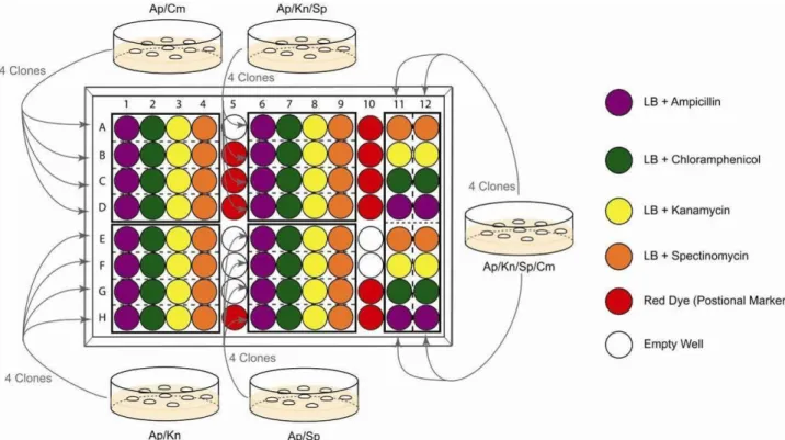
10.During the incubation of colonies, fill a 96 well microtiter plate with 150 µl antibiotic-containing LB media (following Illustration 7). It is recommended to add coloured dye (positional marker) in the wells indicated.
A typical arrangement of the solutions, which is used for parallel selections of multifusion plasmids, is shown in Illustration 7. The concept behind the 96 well plate experiment is that every cell suspension from single colonies needs to be challenged by all four single antibiotics for unambiguous interpretation.
Illustration 7: 96 well analysis of Cre assembly
11.Add 1 µl aliquots of pre-incubated cell culture (Step 9) to the corresponding wells. Then incubate the inoculated 96 well microtiter plate in a 37°C shaking incubator overnight at 180-200 rpm.
Recommended: use parafilm to wrap the plate to avoid drying out.
The remainder of the pre-incubated cell cultures could be kept at 4°C for further inoculations if necessary.
12.Select transformants containing desired multifusion plasmids based on antibiotic resistance, according to the combination of dense (positive) and clear (no growth) cell microcultures from each colony. Inoculate 10-20 µl cell culture into 10 ml LB media with corresponding antibiotics. Incubate in a 37°C shaking incubator overnight.
13.Centrifuge the overnight cell cultures at 4000g for 5-10 minutes. Purify plasmid from the resulting cell pellets with common plasmid miniprep kits, according to manufacturers’ recommendation.
14.Determine the concentrations of purified plasmid solutions by using UV absorption spectroscopy (e.g. by using a NanoDropTM 1000 machine).
15.Digest 0.5~1 µg of the purified plasmid solution in a 20 µl restriction digestion with appropriate endonuclease(s). Incubate under recommended reaction condition for ~2 hours.
16.Use 5-10 µl of the digestion for analytical agarose (0.8-1.2%) gel electrophoresis. Verify plasmid integrity by comparing the experimental restriction pattern to a restriction pattern predicted in silico (e.g. by using program VectorNTI from Invitrogen or similar programs).
C.2.2. Deconstruction of fusion vectors by Cre
The following protocol can be used for example also for the recovery of all four single ACEMBL vectors by deconstructing tetra-fused pACKS plasmid (pACE-pDC-pDK-pDS); which is part of the ACEMBL System kit (Section D). Likewise, the protocol is suitable for releasing any single educt from multifusion constructs (deconstruction). This is achieved by Cre-LoxP reaction, transformation and plating on agar with appropriately reduced antibiotic resistance level (c.f. Illustration 6). In the liberated educt entity, encoding genes can be modified and diversified. Then, the diversified construct is resupplied by Cre-LoxP reaction (C.2.1.).
Reagents:
Cre recombinase (and 10x Buffer)
E. coli competent cells
(pir+ strains, pir- strains could be used only when partially deconstructed Acceptor-Donor fusions are desired).
Antibiotics
1. Incubate ~1 µg multifusion plasmid with 2 µl 10x Cre buffer, 1~2 µl Cre recombinase, add ddH2O to adjust the total reaction volume to 20 µl.
2. Incubate this Cre deconstruction reaction mixture at 30°C for 1-4 hour.
3. Optional: load 2-5 µl of the reaction on an analytical agarose gel for examination.
Heat inactivation at 70°C for 10 minutes before the gel loading is strongly recommended.
4. For chemical transformation, mix 10-15µl De-Cre reaction with 200 µl chemical competent cells. Incubate the mixture on ice for 15-30 minutes. Then perform heat shock at 42°C for 45-60 s.
Up to 20 µl De-Cre reaction (0.1 volumes of the chemical competent cell suspension)
For electrotransformation, up to 2 µl De-Cre reaction could be directly mixed with 100 µl electrocompetent cells, and transformed by using an electroporator (e.g. BIORAD E. coli Pulser) at 1.8-2.0 kV.
Larger volume of De-Cre reaction must be desalted by ethanol precipitation or PCR purification column before electrotransformation. The desalted De-Cre reaction mix should not exceed 0.1 volumes of the electrocompetent cell suspension.
The cell/DNA mixture could be immediately used for electrotransformation without prior incubation on ice.
5. Add up to 400 µl of LB media (or SOC media) per 100 µl of cell/DNA suspension immediately after the transformation (heat shock or electroporation).
6. Incubate the suspension in a 37°C shaking incubator (recovery).
For recovery of partially deconstructed double/triple fusions, incubate the suspension in a 37°C shaking incubator for 1 to 2 hours.
For recovery of individual educts such as single ACEMBL vectors from pACKS plasmid, incubate the suspension in a 37°C whaking incubator overnight or for at least 4 hours.
7. Plate out the recovered cell suspension on agar containing the desired (combination of) antibiotic(s). Incubate at 37°C overnight.
8. Colonies after overnight incubation might be verified directly by restriction digestion at this stage (refer to steps 12-16).
Especially recommended in the case that only one single educt or partially deconstructed multifusion plasmid is desired.
For further selection by single antibiotic challenge on a 96 well microtiter plate, continue with step 9.
Several different single educts/partially deconstructed multifusion plasmids can be processed and selected on one 96 well microtiter plate in parallel.
9. For 96 well analysis, inoculate four colonies each from agar plates containing a defined set of antibiotics into ~500 µl LB media without antibiotics. Incubate the cell cultures in a 37°C shaking incubator for 1-2 hours.
10.During the incubation of colonies, fill a 96 well microtiter plate with 150 µl antibiotic-containing LB media or coloured dye (positional marker) in the corresponding wells.
Refer to Illustrations 7 and 12 for the arrangement of the solutions in the wells, which are used for parallel selection of single educts or partially deconstructed multifusion plasmids. The concept is that every cell suspension from a single colony needs to be challenged by all four antibiotics separately for unambiguous interpretation.
11.Add 1 µl aliquots from the pre-incubated cell cultures (Step 9) into the corresponding wells. Then incubate the 96 well microtiter plate in a 37°C shaking incubator overnight at 180-200 rpm.
Recommended: use parafilm to wrap the plate to prevent dehydration.
The remainder of the pre-incubated cell cultures can be kept in 4°C fridge for further inoculations if necessary.
12.Select transformants containing desired single educts or partially deconstructed multifusion plasmids according to the combination of dense (growth) and clear (no growth) cell cultures from each colony. Inoculate 10-20 µl cell cultures into 10 ml LB media with corresponding antibiotic(s). Incubate in a 37°C shaking incubator overnight.
13.Centrifuge the overnight cell cultures at 4000g for 5-10 minutes. Purify plasmid from cell pellets with common plasmid miniprep kits, according to manufacturers’ information.
14.Determine the concentrations of purified plasmid solutions by using UV absorption spectroscopy (e.g. NanoDropTM 1000).
15.Digest 0.5~1 µg of the purified plasmid solution in a 20 µl restriction digestion (with 5-10 unit endonuclease). Incubate under recommended reaction condition for ~2 hours.
16.Use 5-10 µl of the digestion for analytical agarose gel (0.8-1.2%) electrophoresis. Verify the plasmid integrity by comparing the actual restriction pattern to predicted restriction pattern in silico (e.g. by using VectorNTI, Invitrogen, or any other similar program).
17.Optional: Possibly, a deconstruction reaction is not complete but yields partially deconstructed fusions which still retain entities to be eliminated. In this case, we recommend to pick these partially deconstructed fusions containing and perform a second round of Cre deconstruction reaction (repeat steps 1-8) by using this construct as starting material.
In our hands, two sequential deconstruction reactions were always sufficient to recover all individual modules, for instance all four single ACEMBL vectors from a pACKS plasmid. Liberation of single educts from double/triple fusions were found to be often more efficient than from quadruples such as the pACKS plasmid of the system kit (Section E).
C.3. Coexpression by Cotransformation
Protein complexes can be expressed also from two separate vectors that were cotransformed in expression strains. The cotransformed vectors can have the same or different origins of replication, however, they must encode for different resistance markers. Plasmids pACE (ampicillin resistance marker) and pACE2 (tetracycline resistance marker) have both a ColE1 derived replicon and can therefore be used with all common expression strains. pACE and pACE2 derivatives (also including fused Donors if needed) can be cotransformed into expression strains, and double transformants selected for by plating on agar plates containing both ampicillin and tetracyclin antibiotics.
D. ACEMBL multigene combination: Examples
Examples of multiprotein expressions by ACEMBL are shown in the following illustrating the gene combination procedures detailed in Section C. Reactions presented were carried out manually following the protocols provided, and also on a Tecan Freedom EvoII 200 robot with adapted protocols.
D.1. SLIC cloning into ACEMBL vectors: human TFIIF
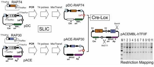
Genes encoding for full-length human RAP74 with a C-terminal oligo-histidine tag and full-length human RAP30 were amplified from pET-based plasmid template9 by using the primer pair TN7InsFor (5’-TCCCGCGAAATTAATACGACTCACTATA GGG-3’) and Tn7Insrev (5’-CCTCAAGACCCGTTTAGAGGCCCCAAGGGGTT ATGCTAG-3’) following the protocols described above. Linearized vector backbones were generated by PCR amplification from pACE and pDC by using primer pair Tn7VecFor (5’CTAGCATAACCCCTTGGGGCCTCTAAACGGGT CTTGAGG-3’) and Tn7VecRev (5’-CCCTATAGTGAGTCGTATTAATTTC GCGGGA-3’) in both cases. SLIC following Protocol 1 (Section C), resulting in pACE-RAP30 and pDC-RAP74his (Fig 8). These plasmids were fused by Cre-LoxP reaction (Section C). Results from restriction mapping by BstZ17I/BamHI double digestion of 11 double resistant (Cm, Ap) colonies are shown by a gel section from 1% E-gel electrophoresis (M: NEB 1kb DNA marker). All clones tested showed the expected pattern (5.0 + 2.8 kb). One clone was transformed in BL21(DE3) cells. Expression and purification by Ni2+-capture and S200 chromatography resulted in
human TFIIF complex (Fig. 3a, main text). Illustration 8: ACEMBLing TFIIF.
D.2. Polycistron by SLIC: human VHL/ElonginB/ElonginC complex.
The gene encoding for Von Hippel Lindau protein (amino acids 54-213), fused at its N-terminus to a six-histidine-thioredoxin fusion tag, was PCR amplified from plasmid pET3-HisTrxVHL by using primers Tn7InsFor (Table I) and SmaBamVHL (5’-GAATTCACTGGCCGTCGTTTTACAGGATCCTTAATCTCCCATCCGTTG ATGTGCAATG-3’). SmaBamVHL primer is a derivative of the SmaBam adaptor sequence (Table I) elongated at its 3’ by the insert specific sequence at the 3’ end of the VHL gene (including a stop codon). The gene encoding for full-length ElonginB was PCR amplified from pET3-ElonginB by using primers BamSmaEB (5’-GGATCCTGTAAAACGACGGCCAGTGAATTCGCTAGCTCTAGAAATAATT TGTTTAAC-3’) and SacHindEB (5’-GAGCTCGACTGGGAAAACCCTGGCG AAGCTTAGATCTGGATCCTTACTGCACGGCTTGTTCATTGG-3’), which are derivatives of the corresponding adaptors (Table I). The gene for ElonginC (amino acids 17-112) wa amplified from pET3-ElonginC by using primers HindSacEC (5’-AAGCTTCGCCAGGGTTTTCCCAGTCGAGCTCCAATTGGAATTCGCTAGCT CTAG-3’) and BspEco5EC (5’GATCCGGATGTGAAATTGTTATCCGCTGG TACCAAGCTTAGATCTGGATCCTTAACAATCTAAGAAG-3’), which are derivatives of the corresponding adaptors (Table I). Vector backbone was PCR amplified by using primers Tn7VecRev and Eco5Bsp, and pACE as a template (Illustr. 9). Multifragment SLIC was carried out according to Protocol 2 (Section C) resulting in pACE-VCB which contains a tricistron. Clones were plated on agar plates containing ampicillin. A positive clone, verified by sequencing, was used in the coexpression experiment described below (section D.5.)
D.3. The Homing endonuclease/BstXI module: yeast RES complex
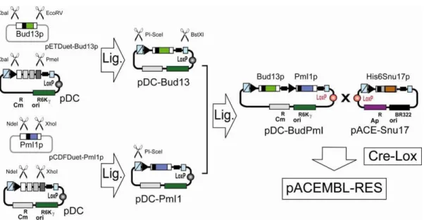
Plasmids pCDFDuet-Pml1p, pRSFDuet-Snu17p-NHis and pETDuet-Bud13p, encoding for yeast proteins (all full-length) PmI1p, Snu17p and Bud13p, respectively, were a kind gift from Dr. Simon Trowitzsch and Dr. Markus Wahl (MPI Göttingen). Snu17p contains a six-histidine tag fused to its N-terminus. The gene encoding for His6-tagged Snu17p was excised from pRSFDuet-Snu17p-NHis by using restriction enzymes NcoI and XhoI, and ligated into a NcoI/XhoI digested pACE construct (containing an unrelated gene between NcoI and XhoI sites) resulting in pACE-Snu17. The gene encoding for Bud13p was liberated from pETDuet-Bud13p by restriction digestion with XbaI and EcoRV, and placed into XbaI/PmeI digested pDC resulting in pDC-Bud13. The gene encoding for Pm1Ip was liberated from pCDFDuet-Pml1p by restriction digestion with NdeI and XhoI, and placed into NdeI/XhoI digested pDC resulting in pDC-PmI1. Next, the expression cassette for Bud13p was liberated from pDC-Bud13 by digestion with PI-SceI and BstXI. The liberated fragment was inserted into PI-SceI digested and alkaline phosphatase treated pDC-PmI1p resulting in pDC-Bud13p-PmI1p.
pACE-Snu17 and pDC-BudPmI were then fused by Cre-LoxP reaction and selected for by plating on agar plates containing ampicillin and chloramphenicol. Fusion plasmids were transformed into BL21(DE3) cells. Expression and purification by Ni2+-capture and S200 size exclusion chromatography resulted in the trimeric RES complex (Supplementary Results, complex S12b).
D.4. Coexpression by cotransformation: human NYB/NYC
Genes encoding for protein NYB (amino acids 49-141) and NYC (amino acids 27-12) were excised from vectors pACYC18411-NYB and pET15-NYC, respectively10.
NdeI and BamHI where used for NFYB. XbaI and BamHI where used for NYC, thus importing a six-histidine tag at the N-terminus of the protein. The NYB insert was ligated into pACE digested with NdeI and BamHI. The NYC insert was ligated into pACE2 digested by XbaI and BamHI. pACE-NFYB and pACE2-NFYC were transformed into BL21(DE3) cells containing the pLysS plasmid. Selection on agar plates containing ampicillin, tetracyclin and chloramphenicol resulted in triple resistant colonies. The complex was expressed and purified by Ni2+ capture (IMAC) and S75HR (Pharmacia) size exclusion chromatography (Supplementary Results, complex S7a).
D.5. Coexpression from Acceptor-Donor fusions
Six heterologous genes encoding for a trimeric protein complex (VHLbc: VonHippel-Lindau protein amino acids 54-213 / full-length ElonginB / ElonginC amino acids 17-112)11, a gene encoding for the AAA ATPase FtsH (amino acids 147-610), and two genes encoding for fluorescent markers (BFP and GFP) were assembled as indicated. In a single Cre reaction, all combinations of one Acceptor (pACE-VHLbc) and three Donors (pDC-FtsH, pDK-BFP, pDS-mGFP) were obtained and selected, including a quadruple fusion containing all six heterologous genes (Main text, Fig. 2). Clones were verified by 96 well microtiter assay as described in Section C. Expression and Ni2+ affinity capture, combined with immunostaining of the untagged fluorescent markers, confirmed successful multiprotein expression (Main text, Figs. 2 and 3b). Proteins were expressed overnight in BL21(DE3) cells in 24 well deep-well plates in small scale using autoinduction media12. Restriction mapping revealed that even large fusion plasmids were stable over many (more than 60) generations, even if challenged by a single antibiotic in the medium only.
10 Romier, C. et al., J. Biol. Chem. 278, 1336-1345 (2003)
11 Stebbins, C.E., Kaelin, W.G. Jr, Pavletich, N.P. Science 284, 455-61 (1999) 12 Studier F.W. Protein Expr. Purif. 41, 207-34 (2005).
E. The ACEMBL System Kit
Reagents to be supplied in ACEMBL system kit: BW23473, BW23474 cells† pACKS quadruple fusion vector* made of: pACE (Acceptor)
pDC, pDK, pDS (Donors) pACE2 vector
pACE-[VHLbc/BFP/mGFP] control plasmid triple fusion vector
made of: pACE-VHLbc pDK-BFP
pDS-mGFP#
† E. coli strains expressing the pir gene for propagation of Donor
derivatives (any other strain with pir+ background can be used).
* This fusion vector was created by Cre-LoxP reaction of pACE, pDC, pDK and pDS. It is resistant to ampicillin, kanamycin, chloramphenicol and spectinomycin. Individual ACEMBL vectors are liberated from this quadruple fusion by Cre-LoxP mediated deconstruction as described in protocol C.2.2. Sequences for single ACEMBL vectors and pACKS quadruple fusion are provided in Appendix.
# pDS-mGFP contains a coiled-coil fused to the N-terminus of eGFP13.
Reagents additionally required:
Antibiotics: ampicillin, chloramphenicol, kanamycin, spectinomycin, tetracycline Enzymes: Cre recombinase
T4 DNA polymerase (for recombination insertion of genes) Phusion polymerase (for PCR amplification of DNA)
Restriction enzymes and T4 DNA ligase (for conventional cloning) Regular laboratory cloning strain (TOP10, HB101, DH5α)
Expression strain(s) of choice
Illustration 11: ACEMBL System Kit: Generating single vectors from pACKS.
pACKS is deconstructed according to the schematic in Illustr. 11 into single vectors pACE, pDC, pDK and pDS. 96 well microtiter assay for identifying single vectors is shown in Illustr. 12.
Illustration 12: 96 well microtiter analysis of pACKS De-Cre reaction.
Clones containing pACE, pDC, pDK and pDS single vectors as identified by microtiter assay, are then used for plasmid generation. The vectors can be further verified by restriction digestion before use for subcloning (see Appendix for vector sequences). pACE2 is provided as a separate vector in the ACEMBL System Kit.
F. Appendix
F.1. DNA sequence of MIE
Below are the sequence and map of the MIE fragment between T7/lac promoter and T7 terminator in ACEMBL vectors. Forward and reverse primers for sequencing can be standard vector primers for T7 and lac. Adaptor primer sequences (c.f. Table I) are indicated. DNA sequences in these homology regions contain tried-and-tested sequencing primers14. Sites of insertion (I1-I4) are shown.. The adaptor sequences, and probably any sequence in the homology regions, can be used as adaptors for multifragment insertions. The ribosome binding site present in the MIE (rbs) is boxed in red.