Laser treatment for toenail fungus
David M. Harris
a,b, Brian A. McDowell
c, John Strisower
b,
a
Univ. of Washington, Seattle WA, USA 98195;
bPathoLase, Inc. 275 Airpark Blvd., Chico, CA
95973;
cNorthern California Orthopedic Centers, 6403 Coyle Avenue, Carmichael, CA, USA
95608
ABSTRACT
An infrared pulsed laser was used to irradiate toenails with visible signs of bacterial/fungal infections. Seventeen subjects with both great toes involved were recruited in a podiatric private practice. Toes were randomized to receive either a single treatment or no treatment. The treatment was tolerated by all subjects without anesthesia and there were no occurrences of serious adverse effects. Eleven out of 14 (79%) treated toes improved. Improvement ranged from 2.1 to 6.1 mm over 90 days following a single treatment. There was a highly significant difference (p<0.001) between treated and untreated toes for nail bed clearing.
Keywords: Laser, onychomycosis, toenail fungus.
1. INTRODUCTION
Onychomycosis is a chronic fungal nail infection that affects 7–10% of the U.S. population and can have serious consequences to the elderly, diabetics, and immunocompromised individuals. Dermatophytes (including the genera Trichophyton, Epidermophyton and Microsporum) are the most common pathogens of onychomycosis,1-3 with T. rubrum accounting for 80% of the infections.4
The efficacy of current treatment options, including topical, oral, mechanical and chemical therapies or a combination of these modalities is low.5,6 Topical drug treatment for onychomycosis is not usually successful because they are unable to penetrate the nail plate7,8 and rapid recurrence can occur after discontinuing use.9,10 Oral antifungal agents are more effective although more toxic with a significant risk of liver toxicity, prolonged loss of taste, and life-threatening drug interactions.11 Fungal resistance can occur when the oral antifungal agents are used on a long-term basis. Topically applied antifungal drugs may work somewhat better adjunctive to surgical removal or chemical dissolution of the nail plate.12 Yet, this often ineffective and traumatic procedure leaves the subject without a nail for months at risk for reinfection.
Given the limitations of current treatment options, there is a great need for a simple, nontoxic and effective alternative treatment.13 In an initial clinical trial an infrared pulsed laser was used to irradiate toenails with visible signs of bacterial/fungal infections. The following is a report of the clinical data from that trial designed to evaluate the safety and efficacy of a prototype antifungal laser, the PinPointe™ FootLaser™ by PathoLase, Inc. (Chico, CA).
2. EXPERIMENT DESIGN
Seventeen volunteer subjects with both great toes involved were recruited in a podiatric private practice. All subjects signed informed consent forms. Subjects were of either sex, 18 years or older, with right and left great toe involvement (e.g., current bacterial/fungal infection classified by the investigator as subungual onychomycosis). Subjects were also required to provide Informed Consent and be available for study follow-up visits and evaluations. Subjects were excluded from study if they were pregnant, had been treated with oral Lamisil (terbinafine hydrochloride) within one year prior to study treatment, had been treated with or taken Penlac (ciclopirox), Sporonox (Itraconazole) or over-the-counter (OTC) remedies for toenail infection within 4 weeks prior to study treatment, were determined by the investigator to be incapable of study compliance, or at the professional discretion of the investigator.
Subject 14 Top: treated Bottom: untreated Days poreath1ent Lea, Ix Leax .b-Tx Subject 02006 0 1 2 3 4 5 6 7 8 0 10 20 30 40 50 60 70 80 90 Da ys post-tre a tment D ist an ce ( m m ) Lesion Tx Lesion No-Tx
Right or left great toes were randomized to receive either a single treatment or no treatment. Nails were not debrided. A notch was placed near the proximal extent of each nail with a triangular file and a calibrated 1-cm scale was placed on the toe. Nails were photographed with a high resolution digital camera before treatment, immediately post-treatment and at 1 week and monthly up to three months when data were analyzed. At that time the untreated toes were treated (crossover design) and subjects were followed an additional three months out to 6 months post-treatment. Nail plate growth and nail bed clearing at 3 months were measured from the photographs using Image J software by an evaluator who was blinded to the treatment condition.
Figure 1. Typical response to treatment. Left: Top treated, bottom untreated. Pretreatment on the left followed by images at 1, 2 and 3 months.
Below: distance of the lesion from the proximal fold (mm). Subject 006 absolute distance and #14 normalized distance. Blue treated, red untreated.
Nail plate growth 0 1 2 3 4 5 6 0 10 20 30 40 50 60 70 80 90 Days post-treatment Di st a n ce ( m m) Tx No-Tx 3. RESULTS
3.1 Demographics and Baseline Characteristics
Mean age of the study patients was 67.3 +/- 12 years (range 46 – 89 years; n=17); 65% (11/17) males and 35% (6/17) females. 94.1% (16/17) subjects were Caucasian and 5.9% (1/17) were Hispanic. The median duration of toenail infection among the study patients was > 10 years (range 2 – 50 years; n=17).
3.2 Safety
The treatment was tolerated by all subjects without anesthesia. However, during the course of the study, a total of 9 adverse events were reported by 5 patients, mostly mild to moderate discomfort during the procedure. There were no reports of nail plate or nail bed damage or discoloration, nor was there any post-operative discomfort related to the treatment.
3.3 Individual data.
The effect of treatment is illustrated by the individual examples in Figure 1. The treated toe of Subject 006 responded well to a single treatment. By 3 months post-treatment there is 4 mm of clear nail growth. The right foot was untreated and the lesion did not “grow out.” Note that the notch on the treated foot moves out at a faster (variable) rate than the lesion whereas, in the untreated foot the notch grows out but the lesion remains stationary. Notice also that the treated toe of subject 14 is onycholytic (nail plate is separated from the nail bed), but this has resolved following treatment.
3.4 Summary data.
The analysis of the data at three months revealed that notch measurements increased in a similar fashion for both toes with no difference between treated and untreated toes (p=0.810). There was a highly significant difference (p<0.001) between treated and untreated toes for the lesion measurement. For treated toes, clear nail average values went from 4.0 mm at baseline to 7.7 mm at 90 days, while the untreated toes went from 5.3 at baseline to 5.6 mm at 90 days. Eleven out of 14 (79%) treated toes improved. Improvement ranged from 2.1 to 6.1 mm over 90 days following a single treatment For the 14 toes that responded to therapy the average improvement was 3.9 mm, or 1.3 mm per month.
3.5 Notch measurements and the rate of nail plate growth.
Each nail had a unique growth rate that would vary from month to month. Also, left and right great toenails would often grow at different rates on the same person. Table 1 shows the summary statistics for notch and measurements for the 16 patients in the analysis and Figure 2 shows that over all there were no differences in nail plate growth rate. Table 2 summarizes the results of repeated measures analysis of variance. The primary factor of interest is the
treatment by time interaction which compares the changes over time between treated and untreated toes (essentially the slopes of the response curve). Notch measurements increased in a similar fashion with no difference between treated and untreated toes (p=0.330).
Figure 2. Growth of the nail plate as the distance in mm from the original notch location at monthly intervals. There is no difference between treated and untreated toes (p=0.330).
TABLE 1 SUMMARY STATISTICS FOR NOTCH MEASUREMENTS. Values are distance of the notch from the proximal fold in mm’s.
The following results are for: VARIABLE = Notch T0 T7 T30 T60 T90 N of cases 15 14 15 15 14 Minimum 3.500 3.660 4.530 5.860 6.680 Maximum 8.270 9.800 10.810 11.200 14.690 Mean 5.149 5.429 6.948 8.279 10.239 Std. Error 0.334 0.439 0.413 0.424 0.530 Standard Dev 1.295 1.643 1.601 1.642 1.984
U0 U7 U30 U60 U90 N of cases 15 14 15 15 14 Minimum 2.330 2.700 4.050 5.190 6.400 Maximum 7.480 8.370 9.620 11.540 13.200 Mean 4.741 5.295 6.751 8.555 10.202 Std. Error 0.339 0.433 0.412 0.555 0.555 Standard Dev 1.311 1.622 1.598 2.148 2.075
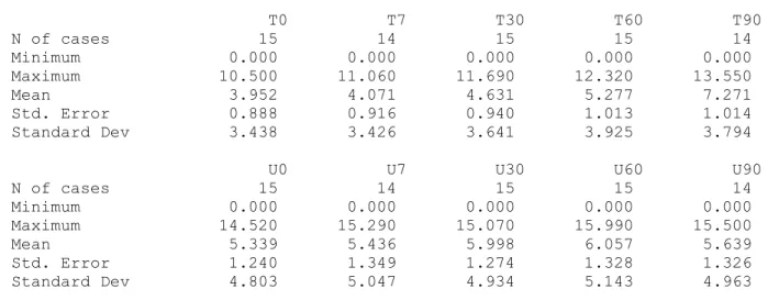
3.5 Lesion measurements and the rate of nail bed clearing
There was a highly significant difference (p<0.001) between treated and untreated toes for the lesion measurement. For treated toes, lesion average values went from 4.0 at baseline to 7.3 mm at 90 days, while the untreated toes went from 5.3 at baseline to 5.6 mm at 90 days. Eleven out of 14 (79%) treated toes improved. Improvement ranged from 2.1 to 6.1 mm over 90 days following a single treatment. For the toes that responded to therapy the average improvement was 4.0 mm, or 1.3 mm per month.
TABLE 2 SUMMARY STATISTICS FOR LESION MEASUREMENTS. Values are distance of the notch from the proximal fold in mm’s.
The following results are for: VARIABLE = Lesion T0 T7 T30 T60 T90 N of cases 15 14 15 15 14 Minimum 0.000 0.000 0.000 0.000 0.000 Maximum 10.500 11.060 11.690 12.320 13.550 Mean 3.952 4.071 4.631 5.277 7.271 Std. Error 0.888 0.916 0.940 1.013 1.014 Standard Dev 3.438 3.426 3.641 3.925 3.794
U0 U7 U30 U60 U90 N of cases 15 14 15 15 14 Minimum 0.000 0.000 0.000 0.000 0.000 Maximum 14.520 15.290 15.070 15.990 15.500 Mean 5.339 5.436 5.998 6.057 5.639 Std. Error 1.240 1.349 1.274 1.328 1.326 Standard Dev 4.803 5.047 4.934 5.143 4.963
PRE-TREATMENT 02006 I MONTH 2 MONTHS 3 MONTHS 4 MONTHS 5 MONTHS
ill I I
02006L1
6 MONTHS Nail bed clearing
0 1 2 3 4 0 10 20 30 40 50 60 70 80 90 Days post-treatment D ist an ce ( m m ) Tx No-Tx
Subjects were followed and photographed out to 6 months post-treatment. Figure 4 shows images for Subject 6 and 14 out to 6 months (Also shown in Figure 1).
In conclusion, there appeared to be no discrimination between treatment and no treatment for notch measurements, however there were highly significant treatment differences for lesion measurements.
Figure 4. Patient 6 improved up to 3 months but after 3 months the lesion progresses proximal. Although still improved, the best result was at 3 months. At six months after a single treatment the treated toe of Subject 14 continues to improve.
4. DISCUSSION
Comparison of nail plate (notch) growth rate and nail bed (lesion) clearing rates reveals that the nail plate grows about twice as fast as the lesion clears out of the nail bed (Figure 5). Time for complete clearing is dependant on the original amount of involvement. We predict that complete clearing of a totally involved great toe should take up to12-18 months and we continue to follow these patients.
We note that the notch grew out at the same rate on the treated as on the untreated toe. This means that there is neither an obvious biostimulatory effect of laser light on nail plate growth, nor is there an inhibitory effect. There were no visible effects of treatment (discoloration, surface texture changes, etc.) seen in any of the photographs or reported by the investigator. In this sample of 17 patients treatment of the toenail with the FootLaser did not damage the nail plate, the underlying nail bed, the matrix, or the surrounding tissue. On the contrary, in a few of the subjects (for example Figure 1 and Figure 4, Subject 0014) an irregular contoured nail grew out looking smooth and natural.
Figure 3. Nail bed clearing plotted as the distance in mm from the original lesion location at monthly intervals. There is a significant difference between treated and untreated toes (p<0.001.)
6 5
@3
U I 0Nail plate growth and
Nail bed clearing
0 10
20 30 40 50 60 70
80 90Days post-treatment
Lesion Tx'
Lesion No-Tx o Notch Tx o Notch Nc-TxFigure 5. Comparison of the rate of nail plate growth (notch) with nail bed clearing (lesion) shows that the nail plate grows out about twice as fast as the lesion clears.
Some subjects (18%) reported mild to moderate intraoperative discomfort although all subjects tolerated the entire procedure. No toxicity, side-effects or drug interactions occurred. Results reported here were from a single treatment so, except for sitting still during the procedure, additional patient compliance was unnecessary.
In this study we selected difficult to treat infections, such as patients with fungal spikes,14,15 onycholytic and/or hypertrophic nails. Even though these patients are typically excluded in drug trials they composed 75% of our sample. It is important to note that most of these difficult cases actually improved following FootLaser treatment, while three hypertrophic nails accounted for the failures.
Treatment with the PinPointe FootLaser resulted in significant nail clearing in most subjects that was similar in clinical appearance to nail clearing seen with terbinafine when it works. In this regard our results support the hypothesis that the laser treatment destroys the pathogens that cause onychomycosis.
The current standard of treatment for onychomycosis is pharmacologic. However, drugs require patient compliance, can be toxic and can induce pathogen resistance. This laser treatment does not require daily compliance and, so far, appears to produce no toxicity. In addition to superior safety the laser treatment promises to be much more effective than drugs. The efficacy of drug therapy is in the range 15-30% versus 80% efficacy observed in this study. Our results are, of course, preliminary and data from a much larger sample in a well-controlled, multi-center trial is essential to substantiate our findings.
5. REFERENCES CITED
[1] Elewski, B.E., Charif, M.A. "Prevalence of onychomycosis in patients attending a dermatology clinic in northeastern Ohio for other conditions," Arch Dermatol 133, 1172-1173 (1977).
[2] Elewski, B.E., Hay RJ. "Update on the management of onychomycosis: highlights of the Third Annual International Summit on Cutaneous Antifungal Therapy," Clin Infect Dis 23, 305-313 (1996).
[3] Clayton, Y.M., "Clinical and mycological diagnostic aspects of onychomycosis and dermatomycoses," Clin Exp Dermatol 17(1), 37-40 (1992).
[4] Ghannoum, M.A., Hajjeh RA, Scher R et al., "A large-scale North American study of fungal isolates from nails: the frequency of onychomycosis, fungal distribution, and antifungal susceptibility patterns," J Am Acad Dermatol 43, 641-648 (2000).
[5] Crawford, F, Young P, Godfrey C et al., "Oral treatments for toenail onychomycosis: a systematic review," Arch Dermatol 138, 811-816 (2002).
[6] Elewski, B.E., "A full ‘cure’ for onychomycosis is not always possible," Arch Dermatol 135, 852-853 (1999). [7] Elewski, B.E., "Onychomycosis: pathogenesis, diagnosis, and management," Clin Microbiol Rev 11, 415-429
(1998).
[8] Gupta, A.K., "Ciclopirox nail lacquer: a brush with onychomycosis," Cutis 68, 13-16 (2001).
[9] Finch, J.J., Warshaw E.M., "Toenail onychomycosis: current and future treatment options," Dermatol Ther 20, 31-46 (2007).
[10] Arrese, J.E., Pierard G.E., "Treatment failures and relapses in onychomycosis: a stubborn clinical problem," Dermatology 207, 255-260 (2003).
[11] Katz, H.I., "Drug interactions of the newer oral antifungal agents," Br J Dermatol 141(Suppl. 56), 26-32 (1999). [12] Grover, C., Bansal S., Nanda S. et al., "Combination of surgical avulsion and topical therapy for single nail
onychomycosis: a randomized controlled trial," Br J Dermatol 157, 364-368 (2007).
[13] Conant, M., "New treatment options for onychomycosis," AIDS Patient Care 9 (Suppl. 1), S19-S21 (1991). [14] Lecha, M., Effendy I., Feuilhade de Chauvin M., Di Chiacchio N, Baran R, "Treatment options--development of
consensus guidelines. Taskforce on Onychomycosis Education" J Eur Acad Dermatol Venereol Suppl 1, 25-33 (2005).
[15] Scher, R.K., Baran, R., "Onychomycosis in clinical practice: factors contributing to recurrence," Br J Dermatol 149 (Suppl 65), 5-9 (2003).
6. ACKNOWLEDGMENT
Disclosure: David M Harris and John Strisower are principals of Patholase, Inc. Brian McDowell, DPM is a podiatrist with a private practice in Sacramento, CA.