Impact of Pretransplantation Risk Factors on Post
Transplantation Outcome of Patients with Acute Myeloid
Leukemia in Remission after Haploidentical Hematopoietic
Stem Cell Transplantation
Yu Wang, Dai-Hong Liu, Kai-Yan Liu, Lan-Ping Xu, Xiao-Hui Zhang, Wei Han,
Huan Chen, Yu-Hong Chen, Xiao-Jun Huang
*Peking University People’s Hospital, Institute of Hematology, No.11 Xizhimen South Street, Xicheng District, Beijing, 100044, People’s Republic of China
Article history: Received 21 July 2012 Accepted 1 October 2012 Key Words:
Acute myeloid leukemia Response
Cytogenetics Risk
Transplantation
a b s t r a c t
The impact of risk-related parameters has not been defined in transplantation settings. We wondered whether the currently recognized predictors could be used to categorize acute myeloid leukemia (AML) patients who underwent transplantation during remission into risk groups. We analyzed the data of 255 consecutive patients (median age, 26) with AML in theirfirst or second remission (CR1 or CR2) after hap-loidentical hematopoietic stem cell transplantation (HSCT). Three parameters were found to be predictive of outcome: response after induction therapy, white blood cell count at diagnosis, and cytogenetics. These three factors were combined to yield two risk groups. The 2-year cumulative incidences of relapse for patients at low and high risk were 8% and 36% (P¼.001), respectively. The 3-year probabilities of leukemia-free survival for these two groups were 80% and 52% (P¼.001), respectively. Multivariate analysis for relapse and for leukemia-free survival showed that not achieving CR after two courses of therapy was the strongest inde-pendent prognostic factor (P¼.001 andP¼.028, respectively). In addition, in a subgroup of patients with quantification of minimal residual disease at the time of HSCT, positive minimal residual disease at this time point was correlated with a poor outcome. Our results suggest that the pretransplantation risk factors influence posttransplantation outcomes of patients with AML in CR after haploidentical HSCT and might be applicable to assist with risk-directed posttransplantation therapy.
Ó2013 American Society for Blood and Marrow Transplantation.
INTRODUCTION
Acute myeloid leukemia (AML) is a heterogeneous disorder characterized by many prognostic parameters. In patients undergoing chemotherapy, the morphology, cyto-genetics, immunophenotype, and white blood cell (WBC) count at diagnosis and response after induction therapy have been proven to affect outcomes [1,2]. Studies have shown
that, compared with chemotherapy alone, a significant
benefit can be achieved with allogeneic hematopoietic stem cell transplantation (allo-HSCT) in AML patients in their first complete remission (CR1) who have intermediate- or poor-risk cytogenetics[3]. Allo-HSCT is currently considered
the treatment of choice for AML patients in their first
complete remission (CR1) without favorable cytogenetics. For patients with favorable cytogenetics, because of the high incidence of transplantation-related mortality (TRM) reported, allo-HSCT is not currently recommended as the preferred choice.
We wondered whether AML patients who underwent transplantation during CR have different prognoses after uniformly performed allo-HSCT. It is widely known that the outcomes are very poor for patients with advanced-stage
AML, even after allo-HSCT [4-6]. Many researchers,
including ourselves, have focused on posttransplantation
management to improve the outcomes of advanced-stage leukemia patients. Recently, we reported that prophylactic use of donor lymphocyte infusion (DLI) can increase the survival of patients with advanced-stage acute leukemia[7]. We were interested in risk factor evaluation of patients undergoing transplantation during CR and the identification of patients with high-risk features whose outcomes might be as poor as those of patients with advanced-stage AML. Therefore, more robust posttransplantation therapies might be applied in these patients with high-risk features, even after transplantation during CR and might further improve the overall outcomes of patients with AML in CR after allo-HSCT.
For AML patients undergoing HSCT during CR, cytoge-netics is the most often investigated predictor[8-11]. To our knowledge, there has been only one study that evaluated the role of risk factors, including cytogenetics, response after induction therapy, and French-American-British (FAB) type, on transplantation outcomes in matched-sibling HSCT settings [10]. We wondered whether the currently recog-nized predictors could be used to categorize into risk groups AML patients who underwent transplantation during remission and whether these predictors would be applicable in HSCT patients. The goal of the current study was to attempt to answer these two questions by analyzing the data on haploidentical HSCT, which is a uniformly performed treatment modality.
METHODS Patient Eligibility
Consecutive patients with AML (n¼255) in theirfirst or second CR who received HSCT from human leukocyte antigen (HLA)-mismatched family
American Society for Blood
ASBMT
and Marrow Transplantation
Financial disclosure:See Acknowledgments on page 289.
*Correspondence and reprint requets: Prof. Xiao-Jun Huang, MD, Peking University People’s Hospital, Institute of Hematology, No. 11 Xizhimen South Street, Xicheng District, Beijing, 100044, People’s Republic of China.
E-mail address:xjhrm@medmail.com.cn(X.-J. Huang).
1083-8791/$esee front matterÓ2013 American Society for Blood and Marrow Transplantation. http://dx.doi.org/10.1016/j.bbmt.2012.10.002
donors between May 2002 and December 2010 were enrolled in this study. Forty-five of the 255 AML patients were previously enrolled in a study in 2009[12]. These patients previously reported were enrolled and followed further in this study. All protocols were approved by the institutional review board of the Peking University Institute of Hematology, and all patients and their donors signed consent forms.
One hundred ninety-three patients (75%) had diagnostic cytogenetic results. These patients were classified into 3 groups, according to the National Comprehensive Cancer Network (NCCN) criteria[13]: better risk (n¼39), intermediate risk (n¼137), and poor risk (n¼17). The reasons for the patients being transplanted with better-risk cytogenetics were as follows: t (8;21) with complex karyotypes or del(9q), according to South-west Oncology Group/Eastern Cooperative Oncology Group (SWOG/ECOG) criteria[14](n¼7); not achieving CR after two courses of chemotherapy (n¼5); CR2 at the time of transplantation (n¼5); AML1/ETO or CBFb/ MYH11 transcript level greater than 103at the time of transplantation (n¼10); and transplanted before 2007 (n¼12). The patients were not evaluated for recently described molecular prognostic factors, such as the Flt3 internal tandem duplication, the NPM1 mutation, and thec-Kit muta-tion, until 2009, so this information is not provided in the present study.
All donorerecipient pairs were typed at the HLA-A, -B, and -DR loci. HLA-A and HLA-B typing was performed by intermediate-resolution DNA typing, whereas HLA-DRB1 typing was performed using high-resolution DNA techniques. For each donorerecipient pair, the patient received stem cells from a family member who shared one HLA haplotype with the patient but who differed to some degree in the HLA-A, -B, and -D antigens of the haplotype that was not shared. In addition, HLA typing was performed on the parents and offspring of each donorerecipient pair to guarantee a true haploid genetic background among the pairs. HLA disparity and other characteristics of the patients and donors are summarized inTable 1. CONDITIONING REGIMEN
The conditioning therapy consisted of cytarabine
(4 g/m2/day) administered intravenously on days10 to9, busulfan (4 mg/kg/day) administered orally on days8 to6 (before January 2008), or busulfan (3.2 mg/kg/day) admin-istered intravenously on days8 to6 (after January 2008),
cyclophosphamide (1.8 g/m2/day) administered
intrave-nously on days5 to4, Me-CCNU (250 mg/m2)
adminis-tered orally once on day3, and, between 2003 and 2004, ATG (either 20 mg/kg/day, porcine [Bioproduct, Wuhan, China] in 5 patients or 2.5 mg/kg/day, rabbit [Sang Stat, Lyon, France] for all other 250 patients) intravenously on days5 to2.
Graft-versus-Host Disease Prophylaxis
All transplantation recipients were administered cyclo-sporine A (CsA), mycophenolate mofetil (MMF), and short-term methotrexate. The dosage of CsA was 2.5 mg/kg/day administered intravenously and was administered from day 9 before transplantation until bowel function returned to normal. At that point, the patient was switched to oral CsA. MMF was administered orally (0.5 g every 12 hours) from day 9 before transplantation to day 30 after transplantation. MMF was tapered from 1 g/day to 0.5 g/day on day 30 and was discontinued on day 60. On day 1 after transplantation,
15 mg/m2 methotrexate was administered intravenously,
and on days 3, 6, and 11 after transplantation, 10 mg/m2 methotrexate was administered. The whole-blood CsA concentration was monitored twice per week usingfl uores-cence polarization immunoassay, and the dosage was adjusted to a blood concentration of 150 to 250 ng/mL. If there was no evidence of graft-versus-host disease (GVHD at days 90 to 100, the CsA dosage was gradually reduced and was discontinued at approximately day 180. If GVHD was observed, the CsA was continued.
Collection of Hematopoietic Cells
The donors were primed with recombinant human granulocyte colony-stimulating factor (filgrastim, Kirin,
Japan; 5
m
g/kg/day) injected subcutaneously for 5 to 6consecutive days. On the 4th day, bone marrow cells were harvested. The target mononuclear cell count was 3108 cells/kg of the recipient’s weight. On the 5th day (and on the 6th day, if necessary, ie, if the target mononuclear cell count was not reached on the 5th day), peripheral blood stem cells were collected with a COBE Blood Cell Separator (Spectra Table 1
Characteristics of Patients and Donors
Characteristics All Patients (n¼255) Age, y, median (range) of the recipient 26 (3-54)
0-20, No. (%) 87 (34) 21-40, No. (%) 130 (51) 41-54, No. (%) 38 (15) Gender, No. (%) Male 151 (59) Female 104 (41)
FAB subtype, No. (%)
M0 6 (2) M1 16 (6) M2 110 (43) M4 46 (18) M5 65 (26) M6 16 (4) M7 1 (1) WBC at diagnosis,109/L, No. (%) <20 165 (65) 20-49 27 (10) 50-99 35 (14) 100 28 (11)
Cytogenetics, NCCN criteria, No. (%)
Better risk 39 (15)
Intermediate risk 137 (54)
Poor risk 17 (7)
Unknown 62 (24)
Remission status, No. (%)
First complete remission (CR1) 228 (89) Second complete remission (CR2) 27 (11) Remission courses among CR1, No. (%)
CR after course 1 145 (64)
CR after course 2 66 (29)
CR after course 3 12 (5)
CR after course 4 5 (2)
Matched HLA locus
3 126
4 98
5 33
6 1
Donorerecipient gender
Maleemale 88
Maleefemale 55
Femaleemale 63
Femaleefemale 49
Donorerecipient blood type
Match 138
Minor mismatch 55
Major mismatch 49
Minorþmajor 12
Donorerecipient relation
Fatherechild 67
Motherechild 61
Siblingesibling 96
Childeparent 19
Other 12
Age, y, median (range) of the donor 40 (13-63) Comorbidity score*
0 186
1 30
2 39
CMV serostatus
Recipient and donor 5
Recipient or donorþ 250
Median CD34þcount,106/kg (range) 2.2 (0.3-55.3) Median CD3þcount,108/kg (range) 1.5 (0.2-8.3) *Comorbidity score was according to published criteria[33].
LRS, COBE BCT, Inc., Lakewood, CO) at a rate of 80 mL/min from a total blood volume of 10 L. The protocol called for the collection of at least 6108mononuclear cells/kg or 4106 CD34þcells/kg. The fresh and unmanipulated bone marrow and PBSCs were infused into the recipient on the day of collection. In instances of ABO major blood group incom-patibility, the red cells were removed from the bone marrow cells by density gradient sedimentation with Hespan (Braun, Irvine, California) according to the manufacturer’s instruc-tions. The surface markers of the cells in the grafts were determined by 2- or 3-color staining using monoclonal antibodies specific for CD34, CD3, CD4, and CD8 cells.
Prevention, Monitoring, Intervention, and Treatment of Relapse
Starting in January 2006, minimal residual disease (MRD) targets were regularly monitored after transplantation, and starting in January 2009, MRD targets were also regularly examined in the 2 weeks before the transplantation. The
bone marrow samples were defined as abnormal if they
contained more than .001% of leukemia-associated aberrant
immune phenotypes[15]or more than 0.6% of Wilms tumor
gene 1 (WT1)[16]. MRD-positive status was defined as either
two consecutive abnormalities in leukemia-associated
aberrant immune phenotypes or WT1 over a 2-week
interval or as an abnormality of both WT1and
leukemia-associated aberrant immune phenotypes in a single bone marrow sample. Starting in 2007, DLI or interleukin-2 was given, according to donor availability, as an intervention for MRD-positive status[17]. We made several modifications to classic DLI, as previously described in detail [18]. When hematologic relapse was diagnosed after HSCT, the relapse was treated with chemotherapy, followed by thera-peutic DLI[19].
Definitions and Assessments
CR was defined as morphologically normal marrow with
less than 5% blasts. Normal findings for peripheral blood were required at the evaluation of the induction course. Relapse was defined as the presence of more than 20% blasts in the bone marrow or blasts at extramedullary sites. Positive MRD was defined as noted above. The patients who had MRD were not classified as having relapsed. Cytogenetic classifi -cation was based on the NCCN criteria. The patients with unknown, unperformed, or unsuccessful cytogenetics were grouped together as an unknown group. In addition, the
classifications used by SWOG/ECOG, Medical Research
Council [20] and International System for Cytogenetic
Nomenclature [21] were also compared in terms of
leukemia-free survival (LFS). With regard to morphology, the
FAB cytological classification was used. Assessments of
engraftment, chimerism, and GVHD and surveillance for infection were previously described in detail[12].
Statistical Analyses
The cumulative incidences were estimated for engraft-ment, GVHD, infection, relapse, TRM, MRD after trans-plantation, and DLI intervention for positive MRD to accommodate the competing risks. Relapse was a competing risk for TRM, and TRM was a competing risk for engraftment, GVHD, infection, relapse, MRD after transplantation, and DLI intervention for positive MRD. The associations among the potential factors and outcomes were evaluated using an add-on package for R statistical software (Bell Labs, New Jersey) that allows for the estimation of a semiparametric
proportional hazards model for the subdistribution of competing risk analyses, as proposed by Scrucca et al[22]. The probabilities of overall survival (OS) and LFS were esti-mated by the Kaplan-Meier method. Potential prognostic factors were evaluated in univariate analyses by the log-rank test, withP<.05 considered statistically significant. In the multivariate analysis, all factors found to influence the outcomes in univariate analysis with aP<.15 were included in a Cox proportional hazard model using time-dependent variables. In these regression models, the occurrence of acute and chronic GVHD was treated as a time-varying co-variate. The potential interactions among the significant covariates were tested. No interactions were detected. SAS, version 8.2 (SAS Institute, Cary, NC) and S Plus 2000 (Math-soft, Seattle, WA) were used for most of the analyses. The endpoint of the last follow-up for all surviving patients was December 31, 2011.
RESULTS Overall Outcome
For the entire study population, up to December 31, 2011, 181 patients were alive, with a median follow-up of 1,075 days (range, 365 to 3,398 days) after transplantation without disease recurrence. The probabilities of OS and LFS were 72.9% (confidence interval [CI], 67.1% to 78.7%) and 70.1% (CI, 64.3% to 75.9%) at 3 years, respectively. Thirty-three patients died from causes other than relapse. The TRM rate at 2 years was 13.1% (CI, 8.9% to 17.3%). Causes of death are shown in
Table 2.
Among patients with regular MRD monitoring after HSCT (underwent transplantation after January 2006, n¼236), 30 patients had positive MRD, among whom 18 patients received DLI as an intervention for positive MRD. In total, 41 patients experienced leukemia relapses at a median time of 210 days (range, 30 to 730 days) after transplantation, reaching a cumulative incidence of relapse of 16.8% (CI, 12.0% to 21.6%) at 2 years. Thirty-three of the 41 cases of relapse occurred within thefirst year after transplantation. At the time of the last follow-up, 33 patients had died after relapses, with a median time to death of 330 days (range, 46 to 817) after HSCT and 100 days (range, 0 to 456) after relapse.
Engraftment
Two-hundred fifty-four patients (99.2%) achieved
sus-tained myeloid engraftment. Polymerase chain reaction DNA fingerprinting of short tandem repeats on recipient periph-eral blood cells was used to confirm 100% donor chimerism in these patients. The median time to reach an absolute neutrophil count above 0.5109cells/L was 13 days (range, 10 to 23 days). The cumulative 30-day myeloid engraftment probability was 99.1% (CI, 98.9% to 99.3%). The one patient who failed myeloid engraftment died from heart failure on day 13 post-HSCT. During the follow-up period, 240 patients Table 2
Causes of Death
Cause No. Patients (n¼66)
Relapse 33 Graft-versus-host disease 3 Infections 29 Bacteria 7 Fungal 9 Viral 13 Organ failure 1
(94.1%) exhibited platelet engraftment, and the median time to reach a platelet count above 20109cells/L was 16 days (range, 7 to 195 days).
GVHD
At 100 days after transplantation, the cumulative inci-dence was 39.5% (CI, 33.3% to 45.2%) for grade 2 to 4 acute GVHD and 11.1% (CI, 9.1% to 13.1%) for grade 3 to 4 acute GVHD. The cumulative incidence was 53.4% (CI, 46.4% to 60.4%) for total chronic GVHD and 19.9% (CI, 14.5% to 25.3%) for extensive chronic GVHD at 2 years after transplantation. Treating the occurrence of GVHD as a time-varying covariate, the occurrence of grade 2 to 4 acute GVHD did not affect relapse (P¼.463), LFS (P¼.935), or OS (P¼.954), whereas the occurrence of grade 3 to 4 acute GVHD did not affect relapse (P ¼.743) but did lower LFS (P ¼.016) and OS (P¼.001) by increasing TRM (P¼.001). The occurrence of chronic GVHD did not affect relapse (P¼.380), LFS (P¼.327), or OS (P¼.103), whereas the occurrence of extensive chronic GVHD did not affect relapse (P¼0.977) but did lower LFS (P¼.001) and OS (P¼.037) by increasing TRM (P¼.006).
Infection Complication
The 1-year cumulative incidence of cytomegalovirus
(CMV) antigenemia, Epstein-Barr virus reactivation,
varicella-zoster virus infection, and fungi infection was 61.2%, 11.5%, 7.2%, and 7.1%, respectively. CMV antigenemia occurred in 17 of 34 patients (50.0%) with HLA mismatch at 0-1 locus, 51 of 95 patients (53.7%) with mismatch at 2 loci, and 88 of 126 patients (69.8%) with mismatch at 3 loci (P¼.018). The incidence of Epstein-Barr virus, varicella-zoster virus, or fungi infection was not associated with the extent of HLA disparity.
Cytogenetics
For patients with known cytogenetics, univariate analysis of outcomes is shown inTable 3. According to published criteria with regard to monosomal karyotype[23], LFS was lower in patients with monosomal karyotype (n¼15, 47%)
than in patients without monosomal karyotype (n ¼178,
75%) (P¼.153), although it did not reach statistical signifi -cance. Only 4 and 5 patients fit in favorable and adverse groups according to the recently published Center for
Inter-national Blood and Marrow Transplant criteria [24], so
patients were not reclassified. For patients with unknown cytogenetics (n¼62), OS and LFS at 3 years was 64% and 59%, respectively, and the cumulative incidence of relapse at 2 years was 21%.
Response to Induction Therapy
Response to induction therapy is shown inTable 3. For patients achieving CR1 after induction therapy course 1, 2, more than 2, or CR2, LFS at 3 years was 76%, 70%, 47%, and 54%, respectively (P ¼.042); the cumulative incidence of relapse at 2 years was 9%, 18%, 53%, and 30%, respectively (P¼.001). LFS was 76% and 65% for patients achieving or not achieving CR after course 1 (P¼.11), whereas LFS was 75% and 47%, respectively, for patients achieving or not achieving CR after 2 courses of therapy (P ¼ .027). Therefore, not
achieving CR after 2 courses of therapy was defined as
difficulty achieving CR and was used as a variable in sub-sequent analyses due to its more prominent differentiation. Univariate analysis of outcomes is shown inTable 3. Table 3
Univariate Analysis of Transplantation Outcomes in 255 Patients Treated With Haploidentical/Mismatched HSCT
Risk Factors Relapse (%) OS (%) LFS (%) Cytogenetics (n¼193)
Better risk 21 63 61
Intermediate risk 11 80 79
Poor risk 34 58 54
Pvalue .047 .051 .035
Response to induction therapy and remission status CR1, CR after course 1 or 2 12 80 75 CR1, CR after course 3 or 4 53 49 47 CR2 30 56 54 Pvalue .001 .031 .023 WBC count at diagnosis,109/L <50 12 78 76 50 22 69 64 Pvalue .064 .183 .085 FAB subtype M2 13 7 78 Other than M2 18 71 65 Pvalue .42 .426 .087 MRD before transplantation (n¼130) Negative 10 78 76 Positive 35 57 52 Pvalue .002 .397 .041
Matched HLA locus
3 15 73 70
4 17 72 66
5 18 91 81
Pvalue .966 .119 .235
Donorerecipient gender
Maleemale 16 75 75
Maleefemale 20 75 69
Femaleemale 11 69 68
Femaleefemale 18 71 67
Pvalue .693 .658 .741
Donorerecipient blood type
Match 17 71 69
Minor mismatch 13 77 75
Major mismatch 16 70 67
Minorþmajor 17 78 69
Pvalue .815 .694 .665
Donorerecipient relation
Fatherechild 15 78 75 Motherechild 18 63 62 Siblingesibling 14 78 76 Childeparent 21 67 62 Other 25 66 50 Pvalue .692 .182 .092
Age of the donor
40 y 16 77 72 >40 y 17 71 70 Pvalue .781 .486 .573 Comorbidity score* 0 18 69 66 1 22 54 54 2 16 71 71 Pvalue .800 .394 .538 CD34þcount
Less than median 15 72 70
At least median 18 72 70
Pvalue .621 .859 .921
CD3þcount
Less than median 16 75 74
At least median 18 70 69
Pvalue .681 .702 .476
Absolute lymphocyte count at day 30*
<300/mL 13 59 58
300/mL 17 81 75
Pvalue .767 .001 .008
*Absolute lymphocyte count at day 30 was chosen as representative of lymphocyte recovery based on our previously published literature[34].
WBC Count at Diagnosis
Among the patients with known cytogenetics, 147 patients had initial WBC counts greater than 50109/L, and the other 46 patients had initial WBC counts less than
50 109/L. For these 2 groups, univariate analysis of
outcomes is shown in Table 3. Among the patients with
better-risk cytogenetics, LFS for patients with initial WBC counts greater than 50109/L and patients with initial WBC counts less than 50109/L were 33% versus 69% (P¼.033); among patients with intermediate-risk cytogenetics, the differences between the 2 groups were not significant.
FAB Subtype
The most frequent FAB subtype was M2 (n¼110). For
patients with an FAB subtype of M2 and for patients with an FAB subtype other than M2, univariate analysis of outcomes is shown inTable 3.
Univariate Analysis of Other Factors
Univariate analysis of transplantation-related character-istics other than pretransplantation charactercharacter-istics, including HLA matching, donorepatient relationship, sex matching, age of the donor, ABO compatibility, CD34 and T cell infused, lymphocyte recovery, and comorbidity are shown inTable 3. CMV serostatus was not considered as a covariate because only 5 patients were low risk (recipient [R]e, donor [D]e) for CMV reactivity (Table 1).
Multivariate Analyses
Among patients with known cytogenetics, not achieving CR after 2 courses of therapy was the only significant prog-nostic factor for relapse, OS, and LFS (Table 4). There was a trend toward LFS with regard to NCCN subgroup (P¼.093) and WBC count (P¼.086), although it did not reach statistical significance.
Risk Groups
A risk group classification was generated according to cytogenetics, not achieving CR after 2 courses of therapy and initial WBC count (Table 5). The small number of patients with poor-risk cytogenetics unavoidably resulted in less statistical power, so in thefinal risk system, the inclusion of
response to induction therapy allowed for tripling of the size of the poor-risk group from 9% of the patients to 25%, and the Pvalue was thus more prominent.
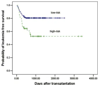
Patients were classified as low or high risk. For these 2 groups, LFS at 3 years was 80% and 52%, respectively (P¼.004,Figure 1); the cumulative incidence of relapse at 2 years was 8% and 36%, respectively (P¼.001,Figure 2). The cumulative incidence of non-relapse mortality at 3 years was 11% and 10%, respectively (P¼.95); the cumulative incidence of positive MRD at 2 years was 7%, and 37%, respectively (P¼.001); and the cumulative incidence of DLI intervention for positive MRD at 2 years was 5% and 25%, respectively (P¼.001).
Comparative Cytogenetic Grouping
Although the outcomes analyzed by NCCN and the other leukemia research groups were slightly different, the esti-mated hazard ratios for the intermediate- versus poor-risk group pointed in the same direction (Table 6). Based on the risk group classification described in“Risk Groups”above, LFS for low-risk and high-risk patients was almost the same (80% vs 52%, 81% vs 53%, 81% vs 49%, and 81% vs 49%, respectively) according to the NCCN, SWOG/ECOG, Medical Research Council, and International System for Cytogenetic Nomenclature cytogenetic criteria systems.
Outcomes in Different Age Groups
For adult patients (>20 years old) and children (20 years old), LFS at 3 years was 71% and 68%, respectively (P¼.694). Table 4
Multivariate Analyses of Relapse, LFS, and OS
Outcome Hazard Ratio
(95% CI)
PValue Relapse
Response and remission status
CR1, CR after course 1 or 2 .26 (.12-.58) .001 CR1, CR after course 3 or 4 or CR2 1.0
LFS
Response and remission status
CR1, CR after course 1 or 2 .35 (.13-.89) .028 CR1, CR after course 3 or 4 or CR2 1.0
Extensive chronic GVHD versus no 3.62 (1.30-1.12) .014 Absolute lymphocyte count at day 30
<300/mL 2.16 (.93-5.02) .073
300/mL 1.0
OS
Response and remission status
CR1, CR after course 1 or 2 .33 (.13-.87) .024 CR1, CR after course 3 or 4 or CR2 1.0
Extensive chronic GVHD versus no 4.44 (1.61-12.19) .004 Absolute lymphocyte count at day 30
<300/mL 2.95 (1.20-7.22) .018
300/mL 1.0
Table 5
Risk Group Definitions
Risk Group Definition No. (%)
Low risk Patients with better-risk cytogenetics and WBC count<50109/L, or patients with better- or intermediate-risk cytogenetics and in CR1, CR after course 1 or 2
145 (75)
High risk Patients with poor-risk cytogenetics, or not in CR1, CR after course 1 or 2 (regardless of cytogenetics), or with better-risk cytogenetics and WBC count50109/L
48 (25)
Figure 1.Probability of LFS with respect to risk group after haploidentical
Based on the risk group classification described in “Risk Groups” previously mentioned, LFS for low-risk and high-risk adult patients and children was 80% versus 57% (P¼.032) and 81% versus 47% (P¼.013), respectively.
MRD at Time of Transplantation
Among the patients with MRD examinations during the 2 weeks before transplantation (underwent transplantation after January 2009, n¼130), 110 patients had negative MRD, and 20 patients had positive MRD before transplantation. For these 2 groups, LFS at 3 years was 76% and 52%, respectively (P¼.041); the cumulative incidence of relapse at 2 years was 10% and 35%, respectively (P¼.002).
DISCUSSION
Allo-HSCT is currently recommended as the preferred treatment choice for AML patients in CR1 without favorable cytogenetics. Whether the risk factors currently recognized in AML have any influence on transplantation outcomes in this cohort of patients is uncertain. In the current report, among all factors investigated, response after induction therapy was the strongest predictor. By combining the
identified risk factors, we divided the entire study pop-ulation into low- and high-risk groups with a survival differential between the two groups of 30%.
In the current study cytogenetics, response after induc-tion therapy, and WBC count in the better-risk cytogenetic group were predictors of outcome in univariate analysis. The results were in accordance with the observations of previous reports [25,26]. The relapse rate of 53% for patients not achieving CR after 2 courses was similar to that of advanced-stage patients reported earlier [4,5]. The results suggested
that identification of this group among other patients
undergoing transplantation during CR is very important and that this group requires closer monitoring and more robust therapy after HSCT. Despite the fact that outcomes are superior among patients with good- or intermediate-risk cytogenetics compared with patients with poor-risk cyto-genetics, LFS for patients with unfavorable cytogenetics appeared to be significantly better than that achieved both
with chemotherapy or auto-HSCT [1,25,26] and with
allo-HSCT [9,23,24] in most published reports. It should be acknowledged that the relatively small number of patients in poor-risk cytogenetic groups might influence the results. When using different classifications of cytogenetic groups, the results showed the same trend, despite a slight difference between the NCCN criteria and the other 3 leukemia cyto-genetic classification criteria. Patients with better-risk cyto-genetics were selected as candidates for transplantations
when AML1/ETO or CBF
b
/MYH11 transcript levels weregreater than 103at the time of the transplantation on the basis of published data[27]and our own unpublished data (Z.-H.H., manuscript in preparation). Apart from cytoge-netics, the possible influence of recently described molecular prognostic factors was not taken into account because data on these factors were not routinely available with long-term follow-up, but they will be the subject of future studies. The effect of WBC count was more prominent in the better-risk
cytogenetic group. The findings were in agreement with
those of other studies [25,28]. Our results revealed that
AML1/ETO or CBF
b
/MYH11 transcript levels greater than103 at the time of transplantation were correlated with
a higher WBC count (data not shown). This finding could
explain the outcome differences with regard to WBC count among better-risk patients.
For patients classified as high risk by our risk stratification who underwent transplantation during CR, our result of a 36% relapse rate was similar to or less than that reported in patients with similar risks[2,9-11]. However, the TRM rate was only 10%, which is much lower than the approximately 30% incidence for patients with similar risks reported by other researchers [2,9,11]and for advanced-stage patients
reported by our group [18]. Better performance status,
compared with advanced-stage AML, and younger age, compared with patients with similar risks reported by other researchers, might have contributed to the lower non-relapse mortality. Infection-related death accounted for 88% of TRM. Regarding the impact of HLA and ATG dose on infection, we found that a higher degree of HLA disparity was associated with higher CMV incidence, which was in accor-dance with the report by Meyers et al.[29]. The development of extensive chronic GVHD and grade 3 to 4 acute GVHD was associated with higher risk for treatment-related mortality, which was in agreement with the results of Kanda et al.[30]. Consequently, a similar relapse rate and lower TRM resulted in a higher LFS of 52% compared with patients with similar risks reported by other researchers and with advanced-stage
Figure 2.Cumulative incidence of relapse characterized by risk group after
haploidentical HSCT.
Table 6
LFS Analysis According to Different Cytogenetic Grouping Systems
Grouping System No. LFS PValue
NCCN Better risk 39 61 .035 Intermediate risk 137 79 Poor risk 17 54 SWOG .051 Favorable 32 61 Intermediate, other 119, 18 78, 82 Unfavorable 24 58
Medical Research Council
Favorable 39 61 .035
Intermediate 144 78
Adverse 10 56
International System for Cytogenetic Nomenclature
.035
Good 39 61
Intermediate, bad 115, 26 79, 79
patients reported by our group, for whom the LFS was re-ported to be 17% to 38%[2,9,11,18].
Similar risk stratification has not been fully studied or universally recognized in allo-HSCT settings, perhaps due to the heterogeneous regimens applied in matched-sibling donor HSCT. In one study reporting on a subgroup analysis, thePvalues were given as .006 and .07, respectively, with regard to relapse and survival for matched-sibling HSCT patients according to risk index[10]. Because thePvalue of .07 was very close to statistical significance, these results revealed that such a risk identification system might be applicable in an allo-HSCT setting to stratify patients into risk groups. Therefore, we initiated the current study to stratify AML patients who underwent transplantation during CR. We chose haploidentical patients as the study population because the regimens (conditioning, GVHD prophylaxis, stem cell source, and harvesting) were homogeneous. Our results suggested that such a risk stratification system might be as applicable for patients undergoing allo-HSCT as it was for patients in a chemotherapy setting. More intensive post transplantation management might be developed for patients with “high-risk” features to improve further the overall outcomes of AML patients who underwent trans-plantation during CR.
MRD examination has been found to be a strong predictor of postremission chemotherapy and postautologous HSCT
[31,32]. Recently, we noted that MRD-directed DLI can improve outcomes for patients who underwent trans-plantation during CR[17]. In the subgroup analysis of the current study, MRD pre-HSCT was a strong predictor of outcomes post-HSCT. Due to the limited patient sample, our results should be considered preliminary. The assessment of MRD should be validated in future studies with larger pop-ulations before and after allo-HSCT, and MRD perhaps should be added into the risk evaluation system, in combination with other factors, as a reliable and generally applicable method for identifying patients at risk for relapse.
In conclusion, the pretransplantation risk factors infl u-ence posttransplantation outcome of patients with AML in CR after haploidentical HSCT and might be applicable to assist with risk-directed posttransplantation therapy. Addi-tional data are needed to validate in other transplantation settings.
ACKNOWLEDGMENTS
The authors thank American Journal of Experts for English editing.
Financial disclosure:This work was supported (in part) by National Natural Science Foundation of China (grant 30971292), National High-tech R&D Program of China (863 Program), Leading Program of Clinical Faculty accredited by the Ministry of Health of China, National Scientific Major Program-major new drug formulation (grant 2008zx09312-026), and Beijing Key Laboratory of Hematopoietic Stem Cell Transplantation.
Conflict of Interest Statement:None of the authors has any potential financial conflict of interest related to this manuscript.
REFERENCES
1. Byrd JC, Mrózek K, Dodge RK, et al. Pretreatment cytogenetic abnor-malities are predictive of induction success, cumulative incidence of relapse, and overall survival in adult patients with de novo acute myeloid leukemia: results from Cancer and Leukemia Group B (CALGB 8461).Blood. 2002;100:4325-4336.
2. Jourdan E, Boiron J-M, Dastugue N, et al. Early allogeneic stem-cell transplantation for young adults with acute myeloblastic leukemia in first complete remission: an intent-to-treat long-term analysis of the BGMT experience. J Clin Oncol. 2005;23: 7676-7684.
3. Koreth J, Schlenk R, Kopecky KJ, et al. Allogeneic stem cell trans-plantation for acute myeloid leukemia infirst complete remission: systematic review and meta-analysis of prospective clinical trials. JAMA. 2009;301:2349-2361.
4. Sierra J, Storer B, Hansen JA, et al. Unrelated donor marrow trans-plantation for acute myeloid leukemia: an update of the Seattle experience.Bone Marrow Transplant. 2000;26:397-404.
5. Aversa F, Terenzi A, Tabilio A, et al. Full haplotype-mismatched hematopoietic stem-cell transplantation: a phase II study in patients with acute leukemia at high risk of relapse.J Clin Oncol. 2005;23: 3447-3454.
6. Horowitz MM, Rowlings PA. An update from the International Bone Marrow Transplant Registry and the Autologous Blood and Marrow Transplant Registry on current activity in hematopoietic stem cell transplantation.Curr Opin Hematol. 1997;4:395-400.
7. Wang Y, Liu DH, Xu LP, et al. Prevention of relapse using granulocyte CSF-primed PBPCs following HLA-mismatched/haploidentical, T-cell-replete hematopoietic SCT in patients with advanced-stage acute leukemia: a retrospective risk-factor analysis.Bone Marrow Transplant. 2011; http://dx.doi.org/10.1038/bmt.2011.213.
8. Ferrant A, Labopin M, Frassoni F, et al. Karyotype in acute myeloblastic leukemia: prognostic significance for bone marrow transplantation in first remission: a European Group for Blood and Marrow Trans-plantation study. Acute Leukemia Working Party of the European Group for Blood and Marrow Transplantation (EBMT).Blood. 1997;90: 2931-2938.
9. Tallman MS, Dewald GW, Gandham S, et al. Impact of cytogenetics on outcome of matched unrelated donor hematopoietic stem cell trans-plantation for acute myeloid leukemia in first or second complete remission.Blood. 2007;110:409-417.
10. Wheatley K, Burnett AK, Goldstone AH, et al. A simple, robust, vali-dated and highly predictive index for the determination of risk-directed therapy in acute myeloid leukaemia derived from the MRCAML 10 trial.Br J Haematol. 1999;107:69-79.
11. Cornelissen JJ, van Putten WL, Verdonck LF, et al. Results of a HOVON/ SAKK donor versus no-donor analysis of myeloablative HLA-identical sibling stem cell transplantation in first remission acute myeloid leukemia in young and middle-aged adults: benefits for whom?Blood. 2007;109:3658-3666.
12. Huang XJ, Liu DH, Liu KY, et al. Treatment of acute leukemia with unmanipulated HLA-mismatched/haploidentical blood and bone marrow transplantation. Biol Blood Marrow Transplant. 2009;15: 257-265.
13. National Comprehensive Cancer Network. Version 2. NCCN clinical practice guidelines in oncology acute myloid leukemia. Available at: www.NCCN.org, 2011.
14. Slovak ML, Kopecky KJ, Cassileth PA, et al. Karyotypic analysis predicts outcome of preremission and postremission therapy in adult acute myeloid leukemia: a Southwest Oncology Group/Eastern Cooperative Oncology Group Study.Blood. 2000;96:4075-4083.
15. Zhao XS, Liu YR, Zhu HH, et al. Monitoring MRD withflow cytometry: an effective method to predict relapse for ALL patients after allogeneic hematopoietic stem cell transplantation. Ann Hematol. 2012;91: 183-192.
16. Zhao XS, Jin S, Zhu HH, et al. Wilms’tumor gene 1 expression: an independent acute leukemia prognostic indicator following allo-geneic hematopoietic SCT. Bone Marrow Transplant. 2012;47: 499-507.
17. Yan CH, Liu DH, Liu KY, et al. Risk stratification-directed donor lymphocyte infusion could reduce relapse of patients with standard-risk acute leukemia after allogeneic hematopoietic stem cell trans-plantation.Blood. 2012;119:3256-3262.
18. Huang XJ, Wang Y, Liu DH, et al. Administration of short-term immu-nosuppressive agents after GPBPCI reduces the incidence of GPBPCI associated acute GVHD without influencing the GVL effect. Bone Marrow Transplant. 2009;44:309-316.
19. Huang XJ, Liu DH, Liu KY, et al. Donor lymphocyte infusion for the treatment of leukemia relapse after HLA-mismatched/haplo-identical T-cell-replete hematopoietic stem cell transplantation.Haematologica. 2007;92:414-417.
20. Grimwade D, Walker H, Oliver F, et al. The importance of diagnostic cytogenetics on outcome in AML: analysis of 1612 patients entered into the MRC AML 10 trial. The Medical Research Council Adult and Children’s Leukaemia Working Parties. Blood. 1998;92: 2322-2333.
21. Mitelman F, editor. ISCN 1995: An International System for Human Cytogenetic Nomenclature. Basel, Switzerland: S. Karger; 1995. 22. Scrucca L, Santucci A, Aversa F. Regression modeling of competing risk
using R: an in depth guide for clinicians.Bone Marrow Transplant. 2010; 45:1388-1395.
23. Breems DA, Van Putten WL, De Greef GE, et al. Monosomal karyotype in acute myeloid leukemia: a better indicator of poor prognosis than a complex karyotype.J Clin Oncol. 2008;26:4791-4797.
24. Armand P, Kim HT, Zhang MJ, et al. Classifying cytogenetics in patients with acute myelogenous leukemia in complete remission undergoing allogeneic transplantation: a Center for International Blood and Marrow Transplant Research study. Biol Blood Marrow Transplant. 2012;18:280-288.
25. Schlenk RF, Benner A, Hartmann F, et al. Risk adapted post-remission therapy in acute myeloid leukemia: results of the German Multicenter AML HD93 treatment trial.Leukemia. 2003;17: 1521-1528.
26. Suciu S, Mandelli F, de Witte T, et al. Allogeneic compared with autologous stem cell transplantation in the treatment of patients younger than 46 years with acute myeloid leukemia (AML) infirst complete remission (CR1): An intention-to-treat analysis of the EORTC/ GIMEMAAML-10 trial.Blood. 2003;102:1232-1240.
27. Perea G, Lasa A, Aventín A, et al. Prognostic value of minimal residual disease (MRD) in acute myeloid leukemia (AML) with favorable cyto-genetics [t(8;21) and inv(16)].Leukemia. 2006;20:87-94.
28. Nguyen S, Leblanc T, Fenaux P, et al. A white blood cell index as the main prognostic factor in t(8;21) acute myeloid leukemia (AML):
a survey of 161 cases from the French AML Intergroup.Blood. 2002;99: 3517-3523.
29. Meyers JD, Flournoy N, Thomas ED. Risk factors for cytomegalovirus infection after human marrow transplantation.J Infect Dis. 1986;153: 478-488.
30. Kanda J, Hishizawa M, Utsunomiya A, et al. Impact of graft-versus-host disease on outcomes after allogeneic hematopoietic cell trans-plantation for adult T-cell leukemia: a retrospective cohort study. Blood. 2012;119:2141-2148.
31. Venditti A, Buccisano F, Del Poeta G. Level of minimal residual disease after consolidation therapy predicts outcome in acute myeloid leukemia.Blood. 2000;96:3948-3952.
32. Venditti A, Maurillo L, Buccisano F, et al. Pretransplant minimal residual disease level predicts clinical outcome in patients with acute myeloid leukemia receiving high-dose chemotherapy and autologous stem cell transplantation.Leukemia. 2003;17:2178-2182.
33. Sorror ML, Maris MB, Storb R, et al. Hematopoietic cell transplantation (HCT)-specific comorbidity index: a new tool for risk assessment before allogeneic HCT.Blood. 2005;106:2912-2919.
34. Chang YJ, Zhao XY, Huo MR, et al. Influence of lymphocyte recovery on outcome of haploidentical transplantation for hematologic malignan-cies.Medicine (Baltimore). 2009;88:322-330.