PDF hosted at the Radboud Repository of the Radboud University
Nijmegen
The following full text is a publisher's version.
For additional information about this publication click this link.
http://hdl.handle.net/2066/202039
Please be advised that this information was generated on 2020-09-10 and may be subject to
change.
Platinum Priority
–
Prostate Cancer
–
Editor
’
s Choice
Editorial by Derek J. Rosario, Thomas J. Walton and Steven J. Kennish on pp. 579–581 of this issue
Head-to-head Comparison of Transrectal Ultrasound-guided
Prostate Biopsy Versus Multiparametric Prostate Resonance
Imaging with Subsequent Magnetic Resonance-guided Biopsy
in Biopsy-naïve Men with Elevated Prostate-specific Antigen:
A Large Prospective Multicenter Clinical Study
Marloes van der Leest
a, Erik Cornel
b, Bas Israe¨l
a, Rianne Hendriks
c, Anwar R. Padhani
d,
Martijn Hoogenboom
a, Patrik Zamecnik
a, Dirk Bakker
b, Anglita Yanti Setiasti
e,
Jeroen Veltman
f, Huib van den Hout
f, Hans van der Lelij
g, Inge van Oort
c, Sjoerd Klaver
h,
Frans Debruyne
i, Michiel Sedelaar
c, Gerjon Hannink
j, Maroeska Rovers
j,
Christina Hulsbergen-van de
[5_TD$DIFF]
Kaa
e[1_TD$DIFF]
,y, Jelle O. Barentsz
a,y,*
aDepartment of Radiology and Nuclear Medicine, Radboud University Medical Center, Nijmegen, The Netherlands;bDepartment of Urology, Ziekenhuis Groep Twente, Almelo-Hengelo, The Netherlands;cDepartment of Urology, Radboud University Medical Center, Nijmegen, The Netherlands;dPaul Strickland Scanner Centre, Mount Vernon Cancer Centre, Northwood, UK;eDepartment of Pathology, Radboud University Medical Center, Nijmegen, The Netherlands; fDepartment of Radiology, Ziekenhuis Groep Twente, Almelo-Hengelo, The Netherlands;gDepartment of Radiology and Nuclear Medicine, Maasstad Hospital, Rotterdam, The Netherlands;hDepartment of Urology, Maasstad Hospital, Rotterdam, The Netherlands;iDepartment of Urology, Andros Men's and Gynos Women's Health Institutes, Arnhem, The Netherlands;jDepartment for Operating Rooms, Radboud University Medical Center, Nijmegen, The Netherlands a v a i l a b l e a t w w w . s c i e n c e d i r e c t . c o m j o u r n a l h o m e p a g e : w w w . e u r o p e a n u r o l o g y . c o m Article info Article history: Accepted November 12, 2018 Associate Editor: James Catto Statistical Editor: Andrew Vickers Please visit www.eu-acme.org/europeanurology
to answer questions on-line. The EU-ACME credits will then be attributed automatically.
Abstract
Background: There is growing interest to implement multiparametric magnetic reso-nance imaging (mpMRI) and MR-guided biopsy (MRGB) for biopsy-naïve men with suspected prostate cancer.
Objective: Primary objective was to compare and evaluate an MRI pathway and a transrectal ultrasound-guided biopsy (TRUSGB) pathway in biopsy-naïve men with
prostate-specific antigen levels of3 ng/ml.
Design, setting, and population: A prospective, multicenter, powered, comparative effectiveness study included 626 biopsy-naïve patients (from February 2015 to February 2018).
Intervention: All patients underwent prebiopsy mpMRI followed by systematic TRUSGB. Men with suspicious lesions on mpMRI also underwent MRGB prior to TRUSGB. MRGB was performed using the in-bore approach.
Outcome measurements and statistical analysis: Clinically significant prostate cancer
(csPCa) was defined as grade group2 (Gleason score3 + 4) in any core. The main
secondary objectives were the number of men who could avoid biopsy after nonsus-picious mpMRI, the number of biopsy cores taken, and oncologic follow-up. Differences
y Christina Hulsbergen-van de Kaa and Jelle O. Barentsz are co-senior authors.
* Corresponding author. Department of Radiology and Nuclear Medicine, Radboud University Medical Center, P.O. Box 9101, Nijmegen, 6500 HB, The Netherlands. Tel. +31(0)24 3619196; Fax. +31 (0)24 3540866.
E-mail address:jelle.barentsz@radboudumc.nl(J.O. Barentsz).
https://doi.org/10.1016/j.eururo.2018.11.023
0302-2838/© 2018 The Authors. Published by Elsevier B.V. on behalf of European Association of Urology. This is an open access article
1. Introduction
International guidelines recommend systematic 12-core transrectal ultrasound-guided biopsy (TRUSGB) in biopsy-naïve men with elevated prostate-specific antigen (PSA) serum levels of >3 ng/ml [1,2]. However, TRUSGB has limitations. First, clinically insignificant prostate cancer (insignPCa) is unnecessarily detected. Second, many men undergo TRUSGB and never have prostate cancer (PCa) diagnosed. Biopsies can lead to complications such as bleeding and infections, leading to increased healthcare costs[3]. Third, clinically significant prostate cancer (csPCa) can be missed. Fourth, multiple risk stratification errors occur, contributing to treatment failures for men undergo-ing active surveillance for presumed low-risk PCa.
Compared with TRUSGB, multiparametric magnetic resonance imaging (mpMRI) has been reported to reduce the detection of insignPCa while increasing the detection of csPCa [4–6]. The opportunity to selectively localize csPCa enables MR-directed biopsy and in so doing use fewer cores. This has improved the diagnostic pathway for men with suspected PCa. It has been shown that if mpMRI is nonsuspicious, immediate TRUSGB can be safely avoided
[7,8]. Multiple single- and multicenter randomized trials
have confirmed the superiority of mpMRI and MR-directed biopsy to TRUSGB [9–14]. However, these studies lacked sufficient standardization of MRI reporting, central quality control review of mpMRI acquisition and reading, central pathology review of biopsies, and adequate oncologic follow-up. In addition, many studies have not performed a comparison of TRUSGB and mpMRI + MR-guided biopsy (MRGB) in the same patients.
Therefore, we conducted a prospective, multicenter, clinical effectiveness study that compared head-to-head mpMRI + MRGB (MRI pathway) with the TRUSGB pathway in biopsy-naïve men at a risk of PCa.
2. Patients and methods 2.1. Study population
Between February 2015 and February 2017, 699 consecutive
biopsy-naïve men, aged 50–75 yr with a PSA level of3 ng/ml were recruited in
this prospective multicenter comparative effectiveness study (
Supple-mentary Fig. 1). Patients were enrolled from four medical centers in the Netherlands: Radboudumc Nijmegen (coordinating, central center;
n= 169) and three nonuniversity hospitals—Ziekenhuis Groep Twente
(n= 357), Maasstad Hospital Rotterdam (n= 152), and Andros Men's
Health Institutes (n= 21).
Exclusion criteria were age<50 or>75 yr, a history of previous
prostate biopsy or PCa, general contraindications for MRI, use of medications known to affect serum PSA levels, symptoms of urinary tract infection, and a history of invasive treatments for benign prostate hyperplasia. The central ethical review board approved this study, and written informed consent was obtained from all patients. The study was
registered in the Dutch Trial register under identifier NTR5555.
2.2. Multiparametric MRI
All patients underwent mpMRI performed at 3 T (Magnetom Skyra, Siemens Healthineers, Erlangen, Germany) compliant with the Prostate Imaging
Reporting and Data System version 2 (PI-RADS v2) standards (
Supplemen-tary Fig. 2)[15,16]. The protocol consisted of T2-weighted imaging in three planes, diffusion-weighted imaging with calculation of apparent diffusion
coefficient (ADC) maps, highb-value images (b>1400 s/mm2), and dynamic
contrast enhanced imaging (Supplementary Table 1). The images were
in proportions were tested using McNemar's test with adjusted Wald confidence
intervals for differences of proportions with matched pairs.
Results and limitations: The MRI pathway detected csPCa in 159/626 (25%) patients and
insignificant prostate cancer (insignPCa) in 88/626 patients (14%). TRUSGB detected
csPCa in 146/626 patients (23%) and insignPCa in 155/626 patients (25%). Relative
sensitivity of the MRI pathway versus the TRUSGB pathway was 1.09 for csPCa (p= 0.17)
and 0.57 for insignPCa (p<0.0001). The total number of biopsy cores reduced from
7512 to 849 (–89%). The MRI pathway enabled biopsy avoidance in 309/626 (49%)
patients due to nonsuspicious mpMRI. Immediate TRUSGB detected csPCa in only 3% (10/309) of these patients, increasing to 4% (13/309) with 1-yr follow-up. At the same
time, TRUSGB would overdetect insignPCa in 20% (63/309).“Focal saturation”by four
additional perilesional cores to MRGB improved the detection of csPCa in 21/317 (7%) patients. Compared with the literature, our proportion of nonsuspicious mpMRI cases is
significantly higher (27–36% vs 49%) and that of equivocal cases is lower (15–28% vs 6%).
This is probably due to the high-quality standard in this study. Therefore, a limitation is the duplication of these results in less experienced centers.
Conclusions: In biopsy-naïve men, the MRI pathway compared with the TRUSGB
pathway results in an identical detection rate of csPCa, with significantly fewer
insignPCa cases. In this high-quality standard study, almost half of men have non-suspicious MRI, which is higher compared with other studies. Not performing TRUS biopsy is at the cost of missing csPCa only in 4%.
Patient summary: We compared magnetic resonance imaging (MRI) with MRI-guided biopsy against standard transrectal ultrasound biopsy for the diagnosis of prostate cancer in
biopsy-naïve men. Our results show that patients can benefit from MRI because biopsy may
be omitted in half of men, and fewer indolent cancers are detected, without compromising the detection of harmful disease. Men also need fewer needles to make a diagnosis.
© 2018 The Authors. Published by Elsevier B.V. on behalf of European Association of
Urology. This is an open access article under the CC BY-NC-ND license (
http://creati-vecommons.org/licenses/by-nc-nd/4.0/). Keywords: Prostate cancer Transrectal ultrasound-guided biopsy Multiparametric magnetic resonance imaging
Prostate Imaging Reporting and Data System
Magnetic resonance-guided biopsy
analyzed by trained radiologists using the PI-RADS v2 to score all detected lesions. A central review before biopsy was performed by two central-center radiologists (J.B. and M.v.d.L., 25 and 5 yr of experience with prostate MRI, respectively) for each case. For patients enrolled in the central center, the images were analyzed independently by these two radiologists. When PI-RADS scores were discordant, a consensus assessment decided on the need for MRGB. Multiparametric MRI was categorized as suspicious in the
presence of PI-RADS 3–5 lesion(s). PSA density (PSAD) was calculated by
dividing serum PSA level by MRI prostate volume.
2.3. Biopsy
Men with a suspicious mpMRI scan underwent in-bore MRGB (using 18G needles with sampling length of 17 mm) followed by a 12-core systematic TRUSGB (using 18G needles with sampling length of 17 mm) preferably the same day, performed by a urologist blinded to the imaging results. MRGB was performed using a commercially available transrectal in-bore MR biopsy device (Invivo, Gainsville, FL, USA). Nonuniversity radiologists received training for the MRGB
procedure. Each PI-RADS 3–5 lesion was biopsied using two to four
cores. The lowest signal areas on ADC maps within a suspicious region were used to target biopsies.
TRUSGB was performed according to international guidelines
[17]. Where a lesion was visible at[6_TD$DIFF]TRUS, it was targeted by using the
core for the relevant prostate zone (no additional cores were performed).
Men with nonsuspicious mpMRI (PI-RADS 1–2) underwent TRUSGB only.
2.4. Histopathology
All biopsies were centrally reviewed at the central center by an experienced uropathologist (C.A.H.K., 25 yr of experience) independent of the results of the nonuniversity pathologists and the mpMRI results. TRUSGB and MRGB of each patient were evaluated separately from each other and blinded to the individual results. TRUSGB cores adjacent to
lesions sampled by MRGB were called“perilesional.”For cores
contain-ing cancer, grade group (GG) and Gleason score (GS) were determined using the 2014 International Society of Urologic Pathology (ISUP) criteria
[18]. Any prostatectomy specimens after radical prostatectomy were
analyzed by the experienced general pathologists.
2.4.1. Definition of clinically significant PCa
Recent EAU guidelines use the definition of GG2 (GS3 + 4) for csPCa
[17,19]. This matches with the newly introduced ISUP scoring system, where no separation is made between large and small GG 1 (GS 3 + 3)
PCa. Therefore, our initial definition of csPCa that included large GG 1 (GS
3 + 3) PCa was changed to GG2 (GS3 + 4). Additional analyses for
the two other csPCa definitions, large GG 1 (GS 3 + 3) and GG3 (GS4
+ 3) were performed.
2.5. Outcome measurements
The primary outcomes comprised the overall detection rates of csPCa and insignPCa for both pathways. The secondary outcomes were the proportion of men in the MRI pathway who did not undergo MRGB after a nonsuspicious scan result, the number of csPCa missed in this group (detected by immediate TRUSGB and 1-yr follow-up), histopathologic details of biopsy and radical prostatectomy specimens, the number of biopsy cores per biopsy session, MRI and histopathology reader performance, biopsy complications, and oncologic follow-up.
2.6. Follow-up
A“safety net”was provided to all patients without csPCa at TRUSGB with
either nonsuspicious mpMRI or suspicious mpMRI without a csPCa at
MRGB. This included a minimum follow-up of 1 yr, with repeated PSA levels every 6 mo. In case of increased or persistent elevated PSA, repeat mpMRI and/or biopsy was performed.
2.7. Statistical analysis
Statistical analyses were performed using R (version 3.5.0; R Foundation for Statistical Computing, Vienna, Austria). Descriptive statistics were used to present clinical and mpMRI characteristics. Continuous variables were presented as median (interquartile range) and categorical variables as numbers with percentages. In patients with multiple lesions on mpMRI, the index lesion with the highest PI-RADS score was used.
To compare the proportions of csPCa and insignPCa in both pathways,
McNemar's tests were used. Adjusted Wald confidence intervals (CIs) for
differences of proportions with matched pairs were calculated. Multi-parametric MRI and histopathology reader agreement was calculated
using Gwet's agreement coefficient (AC)[20].
3. Results
3.1. Patient data and mpMRI results
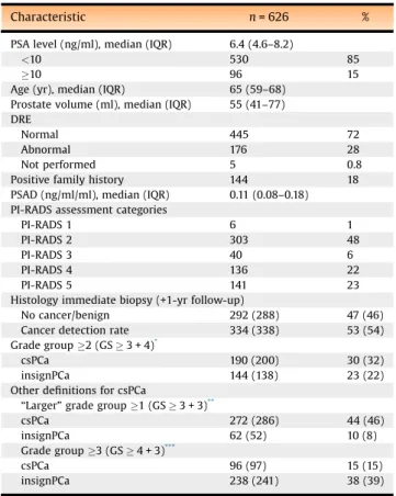
Data of 626 patients were analyzed (Fig. 1). Patient characteristics and mpMRI scores are summarized in
Table 1. The mpMRI was scored PI-RADS 1–2, 3, 4, and
5 in 49%, 6%, 22%, and 23%, respectively. Cancer detection rate (CDR) of the combined pathways was 334/626 (53%). Overall detection rates[7_TD$DIFF](without [8_TD$DIFF]follow-up) were 190/626 (30%) for csPCa and 144/626 (23%) for insignPCa.Table 1and
Supplementary Table 10show the results of two alternate
definitions of csPCa.
3.2. MRGB and TRUSGB results
MRGB was performed in 317/626 (51%) patients. TRUSGB was performed in all patients. Biopsy core analysis details are presented inTable 2. Detection rates of csPCa increased with increasing PI-RADS categories. Differences of csPCa detection between TRUSGB and MRGB for PI-RADS 5 lesions were minimal (2%). These differences were higher for PI-RADS 3–4 lesions (12%;Table 2andSupplementary Fig. 3). 3.3. Clinical performance of MRI and TRUSGB pathways The overall CDR in the MRI pathway was 247/626 (39%) compared with 301/626 (48%) in the TRUSGB pathway. Immediate results, without follow-up, showed that the TRUSGB pathway found csPCa in 146/626 (23%) and the MRI pathway in 159/626 (25%) patients (difference of 2%; 95% CI:–1 to 5); insignPCa was found in 155/626 (25%) and 88/ 626 (14%) patients, respectively (difference of 11%; 95% CI: 7–14). Relative sensitivity of the MRI pathway compared with the TRUSGB pathway was 1.09 for csPCa and 0.57 for insignPCa (Table 3).
The diagnostic impact of biopsy strategies is presented in
Fig. 2. Restricting biopsy to patients with suspicious mpMRI
(PI-RADS 3–5; n= 317) reduced the number of men requiring a biopsy by 309/626 (49%). Not performing biopsy in PI-RADS 1–2 cases resulted in missing 10/309 (3%) of csPCa.Nine patients had a GG 2 (GS 3 + 4) and one a GG 3
(GS 4 + 3;Supplementary Table 2 and Supplementary Fig. 4A). In patients with nonsuspicious mpMRI, TRUSGB overdetected insignPCa in 63/309 (20%). If systematic TRUSGB would be performed in patients with nonsuspi-cious mpMRI and PSAD0.15 ng/ml/ml (n= 55), three cases of csPCa would have been found. This would lower the underdetected rate to 2%, at the cost of 9% more insignPCa. If biopsy would be performed only in patients with PI-RADS 3 and PSAD0.15 ng/ml/ml, four csPCa cases including one GG 3 and one GG 5 would go undetected.
The utility of the MRI pathway alone versus the MRI pathway plus systematic 12-core TRUSGB (combined pathway) was also evaluated. Additional 7% (21/317) csPCa cases were detected with the combined biopsy approach
(Fig. 2,Supplementary Fig. 4B, and Supplementary Table 3).
In 20 of these 21 patients, the csPCa cases detected by TRUSGB were recognized on mpMRI as suspicious lesions. In the remaining patient, the csPCa diagnosed by TRUSGB
(GG 5; GS 4 + 5) was missed by all readers (therefore not specifically targeted) but was retrospectively visible. In the 20 patients with visible lesions diagnosed as csPCa only by TRUSGB cores, the TRUSGB cores were [9_TD$DIFF]obtained from the abnormal mpMRI lesion[10_TD$DIFF]area [11_TD$DIFF]or from neighboring perile-sional TRUSGB areas. In total 72 (peri)leperile-sional TRUSGB cores were taken, yielding 15 patients with GG 2 (GS 3 + 4) and five with GG3 (GS4 + 3) PCa. Thus, the average number of TRUSGB cores was 4 (72/20) to diagnose each extra csPCa.
3.3.1. MRI reader performance
MRI reader performance concordance analysis for PI-RADS score was performed between first nonuniversity center and second central-center reading. The agreement for both readers was 88% (399/456; Gwet's AC = 0.84; 95% CI: 0.80– 0.88) and the agreement for decision whether to perform an MRGB was 94% (428/456; Gwet's AC = 0.88; 95% CI: 0.83– 0.92;Supplementary Table 4).
[(Fig._1)TD$FIG]
Fig. 1–Flow diagram of study design and participants. mpMRI = multiparametric magnetic resonance imaging; MRGB = magnetic resonance-guided biopsy; PI-RADS = Prostate Imaging Reporting and Data System; PSA = prostate-specific antigen; TRUSGB = transrectal ultrasound-guided biopsy.
3.3.2. Histopathologic reader performance
Concordance between the nonuniversity and the central-center histopathologic reading was analyzed with respect to GG/GS and the number of positive biopsy cores. The agreement in GG/GS was 77% for the MRGB and 88% for the TRUSGB group (Gwet's AC = 0.73 and 0.82, respectively). The agreement in the number of positive cancer cores was 94% in the MRGB group and 87% in the TRUSGB group (Gwet's AC = 0.96 and 0.86, respectively; Supplementary Tables 5–8).
3.4. Prostatectomy results
In 131 patients, radical prostatectomy was performed. Between the TRUSGB cores and prostatectomy specimen, there was an agreement in GG in 37%, a downgrade in 38%,
and an upgrade in 25%. For MRGB cores, these values are 44%, 35%, and 21%, respectively (Supplementary Table 9). 3.5. Follow-up
In 39/309 (13%) patients with baseline nonsuspicious mpMRI, repeat biopsies were performed. In these patients, three csPCa cases were found: two had a GG 2 (GS 3 + 4) and one a GG 5 (GS 5 + 4) lesion. The GG 5 (GS 5 + 4) tumor was retrospectively visible on the baseline mpMRI but missed by all readers and thus was a false-negative mpMRI finding. Combined immediate and follow-up results show that in total 13/309 (4%) cases of csPCa were missed in patients with nonsuspicious mpMRI.
In 53/137 (39%) patients with a negative biopsy or insignPCa at combined biopsy and suspicious baseline mpMRI, repeat mpMRI was performed, which was non-suspicious in 26 of these 53 (49%) patients. Seven cases of GG 2 (GS 3 + 4) were found at repeat biopsy (Supplementary Fig. 5).
3.6. Biopsy complications
In total, 6% (41/626) of patients had complications: 3% had a complicated urinary tract infection (UTI/urosepsis) and 3% had other complications including lower urinary tract symptoms (n= 9), bleeding (n= 8), vasovagal episode (n= 3), and transient ischemic attack after discontinuation of anticoagulant medication (n= 1). Fifty percent (20/41) of these complications occurred in patients who underwent only TRUSGB in the nonsuspicious mpMRI group, including 2.9% (nine of 309) with complicated UTI/urosepsis. 4. Discussion
The major strength of this study is its quality-controlled, multicenter, head-to-head design. It confirms the larger body of research and clinical experience on combined mpMRI and MRGB for the detection and localization of csPCa in biopsy-naïve patients [9–14,21–23]. This paper makes multiple contributions to existing literature where there is controversy regarding its use for biopsy-naïve men. Our study provides level 1a evidence that the mpMRI pathway is noninferior to the TRUSGB pathway in biopsy-naïve men with regard to significant disease detection but is superior for detecting fewer insignificant cancers, and supports the “no immediate biopsy approach”after non-suspicious mpMRI scans. Similar to other studies, we show that TRUSGB yields of csPCa in nonsuspicious mpMRI patients are low (4%) [7,8]. Furthermore, not performing TRUSGB in these patients results in avoidance of compli-cated UTI/sepsis in 2.9%.
The proportion of men avoiding biopsy is almost twice that reported by the PROMIS and PRECISION trials—27% and 28%, respectively[9,10]. In the PROMIS study, this was at the cost of underdetection of csPCa of 24% (38/158) found on template mapping biopsy using the csPCa definition of GG2 (GS3 + 4) [9]. However, for TRUSGB the csPCa yield in nonsuspicious mpMRI cases was only 5.1% (H.U. Table 1–Characteristics of patients, PI-RADS assessment
categories, and final pathology (including 1-yr follow-up)
Characteristic n= 626 %
PSA level (ng/ml), median (IQR) 6.4 (4.6–8.2)
<10 530 85
10 96 15
Age (yr), median (IQR) 65 (59–68)
Prostate volume (ml), median (IQR) 55 (41–77)
DRE
Normal 445 72
Abnormal 176 28
Not performed 5 0.8
Positive family history 144 18
PSAD (ng/ml/ml), median (IQR) 0.11 (0.08–0.18)
PI-RADS assessment categories
PI-RADS 1 6 1
PI-RADS 2 303 48
PI-RADS 3 40 6
PI-RADS 4 136 22
PI-RADS 5 141 23
Histology immediate biopsy (+1-yr follow-up)
No cancer/benign 292 (288) 47 (46)
Cancer detection rate 334 (338) 53 (54)
Grade group2 (GS3 + 4)*
csPCa 190 (200) 30 (32)
insignPCa 144 (138) 23 (22)
Other definitions for csPCa
“Larger”grade group1 (GS3 + 3)**
csPCa 272 (286) 44 (46)
insignPCa 62 (52) 10 (8)
Grade group3 (GS4 + 3)***
csPCa 96 (97) 15 (15)
insignPCa 238 (241) 38 (39)
csPCa = clinically significant prostate cancer; DRE = digital rectal
examination; GG = grade group; GS = Gleason score; insignPCa = clinically
insignificant prostate cancer; IQR = interquartile range; mpMRI=
multiparametric magnetic resonance imaging; MRGB = magnetic
resonance-guided biopsy; PI-RADS = Prostate Imaging Reporting and
Data System; PSA = prostate-specific antigen; PSAD = PSA density;
TRUSGB = transrectal ultrasound-guided biopsy.
The prostate volume was measured on mpMRI. Percentages may not total up to 100 because of rounding.
Definitions of csPCa: *
MRGB/TRUSGB: GG2 (GS3 + 4) in any core.
** MRGB: GG 1 (GS 3 + 3) with total tumor core length6 mm or GG2
(GS3 + 4) in any core. TRUSGB: GG 1 (GS 3 + 3) with three or more biopsy
cores or GG2 (GS3 + 4) in any core.
***
Table 2–Biopsy core analysis details for TRUSGB and MRGB
TRUSGB MRGB
Total PI-RADS 1–2 PI-RADS 3 PI-RADS 4 PI-RADS 5 Total PI-RADS 3 PI-RADS 4 PI-RADS 5
n(%) n(%) n(%) n(%) n(%) n(%) n(%) n(%) n(%) Total 626 (100) 309 (49) 40 (6) 136 (22) 141 (23) 317 (100) 40 (13) 136 (43) 141 (44) Biopsy outcome No PCa 325 (52) 236 (76) 23 (58) 50 (37) 16 (11) 70 (22) 26 (65) 38 (28) 6 (4) insignPCa 155 (25) 63 (20) 11 (28) 52 (38) 29 (21) 88 (28) 7 (18) 44 (32) 37 (26) csPCa 146 (23) 10 (3) 6 (15) 34 (25) 96 (68) 159 (50) 7 (18) 54 (40) 98 (70)
Grade group/Gleason score
GG 1/3+2 0 (0) 0 (0) 0 (0) 0 (0) 0 (0) 1 (<1) 0 (0) 0 (0) 1 (<1) GG 1/3+3 155 (25) 63 (20) 11 (28) 52 (38) 29 (21) 87 (27) 7 (18) 44 (32) 36 (26) GG 2/3+4 70 (11) 9 (3) 3 (8) 21 (15) 37 (26) 89 (28) 4 (10) 40 (30) 45 (32) GG 3/4+3 30 (5) 1 (<1) 1 (3) 8 (6) 20 (14) 28 (9) 2 (5) 7 (5) 19 (13) GG 4/4+4 14 (2) 0 (0) 0 (0) 2 (2) 12 (9) 10 (3) 0 (0) 2 (1) 8 (6) GG 4/3+5 3 (<1) 0 (0) 0 (0) 1 (<1) 2 (1) 5 (2) 0 (0) 3 (2) 2 (1) GG 4/5+3 2 (<1) 0 (0) 0 (0) 1 (<1) 1 (<1) 1 (<1) 0 (0) 1 (<1) 0 (0) GG 5/4+5 16 (3) 0 (0) 1 (3) 0 (0) 15 (11) 18 (6) 0 (0) 1 (<1) 17 (12) GG 5/5+4 7 (1) 0 (0) 0 (0) 1 (<1) 6 (4) 6 (2) 1 (3) 0 (0) 5 (4) GG 5/5+5 4 (<1) 0 (0) 1 (3) 0 (0) 3 (2) 2 (<1) 0 (0) 0 (0) 2 (1) Biopsy cores
Total cores sampled 7512 (100) 3708 480 1632 1692 849 105 356 388
Total positive cores 1259 (17) 157 (4) 50 (10) 292 (18) 760 (45) 584 (68) 34 (32) 206 (58) 344 (89)
Median cancer core length (mm, IQR)
4.6 (2.7–7) 2.1 (1–3) 3.0 (1–6) 3.3 (2–5) 6.2 (4.5–9) 6.3 (5–9) 4.0 (3–6) 5.0 (4–7) 7.8 (6–11)
Percentage PCa of positive core length (%)
37 16 27 26 46 57 37 45 65
csPCa = clinically significant prostate cancer; GG = grade group; insignPCa = clinically insignificant prostate cancer; IQR = interquartile range; MRGB = magnetic resonance-guided biopsy; PCa = prostate cancer; PI-RADS = Prostate Imaging Reporting and Data System; TRUSGB = transrectal ultrasound-guided biopsy.
Definition of csPCa: grade group2 (Gleason score3 + 4). Percentages may not total 100 because of rounding.
Table 3–Pathway yield and relative sensitivity for different definitions of clinically significant prostate cancer
Biopsy strategy (n= 626)
TRUSGB pathway MRI pathway Relative sensitivity of
MRI versus TRUSGB pathway
pvalue
TRUSGB (n= 626) No biopsy (n= 309)
MRGB (n= 317)
n(%, 95% CI) n(%, 95% CI)
Grade group2 (GS3 + 4)*[4_TD$DIFF]
Prevalence csPCaa: 200 (32.0, 28–36)
csPCa 146 (23.3, 20–27) 159 (25.4, 22–29) 1.09 0.17
insignPCa 155 (24.8, 21–28) 88 (14.1, 11–17) 0.57 <0.0001
Other definitions for csPCa
“Larger”grade group1 (GS3 + 3)**
Prevalence csPCaa: 286 (45.7, 42–50) csPCa 215 (34.3, 31–38) 229 (36.6, 33–40) 1.07 0.19 insignPCa 86 (13.7, 11–17) 18 (2.9, 2–4) 0.20 <0.0001 Grade group3 (GS4 + 3)*** Prevalence csPCaa: 97 (15.5, 13–19) csPCa 76 (12.1, 10–15) 70 (11.2, 9–14) 0.92 0.46 insignPCa 225 (35.9, 32–40) 177 (28.3, 25–32) 0.79 0.0001
CI = confidence interval; csPCa = clinically significant prostate cancer; GG = grade group; GS = Gleason score; insignPCa = clinically insignificant prostate cancer; MRGB = magnetic resonance-guided biopsy; MRI = magnetic resonance imaging; TRUSGB = transrectal ultrasound-guided biopsy.
Relative sensitivity is the sensitivity (ie, true positive rate) ratio between the MRI pathway and the TRUSGB pathway for each definition of csPCa. A value of
1 shows equal sensitivity. Values>1 indicate greater sensitivity for the MRI pathway, whereas values below 1 show lower sensitivity. Higher relative sensitivity
is desirable for csPCa, and lower for the detection of insignPCa. Thepvalues were calculated with McNemar's test for paired nominal data.
Definitions of csPCa: a
Prevalence of csPCa of both pathways included 1-yr follow-up. *
MRGB/TRUSGB: GG2 (GS3 + 4) in any core.
**
MRGB: GG 1 (GS 3 + 3) with total tumor core length6 mm or GG2 (GS3 + 4) in any core. TRUSGB: GG 1 (GS 3 + 3) with three or more biopsy cores
or GG2 (GS3 + 4) in any core.
***
MRGB/TRUSGB: GG3 (GS4 + 3) in any core.
Ahmed, personal communication). Pokorny et al. [11]
showed that biopsy could be avoided in 36% (81/223), with an underdiagnosis of csPCa in 11% (9/81) found on TRUSGB. The low prevalence of csPCa in this study (30%) compared with contemporary cohorts (38–47%) could contribute to the high number of nonsuspicious MRI scans
[9,10,21,24], which are in line with the MRI screening study
of Grenabo Bergdahl et al. [25]. Another more important explanation for the higher proportion of nonsuspicious mpMRI scans than in other studies may be the high-quality standards achieved in image acquisition and reading. In our study, all mpMRI scans were performed on 3 T scanners, adhering to the PI-RADS v2 protocols, undertaken by trained prostate-MRI technologists. We also attained high quality in mpMRI readings using double expert consensus readings. These high standards helped minimize the proportion of“uncertain”(PI-RADS 3) diagnoses. PI-RADS 3 was present in 6% in our study, versus 28%, 21%, and 15% in the PROMIS, PRECISION, and Pokorny et al's studies[9–11], respectively. That nonuniversity radiologists can perform high-quality reading after appropriate training is illustrated by their high agreement with the central-center radiolo-gists.
This study design can also address the debate regarding the appropriate biopsy action in men with suspicious
mpMRI scans: MRGB alone or MRGB + TRUSGB? In agree-ment with the literature, addition of systematic TRUSGB to MRGB leads to higher rates of csPCa and insignPCa
[6,26]. The majority of csPCa missed by MRGB appears to
be sampling errors related to intratumor heterogeneity. “Focal saturation” by additional four perilesional cores showed to improve csPCa detection when sampling with MRGB.
Some limitations should be discussed. First, reproducing these findings outside expert centers may be a challenge, but as shown in this study, it is not impossible. A well-designed training program can achieve high inter-reader agreements for PI-RADS score allocations as well as for biopsy decision making.
Second, MRGB and TRUSGB were undertaken in se-quence on the same day. The visible MRGB needle track could have influenced the urologist in TRUSGB needle placements. Moreover, when TRUSGB was abnormal[12_TD$DIFF], a needle targeted to the abnormality was undertaken in lieu of the sextant core. This could inflate the PCa detection rates of TRUSGB, although biopsy hemorrhage from MRGB may partly negate this effect.
Third, even though this study used in-bore MRGB, which is considered the optimal MR-targeting technique for smaller lesions, a recent review showed that in-bore MRGB
[(Fig._2)TD$FIG]
Fig. 2–Prostate cancer detection, overdetection, and underdetection for (non)suspicious mpMRI. Definition of csPCa: grade group2 (Gleason score 3 + 4). Percentages may not total 100 because of rounding. csPCa = clinically significant prostate cancer; insignPCa = clinically insignificant prostate cancer; MRGB = magnetic resonance-guided biopsy; NA = not applicable; PCa = prostate cancer; PI-RADS = Prostate Imaging Reporting and Data System; TRUSGB = transrectal ultrasound-guided biopsy. Definitions of csPCa: *underdetection: csPCa not detected by biopsy strategy including cancers detected in 1-yr follow-up; **overdetection: insignPCa detected by biopsy strategy; ***net reduction of insignPCa in MRGB + TRUSGB due to shift from insignPCa to csPCa (underdetection); 11 additional cases of insignificant PCa were detected at TRUSGB where MRGB yielded no PCa.
and MR-TRUS-fusion–guided biopsy are equally accurate, and results are potentially translatable to MR-TRUS-fusion– guided biopsies[27].
Fourth, some investigators have noted that selective sampling of the most aggressive part of a cancer by MRGB may lead to risk-stratification errors and can potentially lead to overtreatments of csPCa detected by MRGB[28]. However, a comparison of TRUSGB and MRGB with prostatectomy specimens within this study did not show marked differences between histologic down- and upgrading.
Finally, the low rate of infection-related complications could be further reduced by utilizing transperineal template mapping biopsies instead of the transrectal sampling route used in this study[9].
An “MRI-first” pathway in biopsy-naïve men has implementation challenges. The recommendation for“no immediate biopsy[13_TD$DIFF]”requires a robust follow-up regimen to minimize missing csPCa that emerge in follow-up. Our approach for a“safety net”is to perform 6-montly PSA tests and repeated mpMRI, MRGB, or TRUSGB when clinical suspicion persists. Panebianco et al.[8] have shown that such a safety net detects most interval cancers after non-suspicious mpMRI and that emerging csPCa are curable at that time (Panebianco, personal communication)[29]. Fur-thermore, an education program and quality control for prostate-MRI technologists, MRGB physicians, and radiol-ogists are needed, to deliver optimized quality of care for men with suspected PCa.
Finally, implementation of all new technologies is always connected with costs; although the MRI pathway, especially when using in-bore MRGB, is initially more expensive, extra costs are compensated for by reduced delays in diagnoses, omittance of biopsies and subsequent biopsy-related morbidities, and treatment costs[30–32].
5. Conclusions
In biopsy-naïve men, the MRI pathway compared with the TRUS pathway results in an identical detection rate of csPCa, with significantly fewer cases of insignPCa. In this high-quality standard study, almost half of men have non-suspicious MRI, which is higher compared with other studies. Not performing immediate TRUS biopsy after negative MRI is at the cost of missing csPCa only in 4%. Author contributions:Jelle O. Barentsz had full access to all the data in the study and takes responsibility for the integrity of the data and the accuracy of the data analysis.
Study concept and design:van der Leest, Rovers, Hulsbergen-van de Kaa, Barentsz.
Acquisition of data: van der Leest, Cornel, Israël, Hendriks, Padhani, Hoogenboom, Bakker, Setiasti, Veltman, van der Hout, van der Lelij, van Oort, Klaver, Debruyne, Sedelaar, Hannink, Hulsbergen-van de Kaa, Barentsz.
Analysis and interpretation of data: van der Leest, Israël, Hendriks, Padhani, Setiasti, Veltman, van der Hout, van der Lelij, Hannink, Rovers, Hulsbergen-van de Kaa, Barentsz.
Drafting of the manuscript: van der Leest, Cornel, Israël, Hendriks, Padhani, Hoogenboom, Zamecnik, Bakker, Setiasti, Veltman, van der
Hout, van der Lelij, van Oort, Klaver, Debruyne, Sedelaar, Hannink, Rovers, Hulsbergen-van de Kaa, Barentsz.
Critical revision of the manuscript for important intellectual content:van der Leest, Cornel, Israël, Hendriks, Padhani, Hoogenboom, Zamecnik, Bakker, Setiasti, Veltman, van der Hout, van der Lelij, van Oort, Klaver, Debruyne, Sedelaar, Hannink, Rovers, Hulsbergen-van de Kaa, Barentsz.
Statistical analysis:Hannink.
Obtaining funding:Rovers, Hulsbergen-van de Kaa, Barentsz.
Administrative, technical, or material support:van der Leest, Cornel, Israel, Hendriks, Hoogenboom, Veltman, van der Hout, van der Lelij, Hannink, Barentsz.
Supervision:Barentsz.
Other:None.
Financial disclosures: Jelle O. Barentsz certifies that all conflicts of
interest, including specific financial interests and relationships and
affiliations relevant to the subject matter or materials discussed in the
manuscript (eg, employment/affiliation, grants or funding,
consultan-cies, honoraria, stock ownership or options, expert testimony, royalties,
or patentsfiled, received, or pending), are the following: None.
Funding/Support and role of the sponsor:This study wasfinanced by a
KWF Kankerbestrijding-Alpe’dHuez grant (KUN 2015-6707)“4M: Met
Prostaat MRI Meer Mans” (Dutch Trial Register under identifier
NTR5555) and was also supported by Guerbet (Paris, France).
Acknowledgments:We would like to thank Professor Fritz Schröder for his expertise and conceptional help with the study design. Without him this study would not have been possible. We also thank the nonuniversity pathologists for the meticulous evaluation of the histopathology.
Appendix A. Supplementary data
Supplementary data associated with this article can be found, in the online version, at https://doi.org/10.1016/j.
eururo.2018.11.023.
References
[1]Heidenreich A, Bastian PJ, Bellmunt J, et al. EAU guidelines on prostate cancer. Part 1: screening, diagnosis, and local treatment with curative intent-update 2013. Eur Urol 2014;65:124–37. [2]Wolf AM, Wender RC, Etzioni RB, et al. American Cancer Society
guideline for the early detection of prostate cancer: update 2010. CA Cancer J Clin 2010;60:70–98.
[3]Loeb S, Vellekoop A, Ahmed HU, et al. Systematic review of com-plications of prostate biopsy. Eur Urol 2013;64:876–92.
[4]Turkbey B, Brown AM, Sankineni S, Wood BJ, Pinto PA, Choyke PL. Multiparametric prostate magnetic resonance imaging in the eval-uation of prostate cancer. CA Cancer J Clin 2016;66:326–36. [5]Venderink W, van Luijtelaar A, Bomers JG, et al. Results of targeted
biopsy in men with magnetic resonance imaging lesions classified equivocal, likely or highly likely to be clinically significant prostate cancer. Eur Urol 2018;73:353–60.
[6] Stabile A, Giganti F, Emberton M, Moore CM. MRI in prostate cancer diagnosis: do we need to add standard sampling? A review of the last 5 years. Prostate Cancer Prostatic Dis. In press.https://doi.org/ 10.1038/s41391-018-0071-8.
[7]Moldovan PC, Van den Broeck T, Sylvester R, et al. What is the negative predictive value of multiparametric magnetic resonance imaging in excluding prostate cancer at biopsy?. A systematic review and meta-analysis from the European Association of Urology Prostate Cancer Guidelines Panel. Eur Urol 2017;72:250–66.
[8] Panebianco V, Barchetti G, Simone G, et al. Negative multipara-metric magnetic resonance imaging for prostate cancer: what's next? Eur Urol 2018;74:48–54.
[9] Ahmed HU, El-Shater Bosaily A, Brown LC, et al. Diagnostic accuracy of multi-parametric MRI and TRUS biopsy inprostate cancer (PROMIS): a paired validating confirmatory study. Lancet 2017;389:815–22. [10]Kasivisvanathan V, Rannikko AS, Borghi M, et al. MRI-targeted or
standard biopsy for prostate-cancer diagnosis. N Engl J Med 2018;378:1767–77.
[11] Pokorny MR, de Rooij M, Duncan E, et al. Prospective study of diagnostic accuracy comparing prostate cancer detection by trans-rectal ultrasound-guided biopsy versus magnetic resonance (MR) imaging with subsequent MR-guided biopsy in men without previ-ous prostate biopsies. Eur Urol 2014;66:22–9.
[12]Castellucci R, Linares Quevedo AI, Sanchez Gomez FJ, et al. Prospec-tive nonrandomized study of diagnostic accuracy comparing pros-tate cancer detection by transrectal ultrasound-guided biopsy to magnetic resonance imaging with subsequent MRI-guided biopsy in biopsy-naive patients. Minerva Urol Nefrol 2017;69:589–95. [13]Siddiqui MM, Rais-Bahrami S, Turkbey B, et al. Comparison of MR/
ultrasound fusion-guided biopsy with ultrasound-guided biopsy for the diagnosis of prostate cancer. JAMA 2015;313:390–7.
[14]Tonttila PP, Lantto J, Paakko E, et al. Prebiopsy multiparametric magnetic resonance imaging for prostate cancer diagnosis in biop-sy-naive men with suspected prostate cancer based on elevated prostate-specific antigen values: results from a randomized pro-spective blinded controlled trial. Eur Urol 2016;69:419–25. [15]Barentsz JO, Weinreb JC, Verma S, et al. Synopsis of the PI-RADS v2
guidelines for multiparametric prostate magnetic resonance imag-ing and recommendations for use. Eur Urol 2016;69:41–9. [16]Weinreb JC, Barentsz JO, Choyke PL, et al. PI-RADS Prostate
Imag-ing—Reporting and Data System: 2015, version 2. Eur Urol 2016;69:16–40.
[17] Mottet N, Bellmunt J, Bolla M, et al. EAU-ESTRO-SIOG guidelines on prostate cancer Part 1: screening, diagnosis, and local treatment with curative intent. Eur Urol 2017;71:618–29.
[18]Epstein JI, Egevad L, Amin MB, et al. The 2014 International Society of Urological Pathology (ISUP) Consensus Conference on Gleason Grad-ing of Prostatic Carcinoma: definition of grading patterns and pro-posal for a new grading system. Am J Surg Pathol 2016;40:244–52. [19]Briganti A, Fossati N, Catto JWF, et al. Active surveillance for low-risk
prostate cancer: the European Association of Urology position in 2018. Eur Urol 2018;74:357–68.
[20] Gwet KL. Computing inter-rater reliability and its variance in the presence of high agreement. Br J Math Stat Psychol 2008;61(Pt 1):29–48.
[21]Hansen NL, Barrett T, Kesch C, et al. Multicentre evaluation of magnetic resonance imaging supported transperineal prostate
bi-opsy in bibi-opsy-naive men with suspicion of prostate cancer. BJU Int 2018;122:40–9.
[22] Panebianco V, Barchetti F, Sciarra A, et al. Multiparametric magnetic resonance imaging vs. standard care in men being evaluated for prostate cancer: a randomized study. Urol Oncol 2015;33:17.e11–1. [23] Porpiglia F, Manfredi M, Mele F, et al. Diagnostic pathway with multiparametric magnetic resonance imaging versus standard pathway: results from a randomized prospective study in biop-sy-naive patients with suspected prostate cancer. Eur Urol 2017;72:282–8.
[24] Boesen L, Nørgaard N, Løgager V, et al. Assessment of the diagnostic accuracy of biparametric magnetic resonance imaging for prostate cancer in biopsy-naive men: the Biparametric MRI for Detection of Prostate Cancer (BIDOC) study. JAMA Network Open 2018;1: e180219.
[25] Grenabo Bergdahl A, Wilderang U, Aus G, et al. Role of magnetic resonance imaging in prostate cancer screening: a pilot study within the Goteborg Randomised Screening Trial. Eur Urol 2016;70:566–73.
[26] Villers A, Marliere F, Ouzzane A, Puech P, Lemaitre L. MRI in addition to or as a substitute for prostate biopsy: the clinician's point of view. Diagn Interv Imaging 2012;93:262–7.
[27] Wegelin O, van Melick HHE, Hooft L, et al. Comparing three different techniques for magnetic resonance imaging-targeted prostate bi-opsies: a systematic review of in-bore versus magnetic resonance imaging-transrectal ultrasound fusion versus cognitive registration. Is there a preferred technique? Eur Urol 2017;71:517–31. [28] Calio BP, Sidana A, Sugano D, et al. Risk of upgrading from prostate
biopsy to radical prostatectomy pathology-does saturation biopsy of index lesion during multiparametric magnetic resonance imag-ing-transrectal ultrasound fusion biopsy help? J Urol 2018;199:976–82.
[29] De Visschere PJ, Naesens L, Libbrecht L, et al. What kind of prostate cancers do we miss on multiparametric magnetic resonance imag-ing? Eur Radiol 2016;26:1098–107.
[30] de Rooij M, Crienen S, Witjes JA, Barentsz JO, Rovers MM, Grutters JP. Cost-effectiveness of magnetic resonance (MR) imaging and MR-guided targeted biopsy versus systematic transrectal ultrasound-guided biopsy in diagnosing prostate cancer: a modelling study from a health care perspective. Eur Urol 2014;66:430–6. [31] Faria R, Soares MO, Spackman E, et al. Optimising the diagnosis of
prostate cancer in the era of multiparametric magnetic resonance imaging: a cost-effectiveness analysis based on the prostate MR imaging study (PROMIS). Eur Urol 2018;73:23–30.
[32] Padhani AR, Weinreb J, Rosenkrantsz AB, Villeirs G, Turkbey B, Barentsz J. Prostate imaging-reporting and data system steering committee: PI-RADS v2 status update and future directions. Eur Urol 2018.http://dx.doi.org/10.1016/j.eururo.2018.05.035.