0095-1137/06/$08.00⫹0 doi:10.1128/JCM.44.4.1219–1223.2006
Copyright © 2006, American Society for Microbiology. All Rights Reserved.
Evaluation of the IDI-MRSA Assay for Detection of
Methicillin-Resistant
Staphylococcus aureus
from
Nasal and Rectal Specimens Pooled in
a Selective Broth
M. Desjardins,
1,2* Christiane Guibord,
2B. Lalonde,
1B. Toye,
1,2and K. Ramotar
1,2Division of Microbiology, Department of Pathology and Laboratory Medicine, The Ottawa Hospital,1and
The Ottawa Hospital Research Institute,2Ottawa, Ontario, Canada
Received 19 December 2005/Returned for modification 10 January 2006/Accepted 19 January 2006
Rapid detection of methicillin-resistantStaphylococcus aureus (MRSA) by PCR can be performed directly
from nasal specimens with the IDI-MRSA assay. To improve the efficiency of screening, we evaluated the performance of the IDI-MRSA assay for the detection of MRSA from pooled and unpooled specimens cultured in a selective broth. Of the 287 specimens evaluated, 71 were culture and PCR positive, 203 were culture and PCR negative, 3 were culture positive and PCR negative, 8 were culture negative and PCR positive, and 2
remained inhibited. A methicillin-susceptibleStaphylococcus aureusisolate was recovered from five of the eight
specimens with false-positive PCR results. Compared to the results of culture, the sensitivity, specificity, and negative and positive predictive values of the IDI-MRSA assay for detection of MRSA from broth were 96%, 96%, 90%, and 98%, respectively. Following implementation of the IDI-MRSA assay, PCR-positive broths were subcultured for evaluation of assay performance. Of the 298 IDI-MRSA assay-positive broths, the results for
103 could not be confirmed by culture. A methicillin-susceptibleS. aureus(MSSA) isolate was recovered from
77 of these 103 broths. Repeat testing by the IDI-MRSA assay directly with the MSSA isolates confirmed the original positive PCR result. The positive predictive value of the IDI-MRSA assay fell from 90% during the evaluation phase to 65% postimplementation. The IDI-MRSA assay performed well for the detection of MRSA from a selective broth compared to the performance of the detection of MRSA from culture. However, because of the burden associated with implementation of infection control precautions, cultures remain essential in confirming positive IDI-MRSA results.
Methicillin-resistant Staphylococcus aureus (MRSA) is an important nosocomial pathogen; and infections due to this organism have been associated with increased morbidity, mor-tality, and prolonged hospitalization (5, 23). According to the Canadian Nosocomial Infection Surveillance Program, MRSA rates across Canada increased from⬍1% in 1995 to 10% in 2003 (4). Although this rate is not as prevalent as that in the United States, a 10-fold increase in MRSA rates over this 8-year period is cause for concern.
Screening for MRSA colonization largely relies on the cul-ture of specimens from appropriate sites, such as the nares, rectum, axilla, and wounds. Reliable detection of MRSA has traditionally depended on culture by using either selective broth or agar medium. However, depending on the culture method, a 2- to 4-day turnaround time for isolation, identifi-cation, and confirmation of MRSA can be expected (2, 3, 26). Increasing MRSA rates place additional burden on hospital infection control programs, leading to increased costs and bur-dens to hospitals and patients (15). Detection of MRSA colo-nization within 24 h would facilitate identification of carriers and allow the early implementation of isolation precautions for colonized patients in hopes of preventing spread to other
pa-tients. Several nucleic acid amplification-based assays for de-tection of MRSA from clinical specimens or for culture con-firmation have been described (6, 9, 10, 12, 24, 25, 31). The IDI-MRSA assay (GenOhm, San Diego, CA) is a multiplex qualitative real-time PCR assay for detection of MRSA from nasal swabs. Differentiation ofmecA-positive S. aureus from coagulase-negative staphylococci (CoNS) is possible by target-ing the right extremity sequences of the staphylococcal cassette chromosomemec(SCCmec) element and the 3⬘end of theS.
aureus orfX gene (12). Five primers targeting the currently
recognized SCCmecright-junction sequences corresponding to SCCmectypes I, II, III, IVa, IVb, and IVc are combined with one primer and three molecular beacons specific for theorfX
gene. Currently, the IDI-MRSA assay is approved for use for the direct detection of MRSA from nasal swabs. The lack of approval for use with pooled specimens and specimens from sites other than the nares presents a logistical problem, since screening of multiple body sites is necessary to achieve optimal detection of MRSA carriers (27). Processing of individual specimens for PCR testing would be cost prohibitive and im-practical. To facilitate processing and to reduce the workload, the performance of the IDI-MRSA assay for detection of MRSA from specimens pooled in a selective enrichment broth was evaluated. We also report on the postimplementation monitoring of this assay and the characterization of isolates with discrepant results.
(This paper was presented at the 2005 Annual Meeting of
* Corresponding author. Mailing address: Division of Microbiol-ogy, The Ottawa Hospital, 501 Smyth Rd., Ottawa, Ontario K1H 8L6, Canada. Phone: (613) 8899, ext. 72242. Fax: (613) 737-8324. E-mail: madesjardins@ottawahospital.on.ca. 1219
on January 28, 2021 by guest
http://jcm.asm.org/
Downloaded from
on January 28, 2021 by guest
http://jcm.asm.org/
Downloaded from
on January 28, 2021 by guest
http://jcm.asm.org/
Downloaded from
the Association of Medical Microbiologist and Infections Dis-ease Canada and the Canadian Association for Clinical Micro-biology and Infectious Disease Conference, Ottawa, Ontario, Canada.)
MATERIALS AND METHODS
Specimen collection.Screening of high-risk patients for MRSA colonization was performed on admission according to established Ottawa Hospital Infection Control policies. The patients included those who were direct transfers from another health care facility, those with a previous admission to any health care facility in the previous 6 months, and those with a previous history of infection with an antibiotic-resistant organism (MRSA, vancomycin-resistant Enterococ-cus, and extended-spectrum beta-lactamase-producing organisms). Nasal and rectal specimens and up to two wound swabs were obtained from at-risk patients and submitted to the laboratory in Amies transport medium (Starplex Scientific, Etobicoke, Ontario, Canada).
MRSA screening by broth culture.Swabs collected at the same time from each individual patient were cultured in a single tube of selective broth consisting of brain heart infusion broth (Becton Dickinson, Sparks, MD) supplemented with 5g/ml of aztreonam (ICN, Costa Mesa, CA) and 75g/ml of ceftizoxime (GlaxoSmithKline, Inc.) (B. Slinger, A. Lee, C. Martel, I. Gaboury, P. Jessamine, K. Ramotar, and B. Toye, 70th Conjoint Meeting, Canadian Association of Clinical Microbiology and Infectious Diseases, abstr. D4, 2002). Because of the low MRSA prevalence in our patient population, specimens for MRSA screening were selected to provide an adequate number of MRSA-positive samples in order to properly evaluate the IDI-MRSA assay. Two swabs were pooled in a single tube of broth, incubated overnight at 35°C in ambient air, and subcultured to 5% sheep blood agar (Trypticase soy agar; Oxoid Inc., Ottawa, Ontario, Canada). TheS. aureusisolates recovered from the broths were screened for methicillin resistance by using oxacillin (6g/ml) salt agar screen plates (MH-Ox; Becton Dickinson). Confirmation of methicillin resistance was performed by disk diffusion testing with 30-g cefoxitin disks (Oxoid, Ottawa, Ontario, Can-ada), according to Clinical and Laboratory Standards Institute (formerly NC-CLS) recommendations (20, 21, 22), and detection of the modified penicillin binding protein (PBP 2⬘) by latex agglutination (Oxoid Inc.).
MRSA PCR testing from broth.Screening for MRSA by the IDI-MRSA assay was performed by transferring a 50-l aliquot of the overnight broth into the sample reagent buffer provided with the assay kit (GenOhm). The PCR was then performed according to the manufacturer’s instructions by using a SmartCycler II device (Cepheid, Sunnyvale, CA). Lysates found to be inhibited on initial PCR testing were frozen and thawed at⫺20°C for 1 h before they were retested. Repeatedly inhibited specimens were reported as unresolved. The sensitivity, specificity, and positive and negative predictive values for the IDI-MRSA assay were calculated by comparison to the results of broth culture of the specimens, which was considered the “gold standard.”
Characterization of discrepantS. aureusisolates.Methicillin-susceptibleS. aureus(MSSA) isolates that were recovered from PCR-positive broths but that failed to grow on MH-Ox were further characterized for oxacillin susceptibility by cefoxitin (30-g) disk diffusion testing and MIC determination by agar dilu-tion according to the Clinical and Laboratory Standards Institute recommenda-tions (21), repeat testing on MH-Ox screen plates, and determination of PBP 2⬘
expression by latex agglutination. Testing of the MSSA isolates was also per-formed by the IDI-MRSA assay directly with the pure colonies.
Postimplementation evaluation of the IDI-MRSA assay.Following implemen-tation of the IDI-MRSA assay as our routine method for MRSA detection, positive broths were subcultured onto blood agar (BA) and incubated overnight at 35°C.S. aureusisolates recovered on subculture were characterized for ox-acillin susceptibility by cefoxitin disk diffusion, growth on MH-Ox screen plates, and determination of PBP 2⬘expression by latex agglutination. Detection of the
mecAgene and confirmation ofS. aureusidentification were performed by a multiplex PCR assay by using a previously described method withmecAandnuc
gene-specific primers and 16S rRNA-specific primers as internal controls (17). The expectednuc(624-bp),mecA(1,235-bp), and 16S rRNA (228-bp) products were separated on a 2% agarose minigel.
PFGE ofStaphylococcus aureus.Genotyping by pulsed-field gel electrophoresis (PFGE) of MSSA isolates that were positive by the IDI-MRSA assay was per-formed as described previously (29). SmaI digests of genomic DNA were elec-trophoresed in a 1% pulse-field-certified agarose gel by using a CHEF DR III apparatus (Bio-Rad, Hercules, CA). The gels were stained in ethidium bromide and photographed after UV transillumination. Restriction fragment profiles were compared visually and interpreted based on the guidelines of Tenover and
colleagues (30). Isolates were classified according to the Canadian nomenclature (C-MRSA), as described previously (28).
RESULTS
MRSA detection by the IDI-MRSA assay.A total of 174 of
287 broths were inoculated with pooled nasal and rectal swabs. The other 113 were inoculated with individual swabs: 53 nasal swabs, 28 rectal swabs, and 32 other swabs (of catheter and tracheal sites and wounds). Of 287 broths, 74 were MRSA culture positive and 213 were MRSA culture negative. For two broths, the PCR assay remained unresolved. Among the MRSA culture-positive broths, 71 were positive and 3 were negative by the PCR assay (Table 1). For the three PCR false-negative broths (culture positive, PCR negative), one was a rectal swab from a patient found to be MRSA PCR and culture positive from a nasal specimen collected on the same day. The second false-negative result was for a patient who was found to be PCR positive on subsequent screening. For the remaining patient, the false-negative IDI-MRSA assay result from pooled nasal and rectal swabs could not be resolved, as additional specimens from the same patient were not available for further testing.
For the 213 culture-negative broths, 203 were negative, 8 were positive, and 2 remained unresolved by the IDI-MRSA assay (Table 1). Of the eight false-positive PCRs (culture neg-ative, PCR positive), three were for previously MRSA culture-positive patients; and for the remaining five, anS. aureusisolate was recovered on culture of the broth. Oxacillin susceptibility was confirmed for all five of these isolates by cefoxitin disk diffusion testing, the absence of growth on the MH-Ox agar, and the lack of PBP 2⬘expression (data not shown). False-positive results were found for all types of specimens and regardless of whether the swabs were pooled or not pooled.
Compared to the results of culture, the sensitivity, specific-ity, and negative and positive predictive values of the IDI-MRSA assay for detection of IDI-MRSA from the selective broth were 96%, 96%, 90%, and 98%, respectively.
Postimplementation evaluation of the IDI-MRSA assay.
Following implementation of the IDI-MRSA assay as routine practice at our hospital for MRSA surveillance swabs, all PCR-positive broths were subcultured to BA. From November 2004 to February 2005, 4,201 broths were processed, with 298 (7.1%) being positive by the PCR assay. Of these, MRSA was
TABLE 1. Evaluation of IDI-MRSA PCR assay for detection of MRSA isolates from 287 nasal and rectal swabs pooled
in a selective broth compared to culture IDI MRSA
PCR resultb
No. of swabs with the following selective broth culture result:
Positive Negative
Positive 71 8a
Negative 3 203
a
Among the eight specimens with false-positive results, two were from pa-tients whose other specimens were PCR and culture positive. For one patient, no additional specimen was available to resolve the PCR assay result. For five patients, an MSSA isolate that was IDI-MRSA PCR was recovered.
b
The sensitivity, specificity, and positive and negative predictive values for the IDI-MRSA PCR assay were 96%, 96%, 90%, and 98%, respectively. Two spec-imens remained inhibited, and both were culture negative (data not included in table).
on January 28, 2021 by guest
http://jcm.asm.org/
cultured from 195 broths (65%) and the result was confirmed (Table 2). Cultures were negative forS. aureusfor 26 (9%) of the PCR-positive broths, and an MSSA isolate was recovered from 77 (26%) broths of specimens from 38 patients (Table 2).
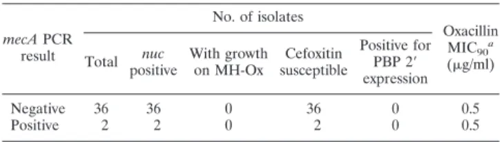
Characterization of IDI-MRSA assay-positive MSSA. We
selected 38 MSSA isolates (one from each patient) for further study. None of these isolates grew on MH-Ox screening agar, all were susceptible to cefoxitin by disk diffusion, and PBP 2⬘ was not detected by latex agglutination (Table 3). The oxacillin MIC at which 90% of isolates are inhibited for all 38 isolates was 0.5g/ml. Repeat testing by the IDI-MRSA assay directly with these MSSA isolates confirmed the original positive PCR result for the pooled specimens. By multiplex PCR,nucwas detected in all 38 isolates and themecAgene was identified in 2 of the 38 isolates (Table 3).
Genetic profile of IDI-MRSA assay-positive MSSA.
Seven-teen different PFGE genotypes were identified among the 38 IDI-MRSA assay-positive MSSA isolates. We found evidence of patient-to-patient transmission for two of these strains in two unrelated events involving four patients (data not shown). For 21 of the isolates, the PFGE patterns were consistent with common the MRSA genotypes found in this region. For 11 of these isolates the genotypes were consistent with variants of C-MRSA type 2 (U.S. MRSA clone USA 500), for 2 isolates the genotypes were consistent with variants of C-MRSA type 1 (U.S. MRSA clone USA 100), and for 8 isolates the profiles were consistent with uncommon MRSA clones previously identified at The Ottawa Hospital. The profiles for 17 isolates were not consistent with those for currently recognized MRSA clones from this region.
DISCUSSION
Rapid detection of MRSA-colonized patients has the poten-tial of improving patient care and positively affecting hospital infection control practices. The development of simple single-step nucleic acid amplification assays for MRSA detection has been complicated by difficulties with the reliable determination of the origin ofmecAin mixed cultures containing both CoNS
andS. aureus. Since optimal screening for MRSA colonization
is not limited to the testing of nasal swab specimens, alterna-tive approaches that allow pooling of multiple specimens and testing of the pooled specimens in a single PCR are desirable. Use of a broth-PCR method for detection of MRSA has been described previously and has been implemented for routine screening for MRSA colonization (24). By this approach, a quantitative real-time PCR assay for detection ofnuc is
re-quired to differentiate the growth of MRSA from that of MSSA from clinical specimens pooled in a selective broth. The IDI-MRSA assay, a commercial PCR method, is designed to be able to differentiate both CoNS and MSSA from MRSA in clinical specimens in a rapid single-step procedure.
We did not find that testing of pooled specimens from broth interfered with the performance of the IDI-MRSA assay, but rather, it was reliable and performed well compared to the performance of culture of nasal and rectal swabs pooled in a selective broth. Although rectal swabs should also be consid-ered for use for screening for MRSA colonization, testing of specimens from rectal sites by amplification methods may be problematic because of the expected higher rates of inhibition from these complex specimens (1, 16). By pooling the rectal and nasal swabs in a broth, potential inhibitors are diluted and are less likely to interfere with the PCR. In fact, we found that the inhibition rate for the IDI assay was less than 1% when we tested samples from broth cultures. One drawback of combin-ing the PCR assay with the broth culture is the delay in the reporting of the results compared to the time to the reporting of the results of direct testing. Overnight incubation of speci-mens in the selective broth was used in the laboratory, as it was convenient for work-flow purposes. Due to the high sensitivity of PCR, overnight incubation may not be necessary and a shorter incubation may be as effective. However, we did not evaluate this assay under these conditions. After incubation of the broth, routine cultures required an additional 40 to 72 h to confirm the presence of MRSA and an additional 48 h if noS.
aureusisolate was recovered, whereas less than 4 h of
addi-tional time was required for a negative result by the MRSA assay. However, culture confirmation of a positive IDI-MRSA assay result required an additional 40 to 72 h. Despite this, pooling of specimens in the selective broth with the over-night incubation prior to PCR testing still provides an improve-ment over the time required for routine culture.
Although the IDI-MRSA assay performed well compared to the performance of culture, we continued to culture PCR-positive broths to recover MRSA isolates for genotyping and epidemiologic purposes. This also provided us with an oppor-tunity to continue monitoring the performance of the assay outside of a research laboratory setting. In the 4 months postimplementation, we found that the positive predictive value of the assay fell from 90% during the evaluation phase to 65%. This significant reduction in performance can be ex-plained in part by the fact that 38 IDI-MRSA assay-positive MSSA isolates were recovered. It is unlikely that the selective broth interfered with the recovery of MRSA by favoring the growth of the MSSA isolates, since for seven of these patients,
TABLE 2. Postimplementation culture outcomes of IDI-MRSA PCR-positive broths Culture outcome No. (%) of broths No. (%) of patients MRSA 195 (65) 93 (62) MSSAa 77 (26) 38 (25) NoS. aureus 26 (9) 19 (12) Total 298 150
aThe positive IDI-MRSA PCR result was confirmed by repeat testing of pure
cultures of the isolated MSSA isolate.
TABLE 3. Characterization of the 38 IDI-MRSA PCR-positive MSSA isolates mecAPCR result No. of isolates Oxacillin MIC90a (g/ml) Total nuc positive With growth on MH-Ox Cefoxitin susceptible Positive for PBP 2⬘ expression Negative 36 36 0 36 0 0.5 Positive 2 2 0 2 0 0.5 a
MIC90, MIC at which 90% of isolates are inhibited.
on January 28, 2021 by guest
http://jcm.asm.org/
the same MSSA isolates were recovered when nasal swabs were directly cultured onto BA (data not shown). We failed to recoverS. aureusfrom 9% of the PCR-positive broths. Some of these were from patients who were previously positive for MRSA and may reflect low-level colonization that may not be detectable by culture. For the seven patients described above in whom MSSA isolates were recovered by direct culture of a nasal specimen onto BA, initial screening cultures failed to recover any S. aureus isolate from the PCR-positive broth. Therefore, the selective broth may suppress the growth of some MSSA isolates and may explain some of the false-posi-tive PCR outcomes in which noS. aureusisolate was recovered. Two of the 38 MSSA isolates were found to bemecApositive but repeatedly oxacillin susceptible. Although the exact nature and significance of these two isolates is not known, they may represent pre-MRSA clones requiring inactivation of themecI
repressor gene before methicillin resistance can be expressed (11). Further characterization of these isolates is ongoing.
For the remaining 36 PCR-positive MSSA isolates, all were found to be negative formecA. Persistent colonization with these strains was demonstrated in seven patients by repeat screening. Furthermore, we found evidence of patient-to-patient transmis-sion in at least two unrelated cases, suggesting that these strains can easily establish themselves and persist in hospital settings. The origins and significance of these isolates are not known. Our findings are consistent with reports of both S.
aureusand CoNS isolates with non-mecA-containing SCC
el-ements (8, 14, 19). Huletsky et al. also reported that 4.6% of MSSA strains tested were positive in a similar PCR assay for MRSA targeting the right-junction element (12). Because SCCmec is a mobile genetic element, complete or partial loss of the cassette may result in isolates harboring a portion of the right-junction sequence while having lost themecAgene complex. In fact, Donnio et al. recently reported on the char-acterization of nine MSSA isolates which likely originated from MRSA epidemic clones but underwent partial excision of SCCmec(8). These isolates lost the mecAgene complex but retained the right extremity of the SCC element integrated in the 5⬘end of theorfXgene (8). Interestingly, 21 of our MSSA isolates did have PFGE profiles consistent with those of com-monly circulating MRSA clones. However, the remaining iso-lates had profiles that were not consistent with those of any MRSA clones from this region, suggesting that the origin of thesemecA-negative SCC elements is manifold. These isolates require further characterization. For a more comprehensive view of the mechanisms of resistance in MRSA, readers are re-ferred to the review article by Hiramatsu (11).
It is possible that other types of SCC elements with similar-ities toSCCmecmay integrate in the same insertion sites. In fact, an SCC element carrying capsular genes (SCCcap1) and with similarities to SCCmectype II but lackingmecAhas been described (18). A PCR assay similar to the IDI-MRSA assay also targeting the right-junction sequence of SCCmecwas pos-itive for anS. aureus isolate carrying SCCcap1 (7). There is evidence that other types of insertion elements do share sim-ilarities with SCCmec and could be a problem for the IDI-MRSA assay. MSSA isolate ATCC 25923, which is commonly used for quality control of in vitro susceptibility testing, has been shown to contain an element that shows similarities to SCCmec and that is inserted in the same integration site as
SCCmec(13). Based on these similarities, we tested this strain with the IDI-MRSA assay and found that it provided a false-positive result (data not shown).
AlthoughmecA-negative SCC elements are not unique, the extent to which thesemecA-negative SCC elements were found in our patient population was surprising. Regardless of their origins, these common and diverse genetic elements will affect the performance of assays targeting the right-junction se-quences as surrogate markers for mecA-mediated resistance. The extent to which these strains will affect the assay will depend on how widespread they are. In settings of high MRSA prevalence, these MSSA strains may be displaced and may not be as relevant as they are in settings of lower MRSA incidence, such as Canada. At our institution, because of the rapid turn-around time and the excellent negative predictive value, the IDI-MRSA assay was implemented for routine screening for MRSA. However, because of the significant burden associated with the implementation of infection control precautions, cul-tures remain essential for confirmation of positive IDI-MRSA assay results. Until broth cultures are finalized, PCR-positive results are considered preliminary and require confirmation. However, pending this confirmation, newly identified patients are placed in isolation because of the potential presence of MRSA. Therefore, it is important that both the microbiology laboratory and infection control evaluate the usefulness of this assay for their local settings. This includes determination of whether IDI-MRSA assay-positive results require culture confir-mation and how these results will be used for determination of when to implement appropriate prevention measures.
REFERENCES
1.Alvarez, J., M. Sota, A. B. Vivanco, I. Perales, R. Cisterna, A. Rementeria, and J. Garaizar.2004. Development of a multiplex PCR technique for detection and epidemiological typing ofSalmonellain human clinical sam-ples. J. Clin. Microbiol.42:1734–1738.
2.Apfalter, P., O. Assadian, A. Kalczyk, V. Lindenmann, A. Makristathis, S. Mustafa, M. Rotter, and A. M. Hirschl.2002. Performance of a new chro-mogenic oxacillin resistance screen medium (Oxoid) in the detection and presumptive identification of methicillin-resistant Staphylococcus aureus. Diagn. Microbiol. Infect. Dis.44:209–211.
3.Blanc, D. S., A. Wenger, and J. Bille.2003. Evaluation of a novel medium for screening specimens from hospitalized patients to detect methicillin-resistant
Staphylococcus aureus. J. Clin. Microbiol.41:3499–3502.
4.Canadian Communicable Disease Report.2005. Surveillance for methicillin-resistantStaphylococcus aureusin Canadian hospitals—a report update from the Canadian Nosocomial Infection Surveillance Program. Can. Communi-cable Dis. Rep.31:33–39.
5.Cosgrove, S. E., E. N. Sakoulas, E. N. Perencevich, M. J. Schwaber, and A. W. Karchmer.2003. Comparison of mortality associated with methicillin-resistant and methicillin-susceptible Staphylococcus aureusbacteremia: a meta-analysis. Can. Infect. Dis.36:53–59.
6.Costa, A.-M., I. Kay, and S. Palladino.2005. Rapid detection ofmecAand
nucgenes in staphylococci by real-time multiplex polymerase chain reaction. Diagn. Microbiol. Infect. Dis.51:13–17.
7.Cuny, C., and W. Witte.2005. PCR for the identification of methicillin-resistantStaphylococcus aureus(MRSA) strains using a single primer pair specific for SCCmecelements and the neighbouring chromosome-borne
orfX. Clin. Microbiol. Infect.11:834–837.
8.Donnio, P.-Y., D. C. Oliviera, N. A. Faria, N. Wilhelm, A. Le Coustumier, and H. de Lencastre.2005. Partial excision of the chromosomal cassette containing the methicillin resistance determinant results in methicillin-sus-ceptibleStaphylococcus aureus. J. Clin. Microbiol.43:4191–4193. 9.Fang, H., and G. Hedin.2003. Rapid screening and identification of
methi-cillin-resistantStaphylococcus aureusfrom clinical samples by selective-broth and real-time PCR assay. J. Clin. Microbiol.41:2894–2899.
10.Francois, P., D. Pittet, M. Bento, B. Pepey, P. Vaudaux, D. Lew, and J. Schrenzel.2003. Rapid detection of methicillin-resistantStaphylococcus au-reusdirectly from sterile or nonsterile clinical samples by a new molecular assay. J. Clin. Microbiol.41:254–260.
11.Hiramatsu, K.2004. Elucidation of the mechanism of antibiotic resistance acquisition of methicillin-resistantStaphylococcus aureus(MRSA) and
on January 28, 2021 by guest
http://jcm.asm.org/
termination of its whole genome nucleotide sequence. Jpn. Med. Assoc. J.
47:153–159.
12.Huletsky, A., R. Giroux, V. Rossbach, M. Gagnon, M. Vaillancourt, M. Bernier, F. Gagnon, K. Truchon, M. Bastien, F. J. Picard, A. van Belkum, M. Ouellette, P. H. Roy, and M. G. Bergeron.2004. New real-time PCR assay for rapid detection of methicillin-resistantStaphylococcus aureusdirectly from specimens containing a mixture of staphylococci. J. Clin. Microbiol.42:1875– 1884.
13.Ito, T., Y. Katayama, K. Asada, N. Mori, K. Tsutsumimoto, C. Tiensasitorn, and K. Hiramatsu.2001. Structural comparison of three types of staphylo-coccal cassette chromosomemecintegrated in the chromosome in methicillin-resistantStaphylococcus aureus. Antimicrob. Agents Chemother.45:1323–1336. 14.Katayama, Y., F. Takeuchi, T. Ito, X. X. Ma, Y. Ui-Mizutani, I. Kobayashi, and K. Hiramatsu.2003. Identification in methicillin-susceptible Staphylo-coccus hominisof an active primordial mobile genetic element for the staph-ylococcal cassette chromosomemecof methicillin-resistantStaphylococcus aureus. J. Bacteriol.185:2711–2722.
15.Kim, T., P. I. Oh, and A. E. Simor.2003. The economic impact of methicillin-resistantStaphylococcus aureusin Canadian hospitals. Infect. Control Hosp. Epidemiol.22:99–104.
16.Kongmuang, U., J. M. Luk, and A. A. Lindberg.1994. Comparison of three stool-processing methods for detection ofSalmonellaserogroups B, C2, and D by PCR. J. Clin. Microbiol.32:3072–3074.
17.Lem, P., J. Spiegelman, B. Toye, and K. Ramotar.2001. Direct detection of
mecA, nuc and 16S rRNA genes in Bact/Alert blood culture bottles. Diagn. Microbiol. Infect. Dis.41:165–168.
18.Luong, T. T., S. Ouyang, K. Bush, and C. Y. Lee.2002. Type 1 capsule genes ofStaphylococcus aureusare carried in a staphylococcal cassette chromo-some genetic element. J. Bacteriol.184:3623–3629.
19.Mongkolrattanothai, K., S. Boyle, T. Murphy, and R. S. Daum.2004. Novel non-mecA-containing staphylococcal chromosomal cassette composite island containingpbp4andtagFgenes in a commensal staphylococcal species: a possible reservoir for antibiotic resistance islands inStaphylococcus aureus. Antimicrob. Agents Chemother.48:1823–1836.
20.National Committee for Clinical Laboratory Standards.2004. Performance standards for antimicrobial susceptibility testing; fourteenth informational supplement. M100-S14. National Committee for Clinical Laboratory Stan-dards, Wayne, Pa.
21.National Committee for Clinical Laboratory Standards.2003. Methods for dilution antimicrobial susceptibility tests for bacteria that grow aerobically; approved standard, 6th ed. M7-A6. National Committee for Clinical Labo-ratory Standards, Wayne, Pa.
22.National Committee for Clinical Laboratory Standards.2003. Performance standards for antimicrobial disk susceptibility tests; approved standards, 8th ed. M2-A8. National Committee for Clinical Laboratory Standards, Wayne, Pa.
23.National Nosocomial Infections Surveillance System.2003. National Noso-comial Infections surveillance (NNIS) system report, data summary from January 1992 through June 2003, issued August 2003. Am. J. Infect. Control
31:481–498.
24.Nilsson, P., H. Alexandersson, and T. Ripa.2005. Use of broth enrichment and real-time PCR to exclude the presence of methicillin-resistant Staphy-lococcus aureusin clinical samples: a sensitive screening approach. Clin. Microbiol. Infect.11:1027–1034.
25.Reischl, U., H. J. Linde, M. Metz, B. Leppmeier, and N. Lehn.2000. Rapid identification of methicillin-resistant Staphylococcus aureus and simulta-neous species confirmation using real-time fluorescence PCR. J. Clin. Mi-crobiol.38:2429–2433.
26.Safdar, N., L. Narans, B. Gordon, and D. G. Maki.2003. Comparison of culture screening methods for detection of nasal carriage of methicillin-resistantStaphylococcus aureus: a prospective study comparing 32 methods. J. Clin. Microbiol.41:3163–3166.
27.Sewell, D., S. A. Potter, C. M. Jacobson, L. J. Strausbaugh, and T. T. Ward.
1993. Sensitivity of surveillance cultures for the detection of methicillin-resistantStaphylococcus aureusin a nursing-home care unit. Diagn. Micro-biol. Infect. Dis.17:53–56.
28.Simor, A. E., M. Ofner-Agostini, E. Bryce, A. McGeer, S. Paton, M. R. Mulvey, and the Canadian Hospital Epidemiology Committee and Canadian Nosocomial Infection Surveillance Program, Health Canada.2002. Labora-tory characterization of Methicillin-resistantStaphylococcus aureusin Cana-dian hospitals: results of 5 years of national surveillance, 1995-1999. J. Infect. Dis.186:652–660.
29.Suh, K., B. Toye, P. Jessamine, F. Chan, and K. Ramotar.1998. Epidemi-ology of methicillin-resistantStaphylococcus aureusin three Canadian ter-tiary-care centers. Infect. Control Hosp. Epidemiol.19:395–400.
30.Tenover, F. C., R. D. Arbeit, R. V. Goering, P. A. Michelson, B. E. Murray, D. H. Persing, and B. Swaminathan.1995. Interpreting chromosomal DNA restriction patterns produced by pulsed-field gel electrophoresis: criteria for bacterial strain typing. J. Clin. Microbiol.33:2233–2239.
31.Warren, D. K., R. S. Liao, L. R. Merz, M. Eveland, and W. M. Dunne, Jr.
2004. Detection of methicillin-resistantStaphylococcus aureusdirectly from nasal swab specimens by a real-time PCR assay. J. Clin. Microbiol.42:5578– 5581.
on January 28, 2021 by guest
http://jcm.asm.org/
0095-1137/06/$08.00⫹0 doi:10.1128/JCM.01091-06
ERRATUM
Evaluation of the IDI-MRSA Assay for Detection of
Methicillin-Resistant
Staphylococcus aureus
from
Nasal and Rectal Specimens Pooled in
a Selective Broth
M. Desjardins, Christiane Guibord, B. Lalonde, B. Toye, and K. Ramotar
Division of Microbiology, Department of Pathology and Laboratory Medicine, The Ottawa Hospital, and The Ottawa Hospital Research Institute, Ottawa, Ontario, Canada
Volume 44, no. 4, p. 1219–1223, 2006. Page 1219, abstract, line 8: “negative and positive” should read “positive and negative.” Page 1220, column 2, line 9 from bottom: “negative and positive” should read “positive and negative.”