UNIVERSITÉ DE MONTRÉAL
IMPLANTABLE MICRO-DEVICE FOR EPILEPSY SEIZURE DETECTION
AND SUBSEQUENT TREATMENT
MUHAMMAD TARIQUS SALAM DÉPARTEMENT DE GÉNIE ÉLECTRIQUE ÉCOLE POLYTECHNIQUE DE MONTRÉAL
THÈSE PRÉSENTÉE EN VUE DE L’OBTENTION DU DIPLÔME DE PHILOSOPHIAE DOCTOR (PH.D.)
(GÉNIE ÉLECTRIQUE) Avril 2012
UNIVERSITÉ DE MONTRÉAL
ÉCOLE POLYTECHNIQUE DE MONTRÉAL
Cette thèse intitulée:
IMPLANTABLE MICRO-DEVICE FOR EPILEPSY SEIZURE DETECTION
AND SUBSEQUENT TREATMENT
présentée par : Muhammad Tariqus Salam
en vue de l’obtention du diplôme de : Philosophiae Doctor a été dûment acceptée par le jury d’examen constitué de :
M. AUDET Yves, Ph.D, président
M. SAWAN Mohamad, Ph.D., membre et directeur de recherche M. NGUYEN Dang Khoa, Ph.D., membre et codirecteur de recherche M. LESAGE Frédéric, Ph.D., membre
DÉDICACE
REMERCIEMENTS
I would like to thank God for giving me strength, wisdom, patience, good health and countless blessing to complete my Ph.D. research. I would also like to express my sincere gratitude and thank to my advisor Professor Mohamad Sawan for all his support, encouragement, generosity and advice during my Ph.D. studies in Polytechnique Montréal. I am grateful to my co-advisor Dr. Dang Khoa Nguyen for his valuable medical advices throughout my research works. Also I am grateful to late Dr. Anas Hamoui who deserves my utmost gratitude for his support and invaluable technical advices. I would like to thank Dr. Alain Bouthillier for teaching me fundamental mechanisms of human brain function and surgical treatment of epilepsy, and giving me many precious feedbacks on my research. It is hard to imagine my animal model experiments without the guidance from Dr. Lionel Carmant. All animal experiments presented in this dissertation conducted in his laboratory. Also, I would like to thank all faculty members, staffs, graduate students, and colleagues of Polystim lab who helped me during my education and research. Special thanks are due to the following people for their helps and collaboration: Mona Safi-Harb, Faycal Mounaim, Ali Hassan Hamie, Marjan Mirzaei, Sébastien Gélinas, people in Notre-dame Hospital and Sainte-Justine Hospital. I am grateful for support from the Canada Research Chair in Smart Medical Devices and le Fonds Québécois de la Recherche sur la Nature et les Technologies (FQRNT). Finally, I wish to thank my mother, wife and brother for their endless support and love.
RÉSUMÉ
L’émergence des micro-dispositifs implantables est une voie prometteuse pour le traitement de troubles neurologiques. Ces systèmes biomédicaux ont été exploités comme traitements non-conventionnels sur des patients chez qui les remèdes habituels sont inefficaces. Les récents progrès qui ont été faits sur les interfaces neuronales directes ont permis aux chercheurs d’analyser l’activité EEG intracérébrale (icEEG) en temps réel pour des fins de traitements.
Cette thèse présente un dispositif implantable à base de microsystèmes pouvant capter efficacement des signaux neuronaux, détecter des crises d’épilepsie et y apporter un traitement afin de l’arrêter. Les contributions principales présentées ici ont été rapportées dans cinq articles scientifiques, publiés ou acceptés pour publication dans les revues IEEE, et plusieurs autres tels que «Low Power Electronics» et «Emerging Technologies in Computing». Le microsystème proposé inclus un circuit intégré (CI) à faible consommation énergétique permettant la détection de crises d’épilepsie en temps réel. Cet CI comporte une pré-amplification initiale et un détecteur de crises d’épilepsie. Le pré-amplificateur est constitué d’une nouvelle topologie de stabilisateur d’hacheur réduisant le bruit et la puissance dissipée. Les CI fabriqués ont été testés sur des enregistrements d’icEEG provenant de sept patients épileptiques réfractaires au traitement antiépileptique. Le délai moyen de la détection d’une crise est de 13,5 secondes, soit avant le début des manifestations cliniques évidentes. La consommation totale d’énergie mesurée de cette puce est de 51 µW.
Un neurostimulateur à boucle fermée (NSBF), quant à lui, détecte automatiquement les crises en se basant sur les signaux icEEG captés par des électrodes intracrâniennes et permet une rétroaction par une stimulation électrique au même endroit afin d’interrompre ces crises. La puce de détection de crises et le stimulateur électrique à base sur FPGA ont été assemblés à des électrodes afin de compléter la prothèse proposée. Ce NSBF a été validé en utilisant des enregistrements d’icEEG de dix patients souffrant d’épilepsie réfractaire. Les résultats révèlent une performance excellente pour la détection précoce de crises et pour l’auto-déclenchement subséquent d’une stimulation électrique. La consommation énergétique totale du NSBF est de 16 mW. Une autre alternative à la stimulation électrique est l’injection locale de médicaments, un traitement prometteur de l’épilepsie. Un système local de livraison de médicament basé sur un nouveau détecteur asynchrone des crises est présenté. Ce dernier système dépendant des calculs
de données réduit jusqu’à 49% la consommation énergétique en comparaison avec le neurostimulateur synchrone précédemment présenté. Le détecteur a été validé à partir d’enregistrements icEEG de sept patients ayant déjà subi des examens intracrâniens pour une chirurgie d’épilepsie. Le déclenchement du système de livraison de médicaments a été testé et une dose prédéfinie de médicament est libérée environ 16 secondes après le début des crises électrographiques.
Dans le but d’améliorer les performances du dispositif implantable, de nouvelles électrodes sous-durales seront présentées. Elles offrent une meilleure qualité d’enregistrement des icEEG et permettent de délimiter la zone épileptogène durant l’évaluation pré-chirurgicale. De plus, le nouveau réseau d’électrodes a une nouvelle forme et permet de minimiser certaines complications (i.e. infection, œdème et hémorragie au niveau du cerveau) habituellement rencontrées avec l’utilisation des électrodes sous-durales actuelles. Ces nouvelles électrodes ont été testées in vitro dans une solution saline et in vivo pendant une période de trois semaines. Le rapport signal sur bruit a été amélioré de 6 dB en comparaison avec des électrodes commerciales actuellement disponibles.
ABSTRACT
Emerging implantable microdevices hold great promise for the treatment of patients with neurological conditions. These biomedical systems have been exploited as unconventional treatment for the conventionally untreatable patients. Recent progress in brain-machine-interface activities has led the researchers to analyze the intracerebral EEG (icEEG) recording in real-time and deliver subsequent treatments.
We present in this thesis a long-term safe and reliable low-power microsystem-based implantable device to perform efficient neural signal recording, seizure detection and subsequent treatment for epilepsy. The main contributions presented in this thesis are reported in five journal manuscripts, published or accepted for publication in IEEE Journals, and many others such as Low Power Electronics, and Emerging Technologies in Computing. The proposed microsystem includes a low-power integrated circuit (IC) intended for real-time epileptic seizure detection. This IC integrates a front-end preamplifier and epileptic seizure detector. The preamplifier is based on a new chopper stabilizer topology that reduces noise and power dissipation. The fabricated IC was tested using icEEG recordings from seven patients with drug-resistant epilepsy. The average seizure detection delay was 13.5 sec, well before the onset of clinical manifestations. The measured total power consumption of this chip is 51 µW.
A closed-loop neurostimulator (CLNS) is next introduced, which is dedicated to automatically detect seizure based on icEEG recordings from intracranial electrode contacts and provide an electrical stimulation feedback to the same contacts in order to disrupt these seizures. The seizure detector chip and a dedicated FPGA-based electrical stimulator were assembled together with common recording electrodes to complete the proposed prosthesis. This CLNS was validated offline using recording from ten patients with refractory epilepsy, and showed excellent performance for early detection of seizures and subsequent self-triggering electrical stimulation. Total power consumption of the CLNS is 16 mW. Alternatively, focal drug injection is the promising treatment for epilepsy. A responsive focal drug delivery system based on a new asynchronous seizure detector is also presented. The later system with data-dependent computation reduces up to 49% power consumption compared to the previous synchronous neurostimulator. The detector was validated using icEEG recordings of 7 patients who have previously undergone intracranial investigations for epilepsy surgery. The triggering of the drug
delivery system was tested and a predefined seizure suppression dose was delivered ~16 sec after electrographical seizure onsets.
In order to improve performances of the whole implantable device, a novel subdural electrode is presented for better icEEG recording quality and delineation of the epileptogenic zone during the presurgical evaluation. Moreover, the new subdural grid electrode has a new shape and several attributes to address some of the problems (e.g. brain infection, swelling and hemorrhage) encountered with currently used subdural electrodes. These electrodes are tested in vitro saline solution and in vivo experiments for 3 weeks, and signal-to-noise ratio is improved up to 6 dB compared to using commercial electrodes.
TABLE DES MATIÈRES
DÉDICACE ... III REMERCIEMENTS ... IV RÉSUMÉ ... V ABSTRACT ...VII TABLE DES MATIÈRES ... IX LISTE DES TABLEAUX ... XIV LISTE DES FIGURES ... XVI LISTE DES SIGLES ET ABRÉVIATIONS ... XXIV LISTE DES APPENDIXES ... XXVIII
INTRODUCTION ... 1
CHAPTER 1 MICROELECTRONIC SYSTEM FOR TREATMENT OF EPILEPSY ... 7
1.1 Low-Power Implantable Device for Onset Detection and Subsequent Treatment of Epileptic Seizures: A Review ... 7
1.2 Microelectronic system design criteria for chronic brain implants ... 8
1.3 IcEEG recording method ... 8
1.4 Noise figures ... 10
1.5 Intracranial electrode contact ... 11
1.6 Neural signal amplifier ... 11
1.7 Low-power seizure detector ... 12
1.8 Implantable devices for the treatment of epilepsy ... 14
1.9 Conclusion ... 16
CHAPTER 2 CMOS BASED EPILEPTIC SEIZURE DETECTOR ... 18
2.2 Introduction ... 19
2.3 Epileptic Seizure Detection algorithm ... 22
2.4 Proposed system ... 24
2.5 Circuit Implementation ... 25
2.5.1 Preamplification ... 26
2.5.2 Voltage Level Detector ... 27
2.5.3 Digital Demodulator ... 28
2.5.4 High-Frequency Detector ... 29
2.6 Experimental results ... 29
2.6.1 Integrated Circuit Measured Performance ... 29
2.6.2 Patient selection methodology ... 31
2.6.3 Method of Case Studies ... 31
2.6.4 Validation of the Seizure Detection Algorithm ... 33
2.6.5 Validation of the SOD Chip ... 36
2.7 Conclusion ... 39
2.8 Acknowledgments ... 40
CHAPTER 3 A RESPONSIVE ELECTRICAL STIMULATOR... 41
3.1 Low-Power Circuit Techniques for Epileptic Seizures Detection and Subsequent Neurostimulation ... 41
3.2 Introduction ... 42
3.3 Method of treatment ... 45
3.3.1 Low-power accurate seizure detector design ... 46
3.3.2 Stimulation parameters ... 48
3.4 Low power system design methodology ... 50
3.4.2 Electrical stimulator ... 52
3.5 Experimental and clinical Results ... 55
3.5.1 Circuit Measured Performance ... 56
3.5.2 Method of case studies and validation ... 58
3.5.3 Validation of the closed-loop neurostimulator ... 59
3.5.4 Performance analysis of the proposed CLNS ... 61
3.6 Conclusion ... 63
CHAPTER 4 IMPLANTABLE CLOSED-LOOP EPILEPSY PROSTHESIS ... 64
4.1 Implantable Closed-loop Epilepsy Prosthesis: Modeling, Implementation and Validation ... 64
4.2 Introduction ... 65
4.3 Methods and materials ... 67
4.3.1 Patient selection and device validation method ... 67
4.3.2 Proposed closed-loop Epilepsy prosthesis ... 69
4.3.3 Seizure detection from icEEG recordings ... 74
4.3.4 In Vitro Experiments ... 75
4.4 Results ... 76
4.4.1 Power dissipation of the proposed device ... 76
4.4.2 Validation of seizure detection ... 77
4.4.3 Validation of electrical stimulation ... 82
4.4.4 In vitro voltage distribution ... 82
4.5 Conclusion ... 85
4.6 Acknowledgments ... 85
5.1 An Implantable Closed-loop Asynchronous Drug Delivery System for the Treatment
of Refractory Epilepsy ... 86
5.2 Introduction ... 87
5.3 Methods and Materials ... 90
5.3.1 Proposed closed-loop epilepsy prosthesis ... 90
5.3.2 Circuits and devices implementation ... 97
5.3.3 Patients description ... 98
5.3.4 Device validation method ... 99
5.3.5 Prosthesis pre-implantation procedure ... 101
5.4 Experimental and clinical results ... 102
5.4.1 Validation of asynchronous front-end detector ... 105
5.4.2 Validation of drug delivery system ... 106
5.5 Discussion ... 107
5.6 Conclusion ... 109
5.7 Acknowledgments ... 109
CHAPTER 6 NOVEL INTRACRANIAL RECORDING SYSTEM ... 110
6.1 Subdural Porous and Notched Mini Grid Electrodes for Wireless Intracranial Electroencephalographic Recordings ... 110
6.2 Introduction ... 111
6.3 Proposed novel intracranial recording system ... 114
6.4 Methods and Materials ... 116
6.4.1 Fabrication of the mini grid electrodes ... 116
6.4.2 IcEEG recording performance of proposed subdural contacts and comparison with commercially available subdural contacts ... 119
6.5.1 Impedance measurement ... 125
6.5.2 IcEEG recording performance ... 126
6.5.3 Magnetic resonance visualization of subdural mini-grids ... 130
6.5.4 Validation of wireless transmission of icEEG recordings ... 130
6.5.5 Post icEEG brain histology ... 131
6.6 Conclusion ... 132
6.7 Acknowledgments ... 132
GENERAL DISCUSSION ... 133
CONCLUSION AND FUTURE WORKS ... 136
Contributions ... 136
Recommendations for future works ... 137
BIBLIOGRAPHIE ... 139
LISTE DES TABLEAUX
Table 1.1: Summary of seizure detection algorithms ... 13
Table 1.2: Comparison of two commercially available neurostimulators for the treatment of epilepsy. ... 15
Table 2.1: Comparison of the conventional and the proposed chopper preamplifier. ... 27
Table 2.2: Measured features for the fabricated SOD ... 30
Table 2.3: Case studies of seven patients with partial epilepsy and Matlab Analysis. ... 32
Table 2.4: The tuneable parameters’ values of the SOD chips and average detection delays. ... 36
Table 2.5: Comparison with latest competitive results. ... 38
Table 3.1: Biphasic stimulation parameters ... 44
Table 3.2: Comparison on simulated power consumption (PC) of traditional and low-power detection cores ... 52
Table 3.3:Measured power consumption (PC) of CLNS ... 57
Table 3.4: Measured stimulation parameters of the electrical stimulator. ... 58
Table 3.5: Seizure detection performance on seven patients and ... 59
Table 4.1:Available parameters in the proposed electrical stimulator ... 72
Table 4.2: Measured power consumption of proposed closed-loop epilepsy prosthesis ... 76
Table 4.3: Average tuneable parameters and detection delay (TSOD) of seizure detector ... 79
Table 4.4: A competitive comparison of different types of seizure detectors ... 81
Table 5.1: Pilot studies on focal drug delivery therapy ... 88
Table 5.2: Comparison of synchronous and asynchronous epileptic seizure detection algorithms ... 92
Table 5.3:Measured power consumption of synchronous and asynchronous prototyped devices at 3.3 V power supply ... 103
Table 5.4: Average seizure detection delay (TDET) of synchronous and asynchronous detectors. ... 106 Table 6.1: 4-AP induced seizure behavior in terms of modified racine’s scale (RACINE, 1972) ... 127 Table 6.2: Comparative study on average icEEG recordings in vivo ... 129
LISTE DES FIGURES
Figure 1-1: Seizure signal analysis: (a) is icEEG recording Vin from the epileptogenic zone during a seizure and (b) is |SF| of Vin. ... 9 Figure 1-2: Various noise sources affect icEEG recording Vin. ... 10 Figure 2-1: IcEEG recordings of two patients with refractory focal epilepsy and signal analyses:
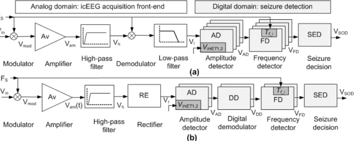
(a) Start of seizure activity characterized by low-amplitude fast activity, (b) Frequency analysis (FSZ) of (a), (c) Mean absolute amplitude (VMA) analysis of (a), (d) Seizure activity of second patient with an initial brief electrical seizures (BES) followed by an electroclinical seizure, (e) FSZ of (d), and (f) VMA of (d)... 22 Figure 2-2: Seizure detection algorithm: (a) Input signal Vin, (b) Modulated signal of Vin, (c)
Output of VLDs Vcomi, and (d) Digital demodulation VDi. ... 23 Figure 2-3: The proposed integrated SOD: (a) Implant configuration which shows the devices
and two sets of electrodes; the sensing subdural electrodes and depth electrodes, and (b) Block diagram of the proposed SOD chip. ... 25 Figure 2-4: Preamplification front-end: (a) Band-pass filter comprising an OTA, a high-pass filter
and a buffer, (b) Circuit of the OTA used in preamplifier. ... 26 Figure 2-5: The dedicated chopper stabilizer circuit and corresponding frequency analysis of
signals in different nodes. ... 26 Figure 2-6: Construction of VLDs: (a) Block diagram of VLDs, (b) Schematic of a VLD, and (c)
Circuit of a comparator. ... 28 Figure 2-7: The digital demodulator (DD): (a) Circuit, (b) Burst of pulses detected by VLD, (c)
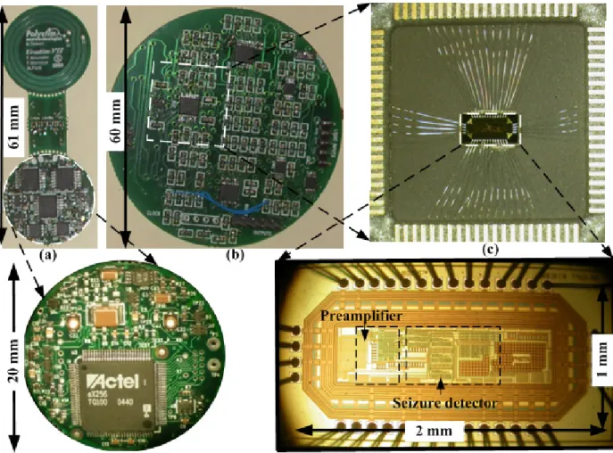
Voltage Veb across the RC circuit, and (d) Output VDi. ... 28 Figure 2-8: Microphotograph of the fabricated SOD chip. ... 29 Figure 2-9: Measured results: (a) Variable gain of the preamplification front-end with changing
of VREF. (b) Gain response of the front-end preamplifier; (c) Comparator threshold levels; (d) Time frame (Tf ) generation. ... 30
Figure 2-10: Seizure onset detections where icEEGs were recorded from different locations in patients, the zoom inset shows signal analysis and detection: Vin is icEEG of seizure recorded using two contacts from the EZ, FSZ is frequency analysis of Vin, VMA is mean absolute amplitude analysis of Vin, VD1 – VD2 are high frequency detections, and VSO is seizure onset detection: (a) Case 1, (b) Case 2, and (c) Case 3. ... 34 Figure 2-11: IcEEG analysis and seizure onset detection using Matlab. The icEEG (Vin) of a
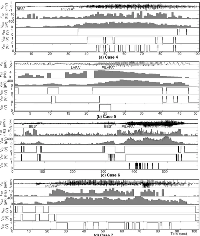
seizure recorded using two contacts from the EZ, frequency analysis FSZ, mean absolute amplitude analysis VMA, the high frequency detections VD1 – VD2, and VSO is seizure onset detection: (a) Case 4, (b) Case 5, (c) Case 6, and (d) Case 7. ... 35 Figure 2-12: Measured seizure onset detection by the SOD chip, where Vin is the icEEG of
seizure from EZ, VD1 – VD2 are high frequency detections in the icEEG, and VSO is seizure onset detection: (a) Case 1, (b) Case 2, and (c) Case 3. ... 37 Figure 2-13: Comparative results of analyzing the same icEEG recordings with several detection
methods (based on 7 patients). ... 39 Figure 3-1: Overview of method of treatment: (a) proposed CLNS interfaces directly to the part
of brain through subdural electrodes; (b) flow chart of CLNS; and (c) graphic representation of automatic triggering neurostimulation, where Vin is icEEG of seizure from epileptogenic zone, VSOD is seizure onset detection, and ISTIM is bipolar biphasic electrical stimulation. ... 45 Figure 3-2: Time-frequency (FSZ) and time-amplitude (VMA) analysis of icEEG recording (Vin) in
(a) normal signal, (b) BES, and (c) electroclinical seizure. ... 48 Figure 3-3: Biphasic stimulation parameters: (a) stimulation waveform profile; and (b) limitation
of maximum pulse width with injected current of biphasic stimulation. ... 49 Figure 3-4:Block diagram of the proposed CLNS. ... 50 Figure 3-5: Schematic diagram of the detection core: (a) detection core based on the classic
chopper preamplifier and (b) low-power detection core. ... 51 Figure 3-6: Flow chart of electrical stimulator ... 53 Figure 3-7: Timing process of electrical stimulation waveform using variable parameters ... 54
Figure 3-8: Output stage of the CLNS: (a) block diagram of output stage and (b) control signals sequence of switches to generate biphasic stimulation waveform. ... 55 Figure 3-9: The proposed CLNS device: (a) is ES PCB and zoom inset shows opposite side; (b) is
SD PCB; and (c) is detection core attached in opposite side of (b); and zoom inset of (c) shows photograph of detection core chip. ... 56 Figure 3-10: Digitally controlled measured results: (a) time frame Tf_i generation using sampling
frequency FS; (b) threshold voltage Vi,HET1,2; and (c) output stimulation current ISTIM. ... 57 Figure 3-11: Case study and validation method: (a) - (b) 3-D reconstruct images of implanted
electrodes using Stellate’s gridview software; (c) identify epileptogenic zones; (d) icEEG recordings from the zone; and (e) the icEEG recording from the epileptogenic zone of patients were used to test the proposed seizure detector and the triggering of predefined electrical stimulation was validated. ... 59 Figure 3-12: Measured electrical stimulation process triggered by seizure onset detection, where
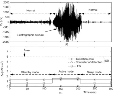
Vin is icEEG of seizure from epileptogenic zone, VSOD is seizure onset detection, VST is electrical stimulation period (offline), zoom inset shows stimulation waveforms, and VNS is no stimulation period. ... 60 Figure 3-13: Demonstration of power dissipation of proposed CLNS: (a) Electrographic seizure
in icEEG recording and (b) Power dissipation densities δP of the devices. ... 61 Figure 3-14: A comparative analysis of the proposed results (based on 7 patients) from different
detectors. ... 62 Figure 4-1: The closed-loop epilepsy prosthesis: icEEG recorded from patients with medically
refractory epilepsy were used to test our seizure detection algorithm and the triggering of an electrical stimulation to cadaveric animal brain tissue. ... 68 Figure 4-2: Illustration of the proposed implantable closed-loop epilepsy prosthesis. ... 69 Figure 4-3:Flowchart of the proposed epileptic seizure detection algorithm, tuneable parameters,
Figure 4-4: Proposed electrical stimulator: (a) seizure detections VSOD (b) two subsequent bursts of biphasic stimulations, (c) the biphasic stimulation, (d) the stimulation waveform profile, and (e) block diagram of the stimulator. ... 72 Figure 4-5: Photographs of main parts of the system: (a) Layout of chip, (b) Fabricated chip, and
(c) Electrical stimulator and zoom inset shows FPGA in the opposite side. ... 73 Figure 4-6: Method of case studies: (a)-(b) is an intracranial study for better delineate the
epileptogenic zone, (c) is axial MRI image and (d) is 3-D reconstruction of implanted electrodes, and (e) electrical seizure onset was marked (‘Seizure’) on icEEG. ... 74 Figure 4-7: in vitro voltage recordings using a cadaveric animal brain tissue in saline solution: (a)
Experimental setup, (b) Equivalent electrical circuit model of (a), and (c) spatial voltage distribution were recorded in 1 mm (VG1), 2 mm (VG2) and 3 mm (VG3) distance. ... 75 Figure 4-8: Demonstration of power dissipation of proposed system: (a) Electrographic seizure in
icEEG recording and (b) Power dissipation densities δP of the devices. ... 77 Figure 4-9: The seizure detection algorithm is validated in (a) normal signal, (b) a BES, and (c)
an electroclinical seizure: Vin is icEEG recording from the two contacts positioned over epileptogenic zone, VD1 – VD2 are abnormalities detection in VLD, and VSOD is seizure detection. ... 78 Figure 4-10: The seizure detection performance: (a) post-layout circuit simulation results and (b)
fabricated chip measured results. ... 79 Figure 4-11: Illustration of proposed seizure detection performance: (a) – (c): Analysis on
training data Vin and detection parameters (ViHET1,2 and fSZ) setting from patients’ specific seizure patterns, and (d) Decision boundaries formation using ViHET1,2 and fSZ and test detection performance using other 5 seizures, 3 brief electrical seizures and 5 normal activities. ... 81 Figure 4-12: Measured and simulation results of electrical stimulator: (a) Output current of the
stimulator for binary encoded amplitude and (b) Measured electrical stimulation triggered by seizure detector. ... 82 Figure 4-13: Current-controlled stimulus waveforms (1 – 12 mA, 100 – 500 Hz, and 100-500
against the distance from the microelectrode tips around the active subdural strip electrodes: (a) – (d) Spatial maps of the voltage distribution in brain when stimulated by 1 – 12 mA ... 83 Figure 4-14: Voltage distribution resulting from biphasic current stimulation: (a) Low-amplitude
current (1mA) at 100 – 500 µsec pulse width, and high-amplitude current (12mA) at 10 – 100 µsec pulse width (b) and 100 – 500 Hz frequency (c). ... 84 Figure 4-15: 3D plot of voltage distribution at 3 mm distance and charge density per phase
resulting from current-controlled bipolar stimulation settings. ... 85 Figure 5-1: Proposed closed-loop drug delivery system: (a) Flow chart, (b) Block diagram, and
(c) Implant configuration. ... 90 Figure 5-2: Block diagram of closed-loop asynchronous drug delivery system. ... 91 Figure 5-3: Asynchronous seizure detection performance analysis using optimum parameters of
different VWDs based on 5 seizures and long normal icEEG of the test patient. ... 93 Figure 5-4: Neural signal amplifier: (a) Schematic diagram of amplifier, (b) Noise reduction
method and corresponding frequency analysis of signals in different nodes, (c) Frequency response, and (d) Measured input-referred noise voltage spectral densities of the proposed and commercial amplifier. ... 95 Figure 5-5: Time frame generation: (a) Schematic diagram of receiver, (b) Transmitted signal
pattern VER from external coil, and (c) Recovered time frame VRST. ... 96 Figure 5-6: Proposed drug delivery system: (a) Seizure detections VSD (b) Drug injection period
TPM, (c) Focal treatment duration TON, (d) Two subsequent drug injections, and (e) – (h): Operation of micropump. ... 96 Figure 5-7: Hybrid subdural electrodes fabrication process: (a) Dimension of mould; (b)
Electrode, wire and fluidic channel assembly on mould; (c) Pour PDMS; (d) Cover up and heating with 110oC; (e) Side view of the released subdural electrodes; and (f) Top view of the electrodes. ... 97 Figure 5-8: Proposed closed-loop drug delivery system: (a) – b) Top and bottom views of
asynchronous front-end detector, (c) External transmitter, (d)–(e) Top and bottom views of drug delivery system, and (f) hybrid subdural electrodes. ... 98
Figure 5-9: The closed-loop epilepsy prosthesis: icEEG recorded from patients with medically refractory epilepsy were used to test the proposed seizure detector and the triggering of predefined drug dose was validated. ... 99 Figure 5-10: Invasive study: 3-D images of implanted electrodes reconstructed with Stellate’s
Gridview software and identification of epileptogenic zones. ... 100 Figure 5-11: The icEEG recordings from the epileptogenic zone as well as adjacent functional
regions and the corresponding behavioural relation with the electro-graphical signals. ... 100 Figure 5-12: Prosthesis pre-implantation settings: (a) Training data Vin and (b) – (c): adjustment
detection parameters FSZ and VT,i,a/b from time-frequency (FS) and time-amplitude (VMA) analysis of Vin. ... 102 Figure 5-13: Demonstration of power dissipation of proposed system: (a) Electrographic seizure
in icEEG recording and (b) Average power dissipation PT and densities δP of the devices, where PT_A and PT_S are power dissipation in asynchronous and synchronous devices, and
δP_A and δP_s are power consumption densities, respectively. ... 104 Figure 5-14: Electrical properties of the hybrid electrode: (a) SEM image of the proposed
electrode contact and (b) Measured impedance sweep of the commercial and proposed electrodes. ... 104 Figure 5-15: Validation of proposed algorithm: (a) icEEG recordings Vin, (b) seizure onset activity
is characterized by progressive amplitude increase, (c) rapid fast-activity, and (d) seizure onset detections... 105 Figure 5-16: Measured seizure onset detection by AFED, where Vamp is icEEG of seizure from
the epileptogenic zone, and VSD is seizure onset detection of case 1. ... 106 Figure 5-17: Monitored drug delivery process triggered by seizure detector, where Vin is neural
signal, Vamp is amplified neural signal, VSD is seizure detection, VPM is drug injection period TPM, and VON is focal treatment duration TON. ... 107 Figure 5-18: Controlled widely programmable measured drug dose by: (a) injection period TPM and (b) micropump needed voltage VDP with different TPM. ... 108
Figure 6-1: Schematic representation of intracerebral EEG recording systems: the traditional procedure using currently available commercial electrodes and the new procedure using the proposed novel subdural electrodes. ... 112 Figure 6-2: Illustration of the proposed novel subdural mini-grid electrodes: (a) new attributes
and dimensions, (b) cross section of the mini grid, (c) placement, and (d) connection of the grid electrodes. ... 114 Figure 6-3: Flow chart of the subdural mini-grid electrode fabrication process. ... 117 Figure 6-4: Fabrication process: (a)-(b) Design of mini-grid electrodes mould structures of top (a)
and bottom (b) parts using CATIA software and (c)-(g) Assembling and packaging process: assembling of electrodes and markers (c), pouring of silicone on the mould (d), attachment of both parts (e) and cross section of assembly (f), and cross section of fabricated mini-grid electrodes (g). ... 118 Figure 6-5: Surgical electrode implantation procedure in rats: (a) animal attached to stereotaxic
instrument, (b) craniotomy windows, (c)-(d) electrode implantation in group 1 rats, and (e)-(f) implantation and coverage with dental cement on group 2 rats. ... 120 Figure 6-6: Method of icEEG recording using wired connections and the wireless system. Zoom
inset illustrates implantation procedure and 4-AP drug injection into the brain for seizure induction. ... 123 Figure 6-7: Fabrication of the mini grid-electrodes: (a)-(b) fabricated mould, (c)-(d) fabricated Au
(c) and Pt (d) grid electrodes, (e) – (f) microanalysis of Au and Pt electrodes: X-ray fluorescence spectra recorded from Au (e) and Pt (f) electrode, and (g) wireless icEEG recording device. ... 124 Figure 6-8: Impedance spectroscopy in vitro: (a) Bipolar impedance analysis configuration, (b)
Equivalent electrical circuit of electrolyte-electrode interface, (c) Frequency analysis of icEEG recording with other noises, and (d) – (f) Measured impedance sweep of different electrodes, where Pt-Pt is Pt-electrode-Pt-wire, Au-Au is Au-electrode-Au-wire, Pt-SE-SS is Pt-electrode- Silver Epoxy (SE)-Stainless steel (SS)-wire, Pt-SE-Pt is Pt-electrode- Silver Epoxy (SE)-Pt-wire, Au-Pt is Au-electrode-Pt-wire, Au-SE-Pt is Au-electrode-Silver Epoxy-Pt-wire, and Au-SE-Au is Au-electrode-Silver Epoxy-Au-wire. ... 125
Figure 6-9: In vivo IcEEG recordings using: (a) Commercial subdural electrodes; (b) Proposed Pt subdural electrodes; (c) Proposed Au electrodes; and (d) Average power spectral density of the icEEG recordings (a)-(c). ... 126 Figure 6-10: Histogram of seizure events induced by 200 nmol 4-AP injections: Durations of 34
seizure events with different intensity (Racine’s scale) recorded in a rat. ... 128 Figure 6-11: Electrographic seizure recording and corresponding frequency analysis using (a) –
(b) commercially available subdural electrodes, (c) – (d) proposed Pt subdural electrodes, and (e) – (f) proposed Au subdural electrodes. ... 128 Figure 6-12: Power spectral density of the electrographic seizure recordings (same seizure as in
Figure 6-11) using commercial subdural electrodes; proposed Pt subdural electrodes; and proposed Au electrodes. ... 129 Figure 6-13: MRI image sequences of the new mini-grid Pt electrodes: Images (a) – (b) were
taken by placing the electrodes into a gel solution (0.9% NaCl with 14gm gelatin) and scanning them using a 1.5 T Phillips MR scanner. ... 130 Figure 6-14: IcEEG recordings using: (a) Stellate Harmonie System (wired setup) and (b)
Wireless system. ... 131 Figure 6-15: Representative micrographs illustrating the effect of the proposed subdural
electrodes on the morphological appearance of cortex, 7 days after the 3 weeks in vivo recording experiment in group 1: (a) Somatosensory cortices (exposed to the electrodes) and (b) Visual cortices (unexposed). ... 131
LISTE DES SIGLES ET ABRÉVIATIONS
Au Gold
ADC Analog-to-Digital Conversion AFED Asynchronous Front-End Detector
AM Amplitude Detectors
ASIC Application-Specific Integrated Circuit
ATN Anterior Thalamic Nucleus
Au Gold
BER Bit Error Rate
BES Brief Electrical Seizures
CAM Current Amplifier
CCPA Canadian Council for the Protection of Animals CHUM Centre Hospitalier de l'Université de Montréal
CIBPAR Comité Institutionel des Bonnes Pratiques Animales en Recherche CLNS Closed-Loop Neurostimulator
CMOS Complementary Metal–Oxide–Semiconductor CNC Computer Numerical Controlled
CSF Cerebrospinal Fluid
DAC Digital-to-Analog Converter
DBS Deep Brain Stimulation
DC DC current
DD Digital Demodulators
DFF D Flip Flop
DPE Depth electrode
DSP Digital Signal Processor
DTD Detection Delay
EBSD Event Based Seizure Detector
EEG Electroencephalography
EEG-fMRI EEG-Functional MRI
eRNS External Responsive Neurostimulator ES Electrical Stimulator
ESD Event-based Seizure Detector
EZ Epileptogenic Zone
FD False Detection
FD Frequency Detectors
FDA Food and Drug Administration FEP Feature Extraction Process FPGA Field-Programmable Gate Array
FQRNT Fonds Québécois de la Recherche sur la Nature et les Technologies
FSK Frequency-shift keying
GTC Generalized Tonic-Clonic
HFB High Frequency Band
HFD High-Frequency Detector
HSE Hybrid Subdural Electrode
icEEG Intracerebral EEG
LFB ow Frequency Band
MEG Magnetoencephalographic
MICS Medical Implant Communication Service
MRE Microelectrode
MRI Magnetic Resonance Imaging
MUX Multiplexer
NLESD Nonlinear Energy Seizure Detector
OLS Open Loop Neurostimulation
OTA Operational Transconductance Amplifier
PC Power Consumption
PCB Printed Circuit Board
PDMS Polydimethylsiloxane
PET Positron Emission Tomography PLCS Post-Layout Circuit Simulation
PS Power Supply
PSD Power Spectral Densities
Pt Platinum
ReSMiQ Microsystems Strategic Alliance of Québec RNS Responsive Neurostimulator
SD Seizure Detector
SDE Subdural Electrode
SE Silver epoxy
SEM Scanning Electron Microscope SESD Spectral Energy Seizure Detector
SNR Signal-to-Noise Ratio
SOD Seizure Onset Detector
SPECT Ictal Single-Photon Emission Computed Tomography
SW Switch
SXT Sensitivity
TFS Time Frame Selector
VLD Voltage Level Detector
VNS Vagus Nerve Simulation
VWD Voltage Window Detector
LISTE DES APPENDIXES
INTRODUCTION
Epilepsy is a neurological disorder affecting 1.3% of Canadians and up to 1% of the world population. Each year, an average of 15,500 Canadians learns that they have epilepsy. Epilepsy is a common chronic neurological disorder characterized by a predisposition to unprovoked recurrent seizures. A seizure is the manifestation of an abnormal, hypersynchronous discharge of a population of cortical neurons. Several causes may disturb the normal pattern of brain activity and trigger an epileptic seizure, such as head injury, brain infection, developmental malformations, brain tumors, cerebrovascular disease, vascular malformations, genetic disorders, and hippocampal sclerosis. Antiepileptic drugs are the mainstay of treatment, but many patients have systemic and central nervous system side effects and a third of them are refractory. Because most drug-resistant epileptics suffer from focal epilepsy, these patients may benefit from epilepsy surgery. Many studies have shown that resection of the epileptogenic zone may lead to seizure freedom (ENGEL, 2009). Success of the surgery is dependent on the accurate localization and complete resection of the epileptogenic zone. Unfortunately, not all refractory patients with focal epilepsy benefit from resective surgery; some have an epileptogenic zone overlying eloquent areas (language, primary motor or visual areas) that cannot be resected without permanent sequelae, while others have multifocal epilepsy (SPENCER, 2009). Therefore, the poor neurological outcome of these cases combined with the lack of efficacy and adverse effects of antiepileptic drugs provide sufficient justification to have an alternative treatment to supplement conventional treatments. The details motivation behind the neurostimulator therapies and the improved presurgical procedure for the treatment of epilepsy are discussed below.
Motivation
Over the last quarter century, the vast progression in neurotechnology has allowed the Food and Drug Administration (FDA) to approve stimulators for the treatment of neurological disorders, such as multichannel cochlear implant for hearing loss, deep brain simulation for Parkinson diseases, and vagus nerve simulation (VNS) for epilepsy. Commercially available VNS provides scheduled stimulation (open-loop) to the left vagus nerve to reduce seizure frequency by a yet unclear mechanism. However, seizure freedom is rare, and only 30 – 40% of patients show a significant decrease in seizure frequency (SCHACHTER, 2009). More recently, there has been growing interest in the development of responsive therapeutic devices to abort seizures at their
onset. The responsive device (detection and treatment) identifies seizures at their onset and triggers focal treatment to the epileptogenic zone to abort the seizure, whether by electrical stimulation, cooling, or drug release. To do so, an efficient seizure detection algorithm is required for accurate seizure onset detection without false alarms, and which can be implemented in a custom integrated circuit.
Several mathematical models have been developed to detect electrographic seizures onset. Intracerebral EEG-based models have shown much better performance than scalp EEG-based models, as lesser artifacts are encountered with the former. Several others studies have shown better seizure detection performances than random predictor. These models were developed using desktop computers for off-time data processing. These types of algorithms cannot be employed in a low-power implantable microchip. However, a power-efficient epileptic seizure detection algorithm can be integrated in novel therapeutic devices for epilepsy that the system could run on a button cell battery for more than 8 years. Currently, only a few clinically applicable seizure detectors exist (e.g. Responsive Neurostimulation System, NeuroPace Inc., and Seizure Advisory System, NeuroVista Corporation). Apart from the seizure detection performance, the other concerns are safety, low-power techniques, miniaturized devices and determination of optimal stimulation parameters. The several proof-of-concept experiments were introduced for the treatment of epilepsy, among them the surgical treatment, focal electrical stimulation, and direct drug delivery.
Surgical treatment of epilepsy
Patients with refractory epilepsy may benefit from epilepsy surgery if the epileptogenic zone (EZ) can be identified and resected without harm. Due to the limited spatial or temporal resolution of currently available non-invasive localization techniques, accurate delineation of the EZ may sometimes be arduous, particularly with nonlesional refractory epilepsy (SPENCER, 2009). As such, invasive EEG recordings may be necessary prior to resective surgery. An intracerebral EEG (icEEG) study consists of implanting intracerebral subdural strip, grid or depth electrodes to sample suspected areas of epileptogenicity. Intracranial electrodes will overcome the sensitivity limitations of extracranial (scalp) electrodes because they are closer to generators of epileptiform activity. Whereas a large cortical surface is required to generate a recordable signal by extracranial electrodes, intracranial electrodes can pick up potential changes occurring over only a few
millimeters of cortex. When seizures occur, the icEEG signal is analyzed to identify the EZ for subsequent resection. But there are some risks associated with icEEG recordings using available commercial intracranial electrodes, such as brain infection, cerebral edema and failure to adequately localize the EZ from inadequate brain sampling
Focal electrical stimulation
In a pilot trial, Osorio et al. (2005) demonstrated the feasibility and short-term safety of automated high-frequency electrical stimulation in blocking seizures using an external prototype composed of an electrocorticographic acquisition system, a desktop computer, and a stimulator (Grass S12). Another external responsive neurostimulator (eRNS) has been shown to terminate electrographic seizures as well (KOSSOFF, 2004). Apart from seizure detection performance, the other concerns are the safety of chronic electrical stimulation and the determination of optimal stimulation parameters. It is generally agreed that limiting the maximum charge density to 30 µC/cm2 per phase can avoid tissue damage (AGNEW, 1990); however, no cell damage in human cortex has been found for 50 to 60 µC/cm2 per phase stimulation (GORDON, 1990; RISINGER, 1995).
Direct drug delivery
The currently available orally administrated antiepileptic drugs are associated with variable systemic side effects (adverse effects in liver, bone marrow, and central nervous system). Focal drug delivery directly onto the epileptogenic zone could enhance the efficacy of drugs while limiting side effects to a minimum. Experiments in the past showed that focal diazepam infusion using a programmable pump in response to automated seizure detection could suppress the seizure (STEIN, 2000; DAVID, 2004). Other drugs such as DP-valproic acid (FISHER, 2002), gabapentin (OOMMENA, 2007), and adenosine (ANSCHEL, 2004) have been used but with less success. Much work lies ahead prior to clinical application in optimal drug selection, the delivery system, automated seizure detection, the refilling procedure, etc. To date, technological barriers limit the exploration of direct drug delivery for the long-term treatment of focal epilepsy.
Thesis Works
The new technologies give us the opportunity to solve drawbacks of the available treatments and extend the traditional neurostimulation therapies and resective surgical treatment procedures
for better treatments of epilepsy. In this thesis, our objectives are to propose new technologies for implantable device therapies and presurgical evaluation for epilepsy. We introduce long-term, safe and reliable low-power microsystems-based implantable devices to perform efficient neural signal recording, seizure detection and subsequent treatment. We have accomplished three specific objectives in the implantable device therapies: (i) Designed and implemented a low-power neural preamplifier and real-time seizure onset detection algorithm in a microchip using advanced integration technology; (ii) Designed a low-power closed-loop electrical stimulators and constructed a responsive neuromodulator by assembling the detector chip and stimulator together; and (iii) Modeled and implementated of an implantable responsive focal drug delivery system as an alternative promising treatment. For the epilepsy presurgical evaluation, we have introduced novel intracranial electrodes and a wireless icEEG recording system in order to improve EZ localization and reduce morbidity.
Contributions
The main contributions of this thesis have been reported in several peer-reviewed scientific journals and international conferences. These contributions are:
1. Design, simulation, implementation, and validation of novel power-efficient epileptic seizure detection algorithms for refractory epilepsy patients (SALAM, 2009(a), (b); SALAM, 2010 (a), (b); SAFI-HARB, 2011(b)).
2. Design, fabrication and testing of new preamplifiers and novel low-power implantable epileptic seizure-onset detectors. The systems were implemented using 0.18µm CMOS technology and discrete components in miniaturized printed circuit boards (PCBs). The implemented devices were validated using icEEG recordings of several refractory epilepsy patients (SALAM, 2011(a), (c); SAFI-HARB, 2011(b)).
3. Modeling, implementation and validation of new implantable closed-loop epilepsy prosthesis. The system was assembled with an embedded microchip (0.18µm CMOS technology) and field-programmable gate array (FPGA) in miniature PCBs, and tested using icEEG recordings of ten refractory epilepsy patients (SALAM, 2010(c), SALAM, 2012(a); SALAM, 2012(c)). 4. Design, implementation and testing of an implantable asynchronous responsive drug delivery
components in miniature PCBs and validated using icEEG recording of refractory epilepsy patients (SALAM, 2011(a); SALAM, 2012(b)).
5. Design and fabrication of new subdural porous and notched mini grid electrodes for wireless icEEG recordings. The electrodes were validated in vivo icEEG recording experiments and demonstrated comparative studies with the commercial electrodes (SALAM, 2011(b); SALAM, 2012(c)).
Thesis organization
This thesis is divided into 8 chapters. The first chapter of this thesis describes a comprehensive literature review on the alternative treatment for epilepsy. Chapters 2 through 6 address the design, implementation and validation of the new implantable device. The last chapters summarize the entire thesis, and draw attention to the contribution of this research and the recommended future works. The following is the brief chapter-wise summary of this thesis.
Chapter 1 reviews the fundamental concepts of implantable device therapies and presurgical evaluation for the treatment of epilepsy. This chapter begins with basic theories, design issues and design criteria of the implantable device. We discuss about subdural electrodes, preamplifiers front-end, seizure detectors and stimulators in the prospect of state-of-the-art, and highlight factors limiting performance in implantable device therapies for epilepsy.
Chapter 2 investigates electrographic seizure onset detection criteria and noises affect for limiting performance in the responsive neuromodulation therapies. A modified chopper stabilized preamplification method and a new power efficient seizure onset detection algorithm are proposed for the responsive neuromodulation therapy. The ultra low-power detector is implemented and validated using icEEG recordings from seven refractory epileptic patients.
Chapter 3 describes electrical stimulation therapy, a new low-power circuit design, implementation and testing of a new closed-loop electrical stimulator. The proposed FPGA embedded neurostimulator is triggered by the seizure onset detector chip for disrupting an upcoming seizure. Experimental results demonstrated safety and reliability for the automated triggering of the bipolar electrical stimulator.
Chapter 4 demonstrates modeling, implementation and validation of a new closed-loop electrical stimulator (second prototype). The new electrical stimulator has lower power
consumption, wider range of parameter variation, better precision, and larger memory. Experimental results show spatial voltage distribution in brain due to the current stimulation and suggest optimum electrical stimulation parameters for the suspected epileptic focus.
Based on the design and implementation trend in the previous chapters, which aims at closed-loop electrical stimulator; chapter 5 presents an asynchronous closed-closed-loop drug delivery system. A new power saving strategy is employed to design this direct drug delivery device. This chapter also describes a new preamplifier, an asynchronous seizure onset detector, a drug delivery system, and a hybrid subdural electrode. The experimental results demonstrate efficiency of the device and wide range of drug dosing capacity at seizure onset.
Chapter 6 describes novel subdural electrodes to improve icEEG recording quality as well as localization of the epileptogenic zone with less morbidity. The new subdural mini grid electrode has new a shape and several attributes to overcome some of the currently encountered problems with intracranial recordings. This chapter also demonstrates the design of the novel electrodes, method of fabrication, architecture of a wireless EEG recording system, and in vitro and in vivo experimental results.
Finally, a discussion chapter is presented in Chapter 7 followed by a conclusion and recommendation for future works in chapter 8.
CHAPTER 1
MICROELECTRONIC SYSTEM FOR
TREATMENT OF EPILEPSY
In the last decade, implantable microelectronic systems have been exploited as unconventional treatment for the conventionally untreatable patients. Several microelectronic devices have been commercialized for treatment of heart disease, Parkinson’s disease, Epilepsy, and deaf. Recent progress in brain-machine-interface (BMI) has led to the idea of using the medical devices to analyze the intracerebral EEG (icEEG) recorded data in real-time in order to provide subsequent treatments. The combination of the online icEEG analysis and responsive treatment approaches ensure consistent link in between neuronal pathways for normal brain activities and break the pathway for the abnormalities. Thus, microelectronic therapeutic devices offer new attractive treatment option for the neurological disorders. However, many challenges are involved in designing the implantable microelectronic devices. The design issues and factors limiting performances of microelectronic therapeutic devices are discussed in the chapter. This review work published in “Journal of Healthcare Engineering” is reproduced in following pages.
1.1
LOW-POWER IMPLANTABLE DEVICE FOR ONSET
DETECTION AND SUBSEQUENT TREATMENT OF EPILEPTIC
SEIZURES: A REVIEW
Muhammad Tariqus Salam1, Mohamad Sawan1, and Dang Khoa Nguyen2 1Polystim Neurotechnologies Laboratory, École Polytechnique de Montréal, Québec 2Neurology service, Notre-Dame Hospital (Centre Hospitalier de l'Université de Montréal) Abstract - Over the past few years, there has been growing interest in neuro-responsive intracerebral local treatments of seizures, such as focal drug delivery, focal cooling, or electrical stimulation. This mode of treatment requires an effective intracerebral electroencephalographic acquisition system, seizure detector, brain stimulator, and wireless system that consume ultra-low power. This review focuses on alternative brain stimulation treatments for medically intractable epilepsy patients. We mainly discuss clinical studies of long-term responsive stimulation and suggest safer optimized therapeutic options for epilepsy. Finally, we conclude our study with the proposed low-power, implantable fully integrated device that automatically detects low-voltage
fast activity ictal onsets and triggers focal treatment to disrupt seizure progression. The detection performance was verified using intracerebral electroencephalographic recordings from two patients with epilepsy. Further experimental validation of this prototype is underway.
1.2
MICROELECTRONIC SYSTEM DESIGN CRITERIA FOR
CHRONIC BRAIN IMPLANTS
Microelectronic systems are generally implanted on the skull under the skin and interface directly with the recording/stimulation sites for the treatment of epilepsy. Thus, the system size needs to be small, lightweight and invisible after surgical implantation. The microelectronic implants are safety-critical in nature; high reliability is an essential system attribute. Moreover, the implants must operate under stringent low-power constraints for minimal heat dissipation in surrounding tissues and to avoid frequent surgical battery replacement. Energy harvesting is essential to recharge the implanted battery from an external power source. The rechargeable implantable battery needs short-circuit protection mechanism and constant current discharge state for longer time. The implant must ideally function for 8 – 10 years without having to replace the battery. The electronic components of an implant must be sealed with biocompatible casing which is implanted in a viable places for energy harvesting, data communication, and battery replacement without a major surgery. Apart from the long lasting implantation facts, the other concerns are proper device functionalities, accurate icEEG monitoring, personalized treatment parameters, and external tuning. The key challenges of a BMI are the icEEG monitoring for true electrographic seizure activity in real-time and the trigger of focal treatment at seizure onset. Thus, noise reduction or isolation from the icEEG recordings is essential for device performance. Moreover, the abnormal icEEG detection criteria and focal treatment parameters may vary from patient to patient, and these criteria and parameters for a patient may change over the time. Therefore, a remote tunability is required in the device for adjusting detection criteria and increase/decrease intensity of the treatment.
1.3
ICEEG RECORDING METHOD
The icEEG records the activity of thousands of neurons in the region across the ∼5 mm diameter of an intracranial electrode contact. A frequency analyzer can decompose the icEEG
Figure 1-1: Seizure signal analysis: (a) is icEEG recording Vin from the epileptogenic zone during a seizure and (b) is |SF| of Vin.
recording and extract brain state information (e.g. normal and abnormal). Recent studies using higher sampling rates (1000 – 2000Hz) have showed high-frequency oscillations in preictal activities (JACOBS, 2010). Until we obtain a better understanding of the significance of these high-frequency oscillations with regards to epileptogenesis, most clinical epileptologists still rely on the analysis of a limited frequency range in the low-frequency bandwidth (10 – 40Hz) of icEEG recording (OSORIO, 2009(b)). In order to acquire the brain state information of a patient, a data acquisition system with low gain-bandwidth product and low noise are required. Commercial instrumentation has poor noise performance for the low-spectral neural signal amplification. Therefore, microelectronic circuit design problems increase due to the various noises located in the low frequency band that degrade the input icEEG recordings. As a result, the icEEG recording may translate erroneous brain state information.
Partial seizure generally begins in the EZ, spreading to adjacent regions. However, electrographic seizure onsets may vary in terms of onset morphology, discharge frequency, focality, and spread pattern. The most common seizure onset pattern in a partial epilepsy patient is characterized by a low-voltage high-frequency discharge. Figure 1-1(a) shows icEEG recording from the EZ (Vin) using a 200 Hz sampling frequency with the sudden appearance of this typical low-voltage fast activity at seizure onset. Figure 1-1(b) illustrates the spectrogram analysis of Vin. Magnitude of short-time Fourier transformation |SF| reveals low frequency activity (< 3 Hz) before seizure onset and high frequency activity (> 10 Hz) at seizure onset and during the seizure. The frequency at seizure onset may vary from patient to patient but generally stands between 10 – 40 Hz in epileptic human icEEG recordings (OSORIO, 2009(b); SALAM, 2011(a)).
Figure 1-2: Various noise sources affect icEEG recording Vin.
1.4
NOISE FIGURES
Unlike scalp EEG recordings, icEEG recordings are relatively devoid of motion and muscle artifacts. However, several other noises may degrade the icEEG recording and subsequent automated evaluation in microelectronic implant may lead to erroneous brain state translation. Figure 1-2 illustrates the noises influence in the input icEEG recording Vin. In this situation, the subdural electrodes placed directly on the brain tissue and the electrode-tissues interface create a capacitive double layer that generates dc offset (VOS)in the millivolts range. But, the front-end amplifier has a low dynamic input range due to the microvolt input neuronal signal and VOS saturates the amplifier. Therefore, VOS needs to be suppressed adequately so that Vin can dominate over VOS in front-end amplification. Moreover, flicker noise VnF contributes a major amount of noise in the low frequency band due to fundamental physics properties of instrumentation. Thermal noise VnT dominates in higher frequency (above the flicker corner frequency) due to internal resistance of instrumentation and wire resistance. Finally, 60 Hz noise Vn60 is a large
source of noise generated from the AC power supply; this noise may completely degrade the Vin.
1.5
INTRACRANIAL ELECTRODE CONTACT
Noises generated from electrodes, electrode-tissues interface, and from instruments may modulate the neural signal and degrade the signal-to-noise ratio (SNR). The custom integrated amplifiers could reduce instrument noise effect for the low-spectral neural signal amplification, but optimum SNR values can be obtained by modifying intracranial electrode contacts (SALAM, 2011(b)). Microelectrode (MRE) arrays are widely used in neurological research to improve spatial resolution in icEEG recording (CHEN, 2011). But MRE arrays have high resistivity in electrode-tissues interface which may impede icEEG neural recording (CHEN, 2011). Macroelectrodes (e.g. subdural (SDE) and depth electrodes (DPE)) have been used for the last sixty years in epilepsy presurgical evaluation. Dimensions of a SDE and a DPE are well suited to delineate the EZ. Due to their larger electrode-tissues interface, these electrodes have different electrical characteristics (compared to microelectrodes) including lower electrode impedance. Platinum (Pt) and stainless steel materials are commercially used in SDE and DPE, with Pt having better biocompatibility properties. Gold (Au) has been used in subdural electrodes (REN, 2010) but no significant improvement on icEEG recording has been reported. The details drawbacks of the available intracranial electrode contacts are described in chapter 6.
1.6
NEURAL SIGNAL AMPLIFIER
Over the past few decades, many neural signal amplifiers have been developed to overcome several challenges (e.g. low-power, low-noise, and minimal chip area). Many integrated amplifiers have been proposed, but very few designs meet the requirements for epilepsy device therapies or presurgical evaluation. The intracranial electrodes potential mismatch creates input dc offset (SALAM, 2011(a)). There are two schemes for the dc offset suppression. The first scheme is capacitive feedback of an operational transconductance amplifier (OTA) (HARRISON, 2003). MOS-bipolar pseudo-resistor elements in parallel with the capacitive feedback were used to provide a very low high-pass cutoff frequency as well as a fast settling time (ZARIFI, 2008). The capacitive feedback scheme removes the dc offset, but it may increase input referred noise (GOSSELIN, 2007(c)).
The second scheme is based on closed loop control of dc level (GOSSELIN, 2007(c); GOSSELIN, 2010(a)). This scheme is made of active components that are placed in the feedback path of an instrumentation amplifier. It can reduce low-spectrum noise and provide high input impedance. Further high-pass and low-pass filters are needed to suppress significant amount of noise. CMOS technology is widely used for bioamplifier due to the low-power dissipation, but this technology has relatively poor noise performance and the amplifier requires low input-referred noise (GOSSELIN, 2007(c); GOSSELIN, 2010(a), (b); GOSSELIN, 2009). Chopper preamplifiers have been constructed using CMOS technology and reported performance have been satisfactory (GOSSELIN, 2007(c); MASAYA, 2010; JAWED, 2009). The chopper preamplifier is based on modulation, amplification, demodulation, and filtering. The restrictions on the power consumption and size of an implantable device limit increasing the biasing current of the amplifier. Therefore, design trade-offs between the biasing current and noise are required to optimize the performance of a device. For the seizure detection application, the neural signal amplifier needs low frequency bandwidth (10 – 40 Hz). However, many amplifiers are made for neural spike detection and the amplifiers bandwidth starts from 100 Hz (GOSSELIN, 2007(c)); GOSSELIN, 2009). But the main challenge in seizure detection application is to record neural signal from the lower frequency band where noises are prominent.
1.7
LOW-POWER SEIZURE DETECTOR
In recent years, there has been growing interest in developing seizure detection algorithm. Several mathematical models have been proposed for this task, such as support vectors (MEIER, 2008), artificial neural networks (GHOSH-DASTIDAR, 2008), wavelet transformation (ZANDI, 2009), wavelet decomposition (SAAB, 2005), nonlinear time series analysis (LEHNERTZ, 1995), and mean nonlinear decision function (TITO, 2009). IcEEG-based models (MEIER, 2008; GHOSH-DASTIDAR, 2008; LEHNERTZ, 1995; TITO, 2009) perform much better than scalp EEG-based models (ZANDI, 2009; SAAB, 2005) owing to motion/muscle artifacts on scalp EEG recordings. Table 1.1 briefly summarizes different detection algorithms. These mathematical models have been implemented in high-speed computer for short-term EEG/icEEG monitoring applications. These models cannot be employed in an implantable device due to heavy mathematical computation involved and high-power dissipation. So far, few seizure detection algorithms have been proposed for low-power implantable microelectronic devices.
The detection algorithm presented in (RAGHUNATHAN, 2009) is based on classifying icEEG data into events, which are defined by two threshold voltages in icEEG recording. An ictal event is translated by high-frequency discharges. Recent studies have showed that high-frequency discharges can be very brief (few seconds), remain very focal (without spread), do not evolve in time or frequency, and are clinically silent (electrical seizures) (SALAM, 2010(a); SALAM 2009(b)). This device may result in false positive detection due to the high-frequency discharge
Table 1.1: Summary of seizure detection algorithms
Reference Method Description Applicat
ion
MCSHARRY, 2002 Multi-modal probability evaluation
Time evolution of the probability density function.
SESD
SHOEB, 2004 Wavelet and
neural network
Wavelet decomposition constructs feature vector and the support-vector machine classification algorithm that determine seizure onset.
SESD
SUFFCZYNSKI,
2006 Bistable mathematical model
Predict time of seizure occurrences using a Poisson process and a random walk process.
IEBSD
MOXON, 2001 Correlation Similarity between a reference and dynamically recorded signal.
IEBSD IASEMIDIS, 2003(a) Nonlinear,
Lyapunov exponent
Convergence and divergence of short-term maximum Lyapunov exponents (STLmax) among critical electrode sites.
IEBSD
IASEMIDIS, 2003(b) Phase changes Measure average angular frequency and optimize the techniques.
IEBSD MORMANN, 2000 Coherence Statistical measurement of phase
synchronization of signals.
IEBSD TETZLAFF, 2006 Features
extraction
Automated technique for the detection of a preseizure state.
IEBSD ARNHOLD, 1999 Signals
synchronization Detect weak interdependences of signal. IEBSD NAGARAJ, 2006 Synchronization Cross-correlationship used to find
dissimilarity of signals between interictal periods and epileptic seizures.
IEBSD
SUKHI, 2005 Bayes’ theorem Spectral feature extraction, Bayes’ theorem and spatiotemporal analysis.
IEBSD SESD: Scalp EEG recording and software in desktop computer.
detection. The support vector seizure detection algorithm (VERMA, 2010) has good flexibility but based on only spectral content of icEEG recording. Moreover it requires a large number of support vectors to define the complex decision boundary between a patient’s seizure and non-seizure activity. This explains its high-power consumption and cost. Similarly, another detector (PATEL, 2009) based on linear discriminant analysis classifiers requires higher complexity of the digital signal processor to improve sensitivity and specificity. In order to avoid redundancy, further discussions on the low-power seizure detectors are described in chapters 2, 3, 4, and 5.
1.8
IMPLANTABLE DEVICES FOR THE TREATMENT OF
EPILEPSY
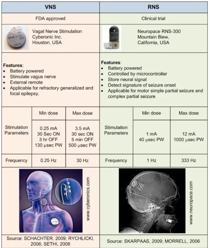
Few implantable devices for the treatment of epilepsy have been commercialized. The only FDA-approved implantable device for the treatment of epilepsy is the vagal nerve stimulator (VNS). The VNS is an open-loop (non-responsive) system comprising an implantable stimuli generator, two leads incorporating two bipolar electrodes, and an external pulse programming system for the stimuli generator (Table 1.2). The stimuli generator of the VNS is inserted through an incision in the left axilla and implanted under the skin in the left chest below the clavicle under general anesthesia. The lead is tunneled to the neck, and the two helical bipolar stimulating electrodes are placed around the left vagus nerve. After the operation, the VNS can be turned on by a computer and programming wand. The most common settings for the stimulator are a frequency of 20 – 30 Hz, a pulse width of 250 – 500 µsec, time on of 30 sec, and time off of 3 – 5 min. The advantages of VNS include (a) no need to exactly delineate the epileptogenic zone, (b) no craniotomy, (c) a lower rate of surgical complications, and (d) mild and infrequent side effects due to stimulation (hoarseness, cough). The main disadvantage is that, while 30 – 40% of cases show a reduction in seizure frequency of more than 50%, only about 3% of them attain seizure freedom (RYCHLICKI, 2006; SETHI, 2008).
Deep Brain Stimulation (DBS) featuring, for example, 1-10 V, 90 µsec pulse width, 100 – 165 Hz frequency for the treatment of Parkinson’s disease has been shown to be safe and effective as evidenced by FDA approval (BLAD, 2007). A 1 min on and 5 min off stimulation paradigm was used during an investigational DBS trial for epilepsy. DBS has also been investigated for the treatment of drug-refractory epilepsy by providing scheduled stimulation (open-loop) via depth electrodes to various sites such as the hippocampus, thalamus, cerebellum, caudate nucleus,
Table 1.2: Comparison of two commercially available neurostimulators for the treatment of epilepsy.
centromedian thalamus, anterior thalamic nucleus, and neocortical seizure foci (LEGA, 2009; ZHONG, 2011; CHABARDES, 2002; HANDFORTH, 2006; HODAIE, 2002). Recently, a large and multicenter trial (SANTE: Stimulation of the Anterior Nucleus of the Thalamus for Epilepsy)