Testosterone, growth and the evolution of sexual size dimorphism
R. M. COX, D. S. STENQUIST & R. CALSBEEKDepartment of Biological Sciences, Dartmouth College, Hanover, NH, USA
Introduction
Biologists dating back to Darwin (1871) have sought to understand why males are larger than females in many species, yet females are the larger sex in numerous others (Fairbairn et al., 2007). Evolutionary studies of sexual size dimorphism (SSD) have focused primarily on infer-ring the selective pressures responsible for its evolution using comparative approaches (Sze´kely et al., 2000; Pe´rez-Barberı´aet al., 2002; Coxet al., 2003) or measuring current selection on body size (Badyaev et al., 2000; Preziosi & Fairbairn, 2000; Schulte-Hosteddeet al., 2002). Recently, physiologists, geneticists and developmental biologists have sought to integrate this ultimate perspec-tive with an understanding of the proximate mechanisms that facilitate the expression of dimorphic phenotypes from a genome that is largely shared between the sexes (Rhen, 2007). A major goal of this integrative approach is to reconcile the observed phylogenetic lability of SSD with theoretical expectations that its evolution should be highly constrained because of inter-sexual genetic
correlations (Lande, 1980, 1987; Fairbairn & Roff, 2006; Bonduriansky, 2007; Delph, 2007; Fedorkaet al., 2007). One resolution to this paradox may be that males and females share most of the same genomic architecture for growth and body size, but that these shared genes are differentially regulated by sex-specific modifiers (Badyaev, 2002).
Sex steroids (i.e. androgens, estrogens and progestins) are excellent candidates for the regulation of sex differ-ences in growth and body size because they are produced and secreted in sex-specific fashion by the gonads. However, if sex steroids are to account for divergent patterns of SSD across species, then either their patterns of secretion or their effects on growth must differ across species. This distinction bears on a fundamental question in evolutionary endocrinology. Are the pleiotropic effects of hormones on their target tissues evolutionary con-served across species, such that trait evolution proceeds primarily via changes to circulating hormone levels (‘evolutionary constraint hypothesis’, Hau, 2007)? Or does selection independently alter the responsiveness of various tissues to hormones, such that traits can evolve independently within a common hormonal milieu (‘evo-lutionary potential hypothesis’, Hau, 2007)? For exam-ple, testosterone is commonly regarded as an anabolic steroid that stimulates male growth. However, the
Correspondence:Robert M. Cox, Department of Biological Sciences, Dartmouth College, 401 Gilman Hall, Hanover, NH 03755, USA. Tel.: 603 646 9916; fax: 603 646 1347; e-mail: robert.m.cox@dartmouth.edu Keywords: comparative studies; hormones; reptiles; sexual dimorphism. Abstract
The integration of macroevolutionary pattern with developmental mecha-nism presents an outstanding challenge for studies of phenotypic evolution. Here, we use a combination of experimental and comparative data to test whether evolutionary shifts in the direction of sexual size dimorphism (SSD) correspond to underlying changes in the endocrine regulation of growth. First, we combine captive breeding studies with mark-recapture data to show that male-biased SSD develops in the brown anole lizard (Anolis sagrei) because males grow significantly faster than females as juveniles and adults. We then use castration surgeries and testosterone implants to show that castration inhibits, and testosterone stimulates, male growth. We conclude by reviewing published testosterone manipulations in other squamate reptiles in the context of evolutionary patterns in SSD. Collectively, these studies reveal that the evolution of SSD has been accompanied by underlying changes in the effect of testosterone on male growth, potentially facilitating the rapid evolution of SSD.
presumed generality of this stimulatory effect may be an artefact of historical focus on the endocrinology of model species (primarily mammals, birds and fishes) in which males happen to be the larger sex (Cox & John-Alder, 2005). By contrast, most studies of reptiles have actually observed an inhibitory effect of testosterone on growth (Crewset al., 1985; Abell, 1998; Klukowskiet al., 1998; Lerner & Mason, 2001; Cox & John-Alder, 2005; Cox
et al., 2005a). However, the majority of these studies have been conducted on species in which females are the larger sex. Thus, a general problem plaguing this area of research is that observed differences in the effects of testosterone on growth cannot be unambiguously attrib-uted to differences in SSD vs. phylogenetic conservatism within lineages.
Recent experiments on squamate reptiles have sought to disentangle these confounding factors by manipulating testosterone levels in closely-related species that differ in the direction of SSD (Cox & Alder, 2005; John-Alder & Cox, 2007; John-John-Alderet al., 2007). Squamates are ideally suited for such studies because they exhibit considerable phylogenetic lability in SSD (Fig. 1). In particular, studies of Sceloporus lizards suggest that
testosterone may act as a ‘bipotential’ regulator by stimulating growth in species with male-biased SSD, but inhibiting growth in species with female-biased SSD. Testosterone manipulations in mammals, fishes, birds and other reptiles hint at the generality of this ‘bipoten-tial regulation hypothesis’ for SSD, but taxonomic disparity and differences in experimental methodology complicate direct comparisons (Cox & John-Alder, 2005; Sockman et al., 2008). Support for this hypothesis is tentative even within squamates, as only one study has found clear evidence that testosterone stimulates skeletal growth in this group (Cox & John-Alder, 2005). More-over, this stimulatory effect was not observed when this same species was studied in captivity (Coxet al., 2006), raising questions as to whether testosterone is truly capable of stimulating growth in this lineage. Here, we address this deficiency by characterizing the effects of testosterone on male growth in a second lizard species with male-biased SSD.
The brown anole (Anolis sagrei) is a small, semi-arboreal lizard that is widely distributed throughout the West Indies. Similar to many other reptile lineages, the genusAnolis (Polychrotidae; Fig. 1) exhibits variation in
Fig. 1Phylogenetic distribution of sexual size dimorphism (SSD) across major squa-mate lineages, illustrating the evolutionary lability of SSD. Bars indicate the relative frequency of male- and female-biased SSD [> 5% difference in mean adult snout-vent length (SVL)] and monomorphism (< 5% difference) within each lineage when counting each individual species as an observation. Squamate phylogeny is based on Vidal & Hedges (2005) and Brandleyet al.
(2008). SSD data are based on Coxet al.
(2003) for lizards and Shine (1994) for snakes, with revisions by Coxet al.(2007). Multiple estimates of SSD per species were reduced to a single mean value to avoid multiple counting of species.
both the direction and the magnitude of SSD (Fitch, 1976; Butler et al., 2000, 2007; Cox et al., 2007). Although West Indian anoles typically exhibit male-biased SSD, the phylogeny of this group reveals repeated evolutionary transitions between relatively modest dimorphisms and extremes that are among the largest for any lizard (Butleret al., 2000, 2007; Coxet al., 2007). Similar to other ‘trunk-ground’ Anolis ecomorphs,
A. sagreiexhibits extreme male-biased SSD (Butleret al., 2000). Given the phylogenetic lability of SSD across
Anolis and the extreme SSD ofA. sagrei, it is likely that sex-specific growth modifiers underlie development and evolution of SSD in this genus in general, and this species in particular. Here, we provide a partial test of the bipotential regulation hypothesis as it applies toA. sagrei.
We do this by (i) testing the underlying assumption that SSD reflects sex differences in growth, (ii) documenting the ontogenetic stages and environmental contexts in which growth divergence occurs and (iii) manipulating testosterone to determine its effect on male growth in this species. If the effect of testosterone is similar to that observed in the majority of reptiles studied to date, then testosterone should inhibit male growth. However, if the bipotential regulation hypothesis applies toAnolislizards, then testosterone should stimulate male growth in this species with extreme male-biased SSD.
A complete test of the bipotential regulation hypoth-esis requires that the effects of testosterone be assessed in multiple species with divergent patterns of SSD (e.g. Cox & John-Alder, 2005). To complement our partial exper-imental test of this hypothesis inA. sagrei, we present a review of published studies measuring growth in response to castration and testosterone manipulation in squamate reptiles. We interpret these studies in light of phylogenetic relationships among species to determine whether the evolutionary lability of SSD in squamates (Fig. 1) corresponds to underlying lability in the effect of testosterone on growth.
Materials and methods
Growth and SSD in wildAnolis sagrei
We characterized SSD over five separate years (2003– 2008) in a wild population ofA. sagreilizards on Kidd Cay near Georgetown (Great Exuma, Bahamas) (2330¢N, 7545¢W). Details regarding the ecology and demography of this population are available elsewhere (Calsbeek & Irschick, 2007; Calsbeek & Smith, 2008; Calsbeek et al., 2008). In May of each year, we captured every adult lizard in the population with a hand-held noose and then measured its snout-vent length (SVL, to the nearest 0.5 mm using a ruler) and body mass (to the nearest 0.05 g using a 10-g Pesola spring scale). To identify individuals, we permanently marked each lizard with a unique combination of coloured elastomer tags (Nauw-elaerts et al., 2000) that were injected subcutaneously
into the ventral surfaces of the limbs (Calsbeek & Marnocha, 2006). We used size measurements to calcu-late an index of SSD (Lovich & Gibbons, 1992) for SVL and body mass: SSD = (male size⁄female size))1. For consistency with conventional usage, and to avoid ambiguity when discussing comparative patterns in SSD, we assigned this index a negative value when males were the larger sex and a positive value when females were the larger sex. We calculated this index for all lizards above the minimal sizes of reproductive maturity for males (39 mm) and females (34 mm) (Lee
et al., 1989). In three of the 5 years of our study (2005, 2006 and 2007), we recaptured all surviving lizards in September and measured them to assess sex differences in adult growth (change in SVL) over the summer breeding season. We did not measure size and growth for juvenile lizards in the wild.
Ontogeny of SSD in captiveAnolis sagrei
We characterized the ontogeny of SSD on the basis of 302 anoles (152 males and 150 females) that we hatched and raised to adult body sizes in captivity. We obtained these hatchlings from an initial sample of 69 gravid adult females that we collected near Georgetown (Great Exuma, Bahamas) (2330¢N, 7545¢W) and returned to our captive breeding facility at Dartmouth College. We housed females individually in 10-gal glass terraria (50·25·30 cm) containing pine mulch bedding and a potted plant in which to oviposit. We watered cages and plants daily and provided crickets ad libitum (dusted weekly with Fluker’s Repta-Vitamin dietary supplement; Fluker Farms, Port Allen, LA, USA). Each cage was placed under a 40-W incandescent bulb in a reflective hood and two Repti Glo 5.0 full-spectrum fluorescent bulbs (5% UVB; Hagen Inc., Montreal, Canada) for heat and ultraviolet radiation. Daytime temperatures within cages spanned a gradient from 26–35C, bracketing the mean body temperature (Tb= 29.2C) of activeA. sagrei
in the wild (Lee, 1980). Every 3 weeks, we searched cages for new hatchlings and measured their SVL (to the nearest 1 mm using a ruler) and body mass (to the nearest 0.01 g using an electronic balance). We assigned each hatchling a permanent identification number by clipping a unique combination of toes.
Female anoles store sperm for up to 3 months and lay a single egg at approximately 1- to 2-week intervals (Calsbeek et al., 2007). Thus, hatchlings in this study were born continuously over a 3-month period, after which time females no longer produced fertilized eggs. For analytical convenience, we assigned each hatchling an age of zero on the first date at which it was measured, although these zero-age animals actually comprised a range of ages (0–21 days) owing to the 3-week census intervals. We housed hatchlings together with their siblings and dam for approximately 7 months until they attained sizes characteristic of reproductive adults, at
which point they were housed individually or, for brief intervals, in breeding male–female pairs as part of a separate experiment. As hatchlings matured, we con-ducted size measurements over less frequent intervals of 4 weeks. The resultant characterization of the ontogeny of SSD covers approximately 1 year of growth in captiv-ity, at which point males and females were sexually mature and approaching mean adult sizes observed in the wild. In the Bahamian populations that we study, approximately 85–95% of all individuals die before their second year, so this captive measurement interval of 1 year encompasses most of the natural lifespan.
Testosterone experiment
We obtained adult A. sagrei males for our testosterone experiment from Carolina Biological Supply (Burlington, NC, USA). These males were collected from wild popu-lations in Florida and thus represent a different genetic stock than the Bahamian population for which we characterized growth and SSD. However, A. sagrei
exhibits extreme male-biased SSD throughout its range (Stamps, 1999; Butleret al., 2000), including populations throughout Florida (Lee, 1987). Thus, we have noa priori
reason to expect that overall sex differences in growth, or the responsiveness of male growth to testosterone, might differ in such a way that our use of these specimens would be problematic.
We measured each male for SVL and body mass and then assigned it to one of three, size-matched treatment groups (n= 14 males per treatment): castrated males receiving a placebo implant (CAST), castrated males receiving a testosterone implant (TEST), and intact control males receiving a placebo implant (CON). Males
ranged in size from 52 to 67 mm (mean ± 1
SE = 57.9 ± 0.4 mm), all of which are within the size range for sexually mature adult males in the wild population. We housed males in glass terraria (described above) for 30 days prior to surgery. Following surgery, we housed three males (one per treatment) together in each cage and provided ad libitum food and water as described above. We measured SVL and body mass at 36 days post-treatment and calculated growth rate by dividing change in size by elapsed time.
We constructed tonic-release testosterone implants from 5 mm lengths of Silastic tubing (1.47 mm i.d., 1.96 mm o.d.; Dow Corning, Midland, MI, USA). After sealing one end of each tubule with silicone adhesive gel (Dow Corning), we used a Hamilton syringe to inject 3lL of a solution of testosterone (T-1500; Sigma-Aldrich Inc., St Louis, MO, USA) dissolved in dimethyl sulfoxide (DMSO, 100lg T-1500 per ll of DMSO) into the open end of each implant. We then sealed each tubule with silicone adhesive and allowed the DMSO to evaporate and diffuse through the tubing over a period of 3 days. This left 300lg of crystalline testosterone within the lumen (approximately 1.5 mm length) of each implant.
We constructed placebo implants in identical fashion, but injected them with pure DMSO, which left an empty tubule after evaporation and diffusion.
We fasted animals for 1 day prior to surgery and then applied local anaesthesia at the site of incision via intraperitoneal injection of lidocaine (2ll of 2% lidocaine HCl; Phoenix Pharmaceutical Inc., St Joseph, MO, USA). We immobilized animals by placing them in a freezer at )20C for approximately 5 min before performing sur-geries atop a slightly thawed chemical ice pack. We exposed the testes with a ventral incision and bilaterally castrated both CAST and TEST males via ligation of the spermatic cords and ablation of the testes. Spermatic cords were cauterized after removal of the testes. We conducted sham surgeries on CON males by making identical incisions to expose and manipulate the testes, which we left intact. Animals then received either a testosterone implant (TEST) or a placebo implant (CAST and CON) that was inserted into the coelomic cavity. We then closed the incisions with Nexaband surgical glue (Veterinary Products Laboratories, Phoenix, AZ, USA) and allowed animals to recover in plastic containers overnight prior to being returned to their cages.
At the conclusion of the experiment (56 days post-treatment), we collected blood samples from the postorbital sinus of each animal using heparinized microhematocrit capillary tubes (cat. no. 22-362-566; Fisher Scientific, Pittsburgh, PA, USA). Samples were centrifuged and the separated plasma was stored at)20C until subsequent assays. Radioimmunoassays were per-formed by following methods reported elsewhere (Smith & John-Alder, 1999; Cox & John-Alder, 2005; Coxet al., 2005a). Samples containing 20ll of plasma were extracted twice in diethyl ether (mean 70.4% extraction efficiency), dried under a stream of ultra-filtered air, and reconstituted in phosphate-buffered saline with gelatin. Reconstituted samples were assayed with3H-testosterone as a radiolabel (PerkinElmer Life Sciences Inc., Boston, MA, USA) and testosterone antiserum (1 : 18 000 initial dilution) developed in rabbits by A.L. Johnson (The University of Notre Dame, IN). Samples were processed in a single assay with a limit of detection of 2.72 pg testosterone per assay tube. Typical intra-assay variation is 7% (Smith & John-Alder, 1999) and inter-assay variation compared to previous assays was 6.9%.
Statistical analyses
We compared body sizes and growth rates of wild adult males and females usingA N O V AA N O V Awith sex and year as main effects with interaction. We also compared growth rates using A N C O V AA N C O V A with initial SVL as a covariate. Prior to
employing A N C O V AA N C O V A, we verified homogeneity of slopes
between sexes by testing for sex·size interactions. For captive animals, we calculated instantaneous growth rates over each individual measurement period (i.e. 0–3 and 3–6 weeks and so forth) by dividing change in body
size (mm) by elapsed time (days). We then compared body size and growth rate between males and females using repeated measuresA N O V AA N O V A with sex as a
between-subjects effect, time as a within-between-subjects effect, and a sex·time interaction term. To account for the fact that offspring were not statistically independent because of shared genetic and⁄or maternal effects within families, we analysed body size and growth rate for each mea-surement interval using repeated measures A N O V AA N O V Awith mean values calculated across all progeny of the same sex from a given dam. We also assessed sex differences in growth over each individual measurement period using
A N O V A
A N O V A with family mean weighted by the number of
offspring contributing to the mean within each family. We assessed the effects of testosterone on growth rate (SVL and mass) usingA N O V AA N O V Awith treatment (CAST, CON and TEST) as the main effect and then compared treatment mean using Tukey’s post hoc tests. When growth rate was related to initial size, we repeated these analyses using A N C O V AA N C O V A with size (SVL or mass) as a
covariate. Prior to employing A N C O V AA N C O V A, we confirmed
homogeneity of slopes across treatment groups by testing for treatment·size interactions. All statistical procedures were implemented inS A SS A S(version 8.2; SAS Institute Inc.,
Cary, NC, USA) orJ M PJ M P(version 6.0.2; SAS Institute Inc.).
Comparative evidence in squamates
We assessed current comparative evidence for the bipo-tential regulation hypothesis in squamate reptiles by reviewing published studies in which changes in SVL and⁄or body mass were assessed following surgical castration and⁄or treatment with exogenous testoster-one. Differences in methodologies and analyses among studies prevented us from conducting formal meta-analysis, so we instead relied upon an assessment of whether castration and testosterone treatment had a significant effect on growth in a given study. For each study, we classified the effect of testosterone as stimula-tory, inhibistimula-tory, non-significant or equivocal. We omit-ted experiments in which testosterone was manipulaomit-ted in eggs or embryos because their relevance to the natural development of SSD is unknown. We classified each species as exhibiting male- or female-biased SSD if adults differed by more than 5% (i.e. SSD > 0.05) in mean SVL, using data reported in several recent reviews (Coxet al., 2003, 2007). We then mapped these data onto a phylogeny to determine whether the evolution of SSD in squamates was accompanied by underlying changes in the effect of testosterone on growth.
Results
Growth and SSD in wildAnolis sagrei
Across 5 years, SSD in wildA. sagreilizards ranged from )0.28 to)0.32 for mean adult SVL and)1.2 to)1.6 for
mean adult body mass (Table 1). Males averaged 55.91 mm in SVL and 4.28 g in body mass, compared to 42.85 mm and 1.77 g in females (Table 1). Sex differences in adult SVL and body mass were highly significant within each year (P< 0.0001 for all com-parisons) and across all years (SVL: F1,1447= 2268.57, P< 0.0001; mass:F1,1440= 1939.35,P< 0.0001). Adult
males also grew significantly more than females over the course of the breeding season (May–September;
F1,276= 36.24, P< 0.0001 for all years combined,
Fig. 2a). Growth differed significantly across years
(F2,276= 165.06, P= 0.0002). A significant sex·year
interaction (F2,276= 3.23,P= 0.041) revealed that male
growth was reduced to a greater extent than female growth in 2007 (Fig. 2a). Growth was negatively corre-lated with initial SVL in both sexes (F1,279= 479.13, P< 0.0001; Fig. 2b) and the inclusion of SVL as a covariate strengthened the effect of sex on growth (A N C O V AA N C O V A F1,279= 574.71, P< 0.0001). These data
pro-vide a benchmark for SSD in wildA. sagreipopulations and show that sex differences in growth are present even in adult lizards nearing their asymptotic sizes.
Ontogeny of SSD in captiveAnolis sagrei
Sexual size dimorphism developed rapidly as a result of highly divergent growth trajectories in captiveA. sagrei
males and females (Fig. 3). Males and females did not differ in size at hatching when treating all individuals as independent observations (SVL: F1,272= 0.09,P= 0.76;
Table 1 Sexual size dimorphism (SSD) in snout-vent length (SVL) and body mass of wildAnolis sagreifrom Kidd Cay near Georgetown (Exuma, Bahamas).
Year Sex N SVL (mm), mean ± SD Mass (g), mean ± SD 2003 Female 99 43.20 ± 2.25 1.86 ± 0.37 Male 132 55.80 ± 5.60 4.41 ± 1.32 SSD )0.292 )1.37 2005 Female 127 42.73 ± 2.82 1.78 ± 0.44 Male 111 54.85 ± 5.12 3.94 ± 1.19 SSD )0.284 )1.21 2006 Female 166 43.12 ± 2.45 1.89 ± 0.33 Male 147 55.93 ± 6.2 4.47 ± 1.52 SSD )0.291 )1.37 2007 Female 169 43.12 ± 2.90 1.75 ± 0.40 Male 119 56.77 ± 6.58 4.34 ± 1.50 SSD )0.317 )1.48 2008 Female 229 42.38 ± 2.47 1.60 ± 0.34 Male 148 56.08 ± 6.43 4.19 ± 1.49 SSD )0.323 )1.62 All Female 790 42.85 ± 2.61 1.77 ± 0.38 Male 657 55.91 ± 6.05 4.28 ± 1.43 SSD )0.304 )1.42
SSD is calculated as (male size⁄female size))1, expressed as a negative number following the convention for species in which males are the larger sex (Lovich & Gibbons, 1992).
mass: F1,272= 1.24, P= 0.27) or when using family
means weighted by sample size (SVL: F1,114= 0.09, P= 0.77; mass:F1,114= 1.17,P= 0.28). However, males
and females differed in growth rate immediately follow-ing hatchfollow-ing (F1,171= 9.12, P< 0.003) and this
differ-ence remained significant when using weighted family means (F1,45= 7.14, P= 0.010). By 6 weeks of age,
males and females differed significantly in size when treating all individuals as independent observations (SVL:
F1,155= 10.22,P< 0.002; mass:F1,155= 8.65,P< 0.004)
and when using family means weighted by sample size (SVL: F1,89= 10.73, P< 0.002; mass: F1,89= 9.86, P= 0.002). Sex differences in growth persisted through-out postnatal ontogeny and were most pronounced from 9 to 30 weeks of age (P< 0.0001 for all comparisons; Fig. 3). The only time points for which males and females did not differ in growth rate were the final two measurement intervals of our study (P >0.05 for all comparisons). Repeated measures analysis of SVL and mass over time confirmed sex differences in growth (i.e. sex·time interactions) when using individual animals as observations (SVL: F1,89= 10.73, P< 0.002; mass: F1,89= 9.86, P= 0.002) and when using family means
(SVL: sexF9,63= 31.29,P< 0.0001; mass:F9,63= 24.50, P< 0.0001).
Testosterone experiment inAnolis sagrei
Prior to surgery, treatment groups did not differ in SVL (F2,39= 0.30, P= 0.74) or body mass (F2,39= 0.06, P= 0.94). Nor did these groups differ in pretreatment growth rates for SVL (F2,39= 1.01, P= 0.38) or body
mass (F2,39= 0.03,P= 0.97). Pretreatment growth rates
(a)
(b)
Fig. 2(a) Mean (+ 1 SE) growth in SVL for adultAnolis sagreimales and females over three separate years in the wild. (b) Growth as a function of initial SVL for adult males and females. For any given initial size, males grow significantly more than females. Data are pooled across three years.
(a) (b)
(d) (c)
Fig. 3Top panels report the mean (± 1 SD) SVL (a) and body mass (b) as a function of age for captiveAnolis sagreimales and females. Bottom panels report mean (± 1 SD) rates of change in SVL (c) and body mass (d) for the intervals between successive size measurements in top panels.
were negatively correlated with initial SVL (F1,38=
18.39,P< 0.0001) but groups did not differ in pretreat-ment growth rate after controlling for SVL (A N C O V AA N C O V A: F2,38= 0.91, P= 0.41). Pretreatment rates of change in
body mass were weakly related to initial mass (F1,38= 2.73, P= 0.11), but rates of mass gain did not
differ across treatment groups after controlling for initial mass (A N C O V AA N C O V A:F2,38= 1.13,P= 0.33).
At 56 days post-treatment, plasma testosterone levels were low in CAST (mean ± 1 SE = 0.67 ± 0.11 ng mL)1) and high in both CON (9.77 ± 2.01 ng mL)1) and TEST (7.83 ± 0.91 ng mL)1). Thus, castration reduced plasma testosterone of CAST males to basal levels that were significantly lower than either TEST or CON males, and hormone implants restored plasma testosterone of cas-trated TEST males to levels that were statistically indis-tinguishable from intact CON males (F1,38= 15.25, P< 0.0001).
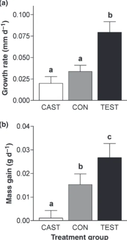
Following treatment, TEST grew significantly more quickly in SVL than either CON or CAST (F2,39= 10.90, P= 0.0002; Fig. 4a). Growth rate was unrelated to initial SVL (F1,38= 2.63,P= 0.11). Treatment effects on growth
in body mass were equally pronounced, with CON intermediate between the rapid mass gain of TEST and the zero mass gain of CAST (F2,39= 7.37, P< 0.002;
Fig. 4b). Rate of mass gain was negatively related to initial body mass (F1,38= 6.98, P= 0.012), but the
treatment effect on mass gain remained strong when including initial mass as a covariate (F2,38= 8.19, P< 0.002). By 36 days post-treatment, TEST males had grown an average of 2.9 mm and 1.0 g, compared to only 0.7 mm and 0.04 g in CAST males. These treatment groups both received castration surgeries and differed only in the implants they received.
Comparative evidence in squamates
Experimental data from castration and testosterone manipulation studies in squamate reptiles are summa-rized in Table 2. Aside from the present results for
A. sagrei, experimental data are available for only one additional species with pronounced male-biased SSD. In a wild population of Yarrow’s spiny lizards (Sceloporus jarrovii, SSD =)0.11), castration inhibited male growth and treatment of castrated males with exogenous testos-terone restored growth to the level of intact controls (Cox & John-Alder, 2005). However, this clear stimulatory effect of testosterone was not observed in two analogous studies conducted in captivity (Cox et al., 2006). Given that natural sex differences in growth were also abolished in captivity (Cox et al., 2006, 2008), we conclude that testosterone stimulates growth in this species, but that this natural effect can be overridden by a surplus of available energy (i.e.ad libitumfeeding in captivity).
Similar studies have been conducted in three species with pronounced female-biased SDD. In the garter snake (Thamnophis sirtalis, SSD = 0.18), castration dramatically increased mass gain in both neonatal and adult males (Crews et al., 1985). Although exogenous testosterone reversed this effect in adult males, it had no effect on neonatal males or females. However, a subsequent study reported that exogenous testosterone inhibited skeletal growth in neonatal females, although effects could not be confirmed in males because of high mortality (Lerner & Mason, 2001). We tentatively conclude that testosterone inhibits growth in this species. Results are more straight-forward for two lizards with female-biased SSD. In wild eastern fence lizards (Sceloporus undulatus, SSD = 0.13), castration of juvenile males had a weak stimulatory effect on growth and replacement of exogenous testosterone strongly inhibited growth (Coxet al., 2005a). Testoster-one also inhibited mass gain in wild adult males of this species (Klukowski et al., 1998). In the striped plateau lizard (Sceloporus virgatus, SSD = 0.11), treatment with exogenous testosterone the onset of sexual maturation inhibited growth of wild males (Cox & John-Alder, 2005). Castration did not influence growth of small juvenile males, but it tended to increase the growth of larger, sexually mature males that presumably had higher endogenous testosterone levels (Cox & John-Alder, 2005). Exogenous testosterone also inhibited growth of juvenile males and females maintained in (a)
(b)
Fig. 4 Mean (+ 1 SE) rate of growth in SVL (a) and body mass (b) forAnolis sagreimales as a function of treatment group (CAST, CON and TEST). Lowercase letters denote statistical separation based
captivity (Abell, 1998). There was no indication that testosterone stimulated growth in any study of these three species with female-biased SSD.
Comparable data are also available for two squamates in which SSD is weak or absent. In a lacertid lizard (Psammodromus algirus, SSD = 0.01), exogenous test-osterone had no effect on growth in SVL or mass of wild adult males (Salvador & Veiga, 2000). In a phrynosoma-tid lizard (Urosaurus ornatus, SSD =)0.05), exogenous testosterone inhibited growth in SVL, but not body mass, of captive male and female neonates. However, castra-tion alone also inhibited growth, so these results are somewhat equivocal (Hewset al., 1994; Hews & Moore, 1995). Moreover, exogenous testosterone resulted in high mortality, leading these authors to suggest that it was elevated to pharmacological levels. We therefore regard the effect of testosterone on growth as equivocal inU. ornatusand conclude that testosterone has no effect on growth inP. algirus.
Placing the comparative data from these seven species in a phylogenetic context reveals a strong association between the evolution of SSD and the effect of test-osterone on growth (Fig. 5). Testtest-osterone stimulates growth in two lizards from separate lineages with male-biased SSD, whereas testosterone inhibits growth in three species from two phylogenetically independent lineages with female-biased SSD. Effects of testos-terone on growth are absent or equivocal for two lizards from separate lineages in which SSD is minor or absent.
Discussion
Growth and SSD inAnolis sagrei
The brown anole (A. sagrei) exhibits extreme male-biased SSD, as illustrated by the 30% sex difference adult SVL and the 140% difference in adult body mass that we consistently documented across 5 years in a wild Baha-mian population (Table 1). Collectively, our results from studies in the wild and in a laboratory common garden environment clearly demonstrate that this extreme SSD develops because of pronounced sex differences in growth rate that are present immediately after hatching (Fig. 3) and persist through adulthood (Fig. 2). Although it is not intuitively surprising that extreme SSD is associated with underlying sex differences in growth, our results actually differ substantially from those observed in several other reptiles, where sex differences that give rise to SSD in the wild are greatly reduced or absent in captivity (Haenel & John-Alder, 2002; Taylor & DeNardo, 2005; Coxet al., 2006; John-Alderet al., 2007). We detected modest interannual variation in growth and SSD in the wild (Table 1, Fig 1), potentially because of corresponding variation in environmental factors (e.g. temperature, rainfall and food availability) that could influence relevant demographic parameters such as growth rate, survival and hatching date. Despite this variation, the overall pattern of extreme male-biased SSD and underlying sex differences in growth remained constant across years. Thus, our growth studies confirm Table 2 Support for the bipotential
regula-tion hypothesis in squamate reptiles. SSD and species Effect of T Treatment Growth Age Sex Site Study
Female-biased SSD
Sceloporus undulatus ) C + T L J, A M Field 1
) T L, M A M Field 2
Sceloporus virgatus ) C + T L, M J, A M Field 3
) T L, M J M, F Lab 4
Thamnophis sirtalis ) C + T L, M N, A M, F Lab 5
) T L N F Lab 6
Male-biased SSD
Anolis sagrei + C + T L, M A M Lab 7
Sceloporus jarrovii + C + T L J, A M Field 3
NS C + T L J, A M Lab 8
Monomorphic
Psammodromus algirus NS T M A M Field 9
Urosaurus ornatus +⁄) C + T L, M N, A M, F Lab 10, 11
SSD, sexual size dimorphism; (+), testosterone-stimulated growth; ()), testosterone inhibited growth; NS, no significant (a= 0.05) effect on growth; (+⁄)) results were equivocal. In some instances, effects of testosterone are inferred indirectly from effects of castration. See text for details. Testosterone manipulated via (T) exogenous testosterone; (C + T) surgical castration and exogenous testosterone. Growth was measured as change in (L) snout-vent length; (M) body mass. Age at the time of manipulation (A) sexually mature adults; (J) sexually immature juveniles; N, neonates. Study conducted in (Field) natural environment or (Lab) captivity. (1) Coxet al., 2005a; (2) Klukowskiet al., 1998; (3) Cox & John-Alder, 2005; (4) Abell, 1998; (5) Crewset al., 1985; (6) Lerner & Mason, 2001; (7) this study; (8) Cox
that SSD develops inA. sagreibecause of sex differences in juvenile and adult growth, as assumed by the bipotential regulation hypothesis.
In the majority of squamate reptiles studied to date, testosterone has a clear inhibitory effect on growth (Crews et al., 1985; Abell, 1998; Klukowskiet al., 1998; Lerner & Mason, 2001; Cox & John-Alder, 2005; Cox
et al., 2005a). However, on the basis of the bipotential regulation hypothesis, we predicted that testosterone would stimulate growth in A. sagrei. Our results clearly show that castration inhibits and exogenous testosterone stimulates growth in both length and mass of adult
A. sagreimales (Fig. 4). Given that adultA. sagreimales and females differ in growth rates (Fig. 1) and in circulating testosterone levels (Tokarzet al., 1997), our experimental testosterone manipulation in adult males is biologically relevant to the issue of sex-specific growth regulation. Moreover, our manipulations are physiologically relevant in the sense that castration surgeries reduced plasma testosterone to basal levels below those of intact males, while exogenous hormone implants restored plasma testosterone to levels identical to those of intact males.
Although our manipulations were conducted at an ontogenetic stage when the sexes are known to differ in both growth rate (Fig. 2) and circulating testosterone levels (Tokarz et al., 1997), it is important to note that SSD begins to develop immediately following hatching (Fig. 3). Thus, our experimental results do not directly address the initial development of SSD inA. sagrei.Given that males and females diverge in growth at such an early age, it would be informative to document the post-natal ontogeny of circulating testosterone levels in each sex. It would also be informative to manipulate testosterone in
juvenile males and females or in eggs prior to hatching, as studies of other lizards suggest that experimental elevation of testosterone in the pre-natal environment can influence post-natal growth (Uller & Olsson, 2003; Uller et al., 2007). Males and females of A. sagrei are sexually dimorphic in colour pattern at hatching, but other dimorphisms (e.g. males develop dorsal crests and orange dewlaps) become pronounced only upon matu-ration. By analogy, sex differences in growth that give rise to SSD may also depend on a combination of early organizational and late activational effects of testosterone (Hewset al., 1994; Hews & Moore, 1995; Hews & Quinn, 2003).
Testosterone clearly stimulates growth inA. sagrei, but the precise physiological and⁄or behavioural mechanisms underlying this effect are unclear. One important caveat to our study is that males were housed together with one other size-matched male from each treatment. Thus, we cannot distinguish between direct effects of testosterone on behaviour and physiology (e.g. increased feeding and⁄or energy allocation to skeletal and muscular growth) vs. indirect effects mediated by social inter-actions (e.g. behavioral dominance of castrated males by testosterone males, resulting in differences in feeding, basking and⁄or stress). However, much of the existing evidence for growth regulation by testosterone comes from studies of free-living animals in which similar behavioural interactions are likely (Klukowski et al., 1998; Cox & John-Alder, 2005; Cox et al., 2005a). Interestingly, the inhibitory effects of castration on growth of free-livingS. jarroviilizards were absent when animals were held in social isolation in captivity (Cox
et al., 2006).
Fig. 5 Evolutionary changes in SSD are accompanied by changes in the effect of testosterone on male growth. SSD is catego-rized as male- or female-biased (> 5% difference in mean adult SVL) or mono-morphic (< 0.05 difference). Arrows indicate the effect of testosterone (T) on growth in each species ( = testosterone stimulates growth; = testosterone inhibits growth;
= results are equivocal; none = no effect). See text for further discussion and Table 2 for a summary of each study.
Bipotential regulation hypothesis
When our results fromA. sagreiare considered alongside those of previous experiments on squamate reptiles, an intriguing pattern emerges. As predicted by the bipoten-tial regulation hypothesis (John-Alder & Cox, 2007; John-Alder et al., 2007), evolutionary changes in the direction of SSD correspond to shifts in the effect of testosterone on male growth (Fig. 5). Testosterone inhib-its growth in three separate species representing two phylogenetically independent occurrences of female-biased SSD, whereas testosterone stimulates growth in two species from separate lineages exhibiting male-biased SSD. In each of these five species, males and females differ by at least 10% in mean adult SVL and sex differences in growth are known to underlie the devel-opment of SSD. In two species with slight or absent SSD, testosterone either has no effect on growth, or results from castration and testosterone addition treatments give equivocal results. Because males are known to exceed females in circulating testosterone levels in each of these seven species, differences in growth regulation across species likely reflect differences in the effect of testoster-one on growth, rather than differences in circulating testosterone.
Two studies of squamates have also reported increased post-natal mass gain following elevation of prenatal testosterone levels, one in a species with moderate female-biased SSD (Lacerta vivipara, Lacertidae, Uller & Olsson, 2003), the other in a species with moderate male-biased SSD (Ctenophorus fordi, Agamidae, Uller et al., 2007). However, in the former case, this stimulatory effect of testosterone was only observed in the absence of tick parasitism (Uller & Olsson, 2003). The relevance of prenatal testosterone to natural post-natal or adult patterns of sex-specific growth and SSD is unknown for either species, so implications with respect to the bipo-tential regulation hypothesis are limited. However, these studies do provide some additional evidence that testos-terone can stimulate growth in squamates.
The novelty of our findings stem largely from compar-ison to other reptiles, but the resultant inference that evolutionary shifts in SSD can be achieved by underlying changes in the effect of testosterone on growth is potentially general across vertebrates. Although testos-terone is commonly regarded as an anabolic steroid that promotes muscular and skeletal growth, most of the evidence supporting this generalization comes from studies of mammals, birds and fishes with male-biased SSD (reviewed by Cox & John-Alder, 2005; John-Alder & Cox, 2007; John-Alderet al., 2007). Interestingly, several studies of birds and mammals with atypical female-biased SSD suggest that testosterone actually inhibits growth in these species (Swanson, 1967; Sockman et al., 2008). Thus, the bipotential nature of testosterone may be general across vertebrates, providing an elegant regula-tory mechanism for sex-specific phenotypic development
from a genome that is largely shared between the sexes. Given that SSD is also common among invertebrates (Fairbairn et al., 2007), which lack testosterone and many other components of the vertebrate endocrine system, the generality of testosterone as a proximate mechanism for SSD has limits. However, other endocrine messengers (e.g. insulin, juvenile hormone and ecdys-teroids) are known to influence growth in arthropods and may also underlie the development of within- and between-sex dimorphisms (Nijhout, 2003; Emlen et al., 2005, 2006). Thus, an analogous version of the bipoten-tial regulation hypothesis could, at least in principle, be extended to invertebrates. However, the evolution of sexual dimorphism in invertebrates may generally involve changes in threshold mechanisms regulating developmental responsiveness to hormones, rather than bipotentiality of the hormones themselves (Emlenet al., 2005).
Although we have focused our discussion on testos-terone, the bipotential regulation hypothesis can also extend to other androgens that may influence male growth (Hewset al., 1994; Hews & Moore, 1995). Even in species where androgens are known to influence male growth, sexual dimorphism may reflect additional effects of estrogens and progestins on female growth (Holloway & Leatherland, 1997; Lerner & Mason, 2001). Moreover, our present experiment does not address the effects of testosterone on growth of females. Whereas some male traits can be induced in females treated with exogenous testosterone (Coxet al., 2005b; Zyslinget al., 2006), other studies reveal that effects of sex steroids can differ between males and females (Holloway & Leatherland, 1997; Lerner & Mason, 2001). However, because males and females of A. sagrei and other squamates differ markedly in circulating testosterone levels, experimental manipulation in males is presumably more relevant to the natural role of testosterone in sex-specific develop-ment. Moreover, given that evolutionary patterns in SSD are often achieved primarily by interspecific changes in male size (Abouheif & Fairbairn, 1997; Fairbairn, 1997; Fairbairn et al., 2007), it may often be appropriate to focus on underlying changes in the regulation of male growth.
Evolutionary implications
Our results illustrate an intriguing endocrine mecha-nism that may help to explain the phylogenetic lability of SSD across reptiles in general (Cox et al., 2007; Fig. 1) and within Anolis lizards in particular (Butler
et al., 2000, 2007). West Indian Anolis lizards comprise one of the most spectacular examples of adaptive radiation on the planet, and the repeated evolution of convergent ecological and morphological specialists (termed ‘ecomorphs’) is mirrored by convergent pat-terns in SSD (Butler et al., 2000, 2007; Butler, 2007). For example, although A. sagrei and other ‘trunk
ground’ ecomorphs are phylogenetically independent species, they have convergently evolved the same pattern of extreme male-biased SSD. It is tempting to speculate that this phylogenetic lability in SSD is facilitated by the bipotential role of testosterone in growth regulation, such that evolutionary changes in SSD could occur simply by altering the direction and⁄or extent to which growth responds to testosterone. Alterations to the interaction between endocrine mod-ifiers and their genetic targets could present a more expeditious evolutionary pathway than the perpetual erosion and reformation of intersexual genetic correla-tions underlying complex polygenic traits such as body size (Lande, 1980; Badyaev, 2002; Fairbairn & Roff, 2006; Rhen, 2007). Future work on Anolis should attempt to identify the sexually antagonistic selection pressures that maintain SSD in wild populations (Cals-beek & Bonneaud, 2008; Cox & Cals(Cals-beek, 2009) and employ a comparative framework to test the prediction that effects of testosterone on growth will be absent in sexually monomorphic species and reversed in main-land species with female-biased SSD.
In a more general context, the bipotential nature of testosterone as a growth regulator illustrates how macro-evolutionary patterns can be linked to the evolution of underlying developmental mechanisms. Evolutionary changes to the interaction between hormones and their target tissues may explain phylogenetic diversity in other sexually dimorphic traits, such as the exaggerated horns of male beetles (Emlenet al., 2006) and aggression and paternal behaviour in male birds (Lynn et al., 2005; Lynn, 2008). The role of testosterone in regulation of squamate growth stands in contrast to the classic view that its phenotypic effects are evolutionarily conserved, which has been termed the ‘evolutionary constraint hypothesis’ (Hau, 2007). Rather, our results support the emerging perspective that selection can alter the linkage between testosterone and male traits, such that individ-ual traits can independently evolve differential respon-siveness to testosterone. This ‘evolutionary potential hypothesis’ is consistent with recent evidence on the hormonal regulation of life history trade-offs (Hau, 2007). Although our results provide a promising proxi-mate context for the evolution of SSD, further work is required to determine whether the bipotential regulation hypothesis provides a general explanation for SSD. We emphasize that such studies should be guided by an explicit consideration of evolutionary patterns in SSD within and among lineages.
Acknowledgments
The authors thank Diane Cheney, Laura Coolidge, Stephen Durham, Samantha Haw, Hari Iyer, Myrtle Karam, Zaneta Thayer and Sarah Wengert for assistance with animal care and data collection. The authors thank Marisol Gutierrez for conducting testosterone assays and
Henry John-Alder for granting use of his laboratory for these assays. Henry John-Alder was also instrumental in the development of the bipotential regulation hypoth-esis. The breeding stock for this study was exported with permission from the Bahamas Department of Agriculture and imported under a permit from the United States Fish and Wildlife Service. All procedures were reviewed and approved by the Dartmouth College Institutional Animal Care and Use Committee (protocol 07-02-03). This project was supported by a grant from the Howard Hughes Medical Institute to D. Stenquist and by funding from Dartmouth College and the National Science Foundation (DEB 0816862) to R. Calsbeek.
References
Abell, A.J. 1998. The effect of exogenous testosterone on growth and secondary sexual character development in juveniles of
Sceloporus virgatus.Herpetologica54: 533–543.
Abouheif, E. & Fairbairn, D.J. 1997. A comparative analysis of allometry for sexual size dimorphism: assessing Rensch’s Rule.
Am. Nat.149: 540–562.
Badyaev, A.V. 2002. Growing apart: an ontogenetic perspective on the evolution of sexual size dimorphism.Trends Ecol. Evol. 17: 369–378.
Badyaev, A.V., Hill, G.E., Stoehr, A.M., Nolan, P.M. & McGraw, K.J. 2000. The evolution of sexual size dimorphism in the house finch. II. Population divergence in relation to local selection.Evolution54: 2134–2144.
Bonduriansky, R. 2007. The genetic architecture of sexual dimorphism: the potential roles of genomic imprinting and condition dependence. In:Sex, Size and Gender Roles: Evolution-ary Studies of Sexual Size Dimorphism (D.J. Fairbairn, W.U. Blanckenhorn & T. Szekely, eds), pp. 176–184. Oxford University Press, Oxford.
Brandley, M.C., Huelsenbeck, J.P. & Wiens, J.J. 2008. Rates and patterns in the evolution of snake-like body form in squamate reptiles: evidence for repeated re-evolution of lost digits and long-term persistence of intermediate body forms.Evolution 62: 2042–2064.
Butler, M.A. 2007. Vive le difference! Sexual dimorphism and adaptive patterns in lizards of the genusAnolis.Integr. Comp. Biol.47: 272–284.
Butler, M.A., Schoener, T.W. & Losos, J.B. 2000. The relation-ship between sexual size dimorphism and habitat use in Greater AntilleanAnolislizards.Evolution54: 259–272. Butler, M.A., Sawyer, S.A. & Losos, J.B. 2007. Sexual
dimor-phism and adaptive radiation in Anolislizards. Nature447: 202–205.
Calsbeek, R. & Bonneaud, C. 2008. Postcopulatory fertilization bias as a form of cryptic sexual selection.Evolution62: 1137– 1148.
Calsbeek, R. & Irschick, D.J. 2007. The quick and the dead: correlational selection on morphology, performance and habitat use in island lizards.Evolution61: 2493–2503. Calsbeek, R. & Marnocha, E. 2006. Context dependent territory
defense: the importance of habitat structure inAnolis sagrei.
Ethology112: 537–543.
Calsbeek, R. & Smith, T.B. 2008. Experimentally replicated disruptive selection on performance traits in a Caribbean lizard.Evolution62: 478–484.
Calsbeek, R., Bonneaud, C., Prabhu, S., Manoukis, N. & Smith, T.B. 2007. Multiple paternity and sperm storage lead to increased genetic diversity in the Cuban anole,Anolis sagrei.
Evol. Ecol. Res.9: 495–503.
Calsbeek, R., Bonneaud, C. & Smith, T.B. 2008. Differential fitness effects of immunocompetence and neighborhood density in alternative female lizard morphs.J. Anim. Ecol.77: 103–109.
Cox, R.M. & Calsbeek, R. 2009. Sexually antagonistic selection, sexual dimorphism, and the resolution of intralocus sexual conflict.Am. Nat.173: 176–187.
Cox, R.M. & John-Alder, H.B. 2005. Testosterone has opposite effects on male growth in lizards (Sceloporus spp.) with opposite patterns of sexual size dimorphism. J. Exp. Biol. 208: 4679–4687.
Cox, R.M., Skelly, S.L. & John-Alder, H.B. 2003. A comparative test of adaptive hypotheses for sexual size dimorphism in lizards.Evolution57: 1653–1669.
Cox, R.M., Skelly, S.L. & John-Alder, H.B. 2005a. Testosterone inhibits growth in juvenile male eastern fence lizards ( Scelop-orus undulatus): Implications for energy allocation and sexual size dimorphism.Physiol. Biochem. Zool.78: 531–545. Cox, R.M., Skelly, S.L., Leo, A. & John-Alder, H.B. 2005b.
Testosterone regulates sexually dimorphic coloration in the eastern fence lizard,Sceloporus undulatus.Copeia2005: 597–608. Cox, R.M., Zilberman, V. & John-Alder, H.B. 2006. Environ-mental sensitivity of sexual size dimorphism: laboratory common garden removes effects of sex and castration on lizard growth.Funct. Ecol.20: 880–888.
Cox, R.M., Butler, M.A. & John-Alder, H.B. 2007. The evolution of sexual size dimorphism in reptiles. In:Sex, Size and Gender Roles: Evolutionary Studies of Sexual Size Dimorphism (D.J. Fairbairn, W.U. Blanckenhorn & T. Szekely, eds), pp. 38–49. Oxford University Press, London.
Cox, R.M., Barrett, M.M. & John-Alder, H.B. 2008. Effects of food restriction on growth, energy allocation, and sexual size dimorphism in Yarrow’s Spiny Lizard,Sceloporus jarrovii.Can. J. Zool.86: 268–276.
Crews, D., Diamond, M.A., Whittier, J. & Mason, R. 1985. Small male body size in snakes depends on testes.Am. J. Physiol.18: R62–R66.
Darwin, C. 1871.The Descent of Man, and Selection in Relation to Sex. J. Murray, London.
Delph, L.F. 2007. The genetic integration of sexually dimorphic traits in the dioecious plant,Silene latifolia. In: Sex, Size and Gender Roles: Evolutionary Studies of Sexual Size Dimorphism(D.J. Fairbairn, W.U. Blanckenhorn & T. Szekely, eds), pp. 115– 123. Oxford University Press, London.
Emlen, D.J., Hunt, J. & Simmons, L.W. 2005. Evolution of sexual dimoorphism and male dimorphism in the expression of beetle horns: phylogenetic evidence for modularity, evolu-tionary lability, and constraint.Am. Nat.166: S42–S68. Emlen, D.J., Szafran, Q., Corley, L.S. & Dworkin, I. 2006. Insulin
signaling and limb-patterning: candidate pathways for the origin and evolutionary diversification of beetle ‘horns’.
Heredity97: 179–191.
Fairbairn, D.J. 1997. Allometry for sexual size dimorphism: pattern and process in the coevolution of body size in males and females.Annu. Rev. Ecol. Syst.28: 659–687.
Fairbairn, D.J. & Roff, D.A. 2006. The quantitative genetics of sexual dimorphism: assessing the importance of sex-linkage.
Heredity97: 319–328.
Fairbairn, D.J., Blanckenhorn, W.U. & Szekely, T. (eds) 2007.
Sex, Size and Gender Roles: Evolutionary Studies of Sexual Size Dimorphism. Oxford University Press, London.
Fedorka, K.M., Winterhalter, W.E. & Mousseau, T.A. 2007. The evolutionary genetics of sexual size dimorphism in the cricket
Allonemobius socius.Heredity99: 218–223.
Fitch, H.S. 1976. Sexual size differences in the mainland anoles.
Univ. Kansas Mus. Nat. Hist. Occas. Pap.50: 1–21.
Haenel, G.J. & John-Alder, H.B. 2002. Experimental and demographic analyses of growth rate and sexual size dimor-phism in a lizard,Sceloporus undulatus.Oikos96: 70–81. Hau, M. 2007. Regulation of male traits by testosterone:
implications for the evolution of vertebrate life histories.
BioEssays29: 133–144.
Hews, D.K. & Moore, M.C. 1995. Influence of androgens on differentiation of secondary sex characters in tree lizards,
Urosaurus ornatus.Gen. Comp. Endocrinol.97: 86–102. Hews, D.K. & Quinn, V.S. 2003. Endocrinology of species
differences in sexually dimorphic signals and aggression: using the organization and activation model in a phylogenetic framework. In:Lizard Social Behavior(S.F. Fox, T.A. Baird & J.C. McCoy, eds), pp. 253–277. Johns Hopkins University Press, Baltimore, MD.
Hews, D.K., Knapp, R. & Moore, M.C. 1994. Early exposure to androgens affects adult expression of alternative male types in tree lizards.Horm. Behav.28: 96–115.
Holloway, A.C. & Leatherland, J.F. 1997. Effect of gonadal steroid hormones on plasma growth hormone concentrations in sexually immature rainbow trout,Oncorhynchus mykiss.Gen. Comp. Endocrinol.105: 246–254.
John-Alder, H.B. & Cox, R.M. 2007. Development of sexual size dimorphism in lizards: testosterone as a bipotential growth regulator. In:Sex, Size and Gender Roles: Evolutionary Studies of Sexual Size Dimorphism(D.J. Fairbairn, W.U. Blanckenhorn & T. Szekely, eds), pp. 195–204. Oxford University Press, London. John-Alder, H.B., Cox, R.M. & Taylor, E.N. 2007. Proximate developmental mediators of sexual size dimorphism: case studies from squamate reptiles.Integr. Comp. Biol.47: 258–271. Klukowski, M., Jenkinson, N.M. & Nelson, C.E. 1998. Effects of testosterone on locomotor performance and growth in field-active northern fence lizards,Sceloporus undulatus hyacinthinus.
Physiol. Zool.71: 506–514.
Lande, R. 1980. Sexual dimorphism, sexual selection, and adaptation in polygenic characters.Evolution34: 292–307. Lande, R. 1987. Genetic correlations between the sexes in the
evolution of sexual dimorphism and mating preferences. In:
Sexual Selection: Testing the Alternatives(J.W. Bradbury & M.B. Andersson, eds), pp. 83–94. John Wiley and Sons, Chichester. Lee, J.C. 1980. Comparative thermal ecology of two lizards.
Oecologia44: 171–176.
Lee, J.C. 1987. Anolis sagrei in Florida: phenetics of a colonizing species II. Morphometric characters.Copeia1987: 458–469. Lee, J.C., Clayton, D., Eisenstein, S. & Perez, I. 1989. The
reproductive cycle ofAnolis sagreiin southern Florida.Copeia 1989: 930–937.
Lerner, D.T. & Mason, R.T. 2001. The influence of sex steroids on the sexual size dimorphism in the red-spotted garter snake,
Thamnophis sirtalis concinnus.Gen. Comp. Endocrinol.124: 218– 225.
Lovich, J.E. & Gibbons, J.W. 1992. A review of techniques for quantifying sexual size dimorphism. Growth Dev. Aging 56: 269–281.
Lynn, S.E. 2008. Behavioral insensitivity to testosterone: why and how does testosterone alter paternal and aggressive behavior in some avian species but not others? Gen. Comp. Endocrinol.157: 233–240.
Lynn, S.E., Walker, B.G. & Wingfield, J.C. 2005. A phylogenet-ically controlled test of hypotheses for behavioral insensitivity to testosterone in birds.Horm. Behav.47: 170–177.
Nauwelaerts, S., Coeck, J. & Aerts, P. 2000. Visible implant elastomers as a method for marking adult anurans.Herpetol. Rev.31: 154–155.
Nijhout, H.F. 2003. The control of body size in insects.Dev. Biol. 261: 1–9.
Pe´rez-Barberı´a, F.J., Gordon, I.J. & Pagel, M.D. 2002. The origins of sexual dimorphism in body size in ungulates.Evolution56: 1276–1285.
Preziosi, R.F. & Fairbairn, D.J. 2000. Lifetime selection in adult body size and components of body size in a waterstrider: opposing selection and maintenance of sexual size dimor-phism.Evolution54: 558–566.
Rhen, T. 2007. Sex differences: genetic, physiological, and ecological mechanisms. In: Sex, Size and Gender Roles: Evolu-tionary Studies of Sexual Size Dimorphism(D.J. Fairbairn, W.U. Blanckenhorn & T. Szekely, eds), pp. 167–175. Oxford University Press, Oxford.
Salvador, A. & Veiga, J.P. 2000. Does testosterone or coloration affect growth rates of adult males of the lizardPsammodromus algirus?Can. J. Zool.78: 1463–1467.
Schulte-Hostedde, A.I., Millar, J.S. & Gibbs, H.L. 2002. Female-biased sexual size dimorphism in the yellow-pine chipmunk (Tamias amoenus): sex-specific patterns of annual reproductive success and survival.Evolution56: 2519–2529.
Shine, R. 1994. Sexual size dimorphism in snakes revisited.
Copeia1994: 326–346.
Smith, L.C. & John-Alder, H.B. 1999. Seasonal specificity of hormonal, behavioral, and coloration responses to within-and between-sex encounters in male lizards (Sceloporus undulatus).Horm. Behav.36: 39–52.
Sockman, K.W., Weiss, J., Webster, M.S., Talbott, V. & Schwabl, H. 2008. Sex-specific effects of yolk-androgens in growth of nestling American kestrels.Behav. Ecol. Sociobiol.62: 617–625. Stamps, J.A. 1999. Relationships between female density and sexual size dimorphism in samples ofAnolis sagrei.Copeia1999: 760–765.
Swanson, H.H. 1967. Effects of pre- and post-pubertal gonadec-tomy on sex differences in growth, adrenal and pituitary weights of hamsters.J. Endocrinol.39: 555–564.
Sze´kely, T., Reynolds, J.D. & Figuerola, J. 2000. Sexual size dimorphism in shorebirds, gulls, and alcids: the influence of sexual and natural selection.Evolution54: 1404–1413. Taylor, E.N. & DeNardo, D.F. 2005. Sexual size dimorphism and
growth plasticity: an experiment on the western diamond-backed rattlesnake (Crotalus atrox). J. Exp. Zool.303A: 598– 607.
Tokarz, R.R., McMann, S., Seitz, L. & John-Alder, H.B. 1997. Plasma corticosterone and testosterone levels during the annual reproductive cycle of male brown anoles (Anolis sagrei).Physiol. Zool.71: 139–146.
Uller, T. & Olsson, M. 2003. Prenatal exposure to testosterone increases ectoparasite susceptibility in the common lizard (Lacerta vivipara).Proc. R. Soc. Lond. B270: 1867–1870. Uller, T., Astheimer, L. & Olsson, M. 2007. Consequences of
maternal yolk testosterone for offspring development and survival: experimental test in a lizard.Funct. Ecol.21: 544–551. Vidal, N. & Hedges, S.B. 2005. The phylogeny of squamate reptiles (lizards, snakes and amphisbaenians) inferred from nine nuclear protein-coding genes.C. R. Biol.328: 1000–1008. Zysling, D.A., Greives, T.J., Breuner, C.W., Casto, J.M., Demas, G.E. & Ketterson, E.D. 2006. Behavioral and physiological responses to experimentally elevated testosterone in female dark-eyed juncos (Junco hyemalis carolinensis).Horm. Behav.50: 200–207.
Received 5 February 2009; revised 20 April 2009; accepted 22 April 2009