BASAL-LIKE BREAST CANCER: MODELING ITS
INITIATION AND CHARACTERIZING
NOVEL EGFR VARIANTS
MAIRA MOURA PIRES
Submitted in partial fulfillment of the requirements for the degree of
Doctor of Philosophy under the Executive Committee of the Graduate School of Arts and Sciences
COLUMBIA UNIVERSITY 2013
© 2012 Maira Pires All Rights Reserved
ABSTRACT
Basal-like breast cancer: modeling its initiation and characterizing novel EGFR variants
Maira Moura Pires
Breast cancer (BC) is now a disease that will affect 1 out of every 8 women worldwide. Due to its highly heterogeneous nature, BC is usually further classified into different subtypes according to gene expression profiling analysis. Basal-like breast cancer (BBC) accounts for 15-20% of all cases of BC. BBC is very aggressive, highly metastatic, and often lethal, mostly due to our poor understanding of the key genetic events that lead to the onset and/or maintain this subtype of BC. As a result, we currently lack targeted therapies that are otherwise very effective in some of the better understood subtypes of BC. Therefore we set to work on deciphering the genetic
alterations that when, expressed altogether, would model the onset of sporadic BBC in human cell lines. We already know that patients with BBC often display mutations in the tumor suppressor p53, as well as over-expression of the epidermal growth factor receptor (EGFR) protein by immunohistochemistry, and that loss of PTEN protein is also seen occurring along with the aforementioned lesions.
However, there is no model for sporadic BBC that accurately reflects the genetic changes that BBC tumor samples display for studying this BC subtype progression and/or providing a base for which pharmacological studies can be done to find an effective therapy. Thus we used a
non-transformed immortalized mammary epithelial cell line, MCF10A, as our model and engineered cells with termed ‘triple modified’ cells that express the three aforementioned alterations (PTEN, EGFR, p53) that highly observed in BBC.
Furthermore, as part of the goal to better understand the genetic underpinnings that occur in BBC progression, we investigated whether variant forms of EGFR existed in cell lines of the basal-like subtype. Prior studies have demonstrated that immunostaining for EGFR in BBC patient tumor samples is elevated, though the molecular mechanisms are not well understood. EGFR is only amplified in BBC in less than 1% of cases thus gene amplification does not account for the high percentage of tumors that express and overexpress EGFR. In the pursuit to better
understand the possible contribution of deregulated EGFR in BBC, we discovered a variety of basal-like breast cancer lines containing genetic alterations in EGFR not yet reported in the literature and demonstrated that a portion of them stimulate growth, invasion and transformation in mammary cells.
In conclusion, the studies on modeling basal-like tumorigenesis and identifying and
characterizing novel variants of EGFR in basal-like breast cancer lines have shed new exciting insights into our current knowledge of BBC. Having a cell line model for the disease provides a new tool for which further genetic and epigenetic manipulations can be exercised to continue asking questions of which other oncogenes and tumor suppressors are important for initiation of BBC. Our data showing that EGFR variants can be found in BBC cell lines and alter breast epithelial growth shows that this is an area that should be further investigated in human tumors
so that targeted therapies can be designed to improve patient survival rates and their quality of life.
i
TABLE OF CONTENTS
TABLE OF CONTENTS ... I LIST OF FIGURES AND TABLES ... III ACKNOWLEDGEMENTS ... V ABBREVIATIONS ... XI CHAPTER I ... 1 INTRODUCTION ... 1 BREASTCANCER ... 5 BREAST GLAND ... 5 BREAST CANCER ... 7 Epidemiology ... 7
Breast Cancer Genetics ... 8
Tumor Suppressors ... 10 p53 ... 10 BRCA1 ... 11 BRCA2 ... 12 PTEN ... 13 Oncogenes ... 14 HER 1/2 ... 14 Cyclin D1 ... 15 MYC ... 16
Molecular Subtypes of Breast Cancer ... 16
Normal-Like Subtype ... 16
ER+ Luminal A and Luminal B Subtypes ... 17
Basal-like Subtype ... 18
HER2+ Subtype ... 19
Breast Cancer Models ... 20
Cell Line Models... 20
Transformation Models in MCF10A cells ... 20
Transformation Models in Hmec-hTERT cells ... 21
PTEN ... 23
Discovery ... 23
structure of pten ... 25
PTEN/PI3K/mtor Pathway ... 27
PTEN Biology and roles ... 32
PTEN: Cell Motility and Polarity... 32
PTEN: Cellular Senescence ... 33
PTEN: Tumor Microenvironment ... 35
PTEN: Metabolism ... 36
PTEN: Nucleus ... 38
PTEN: Stem Cell Maintenance ... 38
Alterations in Cancer ... 40
PTEN: Breast Cancer ... 40
PTEN: All Other Cancers associated with PHTS ... 41
PTEN Mice Models ... 43
ii
Gene and Protein ... 47
Physiological Functions OF P53 ... 50
Deregulation ... 56
Mutant p53 in Breast Cancer... 56
Mechanisms of Action of Mutant p53 Molecules in Breast Cancer ... 57
Transcriptional Activity Mechanism ... 57
Protein Stability Mechanism ... 58
Other post-translational Modifications as Mechanism of Action ... 59
Mutant TP53 Models ... 60
Mouse Models ... 60
EGFR ... 63
INTRODUCTION TO EGFR/HER RECEPTORS ... 63
HER RECEPTORS ... 63
Gene and Protein ... 63
HER Family Members ... 66
Dimerization to EGFR ... 68
Ligands ... 69
Downstream Signaling Pathways of EGFR ... 72
EGFR: Kinase-dependent functions... 73
EGFR: Kinase-independent functions ... 73
EGFR: Nuclear functions... 74
Degradation and Recycling ... 74
EGFR degradation mechanism (1): Exposure and Binding to Clb ... 75
EGFR degradation mechanism (2): EGFR-complex stability ... 76
EGFR/HER FAMILY Mice Models ... 77
CHAPTER II ... 80
Transformation of human mammary epithelial cells through combinatorial alterations of epidermal growth factor receptor, p53 and PTEN ... 80
ABSTRACT ... 81
KEYWORDS... 82
ABBREVIATIONS ... 82
MATERIALSANDMETHODS ... 87
RESULTS ... 90
DISCUSSION ... 96
FIGURESANDLEGENDS ... 101
CHAPTER III: ... 107
Discovery and characterization of novel ... 107
epidermal growth factor receptor variants in basal-like breast cancer cell lines ... 107
INTRODUCTION ... 110
MATERIALSANDMETHODS ... 112
RESULTS ... 119
DISCUSSION ... 129
FIGURESANDLEGENDS ... 133
CHAPTER IV... 150
KEYRESEARCHACCOMPLISHMENTS ... 150
CONCLUDINGREMARKS ... 153
Integrating PTEN/EGFR/p53 in a model for BBC ... 153
The impact of altered EGFR variants in breast tumors ... 154
Therapeutic relevance of this work ... 156
Final Conclusions ... 156
iii
LIST OF FIGURES AND TABLES
Figure 1.1. The anatomy of the female breast. 6
Figure 1.2. Estimated new cases of breast cancer in female patients. 7
Figure 2.1. Timeline of some major findings related to PTEN research since its discovery. 25
Figure 2.2. Structure of the tumor suppressor PTEN. 26
Figure 2.3. An illustration of the canonical PTEN-PI3K pathway in the cell. 31
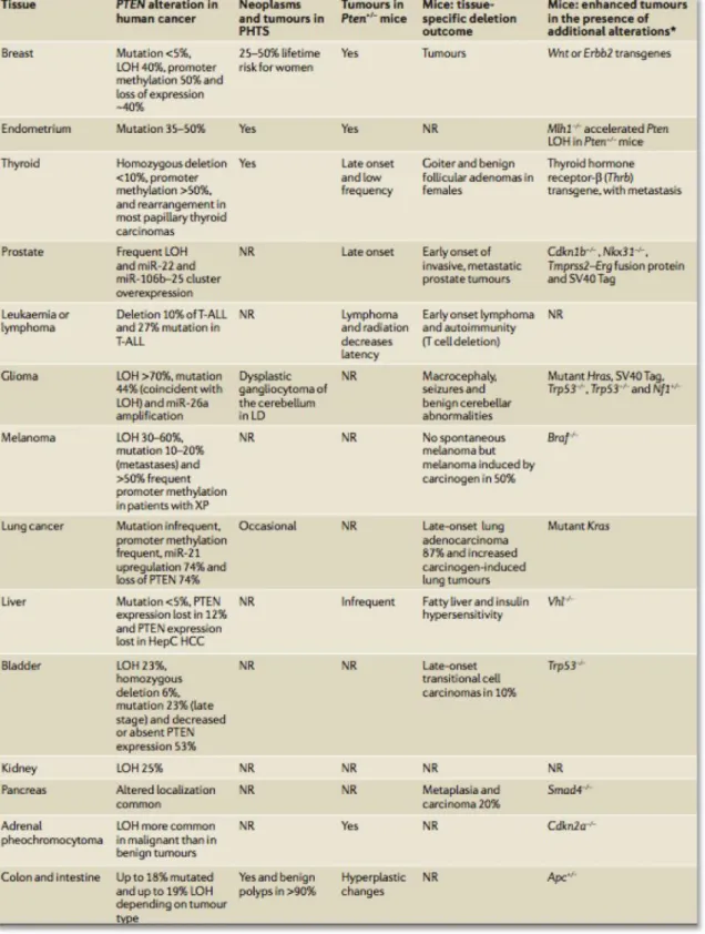
Figure 2.3. List of tissue-specific evidence for PTEN alterations in cancers. 44
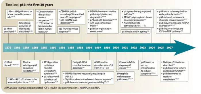
Figure 3.1. Main discoveries in the p53 field in the first thirty years since its discovery. 45
Figure 3.2. p53 structural and functional domains. 48
Figure 3.3. Mutational spectrum of TP53 in human cancers. 49
Figure 3.4. Classic model for p53 activation. 51
Figure 3.5. Downstream targets of p53 activation due to various cellular stresses. 53
Figure 3.6. The two different p53 apoptotic driven pathways and their target genes. 54
Figure 3.7. Most frequent p53 missense mutations in breast cancer. 61
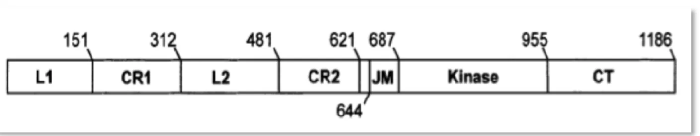
Figure 4.1. Representation of the domains of the EGFR polypeptide. 64
Figure 4.2. The EGFR/HER module is highly conserved through evolution. 65
Figure 4.3. This schematic illustration depicts the HER signaling network. 71
Figure 4.4. A model for signal transduction mediated by epidermal growth factor receptor. 72
Figure 4.5. Interaction with Cbl determines the fate of internalized EGFR. 75
Figure 4.6. The impact of stability on internalized EGFR through interaction with Clb. 76
Table 4.1. List of mice that have been engineered to either have knock-out or knock-in of the multiple
receptor members of the EGFR family as well as their ligands. 78
Table 1. Xenografts of engineered MCF10A cells in immunocompromised scid mice to test in vivo
tumorigenicity 100
Figure 1. Modeling basal-like breast cancer by altering EGFR, p53 and PTEN in MCF10A. 101
Figure 2. In vitro oncogenic properties of singly-modified MCF10A cells. 103
Figure 3. Double- and triple- modified cells grow in absence of stimuli and in soft agar. 104
Figure 4. A model for BBC transformation in MCF10A. 106
Figure 1. Scheme for analyzing the large cDNA of EGFR for alterations in a panel of breast cancer cell
iv
Figure 2. Alterations discovered in the ligand-biding domain of EGFR discovered in breast cancer cell
lines. 134
Figure 3. Schematic illustration of two novel variants of EGFR that are altered either in the tyrosine
kinase domain or the carboxy-terminal domain of this receptor. 135
Figure 4. Cartoon illustrations of the novel variants of EGFR where alteration is localized to the
extracellular domain. 137
Figure 5. Expression of the novel EGFR variants in normal mammary epithelial cells. 138
Figure 6. Immunofluorescence of cells expressing novel EGFR variants for detection of localization
differences between wildtype and variant receptors. 139
Figure 7. Signaling activation post EGF stimulation of mammary epithelial cells over-expressing novel
EGFR variants. 140
Figure 8. Signaling activation of mammary epithelial cells over-expressing novel EGFR variants with or
without loss of PTEN and expression of dominant negative p53 (DDp53). 142
Figure 9. Most of the variants of EGFR are able to grow better in the presence of semi-starvation media
over time as compared to wildtype EGFR. 144
Figure 10. No significant growth advantage of cells expressing variants of EGFR over wildtype
EGFR-expressing cells in media containing semi-complete levels of growth factors. 146
Figure 11. Expression of novel EGFR variants did not show proliferative advantage in the absence of
ligand EGF. 147
Figure 12. Insulin withdrawal does not confer proliferative advantage in most novel EGFR variants. 148
Figure 13. BL29 EGFR variant affects migration, invasion and transformation. 149
Figure 1. Alternative model for BBC tumorigenesis where wt EGFR is replaced with a hyperactive
v
ACKNOWLEDGEMENTS
I am very thankful to many people who have been integral parts of my life in so many ways.
First, and foremost, I want to thank my Ph.D. advisor, Ramon Parsons, for the support he has given me over the years. Ramon was there with me through all of the twists and turns of my projects, guiding me while simultaneously allowing me to learn and grow independently, for which I truly appreciate. As a graduate student, it has been wonderful to see Ramon’s love for science and I aspire to carry this level of passion with me in my future scientific work.
I am so thankful to my committee members Eric Greene and Richard Mann. Eric and Richard, from taking some of the best classes in graduate school from you in my first year, to meeting with you at least once a year thereafter to discuss my doctoral work, thanks for all of your
influence and input into my work. I want to thank Arthur Palmer for his guidance throughout my graduate school years as our Biochemistry chair and for being part of my thesis committee. I also thank Jose Silva for so cheerfully accepting to be the reader of my thesis committee and being supportive in collaborations and in many other ways.
Many thanks for the Institute for Cancer Genetics at Columbia and everyone there who has been supportive in countless ways through the years. I also want to deeply thank two people in the department of Biochemistry and Molecular Biophysics – Ed Johnson and Rachel Hernandez. They went out of their way to make sure the department felt like a very supportive family to me.
vi
I am deeply thankful to the Department of Defense for taking a chance on my proposed projects in breast cancer to fund my work for 3 full years! I am more inspired than ever to keep studying breast cancer, continue the fight that is worth fighting with every penny.
Thanks for the current and past members of the lab who have been great colleagues and friends. Thanks to Kyrie Pappas with whom I have held many conversations of our works while at the same time sharing a wonderful friendship right from the start. Lao Saal – thank you for letting me become part of your graduate work and picking up the baton. Ashley Calahan and Franklin Fenton, you’ve both been the most contagiously friendly people I’ve ever had the pleasure of working every day. Megan Keniry – thank you for the tremendous amount of technical help that you have given me and for your friendship in the lab. Sofia Gruvberger-Saal, you were my first bay mate and I will always cherish the ties we shared – and thanks for being my cheerleader in the start.
Thanks to all my friends who have been part of my life before and during graduate school, in all your loving friendships, caring thoughts and cheers for the progress of my work. Particularly, thanks to Melissa Walker and Mari-Liis Visnapuu – graduate school would have been something completely different if I had never met you. We shared some of the very best memories of graduate school. Thank you both for being there for me through so much and sharing this bond that I know will be with us forever.
I am so thankful to the experience that had me in love with studying how cancer cells work and showed me the utter importance of dedicating oneself to this field – working at Dana Farber
vii
Cancer Institute at Harvard Medical School in Boston. I want to specifically thank Sabina Signoretti and Massimo Loda for their support, guidance and extraordinary passion for studying cancer. I also want to thank the members of both laboratories that were very collegial and supportive in helping me learn the most I could. I want the wonderful partnership that I had working with Meghan Lindauer Vyleta for two of those three years.
I want to acknowledge the influences that previous high school teachers and college professors of Biology and Biochemistry have had on the path that I have taken. Specifically, I want to thank Mrs. Ridenour from Ithaca High School for teaching AP Biology in a way that was the most fruitful – having us really understand the material and giving me my first real taste of what an independent experiment was like, including requiring one of the most extensive lab reports that I have ever had to write! In college, I was highly influenced specifically by my experience with my research advisor Vicki Cameron. She was incredibly talented from her meticulously detailed lecture notes, to her avid love for research using the yeast system. Her encouragement of my independent research in her laboratory was crucial to my success later on at Dana Farber. Lastly, I want to acknowledge and thank Richard Wodzinski. He taught me Biochemistry in a way that was interestingly contextualized. His teaching method has since highly influenced how I teach Biology to others. Thanks also to all those who have gone unmentioned but have paved their influence in my professional career at one point or another.
I am thankful for the influence my grandparents have had in my life and for inspiring me to follow my dreams and to love life. I love you -- Vovo Lucyrene, Vovo Pires and Vovo Lourdes, as you are always in my thoughts and deep within my heart. My grandma Lourdes gave me her
viii
most precious graduation ring years ago for me to wear the day I finish my education and I have never forgotten how this gesture has made me feel the luckiest grandchild in the world. My grandfather Otavio Bittencourt Pires has always been so sweet, cheerful and enormously supportive of me and of my different endeavors – graduate school and also my passion for painting. My grandma Lucyrene is contagiously cheerful and she always has the biggest heart and the warmest smile when you see her. I feel so incredibly lucky to have all my grandparents with me as I finish this important step in my life.
Special thanks go to some of the most supportive members of my family:
Many thanks to Kristina Pires and Stephen Rojas, who have been a wonderful support system and my best friends since college! Both of you are such important parts of my life. Thanks to the sweetest two boys in the whole world to me, my nephews Lucas and Leo Pires. They have shown me how to love in completely new, most wonderful ways that I ever thought imaginable. They have brought the purest joy when I have needed most during graduate school. ‘Auntie’ loves you both more and more each day!
I am so thankful for my two siblings. I want to deeply thank my ‘little’ brother, Marcos Pires, who is such an inspiration to me. Since we were little, we shared so much together and he’s been so instrumental in the last years of graduate school in multiple incredible ways. He has shown me the confidence to grow professionally and is a great role model for achieving what you set your mind to! His passion for science and for research is contagious too. So many thanks to my loving sister, Roberta Pires-Rojas. Her loving words have strengthened me through the years and
ix
she is always there for me whenever I have needed her through graduate school and beyond. Roberta is a wonderful role-model to me of how to best balance personal with professional life. Beta – you’re an inspiration on how to have it all graciously!
Incredibly important – I am deeply thankful to my loving and dedicated parents, Ana Ligia and Jose Otavio Pires. I am grateful for all of their immense support, purest love and important words of encouragement when I most needed, again and again through my whole life. My parents have encouraged me to follow my dreams since I can remember, and whether I followed some passions professionally (science) or as a hobby (painting, sports), they have been my all time biggest cheerleaders. I know to turn to them for advice on anything, and everything. They encourage me to do the best I can, and importantly, they cherish the small victories along with the large ones. We live so far away from each other, but the physical distance only strengthens our bond, our love, and our commitment to stay a close-knit family. My parents are examples to me of how much dedication family and to improving oneself can be so fruitful and essential to true happiness. Their sacrifices, past and current ones, have largely influenced who I am today. I will never stop thanking their commitment to me. I love you so much, mom and dad!
I want to thank Tom Wetmore. He has shown me support in multiple ways when I have needed it most. He’s been there to laugh, to cry and to help me through this graduate school journey. I appreciate his full heartily support all these years but most importantly, in the last few crazy weeks prior to finishing my doctoral thesis defense! Te amo!!
x
To my most beloved parents, Ana Ligia and Otavio
xi
ABBREVIATIONS
a.a. : amino acid
aCGH : array comparative genomic hybridization BBC : basal-like breast cancer
bp : base pair CK : cytokeratin
CDK : cyclin-dependent kinase DBD : DNA-binding domain DBS : double-strand break
EGFR : epidermal growth factor receptor ER : estrogen receptor
ex20d : exon 20 duplication of EGFR ECM : extracellular matrix
GOF : gain-of-function kDa : kilodaltons
LOH : loss-of-heterozygosity
PI3K : phosphatidylinositol 3-kinase
PIP3 : phosphatidylinositol (3,4,5)-triphosphate
PMT : post-translational modification
PTEN : phosphatase and tensin homologue on chromosome 10 RTK : receptor tyrosine kinase
CHAPTER I
INTRODUCTION
Cancer cells arise when a normal cell suffers stepwise accumulations of different genetic alterations able to bypass the homeostatic mechanisms that govern normal cellular proliferation and migration. Once normal cells are transformed, they display different hallmark phenotypes that can be contributed to their oncogenic state – growth independent proliferation (their ability to grow without having growth factors around to signal for growth), loss of proliferation control (aberrant growth rate as compared to their normal cellular state), suppression of apoptosis, growth in an anchorage independent fashion (unlike adherent cells), immortalization of cells (loss of their normal signal to stop dividing after a certain number of passages and therefore growing forever), angiogenesis (the de-novo recruitment of blood vessels to the tumor site), invasiveness (ability to create local tissue destruction), and metastasis (ability to enter the circulatory system to spread) (Hanahan D and Weinberg RA, 2000). Additionally, genes whose alternations lead to cancer predisposition can alter other pathways such as cellular genomic maintenance, which once lost can cause genomic instability, another hallmark in cancer cells. Breast cancer is the second most common type cancer in the world and it affects 200,000 new cases in the United States alone each year (Ries et al, 2005).
Breast cancer (BC) cannot be classified as one disease. Gene expression profiling analysis of breast tumors demonstrates that BC, to be more accurately described, is further classified into five different subtypes due to differential expression of genes between them (Sorlie, Perou et al.
2001, Sorlie 2004). The subtypes for breast cancer are: (1-2) Luminal A and luminal B that are estrogen receptor positive; (3) HER2+ that have amplification of HER2 receptor; (4) basal-like that express basal epithelial keratins; and finally (5) normal-like subtype (Sorlie, Perou et al. 2001, Sorlie 2004, Cancer Genome Atlas 2012). Importantly, each subtype has its own histo- and clinical pathology and prognosis (Sorlie 2004). There is an increasing amount of literature in BC highlighting the extensive heterogeneity not only between all five subtypes, but also within each subtype (Cancer Genome Atlas 2012). The challenge to treat each subtype of BC then becomes more difficult as we do not currently have a consensus for the alterations that can always
accurately identify each subtype due to such high rate of mutation rate (Cancer Genome Atlas 2012). Due to such high genetic and epigenetic possibilities that lead to each subtype of breast cancer, there is recognition for a need for personalized targeted therapy, depending on the prolife of each tumor.
One of the worse subtypes of breast cancer to date is called the basal-like subtype of cancer (BBC). This type of BC is characterized as being one of the most lethal and aggressive subtypes, metastasizing fast and having no targeted therapy for personalized treatment (Sorlie, Perou et al. 2001, Sorlie, Tibshirani et al. 2003, Sorlie 2004). On the other hand, HER2+ amplified breast cancer has Herceptin as a very effective drug targeting the altered HER2 receptor that is the common denominator in all tumors in this subtype and thus this subtype has a very effective targeted therapy (Khasraw and Bell 2012). However, for BBC, even though there are a few known genetic alterations that occur at high frequencies, BRCA1 mutations (~30%), PTEN loss (~15-50%), PIK3CA (7%) and TP53 mutations (>80%), there is no established common
disrupted pathway(s) that we know are responsible for tumor initiation in this lethal subtype (Cancer Genome Atlas 2012).
The work done in this thesis has focused on delineating the genetic alterations that are not only physiologically relevant, meaning that they occur at a high frequency within the population of BBCs, but that they can be shown to initiate tumorigenesis. The knowledge of which
combination of tumor suppressors and oncogenes are able to model cancer initiation in cell lines will not only become a powerful tool to identify the pathways activated or deactivated in a BBC tumorigenesis model, but it can also allow for further studies of how BBC progresses, spreads and behaves to drugs therapies. Therefore, one of the aims is to study the effect of the
combination of alterations that occur at high frequency in BBC: loss of PTEN (phosphatase and tensin homolog deleted on chromosome 10), overexpression of EGFR (epidermal growth factor receptor), and loss of function of the tumor suppressor p53. We have also investigated one of the possible novel mechanisms for the high expression of EGFR in BBC – by analyzing cell lines of the BBC origin for gross alterations of this receptor.
In brief, this thesis will provide a review of breast cancer, its genetics and subtypes, as well as a thorough historical approach to what is known about each gene that we study in our BBC modeling (PTEN, p53, and EGFR). Therefore, we go into details about each of their discovery, structures, functions, altered pathways, known mice/cell line models, and how each of these genes are altered in BC. Following the Introductory chapter, we present our work on BBC cell line modeling in the second chapter, followed by the work on the discovery and characterization of EGFR variants in BBC cell lines in the third chapter. In the last chapter, chapter IV, we
outline the discoveries of all the work performed in this thesis. We also present our concluding remarks and highlight the relevance of this work to the BBC field.
BREAST CANCER
BREAST GLAND
The breast gland mammary epithelium is derived from the embryonic epidermis during development of the fetus in utero in the first six weeks (Tavassoli 1999). During puberty, it continues to grow and elongate from the terminal end buds into ducts and lobules surrounded by fatty tissue, also termed the stroma of the gland (see Figure 1.1) (2002). The breast gland is indistinguishable between female and male prior to puberty, after which released sex hormones, namely estrogen and progesterone, are catalysts for major changes in the breast epithelium to form the mature female gland (Figure 1.1). In men, these changes are inhibited by the release of antagonizing sex hormone testosterone (Tavassoli 1999).
There are four basic structures to the mature female breast: (1) The mammary milk ducts (composed of epithelial cells) that direct milk to be secreted at the nipple; (2) 15-20 lobules per breast that make up the site where milk is produced; these structures are surrounded by
hormonally-responsive specialized periductal stromal cells composed of (3) supportive fibrous connective tissue and (4) fatty tissue (Tavassoli 1999) (Figure 1.1).
Figure 1.1. The anatomy of the female breast.
The post-puberty breast is made out of ducts and lobules surrounded by the stromal cells (fatty tissue). The nipple contains epithelial ducts that can secret milk. Adapted from Mayo Clinic 2011.
The lobules and ducts are mainly made out of two epithelial layers – the luminal and the basal layer. The luminal epithelium makes up the inner secretory layer, facing the lumen of the breast duct, whereas the basal epithelium lines the outer layer of the ducts and can contain contractile myoepithelial cells (Tavassoli 1999). The two aforementioned epithelial layers can be
distinguished typically by the differential expression of cytokeratins (CK). Specifically, luminal epithelial cells express CK 8, 18 and 19 and basal cells express instead CK 5, 6, 14 and 17.
BREAST CANCER
Epidemiology
It is expected about 227,000 women will be newly diagnosed while approximately 39,500 women are expected die of breast cancer yearly in the U.S.A. (Howlader N 2011). Breast
cancer-related deaths comes only after lung cancer among women (Ries LAG). The lifetime risk of a woman developing breast cancer is one in eight, or 12.6% (Greenlee, Hill-Harmon et al. 2001). Men are diagnosed with breast cancer disproportionately less than women but there are still approximately 2,140 cases a year of men diagnosed while approximately 450 will die from this disease.
Figure 1.2. Estimated new cases of breast cancer in female patients.
Number of breast cancer patients per age group in the US in 2011.
Adapted from the American Cancer Society (Surveillance Research 2011).
Mortality rate for breast cancer are the highest among the oldest group of patients diagnosed with breast cancer (see Figure 1.2) (Cosgrove, Chotirmall et al. 2011). It appears that the youngest of the population developing breast cancer have the more aggressive prognosis (Richie and
Swanson 2003). Although chances of recurrence can occur up to 20 years after being treated, most recurrences (60-80%) occur in the first three years (Schapira and Urban 1991, McKay and Langlands 1992).
Breast Cancer Genetics
Cancer is caused by the stepwise accumulation of genetic and/or epigenetic alterations in genes that normally play important roles in the regulation of cell proliferation and whose deregulation can lead to uncontrolled cellular proliferation and cancer. The alterations to the gene or
regulation of a gene can be the result of different factors such as spontaneous, environmentally or chemically induced DNA damage. Genes that affect the evolution of cancer are those that are involved in the following functions: (1) directly regulate cell proliferation by either inhibiting it or promoting it; (2) involved in the repair of DNA that has been damaged; (3) control apoptosis, or cell death capability (Hanahan and Weinberg 2000). There are two classes of genes that involved in cancer genetics: tumor suppressor genes and oncogenes.
Tumor suppressor genes can be defined as genes that encode proteins that normally inhibit the formation of tumors (Klein 1987). Typically, two alleles of a tumor suppressor need to be lost or have lost its function for the gene to cause transformation. Furthermore, tumor suppressor genes can be subdivided into two other groups: the gatekeepers or the caretakers (Kinzler and
Vogelstein 1997). Mutation in gatekeeper tumor suppressor genes, such as TP53 and RB, leads to transformation by directly releasing the ‘breaks’ on cellular proliferation and preventing cell death. On the other hand, however, caretakers are tumor suppressor genes that ensure genomic integrity. Caretaker genes, such as those involved in DNA repair, help in maintaining genomic
instability to prevent the accumulation of deleterious mutations, including other tumor
suppressor genes and oncogenes. Therefore, loss of a caretaker will indirectly affect and lead to tumorigenesis by allowing the accumulation of subsequent mutations. Oncogenes are genes that are growth promoting and thus will promote tumorigenesis if their activity is aberrant (Bishop 1983). For example, in breast cancer, HER2 and PIK3CA are well known oncogenes that are highly mutated in breast cancer (Cancer Genome Atlas 2012). Mutation in only one allele of an oncogene is necessary to lead to tumorigenesis and thus mutations in oncogenes are classified as dominant, unlike in tumor suppressors that are classified as recessive.
In breast cancer, genetic and epigenetic lesions that are found in breast cancer are reported in an ever increasing body of literature with aim to understand how this type of tumor arises and progresses. We currently do not know the initiating events or the sequential order in which the alterations occur in breast cancer due to such high heterogeneity that exists between different types of breast cancer and importantly, also within each subtype of this cancer (Cancer Genome Atlas 2012). A few plausible reasons for the difficulty in deciphering the exact drivers of breast cancer from the passenger mutations as well as delineating the order in which they have to occur for the cells of the mammary epithelium to become transformed are: (1) high heterogeneity that is found across breast cancer tumors (the result of which is sub classifications or different subtypes of breast cancer) (Cancer Genome Atlas 2012), (2) high heterogeneity within each subtype that makes it difficult to predict and study the proper combinations of oncogenes and tumor suppressors that are sufficient to initiate and/or maintain tumorigenesis of breast cells (Cancer Genome Atlas 2012), (3) distinguishing which of the variety of low penetrance
few of the genes and proteins altered in each subtype occur at high frequencies (Cancer Genome Atlas 2012), and finally (4) the need to integrate models that consider both genetic and/or
epigenetic events that are important to drive breast tumorigenesis.
Tumor Suppressors
In breast cancer, as the case of other cancers, tumor suppressors genes typically have a mutation in one allele and a deletion of the remaining allele that is consistent with the ‘two-hit’ hypothesis that was proposed by Alfred Knudson (in reference to the tumor suppressor gene retinoblastoma, RB) (Knudson 1971). However, in some cases, there may not be a mutation in one of the alleles for partial inactivation of a tumor suppressor; there are other mechanisms that lead to their inactivity or loss of expression. Some of the possible ways, aside from a mutation, to inactivate a tumor suppressor allele are (1) abnormalities in the other proteins that interact with the tumor suppressor, (2) methylation of the promoter of the gene, or (3) increased rate of proteosomal degradation, to name a few (Osborne, Wilson et al. 2004). The ‘two-hit’ hypothesis states that both alleles must be lost to allow for the malignant phenotype to be unmasked. There are a few tumor suppressor genes that have been well characterized in breast cancer whose pivotal roles have shown to be important for mammary tumorigenesis – TP53, BRCA1, BRCA2, and PTEN.
p53
The TP53 tumor suppressor gene is located at 17p13.1 and it encodes a 393-kDa protein called p53. Mutation in this gene was detected in lung and colon cancers in 1989, and it has since then become one of the most well studied tumor suppressor genes probably due to TP53 being the
most mutated gene in all human cancers (Nigro, Baker et al. 1989, Takahashi, Nau et al. 1989). In breast cancer, TP53 is mutated in approximately 50% of cases, similar to the mutational rate of TP53 mutations in the rest of cancers (Hussain and Harris 1999). The role of p53 is to sense cellular stress such as DNA damage, oncogene over-expression. As a gatekeeper of the genome, p53 is a transcription factor that regulates the expression of its target genes that regulate the cell cycle, apoptosis, DNA repair response and senescence (Sengupta and Harris 2005, Levine, Feng et al. 2006). The Li-Fraumeni syndrome is a rare hereditary predisposition that stems from germline mutations in TP53 and whose patients have increased risk of a variety of cancers, including breast cancer (Li 1990, Malkin, Li et al. 1990, Srivastava, Zou et al. 1990).
BRCA1
Another important tumor suppressor gene that is highly recognized as an important gene in breast cancer is BRCA1. This gene was identified in 1994, although earlier studies based on linkage analysis of families with multiple breast cancers had identified the locus 17q21 as being altered (Hall, Lee et al. 1990, Miki, Swensen et al. 1994). BRCA1 encodes for a protein that is 1,863 amino acids with various structural domains, including a RING finger domain that encodes for a protein-binding domain (Bienstock, Darden et al. 1996) that associates with
BRCA-associated ring domain (BARD1) for its tumor suppressive functions. Interestingly, BRCA1 mutations in the mammary gland give rise to breast cancers that are nearly always of the basal-like subtype of breast cancer (more information on the subtypes of breast cancer is described below) (Sorlie, Tibshirani et al. 2003, Turner and Reis-Filho 2006, Da Silva, Clarke et al. 2007). More than 5% of all breast cancer cases in women under 40 years of age are those who carry BRCA1 mutations, although this statistics becomes much higher closer to 90% for cases that
arise in families with multiple cases of breast cancer (Ford, Easton et al. 1995, Ford, Easton et al. 1998).
Following genotoxic stress, BRCA1 responds by localizing to the areas of genomic damage to support DNA double-strand break repair in the cell along with Rad51 and BARD1 proteins (Scully, Chen et al. 1997). BRCA1 has also been shown to be involved in transcriptional regulation, cell cycle checkpoint control, chromatin remodeling, X chromosome inactivation, ubiquitination, and centrosome regulation, aside from its DNA repair functions (Chapman and Verma 1996, Monteiro, August et al. 1996, Scully, Anderson et al. 1997, Anderson, Schlegel et al. 1998, Xu, Weaver et al. 1999, Scully and Livingston 2000, Welcsh and King 2001, Ganesan, Silver et al. 2002, Venkitaraman 2002, Narod and Foulkes 2004, Yoshida and Miki 2004).
BRCA2
BRCA2, located at 13q12.3 in the chromosome, is another important tumor suppressor in hereditary breast cancer (Wooster, Neuhausen et al. 1994, Wooster, Bignell et al. 1995,
Tavtigian, Simard et al. 1996). The risk for men developing breast cancer becomes higher when carrying BRCA2 mutation (Ford, Easton et al. 1998, Narod 2005). In the U.S., this gene is mutated in only 4% of men with breast cancer, and like it is with BRCA1, sporadic mutations of BRCA2 are very rare (Friedman, Gayther et al. 1997). Furthermore, having BRCA2 mutations means a person has a higher risk of developing other types of cancers, including melanoma, prostate cancer, and gastric cancer as indicated by the Breast Cancer Linkage Consortium
(1997). An important difference between breast tumors carrying mutations in BRCA2 or BRCA1 is that BRCA2tumors are often of lower grade and ER-positive (Lakhani, Van De Vijver et al.
2002), and on the other hand, BRCA1 breast tumors are mostly higher grade tumors and do not express estrogen receptor (ER) (Johannsson, Idvall et al. 1997).
BRCA2 shares many features with BRCA1; however, their structures are dissimilar. BRCA2 has its own binding partner, Rad51, to function in the high fidelity phase of DNA repair that requires proofreading of chromatid templates for homologous recombination (Scully and Livingston 2000, Narod and Foulkes 2004, Osborne, Wilson et al. 2004).
PTEN
PTEN encodes for a dual protein and lipid phosphatase that is a key negative regulator of
oncogenic phosphatidylinositol 3-kinase (PI3K)-AKT pathway. When PTEN is lost, this pathway becomes unrestrained and the result is the hyper activation of the downstream key protein, AKT, that is responsible for cell survival, growth, proliferation, migration, genomic instability and associated with poor survival in carcinoma (Burke, Daly et al. 1997, Puc, Keniry et al. 2005, Saal, Johansson et al. 2007). Upon heterozygous deletion of one of the Pten alleles in the mammary gland of mice, breast cancer arises that is characterized within the basal-like subtype of breast cancer (Saal, Gruvberger-Saal et al. 2008). Less than 5% of breast cancer tumors have documented coding mutations in PTEN, however, about a quarter of all breast cancers are documented to have significant decrease of PTEN at the protein level by immunohistochemistry (Saal, Gruvberger-Saal et al. 2008), suggesting there are probably other unknown mechanisms by which PTEN is being down-regulated in breast cancer. The Parsons group showed in 2008 that in BRCA1 mutant basal-like breast tumors, PTEN was shown to be
Oncogenes
Given the extraordinarily high level of heterogeneity within breast tumors, it becomes more difficult to identify oncogenes that are prominent in the majority of BCs, however, there are some candidates that have been well studied over time and shown to be influential in mammary tumorigenesis. EGFR, one of its family members, HER2, as well as cell cycle modulator, cyclin D1 and the transcription factor MYC will be discussed in further detail for their important role in the process involved in BC.
HER 1/2
One of the most recognized oncogenes in breast cancer is HER2, a gene located on chromosome 17q21.1 that encodes for the epidermal growth factor receptor (Schechter, Stern et al. 1984). Epidermal growth factor receptor (EGFR), or HER1, another tyrosine kinase receptor highly associated with breast cancer, is found on chromosome 7p12 (Ro, North et al. 1988). These genes can be both amplified in breast cancer – however, while HER2 is amplified in
approximately 25% of breast cancers, EGFR is only amplified in about 1% of them (King, Kraus et al. 1985). The most well known mechanism for EGFR deregulation in breast cancer is its over-expression as measured by immunohistochemistry that can be as high as almost one third of all breast tumors (Spyratos, Delarue et al. 1990, Al-Kuraya, Schraml et al. 2004).
HER2 was discovered in 1984 as a mutant form of a cellular gene derived from rat
neuro/glioblastomas, called neu (Schechter, Stern et al. 1984). Neu was found to encode for a tyrosine kinase receptor and to be a homologue of the avian erythroblastic leukemia viral
oncogene. Furthermore, neu was found to be a homologue of the human EGFR (Schechter, Stern et al. 1984).
Targeted therapy for breast cancer has been mainly successful for the HER2 receptor in patients with HER2-amplified tumors, whereas EGFR targeted therapy has not yet proven to be effective, as EGFR-over amplified breast cancers from patients whose tumors fall within the basal-like subtype still lack targeted therapy that can significantly hinder cancer progression. HER2 targeted therapy, such as humanized antibody trastuzumab, also termed Herceptin, works well to improve patient survival and reduce the recurrence of the tumor (Piccart-Gebhart, Procter et al. 2005, Romond, Perez et al. 2005).
Further information on EGFR and HER2 regarding their structures, their modes of activation, their specificity to ligands for receptor activation (or lack thereof for HER2), the proteins they interact with, and the downstream signaling pathways activated will be detailed in a later section in this thesis.
Cyclin D1
An important gene that regulates the cell and is highly altered in breast cancer is CCDN1, which encodes for cyclin D1. CCDN1 is found on 11q13 in the chromosome and is amplified in about a quarter of breast cancers (Gillett, Fantl et al. 1994). Breast tumors also over-express the protein product of this gene in approximately half of all patients with this disease (Ormandy, Musgrove et al. 2003).
MYC
MYC is a homolog of the avian v-myc myelocytomatosis viral oncogene, which is located on chromosome 8q24. This gene is amplified in about 15% of breast cancer cases (Escot, Theillet et al. 1986). This oncogene is a transcriptional factor that regulates cell growth and it promotes cell cycle progression, metastasis, genomic instability, while inhibiting apoptosis (Bishop, Eilers et al. 1991, Eilers, Schirm et al. 1991, Felsher and Bishop 1999, Johnston, Prober et al. 1999, Pelengaris, Khan et al. 2002).
Molecular Subtypes of Breast Cancer
Breast cancer is a disease that, due to its highly heterogeneous nature, can be classified further down into five different subtypes according to gene expression profiling (Sorlie, Perou et al. 2001). Groups were also able to show that the different subtypes of breast cancer were clinically relevant and distinct, with their own patient outcome, therapy, and prognosis (Sorlie, Tibshirani et al. 2003). The five different subtypes – ER+ luminal A, ER+ luminal B, basal-like subtype, HER2+, and normal-like – will be discussed in further detail below. Recently a new study by The Cancer Genome Atlas Network comprising of multiplatform analysis that has not yet been done yet was able to do a more extensive analysis of each subtype of breast cancer (Cancer Genome Atlas 2012).
This subtype is named ‘normal-like’ breast cancer because these tumors express clusters of genes that are also shared by the normal breast specimen by gene expression profiling (Perou, Sorlie et al. 2000). This subtype of BC encompasses the least understood of all subtypes of breast cancer, fall within the ER- expressing tumors and generally has very low occurrence such that analysis of its molecular underpinnings is very difficult due to low statistical strength (Perou, Sorlie et al. 2000) (Cancer Genome Atlas 2012).
ER+ Luminal A and Luminal B Subtypes
The BC subtype called Luminal A expresses the estrogen receptor (ER+). In general, this subtype expresses the highest levels of ER status genes, aside from the ER receptor itself, as compared to the other ER+ luminal subtype B (Cancer Genome Atlas 2012). An important aspect of this subtype is that patients who have breast cancer of the luminal A subtype have relatively better prognosis compared to any of the other subtypes (Sorlie, Perou et al. 2001, Sorlie, Tibshirani et al. 2003). In terms of mutations, luminal A subtype has approximately the highest percentage of PIK3CA mutations among all subtypes (45%), followed by mutations in TP53 (12%) and MAP3K1 (13%) (Cancer Genome Atlas 2012). Interestingly, luminal A subtype has the lowest mutation rate of all BC tumors, in contrast to the basal-like and HER2+ which have the highest (Cancer Genome Atlas 2012).
The luminal B subtype, unlike luminal A, expresses less ER status genes, although it is still significantly more than the other subtypes (87% for luminal A and 82% for luminal B) (Sorlie, Perou et al. 2001, Cancer Genome Atlas 2012). However, both luminal A and B subtypes have been recently characterized as having more diverse and recurrent significantly mutated genes
than either the basal-like or HER2+ subtypes of breast cancer (Cancer Genome Atlas 2012). Prognosis for luminal B tumor-bearing patients is generally worse than luminal A tumor-bearing patients (Sorlie, Perou et al. 2001). Luminal B breast cancers express 29% of TP53 mutations as well as the same percentage of tumors that have PIK3CA mutations (Cancer Genome Atlas 2012).
Basal-like Subtype
The basal-like subtype of breast cancer was identified in the early 1980’s prior to gene
expression profiling (Moll, Franke et al. 1982). The basal-like breast subtype is still today one of the least well understood mechanistically, it is associated with high risk for metastasis and very poor prognosis (Sorlie, Perou et al. 2001, Sorlie 2004). The basal-like subtype of breast cancer (BBC) has the name ‘basal-like’ due to its expression of high levels of cytokeratin (CK) proteins (typically CK5, 6, 14 and 17) found in the basal compartment of mammary epithelial cells in the normal mammary duct.
The basal-like subtype has the highest rate of TP53 mutations, reaching over 80% of the tumors with some type of p53 mutation (Sorlie, Perou et al. 2001). Interestingly, BBC has different types of TP53 mutations than the other subtypes of BC – BBC generally has non-sense and frame-shit TP53 mutations whereas the luminal A and B subtypes carry missense mutations (Cancer Genome Atlas 2012). BBC tumors also generally have a poorer outcome and prognosis (Sorlie, Perou et al. 2001, Sorlie, Tibshirani et al. 2003). BBC tumors display other markers such as loss of PTEN protein, overexpression of EGFR, MYC focal gain (40%) and mutations in BRCA1/2 (30%), PIK3CA (7%), RB (20%) and INPP4B (30%) (Foulkes 2004, Nielsen, Hsu et
al. 2004, Cancer Genome Atlas 2012). BBC tumors that contain a germline BRCA1 mutation classify within the basal-like subtype (Sorlie, Tibshirani et al. 2003), suggesting that lesions in the BRCA1 gene may be one of the first genetic hits to regulate basal-like tumorigenesis in stem cells (Foulkes 2004). Basal-like breast cancer is also sometimes termed ‘triple negative’ for lacking estrogen receptor, progesterone receptor, and lacking HER2 ( ‘ER-/PgR-/ERBB2- group). The triple negative falls within the basal-like subtype but they are distinct groups.
HER2+ Subtype
By gene expression profiling, ERBB2 group is correlated with amplification of the receptor itself as well as with high expression of genes in the ERBB2 amplicon that includes GRB7, aside from ERBB2 receptor itself (Perou, Sorlie et al. 2000, Sorlie, Perou et al. 2001, Sorlie, Tibshirani et al. 2003). ERBB2/HER2+ breast tumors are classified generally as ER- (Sorlie, Tibshirani et al. 2003). TP53 is mutated in approximately 72% of the cases, while PIK3CA is mutated about 39%, thus expressing ERBB2 renders this a very poor outcome group (Sorlie, Perou et al. 2001, Sorlie, Tibshirani et al. 2003, Cancer Genome Atlas 2012).
BREAST CANCER MODELS
Cell Line Models
Transformation Models in MCF10A cells
Multiple in vitro cancer models using MCF10A cell lines have been proposed that illustrate the diversity of the genetic modifications required for non-malignant cells to become transformed. Interestingly, loss of PTEN alone has been investigated comprehensively, such as the case in MCF10A cells deleted for PTEN in either one or two alleles and showed that PTEN loss causes enhanced growth in the absence of growth factors, resistance to anoikis and oncogenic signaling (Vitolo et al, 2009).
A knock-in mutant EGFR (delE746-A750) in MCF10A cells has shown that EGFR alone increases total EGFR and phosphorylated EGFR levels in the lack of EGF (Nicolantonio FD et al, 2008). Two important downstream signaling molecules of EGFR and PTEN have been studied in MCF10A cells -- serine/threonine kinase Akt/PKB and 3-phosphoinositide-dependent kinase 1 (PDK1). Over-expressing Akt showed enhanced proliferation and disruption of
mammary acinar architecture in MCF10A cells (Debnath J et al, 2003) whereas overexpressing PDK1 showed increased Akt signaling and enhanced migration, as shown (Maurer M et al, 2009). We demonstrated that only when PDK1 was co-expressed with a potent oncogene, NeuT (rat c-neu with single point activating mutation), did MCF10A cells also show higher migration, enhanced proliferation as well as tumor formation.
Deletion of the tumor suppressor p53 have shown to cause chromosomal instability and altered response to therapies in MCF10A cells (Weiss MB et al, 2010), whereas mutant p53 expression in MCF10A was shown to promote invasion, loss of directionality of migration and increased EGFR trafficking (Muller et al, 2009). A more recent study showed that knocking down p53 in MCF10A cells altered acinus formation by leading to partial clearance of lumen cells due to decreased apoptosis and mutant p53 expression led to epithelial-to-mesenchymal transition (EMT) in MCF10A cells, as reported by previous groups (Zhang Y et al, 2011). Taken together, previous cell line models studying the transforming effect elicited by PTEN, EGFR and p53 in MCF10A cells have been critical for understanding each of their roles in cancer progression.
Transformation Models in Hmec-hTERT cells
Multiple in vitro cancer models using Hmec-hTERT (non-transformed human mammary epithelial) cell lines have been proposed and studied that illustrate the diversity of the genetic modifications that are required for non-transformed cells to become transformed. William Hahn and Robert Weinberg pioneered methods to transform HMEC cells (human mammary epithelial cells) into transformed cells by expressing a combination of viral and mammalian proteins (Hahn WC et al, 1999). The tumorigenesis in this particular model was mediated by the following collection of alterations: large T antigen (it that binds and inactivates RB and p53), small T antigen (it inhibits protein phosphatase 2A leading to increase level of important phosphorylated targets such as MEK and MAPK (Gretarsdottier, S et al., 1998)), hTERT (it contributes to the oncogenic state by allowing cells to divide indefinitely) and activated h-RasG12V (it induces strong signaling for cell survival, angiogenesis, proliferation in the absence of growth factors, and metastasis (Shay JW and Wright WE, 2002; Downward J, 2003)).
Since the seminal work by Hahn and Weinberg, a number of other studies have sought out for the mammalian genes that could function in the same pathways and thus be able to substitute for the viral oncoproteins that are not normally expressed in human breast cancer. It was soon established that phosphatidylinositol 3’-kinase (PI3K) was the major target of SV40 t-Ag in the transformation of HMEC cells. The oncoproteins sufficient to transform human mammary cells in this model were hTERT, elevated myc levels, SV40 T-Ag, activated alleles of p110 in HMEC cells (Zhao JJ et al, 2003). Their transformation model was subsequently improved by excluding the need to express any viral proteins. They were able to create a human epithelial transformation system that was more physiologically relevant to the human disease. High-passaged HMEC cells that had lost p16 expression and exogenously expressed dominant negative p53 were able to bypass the need for SV40 T-Ag. Additionally, expression of
h-RasG12V was crucial for the tumor formation aspect of their model (Elenbaas et al, 2001; Zhao JJ et al, 2003).
In 2005, Kendall’s group also demonstrated that they could transform HMEC cells with the exogenous over-expression of only mammalian oncoproteins (hTERT, p53DD (truncated mutant p53), cyclin D1, CDKR24T(mutant CDK4), c-MycT58A (stabilized mutant form) and h-RasG12V). Indeed, multiple studies were able to show that it is possible to transform human mammary epithelial cells by targeting components of the pathways that were affected in Hahn and Weinberg’s model.
PTEN
DISCOVERY
The tumor suppressor PTEN, phosphatase and tensin homolog deleted on chromosome 10, was first identified by the Parsons group in 1997 by deletion mapping of brain, breast and prostate cancer and since then PTEN has since been shown to be one of the most frequently altered genes in human cancers (Li, Yen et al. 1997). In the same year, an independent group also showed that PTEN was lost in multiple advanced cancers (Steck, Pershouse et al. 1997) (see Figure 2.1).
Specifically, PTEN was discovered at the 10q23 locus via mapping of homozygous mutations in chromosome 10. In sporadic cancers, such as glioblastoma, and prostate cancer, endometrial carcinoma, the frequency of mono-allelic mutations at this locus has been estimated to be between 50 to 80 percent in sporadic tumors, and between 30 to 50 percent in breast, colon, and lung tumors (Salmena, Carracedo et al. 2008). Complete PTEN loss is generally associated with advanced cancers and metastasis (Alimov, Li et al. 1999).
Shortly after mutations in PTEN were found in variety of cancers, the Parsons group also showed that there was also a link of germline PTEN mutations to the cancer predisposition Cowden syndrome (Liaw, Marsh et al. 1997), an autosomal dominant multiple hamartoma condition that increases the risk of acquiring some forms of cancer including breast, and other proliferative diseases (Hobert and Eng 2009) (Figure 2.1). The term PHTS, standing for PTEN hamartoma tumor syndrome, now unifies these clinical syndromes into one class. Because PTEN loss seems to drive many of the phenotypic features of PHTS, patients with this syndrome are considered an
ideal population to study the biology of PTEN. Furthermore, as a common feature of most tumors, sporadic tumors with somatic PTEN alterations also carry other genetic alterations, making it more difficult to isolate the role of PTEN in tumor settings (Hollander, Blumenthal et al. 2011).
PTEN is a phosphatase whose principal catalytic function is to de-phosphorylate
phosphatidylinositol-3,4,5-trisphosphate (PIP3), a second messenger in the cell that is a potent
activator of 3-phosphoinositide-dependent kinase (PDK) and AKT (Maehama and Dixon 1998). In tumors or syndromes displaying loss of PTEN function, the consequence is increased pool of PIP3 in the cytoplasm, which leads to a potent de-repression of the PTEN--phosphoinositide
3-kinase (PI3K)—AKT pathway that in turn stimulates survival and growth (Stambolic, Suzuki et al. 1998, Sun, Lesche et al. 1999).
Figure 2.1. Timeline of some major findings related to PTEN research since its discovery.
The tumor suppressor PTEN was discovered to be missing at 10q23 locus, one which is highly susceptible
to mutations by two independent groups, including the Parsons group (Li et al, 1997) and the Steck group (Steck et al, 1997). Since then, PTEN has been extensively studied and its importance has been illustrated to affect so many aspects of tumor biology. Adapted from Song et al, 2012.
STRUCTURE OF PTEN
The PTEN gene spans 105 kb and includes nine exons on chromosome 10q23. PTEN is a phosphatase that can act on both phosphoinositide substrates as well as polypeptide ones. PTEN can de-phosphorylate highly acid substrates on Ser-, Tyr- and Thr-phosphorylated peptides in vitro (Myers, Stolarov et al. 1997). The phosphatase domain of PTEN is homologous in
sequence to those of other dual specificity protein phosphatases (DUSPs), including cell division cycle 14, baculovirus phosphatase, DUSP2 (PAC1), DUSP3 (VHR), and DUSP4 (MKP2) (Li, Yen et al. 1997, Steck, Pershouse et al. 1997). PTEN contains a catalytic signature motif, HCXXGXXR (where X is any a.a.), that is characteristic of other active sites of other protein tyrosine phosphatases (PTP) (Denu, Stuckey et al. 1996). The first 190 amino acids of PTEN in
the amino-terminal end of the protein, encompassing the catalytic domain motif, has homology to proteins such as actin-binding protein tensin1 (TNS1) and auxilin (cofactor of ATPase heat shock cognate 70), unlike other PTPs that would have homology to the proteins related to its catalytic activity (Li, Yen et al. 1997, Steck, Pershouse et al. 1997).
The PTEN protein consists of a total of 403 amino acids (a.a.) and five functional domains (Figure 2.2). The domains of PTEN are the PBD domain (phosphatidylinositol-4,5-bisphosphate binding domain), a phosphatase domain, a C2 domain (targets proteins to cell membranes), a carboxy-terminal tail and a PDZ-binding domain (protein-interacting domain in scaffolding proteins). More specifically, in 1999, crystallographic analysis of a portion of PTEN spanning from residues 7 to 353 provided detailed information into how PTEN folds three dimensionally (Lee, Yang et al. 1999). The amino-terminal end of PTEN contains a short PtdIns (4,5)-binding domain (PBD).
Figure 2.2. Structure of the tumor suppressor PTEN.
PTEN, phosphatase and tensin homolog deleted on chromosome 10, is made up of 403 amino acids containing five functional domains. The domains are an N-terminus PBD domain (phosphatidylinositol-4,5-bisphosphate binding domain), a phosphatase domain, a C2 domain, a carboxy-terminal tail and a
Furthermore, the HCKAGKGR catalytic signature motif in PTEN is located at the bottom of the pocket of the active site of the enzyme and forms the phosphate-binding loop that is comprised of residues 123 to 130. The phosphatase domain of PTEN has a central five-stranded -sheet, grouping four -helices on one side and two -helices on an opposite side. The substrate-binding pocket in PTEN is deep and contains a positive charge, which is important for binding to
phosphoinositide substrates. This is an important area for PTEN phosphatase activity as observed by high frequency of cancer-associated mutations that can lead to the reduction of phosphatase activity by PTEN (Maehama and Dixon 1998, Stambolic, Suzuki et al. 1998).
Additionally, the most carboxy-terminal 170 amino acids of PTEN consists of a -sandwich containing two anti-parallel -sheets with two short -helices intervening between the strands (Lee, Yang et al. 1999). In vitro, the C2 domain of PTEN can bind to phospholipid membranes and it has been shown to enable PTEN to inhibit cell migration (Lee, Yang et al. 1999,
Raftopoulou, Etienne-Manneville et al. 2004). This structure is similar to C2 domains of
phospho-lipase C1, protein kinase C, and PLA2 (Lee, Yang et al. 1999). Lastly, PTEN contains a C-terminal tail containing PEST sequences (Pro, Glu, Ser, Thr) as well as a PDZ-domain interaction motif.
PTEN/PI3K/MTOR PATHWAY
PTEN is one of the most important known tumor suppressor genes for its unique PIP3
of AKT and its plethora of downstream target proteins that control a variety of important functions to maintain proper cell growth, cell cycle progression, proliferation and escape from apoptosis (Figure 2.3). Specifically, when growth factors bind to receptor tyrosine kinases (RTKs; such as EGFR and Her2 in breast cancer) on the surface of a cell’s lipid bilayer, they activate them, leading to the creating of PIP3 second messengers by PI3K enzyme (Hollander,
Blumenthal et al. 2011). PTEN is critically important to attenuate RTK signaling toward activation of the AKT pathway by antagonizing the action of PI3K enzyme to decrease the pool of PIP3. PTEN can de-phosphorylate PIP3 to PIP2, with specific affinity for the phosphate group
at the D3 position of the inositol ring in vitro (Maehama and Dixon 1998). The increased level of PIP3 at the plasma membrane not only recruits, but also allows for the activation of a group of
proteins containing pleckstrin homology domains (PH domains) that are capable of binding phosphatidylinositol lipids on the cell membrane. Namely, the PH-containing proteins recruited to the cell membrane are AKT family members and PDK1.
AKT is activated, once brought to the plasma membrane by increased PIP3 pool, by phosphorylation on two specific residues, serine 473 and threonine 308. Ser473 is
phosphorylated by the mammalian target of rapamycin complex 2 (mTORC2) that itself is composed of mTOR, rapamycin insensitive companion of mTOR (RICTOR), DEP domain-containing mTOR-interacting protein (DEPTOR), stress-activated MAP kinase –interacting protein 1 (mSIN1), mammalian lethal with SEC13 protein 8 (mLST8), and pro-rich protein 5 (PRR5) (Zoncu, Efeyan et al. 2011). On its other important residue, AKT is activated by PDK1 on Thr308 (Manning and Cantley 2007). Once AKT is activated, out of which there are three different isoforms (AKT1, AKT2, and AKT3), it phosphorylates downstream signaling proteins
such as glycogen synthase kinase 3 (GSK3 and GSK3 ), B cell lymphoma 2 (BCL-2)
antagonist of cell death (BAD), members of the forkhead transcription family (FOXO1, FOXO3 and FOXO4), p21 (encoded by CDKN1A), p27 (encoded by CDKN1B), caspase 9 and the E3 ubiquitin –protein ligase MDM2, apoptosis signaling regulating kinase 1 (MAP3K5), and PAWR (WT1 regulator) to drive cellular proliferation, cell cycle progression, cell survival (via inhibition of apoptosis), metabolism and migration (Manning and Cantley 2007).
Furthermore, when AKT is activated, it can also inhibit some of its downstream targets. For instance, by direct phosphorylation, AKT inhibits the tuberous sclerosis protein 2 (TSC2 or also known as tuberin) component of the complex composed of TSC2 with TSC1. Upon
phosphorylation of TSC2 by AKT on specific residues, including Ser939, Ser981 and Thr1462, the inhibition of RAS-related small GTPase RAS homologue enriched in brain (RHEB) is lost. When RHEB is no longer inhibited, it activates the phosphotransferase activity of mTOR (Guertin and Sabatini 2007).
Another protein inhibited by AKT is PRAS40 (40-kDa pro-rich AKT1 substrate 1 or also known as AKT1S1). AKT inhibits PRAS40 by phosphorylation, and the resulting effect is activation of another important complex in the AKT pathway, mTORC1 (Vander Haar, Lee et al. 2007, Zoncu, Efeyan et al. 2011). mTORC1 is composed of mTOR, mLST8, DEPTOR, regulatory associated protein for mTOR (RAPTOR), and PRAS40, which negatively regulates mTORC1 (Song, Salmena et al. 2012). Once mTORC1 is activated via inhibition of PRAS40 by AKT, active mTORC1 phosphorylates has two main targets to active protein synthesis and translation: p70 ribosomal protein S6 kinase (S6K) and the eukaryotic initiation factor 4E-binding protein 1
(4EBP1) (Ma and Blenis 2009). The inactivation of 4EBP1, a key negative regulator of proliferation downstream of mTORC1, may directly lead to cancer by promoting growth of sporadic cancers. (Dowling, Topisirovic et al. 2010, Hsieh, Costa et al. 2010, She, Halilovic et al. 2010). Therefore, as a consequence of PTEN inactivation in the cell, mTORC1 is activated and the result is translation of specific mRNAs that are important for cell proliferation and growth.
Interestingly, activation of mTORC1 signaling can trigger a negative feedback loop to inhibit the PTEN-PI3K-AKT pathway and thus limit tumorigenesis (Figure 2.3). The mechanism for this observation is that activated mTORC1 –S6K1 signaling in turn triggers a negative feedback loop of the insulin-PI3K-AKT pathway caused by phosphorylation and thus degradation of the insulin receptor substrate 1 (IRS1) (Harrington, Findlay et al. 2004, Um, Frigerio et al. 2004). If the mTORC1 complex is inhibited, then a hyperactive IRS1-PI3K-AKT pathway results that increases signaling towards the RAS-mitogen-activated-protein kinase (MAPK) pathway (Carracedo, Ma et al. 2008, Kinkade, Castillo-Martin et al. 2008). However, PTEN inactivation can override the negative feedback loop of the PI3K-AKT pathway that is mediated by
mTORC1, the result of which is the activation of the PI3K-AKT pathway. This has been demonstrated by work done in Pten+/-Tsc2+/- and Pten+/- mice overexpressing RHEB
(Manning, Logsdon et al. 2005, Nardella, Chen et al. 2008). Therefore, the feedback inhibition of the PI3K signaling pathway by PI3K independent mTORC1 activation that can be mediated by loss-of-function of the inhibitor of mTORC1, the TSC1-TSC2 complex, or by the gain-of-function of RHEB that can constitutively activate mTORC1 (Manning, Logsdon et al. 2005, Nardella, Chen et al. 2008).
Figure 2.3. An illustration of the canonical PTEN-PI3K pathway in the cell.
PTEN is a central inhibitor of the PI3K (phosphoinositide-3-phosphate) pathway that signals down to AKT and mTOR to control many essential functions for maintaining proper cell signaling including proliferation, cell cycle, apoptosis, protein synthesis and metabolism. Specifically, under physiological conditions, after ligand activation of a protein tyrosine kinase receptor (such as Her2 or EGFR in mammary epithelial cells) on the cell surface, PTEN can oppose the action of the PI3K enzyme, thus
antagonizing the action of AKT. However, when PTEN is lost in cancers, downstream targets of AKT
and mTOR are activated that are important for tumor cell growth and survival. Adapted from Hollander et al, 2011.
PTEN BIOLOGY AND ROLES
Due to the importance of PTEN and its role in the cell, as well as the high incidence of functional disruption of PTEN in a variety of cancers, the last ten years has witnessed a major focus in deciphering details on classical and novel roles for this tumor suppressor inhibitor of the PI3K-AKT pathway. PTEN antagonizes the function of PI3K enzyme that produces PIP3 from PIP2;
thus if PTEN is lost, excessive PIP3 second messengers accumulate. The consequence of an
increased PIP3 pool on the plasma membrane is the hyper activation of AKT members that can
lead to cell survival and cell (Manning and Cantley 2007, Salmena, Carracedo et al. 2008). The various cellular functions of PTEN that demonstrates both its novel and conventional roles in the cell are described below in further detail.
PTEN: Cell Motility and Polarity
PTEN has been known to have conserved roles in cell polarity in a variety of cell types and species (including neutrophils, neurons, and Dictyostelium discoideium) (Liliental, Moon et al. 2000). PTEN-PI3K pathway is known to drive membrane ruffling, cell spreading and cell motility via key downstream effectors such as RAC1, RHO, and CDC42 (Liliental, Moon et al. 2000).
PTEN is localized to the apical plasma membrane during epithelial morphogenesis where it catalyzes the conversion of PIP3 to PIP2, whereby de-activating the PI3K-AKT pathway. To
establish and promote polarity in the cell, PIP2 recruits annexin2 (ANXA2), that in turn, brings
PKC ( PKC) complex (Martin-Belmonte, Gassama et al. 2007). PTEN may also inhibit cell migration of glioma cells independently of its phosphatase activity (Raftopoulou, Etienne-Manneville et al. 2004).
Losing PTEN can result in the loss of normal development of the apical surface and lumen, which suggests that aberrant PTEN-PI3K pathway activation might lead to a phenotype known as epithelial-mesenchymal transition (EMT). Loss of regulation of the PTEN-PI3K pathway may induce EMT due to the following consequences: (1) loss of epithelial characteristics, (2) higher chances of cells acquiring instead mesenchymal characteristics, (3) increased cell motility, and (4) invasiveness (Song, Li et al. 2009). Therefore, PTEN has been shown to play important roles in both cell motility and polarity; situations where the PI3K is hyperactive due to functional loss of PTEN, cells can suffer dire consequences, eventually leading to cancer.
PTEN: Cellular Senescence
Cellular senescence is defined as a cellular program that can trigger irreversible growth arrest and a limited replicative lifespan of primary cells (Hayflick 1965). This safety net program is triggered when cells are exposed to acute stresses such as the activation and expression of oncogenes (this type of senescence is particularly known as oncogene-induced senescence), or the loss of tumor suppressor genes, such as PTEN (other examples shown are retinoblastoma 1, RB1, or neurofibromin, NF1) (Song, Salmena et al. 2012). Complete PTEN loss is not frequently present in cancer due to its potential to drive senescence, which is not a desirable effect for the process of tumorigenesis.