SELECTIVE CYTOTOXIC ACTIVITY OF
Anredera cordifolia
LEAVES EXTRACT TOWARDS HELA CELLS
DITA MARIA VIRGINIA1, RONI PERMANA SAPUTRA2,3 and AGUSTINA SETIAWATI1, 4, 5*
1 Faculty of Pharmacy, Sanata Dharma University, Yogyakarta 55281, Indonesia
2 Research Center for Electrical Power and Mechatronics, Indonesian Institute of Sciences, Bandung 40135, Indonesia 3Dyson School of Design Engineering, Imperial College of London, United Kingdom
4Institute of Biological Interfaces, Department of Chemistry, Sogang University, Republic of Korea 5 Department of Life Science, Sogang University, Republic of Korea
*Corresponding author, e-mail: nina@usd.ac.id
Received ……….. / Accepted ………..
ABSTRACT
Cervical cancer is second rank cancer in female cancer incidence all over the world and the strategy therapy against this disease is addressing cancer cells without endanger normal cells. Discovering potentially selective anticancer agent from plants for the treatment of cervical cancer has become a very challenging area of research worldwide. Our previous study Anredera cordifolia, commonly named as binahong in Indonesia, revealed cytotoxic activity on HeLa cervical cancer cells with IC50 75 µg/mL. However, the selectivity of the chemical agent and its
molecular target has still remained a question. The current study was aimed at investigating the selectivity of ethanolic extract of A. cordifolia leaf (EAC) on Vero cells and its molecular target on HeLa cells. The extracts were prepared by macerating Anredera cordifolia leaf powder in 70% ethanol. The assessment of the viability of Vero cells was carried out using 3-(4,5-dimethylthiazol-2-yl)-2,5-diphenyltetrazolium bromide (MTT) assay; while the cell cycle analysis of HeLa cells was probed by using flow cytometry. Based on the cell cycle analysis, the molecular target of the extract was investigated by immunocytochemical staining. The present study exhibited the selective cytotoxicity of EAC on HeLa cells compared to Vero cells with a Selectivity Index (SI) of 17.36. It arrested cell cycle on G1/S phase and suppressed Bcl–2 expression, anti-apoptotic protein, which also regulates cell cycle. Therefore, the current piece of work endorses the use of EAC as a promising anticancer agent in the treatment of cervical cancer. EAC may be used as a selective anticancer agent on HeLa cells.
Keywords: Anredera cordifolia, cytotoxic, HeLa, selective
INTRODUCTION
According to the WHO report (2013, 2014) cervical cancer is one of the most prevaling type of cancers among women all over the world and ranks second in the female cancer incidence in ASEAN (Jan et al. 2012). It is one of the major causes of deaths among female cancer patient in Indonesia, with an addition of 20,928 new cervical cancer cases annually (Bruni et al. 2017). Its pattern of occurrence gradually shifted from developed to developing countries (Torre et al. 2015). Since the number of cases of cervical cancer has alarmingly gone up, several strategies have been, therefore, executed for the treatment
of cervical cancer such as chemotherapy, radiation, and their combination (Hong 2006).
Currently, cisplatin;
cis-[Pt(II)(NH(3))(2)Cl(2)] ([PtCl2(NH3)2] is considered one of the most widely used chemotherapeutic agents for the treatment of cervical cancer (Hu et al. 2012). It has also been clinically tested for the other types of cancer, such as bladder cancer (Drayton & Catto 2012), head and neck cancer (Pendleton & Grandis 2012), lung cancer (Ahmadzadeh et al. 2015) and gastric cancer (Ahmadzadeh et al. 2015; Huang et al. 2016). However, there are several reports of severe side effects in patient associated with this chemical such as nephrotoxicity and bone marrow suppression resulting in hematologic toxicity as well as increased resistance in cancer * Corresponding author: nina@usd.ac.id
Selective cytotoxic activity of Anredera cordifolia
cells (Drayton & Catto 2012; Huang et al. 2016; Florea & Büsselberg 2011; Hu et al. 2012; Prasaja et al. 2015). This setback can be attributed to its limited selectivity between cancer cells and normal cells. Therefore, extensive studies have been conducted so far in order to discover and develop a novel selective anticancer agent for using in anticancer therapies.
Indonesia has natural products that are potentially active against cervical cancer cells (Larasati et al. 2014) including Anredera cordifolia (Ten) Steenis which is commonly named binahong in Indonesia. Its leaves contain a
compound 8-glucopyranosil 1-4’,5,7
trihydroxyflavone that has previously been demonstrated as an active antioxidant (Jamil 2012). Natural antioxidant compounds can selectively inhibit tumor cell proliferation and have the potential to be explored as chemopreventive agent on cervical cancer (Di Domenico et al. 2012). In our previous studies, A. cordifolia extracts successfully exhibited cytotoxic activity as well as apoptosis induction in HeLa cells with an IC50 value 75 µg/mL without interfering with p53, a tumor suppressor protein (Yuliani et al. 2015). However, its selectivity of cytotoxic effect towards on HeLa than normal cells has yet to be ascertained.
The current study was conducted to explore the selective cytotoxic effect of ethanolic extracts of A. cordifolia leaves (EAC) towards Vero cells, as a model system of normal cells, by calculating Selectivity Index. Furthermore, the molecular pathway of EAC in HeLa cells was also determined by cell cycle analysis using flow cytometric method and more specifically using immunocytochemical staining.
MATERIALS AND METHODS Extract Preparation
The A. cordifolia leaves were collected from the Herbal Garden located in the Faculty of Pharmacy, Sanata Dharma University, Maguwoharjo, Depok, Yogyakarta. The extracts were prepared according to the previous report (Yuliani et al. 2015). First, the leaves were dried and extracted using maceration method with
70% ethanol (Merck Milipore Cat
No.1.00983.2511). The extract was soaked in 70% ethanol in erlenmeyer flask for 72 hours and repeat it until get clear macerat. The extract was collected and concentrated using a rotary evaporator (Bucchi, Rotavapor R-300) followed by freeze drying in a lyophilizer (VirTis BTK, SP Scientific, Gardiner, NY, USA). All these processes were carried out in the Phytochemistry Laboratory, Sanata Dharma University. The extract was stored in tightly amber bottles in refrigerator 4°C (Samsung Type RT32FARCDSA). For longer use, the extract was kept in -20°C.
Materials: Cytotoxicity, Flow cytometry, and Immunocytochemical Assay
The Vero cells and HeLa cervical cancer cells used in the current research were procured from the Parasitology Laboratory, Faculty of Medicine, Universitas Gadjah Mada. The cells were cultured and maintained in Roswell Park Memorial Institute Medium (RPMI) (Gibco, USA Cat.No.11875093) supplemented with Fetal Bovine Serum (FBS) 10% (v/v) (Gibco, GI, USA Cat. No.16000044) and 1% (v/v) penicillin-streptomycin (Gibco, USA Cat. No. 15070063). Dimethyl sulfoxide (DMSO) (Merck, Germany Cat. No. 1.02952.2500) was used to dissolve stock solution of extract and cisplatin (Cisplatin Kalbe 10 mg/10mL) was used as a positive control. The cytotoxic effect was determined using 3-[4,5-dimethylthiazol–2-yl]–2,5 diphenyl tetrazolium bromide (MTT) (Sigma, Missouri, USA Cat. No.M2003-1G) and the formazan complex was dissolved in 10% sodium duodecyl sulphate (SDS) (Sigma Cat. No. L3771). The flowcytometric analysis was carried out using propidium iodide kit (BD Bioscience Cat. No.556463), 0.1% Triton X solution (Sigma-Aldrich, Cat. No. T9284), 0.2% RNAse solution (Sigma, MO, USA Cat. No. R4642) and 5% propidium iodide solution (Sigma-Aldrich, Cat.No. P4170) in Phosphate Buffer Saline
(PBS) (Gibco Cat No.70011044). For
Immunocytochemical staining, BCl–2 primary antibodies were purchased from Novus Biologicals (Cat. No. NB100–56098, Littleton USA), while secondary antibody was diluted from Starr Trek HRP universal detection system (Control No. 901-STUHRP700–090314, USA). The 96-well plate and 6 well plate used in this
study were supplied by Iwaki®, and micropipette tips of all sizes were supplied by Axygen®. FACS tubes were supplied by BD Biosciences, Falcon Cat.No.352054.
MTT Cytotoxicity Assay on Vero Cells
Cytotoxicity assay on Vero cells was designed based on our previous studies (Yuliani et al. 2015; Setiawati 2016). Vero cells were cultivated in a culture flask up to 80% confluency, and then 5 × 10³ cells in 100 μL medium were seeded in a 96-well microplate. The cells were cultured in an incubator at 37°C and 5% CO2. The medium was discarded and the cells were soaked twice using PBS. The initial stock solution of EAC and cisplatin were prepared by dissolving them into DMSO and later diluted with DMEM to obtain different concentrations. The medium was replaced with 100 µL medium containing EAC and cisplatin concentration into each well of the 96-well microplate and analyzed three times in each set. The range concentration of EAC was 500, 1000, 1500, 2500, 3000 and 4000 µg/mL while the range of cisplatin concentrations were 20, 30, 40, 60, 70 and 80 µM. The treated cells were incubated at the same condition as the previous step. The medium was then eliminated and replaced with the medium containing 10% MTT in each well. The reaction between MTT and succinate hydrogenase of cells leads to the formation of formazan crystals within 3 to 4 h. After 4 h of incubation, 100 µL of 10% SDS solution was added to each well to dissolve formazan crystals. The microplates were wrapped with aluminum foil to avoid light exposure and incubated for 12–24 h, finally the formazan complex was determined in 595 nm visible wavelength by using ELISA reader (Bio-Rad).
Flowcytometric Assay
In order to analyze the cell cycle of HeLa cells, the flow cytometric assay was performed (Setiawati 2016). The cells were cultured in a 6 well plate at the density of 1 × 10⁶ cells in 2000 µL medium per treatment and incubated at 37 oC under 5% CO2 for 24 h. The extracts were prepared at a concentration of 75 µg/mL, showing the IC50 towards HeLa cells (Yuliani et al 2015). They were poured into the precise flask and incubated for 12 and 24 h. The cells were
separated from the flask by pouring trypsin solution and eventually were washed and collected by centrifugation at 2000 rpm for 3 min. Finally, the cell pellets were rinsed with phosphate buffer saline (PBS) solution three times at 5 °C. The cells were suspended in a
propidium iodine solution (10 μg/mL)
containing 300 μg/mL RNase and incubated for 10 min in a water bath. The cells were transferred into a flowcyto vial and analyzed by FACSCalibur flow cytometer (Becton, Dickinson and Company).
Immunocytochemistry Assay
The HeLa cells at a density of 105 per 1000 µL were cultured on a coverslip inside 24-well plate and incubated at 5% CO2 and 37 °C for 24 h. Subsequently, the cells were rinsed in PBS solution three times. The solution of EAC and cisplatin at IC50 concentration (Yuliani et al. 2015) were added to the cells and incubated for 24 h and rinsed in PBS solution three times. Cold methanol was added to the cells on the coverslips and incubated for 10 min and then methanol was removed and the cells were washed three times using PBS solution. The coverslips containing cells were transferred to the object glass. Hydrogen peroxidase was added to the object glass and it was incubated at room temperature for 10–15 min. The PBS solution was gently dropped onto the coverslips to wash the cells. The monoclonal antibody of Bcl–2 was added to the coverslips and incubated for 1 h at room temperature. The cells were rinsed three times with PBS and following this, the secondary antibody was also added to the coverslips, incubated at room temperature for 15 min. The traces of secondary antibody were removed by pouring PBS solution three times. The cells were stained with the chromogen solution of 3, 3'-diaminobenzidine (DAB) for 8 min. Finally, the cells were gently rinsed in distilled water and stained with hematoxylin-eosin for 4 min. The intensity of Bcl–2 expressions was observed under an inverted microscope (Axiovert 40 CFL, Zeiss).
Data Analysis
The viability of Vero cells was calculated from MTT data using the following formula:
Sample treatment absorbance – Medium absorbance
X 100%
Selective cytotoxic activity of Anredera cordifolia
The cell viability data were analyzed using regressions fit on Microsoft Excel 2013 to calculate IC50 of EAC and cisplatin. Furthermore, the Selectivity Index (SI) was used to calculate and determine the differential selectivity of extracts and cisplatin towards cancer cells and normal cells. This SI was calculated by the ratio of IC50 towards Vero cells and IC50 towards HeLa cells (Calderón-arancibia et al. 2015). The number of cells, out of 10,000 HeLa cells in each phase, was determined using ModFit LT 3.0TM software using FACS caliber. In addition to that, the number of Bcl–2 expressing cells was qualitatively analyzed.
RESULTS AND DISCUSSION
The present study evaluated the selective cytotoxic effects of ethanolic extracts of A. cordifolia leaves (EAC) towards Vero cells and
also determined the molecular targets of EAC in HeLa cervical cancer cells. Vero cells represent a mammalian cell line derived from the kidney of the African green monkey (Cercopithecus aethiops) and are recommended for in vitro screening the chemical toxicity (International Standard ISO 2009; Menezes et al. 2013). Cisplatin is applied as first-line chemotherapy in cervical cancer in
single or combination with other
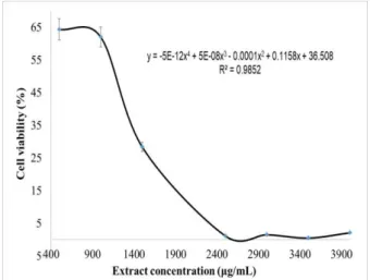
chemotherapeutic agents (Sundar et al. 2003). The cytotoxic effect of A. cordifolia extracts and cisplatin on Vero cells was measured by MTT assay. Figures 1 and 2 revealed the cytotoxic effect of EAC and cisplatin on Vero cells. The extract showed the cytotoxic effect at a concentration applied above 1000 µg/mL and its cytotoxic profile followed the polynomial order 4 with IC50 value at 1302 µg/mL [Figure 1]. On the other hand, cisplatin demonstrated strong cytotoxic effect with IC50 value at 64 µM on Vero cells [Figure 2].
Figure 1 Effect of EAC on Vero cell viability
In this study, the calculated Selectivity Index (SI) was used to determine selectivity cytotoxic effect of EAC and cisplatin. Higher SI value refers to a higher selective cytotoxic effect of an extract or a compound, a selective extract or
compound has SI higher than 3
(Mahavorasirikul et al. 2009). In this study, SI was calculated based on IC50 value of extract (75 µg/mL) and cisplatin (40 µM or equal to 11.9 µg/mL) on HeLa cells concluded from our previous studies (Yuliani et al. 2015; Setiawati 2016). The data recorded that EAC had a significantly higher selectivity to the HeLa cells (SI 17.36) than cisplatin (SI 1.60). Therefore, EAC has enormous potential to be developed as an anticancer agent for the treatment of cervical cancer. On the other hand, a lower selectivity is shown by cisplatin than EAC may be attributed to the formation of active species that interacts primarily with DNA via formation of DNA crosslinks (Hassan et al. 2014), so that cisplatin interferes with DNA both in normal as well as cancer cells. However, the investigation of selectivity of A. cordifolia extracts endorsed with the molecular target in cervical cancer cells is an interesting observation.
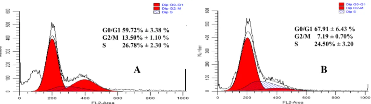
In addition, flow cytometric method was employed in this study to assess the cell cycle of HeLa cells during EAC treatment. The cells were accumulated more in G0/G1 phase due to
the extract treatment (67.91 ± 6.43 %) [Figure 3] compared to the untreated cells (59.72 ± 3.38%); while in our previous studies cisplatin treatment on HeLa cells triggered the cell accumulation on G2/M (Setiawati 2016). Investigating the molecular target of G0/G1 cell cycle arrest was very interesting in this study. The regulation of cell cycle is an important molecular target in cervical cancer therapy that is induced by Human Papilloma Virus (HPV) (Fernandes et al. 2003). Cell cycle analysis demonstrated that EAC stimulated HeLa cells to delay the G0/G1 phase, while our preceding studies presented that cisplatin-induced HeLa accumulation in the G2/M phase (Setiawati 2016). Furthermore, the molecular mechanism of triggered by the extract was further elucidated by immunocytochemical staining method on Bcl–2. Bcl–2 protein is significantly over-expressed in cervical cancer than normal cells (Eifler et al. 2014; Zhou & Wang 2015). The Bcl–2 protein family plays an important role in the regulation of the mitochondrial apoptotic pathway, Bcl–2 expressing cells showed brown color while non-expressing cells showed purple color staining (Figure 4). Untreated HeLa cells were stained brown color, while EAC and cisplatin treated cells were stained in blue color. This result indicated that Bcl–2 expression was suppressed by extract and cisplatin.
Figure 3 Cell cycle analysis of HeLa cells; (A) Untreated cells, (B) EAC 75 µg/mL treated cells
A B C
Figure 4. The expression of Bcl–2 in HeLa cells detected by immunohistochemistry method under a light microscope at 400X magnification. Brown color (arrow) in cells refers to a positive result. (A) Untreated cells, (B) EAC 75 µg/mL treated cells, (C) Cisplatin 40 µM treated cells
A B G0/G1 59.72% ± 3.38 % G2/M 13.50% ± 1.10 % S 26.78% ± 2.30 % G0/G1 67.91 ± 6.43 % G2/M 7.19 ± 0.70% S 24.50% ± 3.20
Selective cytotoxic activity of Anredera cordifolia
This study showed that both of EAC and cisplatin suppressed the Bcl–2 expression in HeLa cells. The down-regulating effect of Bcl–2 in cisplatin-treated HeLa cells indicated that the cells are not resistant to cisplatin (Leisching et al. 2015). The Bcl–2 protein family contain two functional subfamilies of proteins: pro-apoptotic proteins (Bax and Bid) and anti-apoptotic proteins (Bcl–2 and Bcl-xL). The subfamily of Bcl–2 proteins binds to apoptosis protein and neutralizes its activity and induces apoptosis (Terrano et al. 2010). The downregulation of Bcl–2 contributes to the escape of cancer cells from death (Koff et al. 2015) and play an important role in cell cycle regulation (Naser et al. 2015). However, anti-apoptotic activity of Bcl–2 is prevented by Bax activity, a dominant pro-apoptotic protein of Bcl–2 family (Weinberg 2007). Bax protein induces cytochrome C to be released from mitochondria and cause apoptosis. Therefore, a comprehensive assessment of both Bcl–2 and Bax protein expression gives a clearer molecular mechanism of the extract. The ratio of Bax/Bcl–2 expression indicated the intrinsic pathway of apoptosis induction through the release of cytochrome C from mitochondrion. This ratio is a critical determinant of a cell’s threshold for undergoing apoptosis (Zhang et al. 2014). The higher value of this ratio indicates a compound or an extract with an apoptotic induction activity through the intracellular mitochondrial pathway (Peng et al. 2015). However, the ratio of Bax/Bcl–2 expression was not determined in this study. Therefore, efforts will be made in the futures studies in order to delineate the molecular mechanisms further.
CONCLUSION
Anredera cordifolia showed selective cytotoxic
activity in HeLa cells compared to Vero cells through arrested cell cycle at G1/S phase by down regulating Bcl–2 expression. Further, a clear molecular mechanism of Anredera cordifolia should be determined.
ACKNOWLEDGEMENTS
This work was funded by Indonesian Directorate General of Research and Higher
Education (Decentralization Grant No.: 027a/ Penel.LPPM USD/IV 12016).
REFERENCES
Ahmadzadeh A, Shahbazian H, Safapour N, Tulabi M, Zandifar S. 2015. Comparison between the effects of one-day treatment regimen with cisplatin on renal function and various biochemical parameters in patients with gastric and lung cancer compared with two-days divided cisplatin treatment regimen, J Renal Inj Prev.4(3): 87–91.
Bruni L, Barrionuevo-Rosas L, Albero G, Serrano B, Mena M, Gómez D, Muñoz J, Bosch FX, de Sanjosé S. ICO Information Centre on HPV and Cancer (HPV Information Centre). Human Papillomavirus and Related Diseases in the World. Summary Report 19 May 2017
Calderón-arancibia J, C. Espinosa-bustos, Cañete-molina Á, Tapia RA, Faúndez M, Torres MJ, Aguirre A, Paulino M, Salas CO. 2015. Synthesis and Pharmacophore Modelling of 2,6,9-Trisubstituted Purine Derivatives and Their Potential Role as Apoptosis-Inducing Agents in Cancer Cell Lines. Molecules. 20: 6808–6826.
Di Domenico F, Foppoli C, Coccia C, Perluigi M. 2012. Antioxidants in cervical cancer: Chemopreventive and chemotherapeutic effects of polyphenols. Biochimica et Biophysica Acta – Molecular Basis of Disease. 1822(5): 737–747.
Djamil R. 2012. Antioxidant Activity of Flavonoid From Anredera Cordifolia (Ten) Steenis Leaves. International Research Journal of Pharmacy. 3(9): 241–243
Drayton RM, Catto JW. 2012. Molecular mechanisms of cisplatin resistance in bladder cancer. Expert Rev. Anticancer Ther. 12(2), 271–28.
Eifler A, Godoy G, Bazzo KO, De Moura LB. 2014. Expression Analysis of p53 , Ki–67 and bcl–2 in Pre-Malignant Lesions of the Cervix. Open Journal of Obstetrics and Gynecology. 4: 462–469. Fernandes SM, Syrjänen KY. 2003. Regulation of cell
cycles is of key importance in human papillomavirus (HPV)-asociated cervical carcinogenesis. São Paulo Med J. 12(3): 128–132. Florea A, Büsselberg D. 2011. Cisplatin as an anti-tumor
drug: cellular mechanisms of activity, drug resistance and induced side effects. Cancers. 13: 2315–2325.
Hassan M, Watari H, Abualmaaty A, Ohba Y, Sakuragi N. 2014. Apoptosis and Molecular Targeting Therapy in Cancer. BioMed Research International. 150845 Hong J. 2016. Concurrent Chemotherapy for Cervical Cancer Patients, Chang Gung Med J. 29(6): 550- 554.
Huang H, Duan H, Huang H, Tong X, Han Y, Ru Y, Qu L, Shou C, Zhao Z. 2016. Cisplatin resistance in gastric cancer cells is associated with HER2 upregulation-induced epithelial-mesenchymal transition. Scientific reports. DOI: 10.1038/srep20502.
Hu Y, Cai ZQ, Su XY. 2012. Concurrent weekly cisplatin versus triweekly cisplatin with radiotherapy in the treatment of cervical cancer: A metaanalysis result,
Asian Pacific J Cancer Prev. 13(9): 4301–4304. International Standard ISO/EN10993–5.2009. Biological
evaluation of medical devices – Part 5: Tests for cytotoxicity: in vitro methods. 3: 42.
Jan S, Kimman M, Kingston D, Woodward M. 2012. The socioeconomic burden of cancer in member countries of the Association of Southeast Asian Nations (ASEAN) stakeholder meeting report.
Asian Pacific J Cancer Prev. 13: 411–420
Koff JK, Ramachandiran S, Bernal-mizrachi L. 2015. A Time to Kill : Targeting Apoptosis in Cancer. Int. J. Mol. Sci. 16: 2942–2955.
Larasati YA, Dewi D, Putri P, Utomo RY, Hermawan A, Meiyanto E. 2014. Combination of Cisplatin and Cinnamon Essential Oil Inhibits HeLa Cells Proliferation through Cell Cycle Arrest. J Appl Pharm Sci. 4(2): 014–019.
Leisching G, Loos B, Botha M, Engelbrecht AM. 2015. Bcl–2 confers survival in cisplatin treated cervical cancer cells: circumventing cisplatin dose-dependent toxicity and resistance. J Transl Med. 13:328.
Mahavorasirikul W, Chaijaroenkul W, Itharat A, Na-Bangchang K. 2009. Screening of cytotoxic activity of Thai medicinal plants against human cholangiocarcinoma cells In Vitro. Drug Metabolism Reviews. 41: 85.
Menezes C, Valério E, Dias E. 2013. The Kidney Vero-E6 Cell Line: A Suitable Model to Study the Toxicity of Microcystins. New Insights into Toxicity and Drug Testing. 29–48.
Naser MH, Mahdavi M, Davoodi J, Tackallou SH, Goudarzvand M, Neishabouri SH. 2015. Up regulation of Bax and down regulation of Bcl2 during 3-NC mediated apoptosis in human cancer cells. Cancer Cell International. 15:55
Prasaja Y, Sutandyo N, Andrajati R. Incidence of Cisplatin-Induced Nephrotoxicity and Associated
Factors among Cancer Patients in Indonesia. 2015.
Asian Pacific J Cancer Prev. 16(3): 1117–22.
Pendleton KP, Grandis JR. 2013. Cisplatin-based chemotherapy options for recurrent and/or metastatic squamous cell cancer of the head and neck. Clinical Medicine Insights: Therapeutics. 3:103–116.
Peng Y, Fu Z, Guo CS, Zhang YX, Di Y, Jiang B, Li QW. 2015. Effects and Mechanism of Baicalin on Apoptosis of Cervical Cancer HeLa Cells In-vitro. IJPR, 14 (1): 251–26
Setiawati A. 2016. Celecoxib , A COX–2 Selective Inhibitor , Induces Cell Cycle Arrest at the G2 / M Phase in HeLa Cervical Cancer Cells. Asian Pacific J Cancer Prev. 17(4): 1655–1659.
Sundar S, Horne A, and Kehoe S. 2003. Cervical cancer. Clin Evid.13: 2285– 2292.
Terrano DT, Upreti M, Chambers TC. 2010. Cyclin-dependent kinase 1-mediated Bcl-xL/Bcl–2 phosphorylation acts as a functional link coupling mitotic arrest and apoptosis. Molecular and cellular biology. 30(3): 640–656.
Torre LA, Bray F, Siegel RL, Ferlay J, Tieulent JL, Jemal A. 2015. Global Cancer Statistics, 2012, CA Cancer J Clin. 65: 87- 107.
Weinberg RA. The Biology of Cancer. Garlans science. New York, 2007; 339.
WHO. World Cancer Report. International Agency for Research on Cancer. 2014.
WHO. Latest World Cancer Statistic. IARC. 12 December 2013.
Yuliani SH, Anggraeni CD, Sekarjati W, Panjalu P, Istyastono EP, Setiawati A. 2015. Cytotoxic Activity Of Anredera Cordifolia Leaf Extract On Hela Cervical Cancer Cells Through p53-Independent Pathway. Asian J Pharm Clin Res. 8(2): 328–331.
Zhang W, Zhang D, Ma X, Liu Z, Li F, Wu D. 2014. Paris saponin VII suppressed the growth of human cervical cancer Hela cells. European journal of medical research. 19: 41.
Zhou XL, Wang M. 2015. Expression levels of survivin, Bcl–2, and KAI1 proteins in cervical cancer and their correlation with metastasis. Genetics and Molecular Research. 14(4): 17059–17067.