1
Immunohistochemistry
Application Guide
Common abbreviations
ABC: Avidin biotin complex AEC: 3-amino-9-ethylcarbazole AP: Alkaline phosphatase
BCIP: 5-bromo-4-chloro-3-indolyl phosphate BSA: Bovine serum albumin
DAB: 3,3’ diaminobenzidine
FISH: Fluorescence in situ hybridization FITC: Fluorescein isothiocyanate
FFPE: Formalin-fixed, paraffin embedded HIER: Heat induced epitope retrieval HRP: Horseradish peroxidase
H2O2: Hydrogen peroxide ICC: Immunocytochemistry IHC: Immunohistochemistry IP: Immunoprecipitation ISH: In situ hybridization
LSAB: Labeled steptavidin biotin
KO: Knock out
NBF: Neutral buffered formalin NBT: p-nitroblue tetrazolium chloride PBS: Phosphate buffered saline PFA: Paraformaldehyde
PIER: Proteolytic induced epitope retrieval RabMAb: Rabbit monoclonal antibody
TMA: Tissue microarray
TMB: 3,3’,5,5’-tetramethylbenzidine WB: Western blot
3
Contents
Common abbreviations . . . .2
Introduction . . . .4
- Tips for designing a successful IHC experiment . . . .4
Sample preparation . . . .6
Fixation, embedding and sectioning . . . .7
- Paraffin embedded tissue . . . .8
- Frozen tissue . . . .9
Antigen retrieval . . . .10
- Buffers and epitope retrieval reagents . . . .11
Permeabilization . . . .13
Blocking . . . .14
- Protein blocking . . . .14
- Biotin blocking . . . .15
- Blocking endogenous enzymes . . . .16
- Peroxidase blocking . . . .16
- Alkaline phosphatase blocking . . . .16
- Reduction of autofluorescence in IHC . . . .16
- Blocking cross-reactive antigens . . . .17
- Reagents for blocking . . . .18
Immunostaining . . . .19
Primary antibody selection and optimization . . . .21
- Antibody specificity . . . .21
- Proof of use in IHC . . . .21
- Clonality . . . .21
- Antibody optimization . . . .22
- RabMAb advantages for IHC . . . .22
- KO validated antibodies . . . .22
Direct vs indirect detection . . . .24
- Secondary antibodies for IHC . . . .24
Chromogenic detection . . . .26
- ABC . . . .26
- LSAB . . . .26
- Polymer . . . .28
- Micro-polymer . . . .28
- Chromogenic multicolor detection systems . . . .29
- Enzymes and chromogens . . . .31
Fluorescent detection . . . .33
Counterstaining . . . .34
Mounting media . . . .37
IHC controls . . . .38
- Antigen (tissue) controls . . . .38
- Reagent controls . . . .38
- Tissue slides . . . .39
- Tissue microarrays . . . .39
Troubleshooting IHC experiments . . . .40
- No staining . . . .40
- High background . . . .41
- Non-specific staining . . . .42
- Poorly resolved or damaged tissue morphology . . . .42
- IHC Worksheet . . . .43
Introduction
Immunohistochemistry (IHC) is a method for detecting the location of proteins and other antigens in tissue sections using antibodies . Though less quantitative than other immunoassays such as western blotting or ELISA, IHC shows where proteins are expressed in the context of intact tissue . This is especially useful for assessing the progression and best treatment options of diseases such as cancer . In general, IHC data provide a valuable perspective that can help interpret data obtained using other methods . The key to high quality immunohistochemical staining is the specificity of the antibody used . A highly specific antibody will bind only to the protein of interest in the tissue section . The antibody-antigen interaction is visualized using either chromogenic or fluorescent detection . In chromogenic detection, the antibody is conjugated to an enzyme that cleaves a substrate to produce a colored precipitate at the location of the protein . In fluorescent detection, the antibody is conjugated to a fluorophore that can be visualized using fluorescence microscopy .
Tips for designing a successful IHC experiment
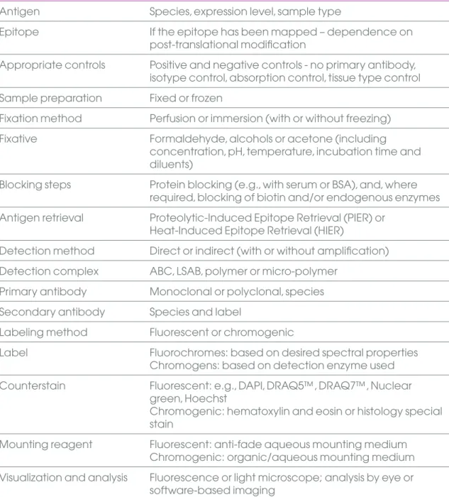
There are a number of variables that have to be considered and optimized for every IHC experiment to ensure consistent and reproducible results . These variables are listed in Table 1 .
5
Table 1. Immunohistochemistry variables
Variable Factors to consider
Antigen Species, expression level, sample type
Epitope If the epitope has been mapped – dependence on
post-translational modification
Appropriate controls Positive and negative controls - no primary antibody, isotype control, absorption control, tissue type control Sample preparation Fixed or frozen
Fixation method Perfusion or immersion (with or without freezing)
Fixative Formaldehyde, alcohols or acetone (including
concentration, pH, temperature, incubation time and diluents)
Blocking steps Protein blocking (e .g ., with serum or BSA), and, where required, blocking of biotin and/or endogenous enzymes Antigen retrieval Proteolytic-Induced Epitope Retrieval (PIER) or
Heat-Induced Epitope Retrieval (HIER)
Detection method Direct or indirect (with or without amplification) Detection complex ABC, LSAB, polymer or micro-polymer
Primary antibody Monoclonal or polyclonal, species Secondary antibody Species and label
Labeling method Fluorescent or chromogenic
Label Fluorochromes: based on desired spectral properties
Chromogens: based on detection enzyme used Counterstain Fluorescent: e .g ., DAPI, DRAQ5™, DRAQ7™, Nuclear
green, Hoechst
Chromogenic: hematoxylin and eosin or histology special stain
Mounting reagent Fluorescent: anti-fade aqueous mounting medium Chromogenic: organic/aqueous mounting medium Visualization and analysis Fluorescence or light microscope; analysis by eye or
Sample preparation
Sample preparation for an IHC experiment may include processes such as fixation, dehydration, embedding and sectioning . The two main methods of preserving tissues for IHC are paraffin embedding and freezing of the tissue . The most appropriate route of sample preparation is usually determined by one or two experimental variables . For example, if a phosphorylated epitope is being studied, tissues may need to be snap-frozen . The method of fixation often drives the design of the sample preparation workflow . Additional steps in sample preparation include antigen retrieval to unmask epitopes that have been altered by fixation, permeabilization to grant the antibody access to intracellular proteins and blocking to prevent non-specific staining .
7
Fixation, embedding
and sectioning
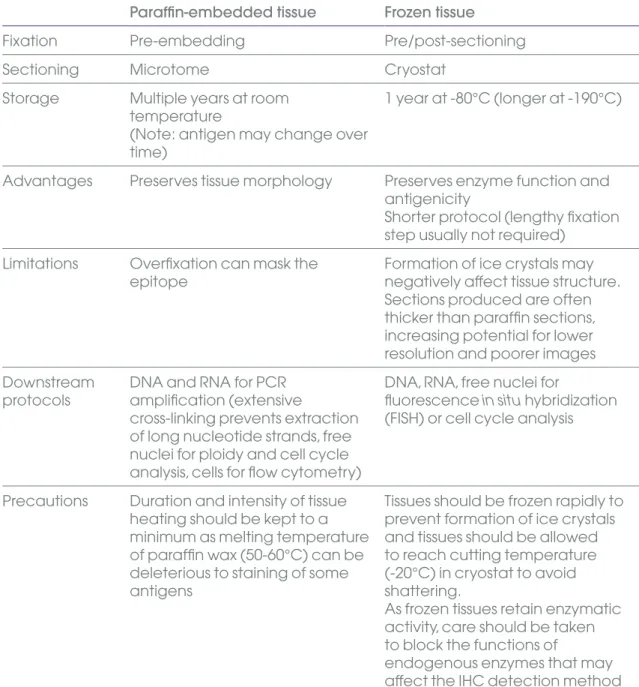
Fixation prevents the autolysis and necrosis of excised tissues, preserves antigenicity, enhances the refractive index of tissue constituents and helps to preserve cellular elements during tissue processing . The fixative used is influenced by the target antigen as well as the desired detection technique (fluorescent or chromogenic) . After fixation, the tissue sample is either embedded in paraffin or frozen . Embedding is important in preserving tissue morphology and giving the tissue support during sectioning (microtomy) . Some epitopes may not survive harsh fixation or embedding . Some guidelines for tissue embedding are given in Table 2 . During sectioning, the tissue is typically cut into thin sections (5-10 µm) or smaller pieces (for whole mount studies) to facilitate further study .
Table 2. Tissue preservation guidelines
Paraffin-embedded tissue Frozen tissue
Fixation Pre-embedding Pre/post-sectioning
Sectioning Microtome Cryostat
Storage Multiple years at room temperature
(Note: antigen may change over time)
1 year at -80°C (longer at -190°C)
Advantages Preserves tissue morphology Preserves enzyme function and antigenicity
Shorter protocol (lengthy fixation step usually not required)
Limitations Overfixation can mask the
epitope Formation of ice crystals may negatively affect tissue structure . Sections produced are often thicker than paraffin sections, increasing potential for lower resolution and poorer images Downstream
protocols DNA and RNA for PCR amplification (extensive
cross-linking prevents extraction of long nucleotide strands, free nuclei for ploidy and cell cycle analysis, cells for flow cytometry)
DNA, RNA, free nuclei for
fluorescence in situ hybridization (FISH) or cell cycle analysis
Precautions Duration and intensity of tissue heating should be kept to a minimum as melting temperature of paraffin wax (50-60°C) can be deleterious to staining of some antigens
Tissues should be frozen rapidly to prevent formation of ice crystals and tissues should be allowed to reach cutting temperature (-20°C) in cryostat to avoid shattering .
As frozen tissues retain enzymatic activity, care should be taken to block the functions of
endogenous enzymes that may affect the IHC detection method
Paraffin-embedded tissue
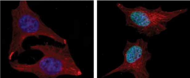
When generating paraffin-embedded tissue samples, the tissue must be fixed before embedding in paraffin . Fixation is achieved by perfusion or immersion immediately following dissection . The process typically takes 4 - 24 hours . Fixation for longer than 24 hours is not recommended as it may lead to overfixation, which may mask the antigen . The most suitable fixative for an IHC experiment depends on the antigen, as illustrated in Figure 1 . Standardized fixatives for each type of antigen are essential for reproducible staining - an antigen that has been inappropriately fixed may not be detected . Some guidelines for the type of fixative to use are given in Table 2 .
Formaldehyde Methanol
Figure 1. Effect of fixative on immunostaining patterns
Crotonylation of histone H2B K5 is clearly detected in immunocytochemistry of HeLa cells fixed with methanol (right), while no staining is observed in cells fixed in formaldehyde (left) . Samples are stained with rabbit polyclonal to histone H2B (crotonyl K5) (ab177139), 1 µg/mL, and goat anti-rabbit IgG H&L (Alexa Fluor® 488) preadsorbed (ab150081), 0 .5 µg/mL . Nuclei are stained with DAPI and tubulin (red) is stained with anti-alpha tubulin antibody (ab7291) and goat anti-mouse IgG H&L (Alexa Fluor® 594), preadsorbed (ab150120) .
Table 3. Guidelines for choosing a fixative
Antigen Fixative
Most proteins, peptides and enzymes of low
molecular weight Cells / cytological preparations: 4% formaldehyde Tissue sections: 10% Neutral-buffered formalin (NBF)
Delicate tissue Bouin’s fixative
Small molecules such as
amino acids 4% formaldehyde
Blood-forming organs
(liver, spleen, bone marrow) Zenker’s solution
Connective tissue Helly’s solution
Nucleic acids Carnoy’s solution
Large protein antigens (e .g ., immunoglobulin) Ice-cold acetone or methanol (100%)
Nuclear morphology Zinc formalin
9 After fixation, the tissue is dehydrated to enable embedding with paraffin, which
is water-insoluble . The tissue is dehydrated gently by immersion in increasing concentrations of a dehydrating agent such as alcohol . This gradual change in hydrophobicity minimizes cell damage . The dehydrating agent is then cleared by
incubation in xylene prior to paraffin embedding . Paraffin is typically heated to 60°C and then allowed to harden overnight . Finally, the tissue is sectioned using a microtome . Tissue sections may be dried onto microscope slides and stored for extended periods at room temperature . The tissue is rehydrated before commencing the immunostaining protocol .
Frozen tissue
Frozen tissues are prepared by immersing the tissue in liquid nitrogen or isopentane, or by burying the sample in dry ice . Snap-freezing is frequently used when detecting post-translation modifications such as phosphorylation . The frozen tissue is cut using a cryostat . The sections can be stored at -80°C for up to 1 year . The frozen tissue sections may then be fixed, typically with an alcohol e .g ., methanol or ethanol . As alcohols do not mask epitopes, their use avoids the need for antigen retrieval .
Antigen retrieval
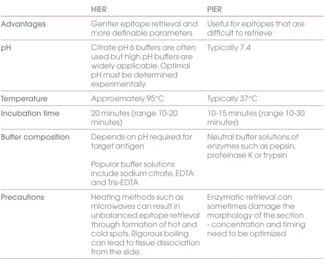
Fixation can lead to protein cross-linking, which masks epitopes and can restrict antigen-antibody binding . Such masked epitopes can be retrieved (unmasked) by antigen retrieval . In the Proteolytic-Induced Epitope Retrieval (PIER) method, enzymes (such as proteinase K, trypsin or pepsin) or commercially available PIER reagents are used to restore antibody access to an epitope . The Heat-Induced Epitope Retrieval (HIER) method uses heat from a variety of sources (microwave, pressure cooker, steamer, waterbath or autoclave) to unmask epitopes . Some antigens (e .g ., some cytokeratins and immunoglobulin light chains) can be retrieved more efficiently by a combination of heating and enzyme digestion .
The preferred method for optimal retrieval is dependent on the tissue, fixation and/or primary antibody and must be optimized by the histologist . A starting point for selecting the appropriate antigen retrieval method is to test two methods of HIER (for example, using citrate buffer pH 6 and Tris-EDTA pH 9) and one or two methods of PIER (for example, using proteinase K and/or trypsin) . Some suggested guidelines are shown in Table 4 . Conditions must be optimized for each antigen, as illustrated in Figure 2 . Antigen retrieval may not be required for frozen sections .
Table 4. Epitope retrieval guidelines
HIER PIER
Advantages Gentler epitope retrieval and
more definable parameters Useful for epitopes that are difficult to retrieve
pH Citrate pH 6 buffers are often
used but high pH buffers are widely applicable . Optimal pH must be determined experimentally .
Typically 7 .4
Temperature Approximately 95°C Typically 37°C
Incubation time 20 minutes (range 10-20
minutes) 10-15 minutes (range 10-30 minutes) Buffer composition Depends on pH required for
target antigen
Popular buffer solutions include sodium citrate, EDTA and Tris-EDTA
Neutral buffer solutions of enzymes such as pepsin, proteinase K or trypsin
Precautions Heating methods such as microwaves can result in unbalanced epitope retrieval through formation of hot and cold spots . Rigorous boiling can lead to tissue dissociation from the slide .
Enzymatic retrieval can sometimes damage the morphology of the section - concentration and timing need to be optimized
11 Progesterone receptor E-cadherin
No heating pH 3.0 (20 mM glycine-HCI buffer) pH 4.5 (20 mM citrate buffer) pH 6.0 (20 mM citrate buffer) pH 7.5 (20 mM TB) pH 9.0 (20 mM TB) pH 10.5 (20 mM TB)
Figure 2. Effects of pH on heat-induced antigen retrieval in human tissues
Adapted from Emoto K, Yamashita S and Okada Y (2005) . Mechanisms of Heat-induced Antigen Retrieval: Does pH or Ionic Strength of the Solution Play a Role for Refolding Antigens? J Histochem Cytochem 53 (11):1311-21 .
Buffers and antigen retrieval reagents
IHC Buffers
Product name Size Product code
Background reducing buffer 50 mL ab64234
10x Citrate Buffer pH 6 .0 125 mL ab64214
100x Citrate Buffer pH 6 .0 50 mL ab64236
10x EDTA Buffer pH 8 .0 125 mL ab64216
100x EDTA Buffer pH 8 .0 50 mL ab64239
10x PBS Buffer 1 L ab128983
25x PBS Buffer pH 7 .6 125 mL ab64026
20x PBS Buffer with Tween® 20 125 mL ab64028
25x PBS Buffer pH 7 .6 1 L ab64246
20x PBS Buffer with Tween® 20 1 L ab64247
25x TBS pH 7 .4 125 mL ab64203
20x TBS-T with Tween® 20 125 mL ab64204
25x TBS pH 7 .4 1 L ab64248
20x TBS-T with Tween® 20 1 L ab64250
10x Tris Buffer pH 10 .0 125 mL ab64222
100x Tris Buffer pH 10 .0 50 mL ab64251
Antigen retrieval reagents / tissue pretreatments
Product name Size Product code
Antigen Retrieval Buffer (100X Citrate Buffer pH 6 .0) 125 mL ab93678 Antigen Retrieval Buffer (100X Citrate Buffer pH 6 .0) 250 mL ab94674 Antigen Retrieval Buffer (100X EDTA Buffer, pH 8 .0) 125 mL ab93680 Antigen Retrieval Buffer (100X EDTA Buffer, pH 8 .0) 250 mL ab94677 Antigen Retrieval Buffer (100X Tris-EDTA Buffer, pH 9 .0) 125 mL ab93684 Antigen Retrieval Buffer (100X Tris-EDTA Buffer, pH 9 .0) 250 mL ab94681 Antigen Retrieval Buffer (100X Tris Buffer, pH 10 .0) 125 mL ab93682 Antigen Retrieval Buffer (100X Tris Buffer, pH 10 .0) 250 mL ab94680
10X Tris-HCl Buffer for HIER 50 mL ab128986
10x Heat Mediated Antigen Retrieval Solution pH 6 .0 250 mL ab973 Heat Mediated High pH Antigen Retrieving Solution 1 L ab972
HistoReveal 15 mL ab103720
Pepsin Solution 7 mL ab64201
Pepsin Solution 15 mL , 125 mL ab128991
Proteinase K 4 mL ab64220
Trypsin (Enzymatic Pretreatment) 15 mL , 125 mL ab128214
Trypsin Enzymatic Pretreatment Kit 7 mL ab64205
Trypsin Enzymatic Antigen Retrieval Solution 1 .6 mL
concentrated liquid trypsin & 25 mL trypsin buffer
ab970
HistoReveal (ab103720) is an enzymatic antigen retrieval reagent that provides superior staining when compared to PIER using standard enzymes such as trypsin or pepsin, as shown in Figure 3 . HistoReveal is specially formulated to preserve tissue morphology better than standard enzymes . As a result, less primary antibody is required .
Figure 3. HistoReveal enhances staining
Cytokeratin 20 staining in colon tissue . Formalin/PFA-fixed paraffin-embedded sections following antigen retrieval using HistoReveal (ab103720) .
13
Permeabilization
Permeabilization allows the antibody to access intracellular antigens . These include the cytoplasmic epitopes of transmembrane proteins . Permeabilization is achieved using solvents or detergents .
Solvents: Can be used after fixation with cross-linking agent e .g ., paraformaldehyde . Recommended for cytoskeletal, viral and some enzyme antigens .
Detergents: These can be harsh (e .g ., Triton™ X-100 or NP-40) to disrupt proteins or mild (e .g ., Tween® 20, saponin or digitonin) so as not to dissolve plasma membranes .
Detergent permeabilization can significantly improve antibody access to soluble nuclear antigens and antigens in the cytoplasm or on the cytoplasmic face of the plasma membrane .
Table 5. Solvent and detergent guidelines
Solvents Comments
Solvents Acetone Acetone fixation will also
permeabilize
Methanol Methanol fixation can be
used to permeabilize but is not always effective
Detergents Triton™ X-100 or NP-40 Use 0 .1 - 0 .2% in PBS for 10 min only
Tween® 20, saponin,
Blocking
Protein blocking
Blocking with sera or a protein blocking reagent is essential to prevent non-specific binding of antibodies to tissue or Fc receptors (receptor that binds the constant region (Fc) of an antibody) . Theoretically, any protein that does not have binding affinity for the target epitope can be used for blocking . In practice, some proteins are better than others because they bind more readily to non-specific sites . Serum is a common blocking agent as it contains antibodies that bind to non-specific sites . A serum matching the species of the secondary antibody is recommended . When performing multiple stains using secondary antibodies from different species, it may be necessary to use blocking sera from the species of both secondary antibodies .
Proteins such as BSA or casein may also be used to block non-specific antibody binding . These are simpler to use as there is no need to match the reagent to the species of the secondary antibody . Commercial blocking buffers, such as our 10X Blocking Buffer (ab126587), are frequently used to block non-specific antibody binding . These often have proprietary formulations that optimize performance and/or shelf life .
Sterile blocking sera
Product name Size Product code
Bovine Calf Serum (sterile) 25 mL ab138477
Cat Serum (sterile) 50 mL ab139511
Chicken Serum (sterile) 25 mL ab138577
Donkey Serum (sterile) 50 mL ab138579
Goat Serum (sterile) 50 mL ab138478
Guinea Pig Serum (sterile) 50 mL ab138480
Hamster Serum (sterile) 50 mL ab139500
Horse Serum (sterile) 50 mL ab139501
Mouse Serum (sterile) 50 mL ab138705
Rabbit Serum (sterile) 50 mL ab7487
Rat Serum (sterile) 50 mL ab138328
15
Non-sterile blocking sera
Product name Size Product code
Bovine Calf Serum 25 mL ab7479
Cat Serum 10 mL ab139724
Chicken Serum 25 mL ab7477
Dog Serum 10 mL ab139737
Donkey Serum 25 mL ab7475
Goat Serum 50 mL ab7481
Guinea Pig Serum 10 mL ab7482
Hamster Serum 10 mL ab7483
Horse Serum 25 mL ab7484
Llama Serum 20 mL ab139738
Mouse Serum 10 mL ab7486
Rabbit Serum 25 mL ab7487
Rat Serum 10 mL ab7488
Sheep Serum 50 mL ab7489
Biotin blocking
Blocking endogenous biotin is recommended when using an avidin/biotin-based detection system . Endogenous biotin is present in many tissues, with particularly high levels in tissues such as kidney, liver and brain .
It is blocked by pre-incubation of the tissue with avidin, followed by incubation with biotin to block additional biotin binding sites on the avidin molecule . For robust and reproducible blocking, a biotin-blocking reagent such as our Endogenous Avidin + Biotin Blocking System (ab3387) is recommended .
Protocol recommendations:
1 . Block endogenous biotin prior to, or after incubation with, primary antibody but NOT after incubation with a biotinylated secondary .
Blocking endogenous enzymes
Chromogenic detection methods usually use an enzyme conjugated to a secondary antibody to visualize antibody localization . If the enzymatic activity is also endogenous to the tissue being studied, the endogenous enzymes must be blocked before the detection step .
Peroxidase blocking
When using a horseradish peroxidase (HRP)-conjugated antibody for detection, non-specific or high background staining may occur due to endogenous peroxidase activity in tissues such as kidney, liver and those containing red blood cells (such as vascular tissue) . To check for endogenous peroxidase activity, tissues can be incubated with 3,3’-diaminobenzidine (DAB) substrate prior to primary antibody incubation . If the tissues turn brown, endogenous peroxidase is present and a blocking step is required . Hydrogen peroxide (H2O2) is the most common peroxidase blocking agent .
Protocol recommendations:
1 . Block at preferred stage of IHC protocol:
- after rehydration to water and before antigen retrieval;
- or after antigen retrieval / before primary antibody incubation;
- or after primary antibody incubation / before secondary antibody incubation; - or after secondary antibody incubation .
For certain antigens, like CD4 and CD8, blocking after primary and secondary incubation is recommended as H2O2 is detrimental to key epitopes on these proteins .
2 . Incubate section in H2O2 (typically 0 .3%) for 10-15 minutes (incubation time may be
5-60 minutes, depending on H2O2 concentration) .
Alkaline phosphatase blocking
Endogenous alkaline phosphatase (AP) can produce high background when using an AP-conjugated antibody for detection . It can be found in kidney, intestine, osteoblasts, lymphoid tissue and placenta . Endogenous AP activity is often more prevalent in frozen tissue . Tissue can be tested for endogenous AP by incubating with 5-bromo-4-chloro-3-indolyl phosphate/ nitro blue tetrazolium chloride (BCIP/NBT); if a blue color is observed, endogenous AP is present and blocking is necessary .
Protocol recommendations:
1 . Endogenous AP can be blocked by adding levisamole with the chromogen substrate . Chromogens containing levisamole are commercially available .
2 . In order to block intestinal AP, which is unaffected by levisamole, the tissue may be treated with a weak acid prior to application of the primary antibody .
Reduction of autofluorescence in IHC
When using a fluorescent label for detection, high background may be observed if the tissue is autofluorescent . Autofluorescence may be caused by the presence of fluorescent compounds in the tissue, such as flavins and porphyrins . These compounds may be extracted from the tissue by the solvents used to generate fixed, dehydrated sections . However, they persist in frozen sections that have been processed using aqueous reagents . The fixation step may also induce autofluorescence . This often occurs when using aldehyde fixatives, which react with amines to generate fluorescent products . Tests should be carried out to ensure that the tissue being studied is not inherently fluorescent or that fixation steps do not induce autofluorescence . Potential
17
Table 6. Autofluorescence blocking guidelines
Method 1 Use frozen tissue sections to reduce possibility of induced autofluorescence during fixation
Method 2 Use fixatives that do not contain aldehydes, such as Carnoy’s solution (if generating paraffin sections)
Method 3 Block aldehydes during fixation by treating tissue with sodium borohydride or glycine/lysine
Method 4 Treat tissue with quenching dyes such as pontamine sky blue, Sudan black, trypan blue or FITC block
If endogenous autofluorescence cannot be blocked, a chromogenic detection system may be preferable .
Blocking cross-reactive antigens
When staining mouse tissues with mouse primary antibodies, high background is often observed as endogenous mouse IgG and Fc receptors are detected by the secondary antibody targeting the (exogenous) mouse primary antibody . This background binding can be reduced using F(ab) fragments . (Note: a F(ab) fragment is a single variable domain of an antibody that binds to an antigen but has no Fc region .) For robust and reproducible staining, a Mouse on Mouse Polymer IHC kit such as ab127055 is recommended, as shown in Figures 4 and 5 .
Key benefits of our Mouse on Mouse Polymer IHC kit (ab127055) are: - Minimal background
- Strong staining through polymer-based detection - Biotin-free detection in biotin-enriched tissue
Figure 4. Reduced endogenous mouse IgG background using ab127055
Negative control images using Mouse on Mouse Polymer IHC kit (ab127055) (left) and EXPOSE detection kit (ab80436), which is not suitable for use on mouse tissues (right) . ab127055 reduces endogenous mouse IgG background staining . Both images show mouse spleen formalin/PFA-fixed paraffin-embedded tissues .
Figure 5. Stronger staining with Mouse on Mouse detection using ab127055
Mouse Pan CK (Clone AE1/AE3) antibody staining using Mouse on Mouse Polymer IHC kit (ab127055) (left) compared with ABC Mouse on Mouse system (ab80436) . (right) ab127055 gives stronger staining . Both images show mouse colon formalin/PFA-fixed paraffin-embedded tissues .
Reagents for blocking
Product name Size Product code
Endogenous Avidin/Biotin Blocking Kit 15 mL ab64212
Endogenous Avidin + Biotin Blocking System 15 mL avidin,
15 mL biotin ab3387
FITC Protein Blocking Agent (PBA) 6 mL ab128980
Hydrogen Peroxide Blocking Reagent 125 mL ab64218
Hydrogen Peroxide Blocking Reagent 60 mL ab94666
Protein Block 125 mL ab64226
Protein Block 60 mL ab156024
Sea Block Buffer 500 mL ab166951
19
Immunostaining
Immunostaining (or immunodetection) relies on the specificity of the primary antibody for the target antigen . The antibody is detected either directly, through a label that is directly conjugated to the primary antibody, or indirectly, using a labelled secondary antibody that has been raised against the host species and antibody type and subtype of the primary antibody . The antibody is visualized using either a fluorescent label or an enzyme that converts a soluble substrate into an insoluble chromogenic product . An outline of immunostaining is show in Figure 6 . Direct immunostaining methods do not require a secondary antibody incubation . In fluorescent detection, the addition of substrate is omitted .
Block with 5% serum or BSA for 30 - 60 min Wash with PBS 0.2% Tween 4 times for 5 min
Wash with PBS 0.2% Tween 4 times for 5 minutes
Enzymatic detection Incubate with conjugated secondary antibody 30 min to 2 hr RT Incubate with primary antibody 30 min to 2 hr RT or overnight 4˚C
Permeabilize the cells if detecting an intracellular target Antigen retrieval
- Heat in citrate buffer pH 6 for 5 - 20 min - Or enzymatic (trypsin, proteinase K)
Fix slides:
- 4% PFA for 10 min
- Or methanol (ice cold) for 10 min - Or acetone (ice cold) for 10 min Deparaffinization and dehydration:
- xylene
- xylene 1:1 100% ethanol
- 100% ethanol down to 50 % ethanol
IHC-Fr and ICC
IHC-P
0.2% Triton for 10 min (not necessary if fixed in acetone or methanol) Cell Antigen Primary antibody Conjugated secondary antibody
21
Primary antibody selection
and optimization
The most critical decision when designing an IHC experiment is the selection of the primary antibody .The key factors that must be considered are discussed below . Once the primary antibody has been chosen, optimization is essential .
Antibody specificity
Specificity of the primary antibody for the epitope of interest is usually determined experimentally . The most conclusive demonstration of antibody specificity is lack of staining in tissues or cells in which the target protein has been knocked out . Other indicators are (1) recognition of a single band in western blotting, (2) staining patterns that are consistent with known localization of the protein of interest in control cells or tissues and (3) lack of staining in tissues or cells known not to express the protein . Comparison of the immunogen sequence to other proteins using alignment tools such as BLAST may give some indication about antibody specificity but is not conclusive . Ideally, the antibody should recognize the target antigen in the species of interest . If this information is unavailable, sequence comparison of the immunogen with the corresponding region in the protein from the species of interest may give some indication of specificity .
Proof of use in IHC
An antibody that recognizes its target protein in western blotting experiments, which are run under denaturing conditions, may not always recognize the antigen in IHC, where the antigen is more likely to be in its native (3D) form . An antibody that has been shown to work in IHC or immunocytochemistry (ICC) is preferable . It is important to note that fixation and antigen retrieval methods also have significant impact on the ability of an antibody to recognize the epitope of interest in an IHC experiment .
Clonality
As monoclonal antibodies are produced from a single B cell clone, they represent a homogeneous population that binds to a single epitope . As a result, they are less likely to cross-react with other proteins (provided that the clone recognizes a unique epitope) and therefore produce less background staining than polyclonal antibodies . The production of monoclonals from a hybridoma also means that there is less variation between different antibody lots than with polyclonal antibodies .
Polyclonal antibodies are heterogeneous populations that can recognize multiple epitopes . They are therefore more tolerant of changes in protein conformation (resulting, for example, from fixation or changes in temperature) . They are also more stable over a range of pH and salt concentrations than monoclonal antibodies .
Host species
Ideally, the primary antibody should be raised in a host species that is different to the species of the sample in order to avoid cross-reactivity with endogenous immunoglobulins in the tissue .
Antibody optimization
The quality of staining is influenced by the primary antibody concentration, the diluent used, the incubation time and temperature . All of these variables may need to be optimized for each antibody and sample in order to achieve specific staining with minimal background . Typically, antibody concentration is varied while maintaining a constant incubation time and temperature . Longer incubation times may be used to ensure that the antibody penetrates the tissue . Longer incubation times can be combined with lower temperatures to promote specific binding .
RabMAb
®antibody advantages for IHC
We offer high-quality rabbit monoclonal antibodies developed using our patented RabMAb technology . As monoclonals, RabMAb® primary antibodies detect a single
epitope and are therefore less likely to cross-react with other proteins . At the same time, we have observed that RabMAb® primary antibodies bind to their target with greater
affinity, enabling higher signal-to-noise ratio than mouse monoclonal antibodies at a given antibody concentration . RabMAb® primary antibodies typically provide more
specific and sensitive detection of their target protein with low background, making them ideal for demanding applications like IHC on FFPE tissues (Figure 7) . RabMAb®
antibodies typically allow higher working dilutions (5 - 10X on average) and can be used with various tissue fixatives such as FFPE at a minimal level of pretreatment . Additionally, when used along with a mouse monoclonal, dual staining with two monoclonal
antibodies can be performed for high quality double staining on the same tissue sample .
HER2/ ErbB2 RabMAb® Rabbit polyclonal antibody Mouse monoclonal antibody
antibody 3 ng/mL 20 ng/mL (vendor A) 30 ng/mL (vendor B)
Figure 7. HER2 RabMAb
®primary IHC antibody comparison
A comparison of our HER2/ ErbB2 RabMAb® antibody against leading commercially available HER2/ErbB2
rabbit polyclonal (Vendor A) and mouse monoclonal (Vendor B) on FFPE human breast carcinoma tissue . Recommended IHC protocol and dilution factor were used for each case . The antibody concentration used is shown next to the image for each stain .
Knockout (KO) validated antibodies
With the increasing need for highly specific antibodies, we are introducing KO validation as a standard quality control test on our antibodies . Antibody specificity is confirmed by testing the antibody of interest in a knockout cell line (which does not express the protein) . This data is compared side-by-side against a normal (wild type) cell line . If the antibody is truly specific, the antibody should show no staining in the knockout cell line and a specific signal in the normal cell line (Figure 8) . Knock out validations offer a true negative control to confirm the antibody specificity to the protein of interest . When you purchase one of our KO validated antibodies, you can trust that the antibody has been validated in the recommended applications and species, and that its specificity has also been confirmed through our in-house knockout validation approach .
23
Merged ab92742
Wild-type HAP1 cells
Ki67 Knockout HAPI cells
Figure 8. Knockout specificity testing by immunohistochemistry for Ki67
antibody (ab92742)
ab92742 specifically recognizes Ki67 in wild-type HAP1 cells (top right panel) and not in Ki67 knockout HAP1 cells (bottom left panel) . The cells were fixed with 100% methanol (5 min), permeabilized with 0 .1% Triton™ X-100 for 5 mins and blocked with 1% BSA/10% normal goat serum/0 .3 M glycine in 0 .1% PBS-Tween for 1 hr . Cells were incubated with ab92742 at 1µg/mL and anti-alpha tubulin antibody (Alexa Fluor® 594) (ab195889)
at 1:250 dilution (shown in pseudo color red) overnight at +4°C, followed by a further incubation at room temperature for 1 hr with a goat secondary antibody to rabbit IgG (Alexa Fluor® 488) (ab150081) at 2 µg/mL
Direct vs indirect detection
Once a primary antibody is bound to the antigen of interest, it can be detected either directly or indirectly . For direct detection methods, the primary antibody is directly conjugated to a label . For indirect detection, the primary antibody is detected by a labeled secondary antibody . Indirect methods may include amplification steps to increase signal intensity .
Direct detection is suitable for studying highly expressed antigens . For direct detection, the primary antibody can be conjugated to an enzyme, such as HRP or AP, or a
fluorochrome . The benefit of direct detection is that an additional incubation step with a secondary reagent is unnecessary . Another significant benefit of direct detection is increased flexibility in the design of multicolor experiments . Our rapid one-step antibody conjugation kits allow you to create your own directly-labeled primary antibodies . Choose from enzymatic, fluorescent, biotin or gold labels .
For additional information, visit www .abcam .com/conjugation
Indirect detection is more suitable for studies of poorly expressed antigens, which benefit from the signal amplification provided by the secondary reagent . Signal amplification occurs through the potential for two or more labeled secondary antibodies to bind to each primary antibody . However, the use of a secondary antibody requires additional blocking steps and controls . The signal may be amplified further by using avidin or streptavidin with biotinylated secondary antibodies (and is discussed in the following sections) .
Secondary antibodies for IHC
During indirect detection, the secondary antibody should be directed against the species in which the primary antibody was raised . For example, if using a primary antibody raised in rabbit, an anti-rabbit secondary antibody raised in a species other than rabbit must be used . It is also important that the secondary antibody has been raised against the isotype of the primary antibody . Affinity-purified antibodies are the most popular as they provide the lowest amount of non-specific binding . However, IgG fractions sometimes contain very high affinity antibodies and may be useful when an antigen is in low abundance .
The use of pre-adsorbed secondary antibodies can reduce non-specific background as they are less likely to show species cross-reactivity or to react with endogenous antigens of the species against which they have been pre-adsorbed . The secondary antibody should be pre-adsorbed against the species in which the sample originated . For example, a secondary antibody pre-adsorbed against human serum should be used when staining human tissues or cell lines .
For more information, please go to www .abcam .com/pre-adsorbed
F(ab’)2 fragment secondary antibodies are recommended for staining of tissues rich
in Fc receptors (e .g ., spleen, thymus, blood) . (Note: F(ab’)2 fragments have 2 antigen
binding domans linked by disulfide bonds, but no Fc region .) As these secondary antibodies are smaller and therefore penetrate tissues more easily, they are particularly useful for multiple IHC staining .
25 Secondary antibodies can be labeled with enzymes (HRP, AP), fluorochromes or biotin . We offer a range of biotinylated secondary antibodies for use in ABC (avidin-biotin complex) detection systems .
Optimized IHC secondary antibodies
Product name Size Product code
Biotinylated Goat anti Mouse IgG (H+L) (Ready-To-Use) 125 mL ab64255 Biotinylated Goat anti Rabbit IgG (H+L) (Ready-To-Use) 125 mL ab64256 Biotinylated Goat anti Mouse & Rabbit IgG (H+L) 125 mL
(Ready-To-Use) ab64257 Biotinylated Goat anti Mouse & Rabbit IgG (H+L) 60 mL
(Ready-To-Use) ab128977 Mouse polyclonal to Peroxidase anti-Peroxidase
complex/PAP antibody 100 µL ab21867
Chromogenic detection
Chromogenic detection uses enzymes that convert soluble substrates into insoluble, chromogenic products . These enzymes are usually attached to secondary antibodies but can also be attached to primary antibodies for direct detection . The most commonly used enzymes are HRP, which converts DAB into a brown product, and AP, which converts 3-amino-9-ethylcarbazole (AEC) into a red product . Chromogenic detection is usually more sensitive than fluorescent detection . Furthermore, unlike fluorophores, the colored precipitates are photostable, allowing storage of the slides for many years . Unlike fluorescent detection, which requires specialized light sources and filters, chromogenic detection only requires a standard microscope . However, the experimental procedure is longer as it includes more incubation and blocking steps than fluorescent methods . Four main methods of indirect chromogenic detection are widely used today .
Avidin-Biotin Complex (ABC)
The ABC method (Figure 9) relies on biotinylated secondary antibodies that act as links between a tissue-bound primary antibody and an avidin-biotin-reporter enzyme complex . As avidin is tetravalent, it forms large complexes containing multiple copies of the biotinylated reporter enzyme, resulting in high signal intensity .
Biotinylated secondary antibody Primary antibody Tissue antigen Reporter enzyme Avidin Biotin
Figure 9. Avidin-Biotin Complex (ABC) method
Labeled Steptavidin-Biotin (LSAB)
The LSAB method (Figure 10) is similar to the ABC method because it uses a biotinylated secondary antibody that links a primary antibody to a streptavidin-enzyme conjugate . While streptavidin is also tetravalent, it is not glycosylated and has a more neutral isoelectric point than avidin, resulting in less non-specific tissue binding .
27
Streptavidin
Figure 10. Labeled Streptavidin-Biotin (LSAB) method
We offer a number of kits that use the LSAB detection system . Choose our HRP Plus detection kit for enhanced sensitivity (ab93697) .
Mouse and rabbit specific kits (anti-polyvalent)
Product name Size Product code
Mouse and Rabbit specific HRP/DAB (ABC)
Detection IHC Kit 15 mL ab64264
Mouse and Rabbit specific HRP/DAB Plus (ABC)
Detection IHC Kit 60 mL,125 mL ab93697
Mouse and Rabbit specific HRP/AEC (ABC)
Detection IHC Kit 15 mL ab93705
Mouse and Rabbit specific HRP (ABC) Detection IHC Kit 125 mL, 1 L ab93677 Mouse and Rabbit specific AP (ABC) Detection IHC Kit 60 mL, 125 mL ab93695 Mouse and Rabbit specific AP/Fast Red (ABC)
Detection IHC Kit 15 mL ab128967
Rabbit specific kits
Product name Size Product code
Rabbit specific HRP/DAB (ABC) Detection IHC Kit 15 mL ab64261 Rabbit specific HRP/AEC (ABC) Detection IHC Kit 15 mL ab64260 Rabbit specific AP (ABC) Detection IHC Kit 60 mL, 125 mL ab128972
Mouse specific kits
Product name Size Product code
Mouse specific HRP/DAB (ABC) Detection IHC Kit 15 mL ab64259 Mouse specific HRP/AEC (ABC) Detection IHC Kit 15 mL ab64258 Note: Our ABC detection IHC kits use an LSAB detection system .
Polymer
Although biotin-based detection systems are still widely used, there are a number of limitations associated with these methods . The key challenge is that the presence of endogenous biotin can lead to significant background staining in certain tissues (e .g ., kidney or brain) . Furthermore, while formalin fixation and paraffin embedding reduce the levels of endogenous biotin, procedures such as HIER can result in the recovery of endogenous biotin as an undesirable side effect . Background staining from biotin is a significant problem when staining frozen sections, where levels of endogenous biotin tend to be higher than in paraffin-embedded specimens . Methods to block endogenous biotin are often partially effective and add another level of complexity to the IHC procedure . Non-biotin based detection methods were introduced to offer a solution to the challenge of endogenous biotin background . These methods also have simpler protocols and generate comparable, if not better, staining to ABC methods . Polymer-based methods use a dextran backbone to which multiple enzyme molecules and secondary antibodies are attached . The dextran backbone-secondary antibody complex then binds to the primary antibody (Figure 11) .
Dextran backbone Secondary antibody Enzyme Primary antibody Tissue antigen
Figure 11. Polymer method
Micropolymer
More recently, micropolymer (or compact polymer)-based detection methods have been developed (Figure 12) . The enzyme is polymerized directly onto the secondary antibody, forming a smaller detection complex . Unlike conventional detection polymers, micropolymers do not have a tendency to aggregate as they do not contain a dextran backbone . The main advantages offered by the smaller micropolymers are greater sensitivity through better tissue penetration than the polymer methods and improved signal-to-noise ratio with no background staining from endogenous biotin .
Polymerized enzymes
Figure 12. Micro-polymer method
Discover the advantages of using a micro-polymer/compact-polymer detection system . Our EXPOSE range offers greater sensitivity as well as improved signal-to-noise ratio .
29
Product name Size Product code
EXPOSE Mouse and Rabbit specific
HRP/DAB detection IHC kit 15 mL, 60 mL, 125 mL ab80436
EXPOSE Mouse and Rabbit specific
HRP/AEC detection IHC kit 15 mL, 60 mL, 125 mL ab93686
EXPOSE Mouse and Rabbit specific AP
(red) detection IHC kit 15 mL, 60 mL, 125 mL ab94734
EXPOSE Mouse and Rabbit specific
HRP/DAB or AEC detection IHC kit 1 L ab93702
EXPOSE Rabbit specific HRP/DAB
detection IHC kit 15 mL, 60 mL, 125 mL ab80437
EXPOSE Rabbit specific HRP/AEC
detection IHC kit 15 mL, 60 mL, 125 mL ab94361
EXPOSE Rabbit specific AP (red)
detection IHC kit 15 mL, 60 mL, 125 mL ab94737
EXPOSE Mouse specific AP (red)
detection IHC kit 15 mL, 60 mL, 125 mL ab94740
Chromogenic multicolor detection systems
Detection of multiple antigens using chromogenic methods usually involves long and complicated protocols . Our muticolor enzymatic IHC kits have been designed for optimal staining of two or three antigens on mammalian tissue sections using an optimized protocol that avoids multiple sequential staining steps . Other benefits of our multicolor enzymatic IHC kits are:
- Biotin blocking not required: polymer-based system
- Better signal-to-noise ratio: clear visualization of staining and morphology - Choice of color combinations: red, green, purple, brown and black
- Use of rodent antibodies on rodent tissue: protocols for blocking endogenous mouse or rat IgG
Kit name and selection key
Kit primary antibody key:
- G: Goat, M: Mouse, R: Rabbit, Rt: Rat Kit chromogen name and color key: - DAB: Brown
- AP/Red & Fast-Red: Red - HRP/Green: Green - DAB/Ni: Black - BCIP: Blue - AEC: Red
Size guidelines if using 100 µL polymer conjugate per slide:
- DoubleStain kits: 12 mL (~120 slides), 36 mL (~360 slides), 120 mL (~1200 slides) - TripleStain kits: 24 mL (~120 slides), 72 mL (~360 slides), 240 mL (~1200 slides)
Double-staining kits for human tissue
Product name Size Product code
DoubleStain IHC kit: G&M on human
tissue (DAB & AP/Red) 12 mL, 36 mL, 120 mL ab183159
DoubleStain IHC kit: G&M on human
tissue (BCIP & AEC) 12 mL, 36 mL, 120 mL ab183271
DoubleStain IHC kit: G&M on human
tissue (HRP/Green & AP/Red) 12 mL, 36 mL, 120 mL ab183272
Double-staining kits for animal tissue
Product name Size Product code
DoubleStain IHC kit: M&M on rodent
tissue (DAB & AP/Red) 12 mL, 36 mL, 120 mL ab183273
DoubleStain IHC kit: M&M on rodent
tissue (BCIP & AEC) 12 mL, 36 mL, 120 mL ab183274
DoubleStain CL IHC kit: M&M on
rodent tissue (HRP/Green & AP/Red)* 12 mL, 36 mL, 120 mL ab183275 DoubleStain CL IHC kit: M&M on
rodent tissue (DAB & FastRed)* 12 mL, 36 mL, 120 mL ab183276 DoubleStain IHC kit: M&Rt on mouse
tissue (DAB & AP/Red) 12 mL, 36 mL, 120 mL ab183277
DoubleStain IHC kit: M&Rt on mouse
tissue (BCIP & AEC) 12 mL, 36 mL, 120 mL ab183278
DoubleStain IHC kit: M&Rt on mouse
tissue (HRP/Green & AP/Red) 12 mL, 36 mL, 120 mL ab183279 * Note: CL kits are designed for ease of use in co-localization experiments
Double-staining kits for human and rodent tissue
Product name Size Product code
DoubleStain IHC kit: G&Rt on human/
mouse tissue (DAB & AP/Red) 12 mL, 36 mL, 120 mL ab183280 DoubleStain IHC kit: G&Rt on human/
mouse tissue (BCIP & AEC) 12 mL, 36 mL, 120 mL ab183281 DoubleStain CL IHC kit: G&Rt on
human/mouse tissue (HRP/Green & AP/Red)*
12 mL, 36 mL, 120 mL ab183282
DoubleStain IHC kit: R&Rt on human/
mouse tissue (DAB & AP/Red) 12 mL, 36 mL, 120 mL ab183283 DoubleStain IHC kit: R&Rt on human/
mouse tissue (BCIP & AEC) 12 mL, 36 mL, 120 mL ab183284 DoubleStain IHC kit: R&Rt on human/
mouse tissue (HRP/Green & AP/Red) 12 mL, 36 mL, 120 mL ab183285 * Note: CL kits are designed for ease of use in co-localization experiments
31
Triple-staining kits for human tissue
Product name Size Product code
TripleStain IHC kit: M&M&R on human tissue
(DAB, AP/Red & HRP/Green) 24 mL, 72 mL, 240 mL ab183286
TripleStain IHC kit: M&M&R on human tissue
(DAB, AP/Red & DAB/Ni) 24 mL, 72 mL, 240 mL ab183287
TripleStain IHC kit: R&R&M on human tissue
(DAB, AP/Red & HRP/Green) 24 mL, 72 mL, 240 mL ab183288
TripleStain IHC kit: R&R&M on human tissue
(DAB, AP/Red & DAB/Ni) 24 mL, 72 mL, 240 mL ab183289
TripleStain IHC kit: M&R&G on human tissue
(DAB, AP/Red & HRP/Green) 24 mL, 72 mL, 240 mL ab183290
TripleStain IHC kit: M&R&G on human tissue
(DAB, AP/Red & DAB/Ni) 24 mL, 72 mL, 240 mL ab183291
Enzymes and and chromogens
Additional factors for consideration in chromogenic detection are the choice of enzyme and chromogenic substrate . A number of different chromogens are available for each detection enzyme . Table 7 summarizes the key features of commonly used enzymes and chromogens/substrates .
Table 7. Enzymes and substrates / chromogens for IHC
Enzyme Chromogen /
substrate Color Mounting media Advantages Disadvantages
HRP AEC Red Aqueous Intense color;
contrasts well with blue in
double staining Endogenous peroxidase activity in tissue can lead to false positive staining
DAB Brown Organic Intense color;
permanent DAB + nickel
enhancer Black Organic Intense color; permanent
TMB Blue Aqueous Intense color;
permanent
AP BCIP/NBT Blue Organic Intense color
Endogenous AP activity in tissue can lead to false positives Fast-Red and Fast Blue TR prone to fading Naphthol AS-MX phosphate + Fast Blue BB
Blue Aqueous Less intense, good for double staining
Naphthol AS-MX phosphate + Fast-Red TR
Red Aqueous Less intense, good for double staining
Naphthol AS-MX phosphate + new fuchsin
Red Organic Intense color
Glucose
oxidase NBT Blue Organic No endogenous enzyme activity Low staining intensity High antibody concentrations needed
Substrate and chromogen products
Product name Size Product code
AEC Single/Plus 30 mL ab103742
AEC Substrate System (Ready to Use) 125 mL ab64252
Alkaline Phosphatase chromogen (BCIP/TNBT) 100 mL ab7413 Alkaline Phosphatase chromogen (BCIP/NBT) 100 mL ab7468
Alkaline Phosphatase Enhancer 250 mL ab671
DAB Enhancer 10 mL ab675
DAB Substrate Kit 125 mL ab64238
DAB Substrate Kit 60 mL ab94665
Fast-Red Substrate System 60 mL ab128979
Permanent Fast-Red Substrate System 60 mL, 125 mL ab128992
Liquid Fast-Red Substrate Kit 60 mL ab128981
Liquid Fast-Red Substrate Kit 125 mL ab64254
StayGreen/AP (Alcohol and xylene substitute
compatible) 30 mL ab156428
StayRed/AP (Alcohol and xylene compatible) 30 mL ab103741
Steady DAB/Plus 200 mL ab103723
Streptavidin Alkaline Phosphatase
(Ready to Use) 125 mL ab64268
33
Fluorescent detection
Fluorescent detection (immunofluorescence) methods use fluorochrome labels, which emit light of a longer wavelength when excited by light of a specific wavelength .
Fluorescent detection is commonly used to visualize multiple antigens simultaneously . The fluorochrome may be conjugated directly to the primary or secondary antibody or to streptavidin .
When designing multicolor experiments, two key parameters must be considered . Firstly, spectral overlap between the fluorochromes should be limited as much as possible . Secondly, if indirect detection methods are employed, cross-reactivity between the detection reagents should be avoided . This is usually achieved by selecting primary antibodies from different species, ensuring that each secondary antibody only recognizes one primary antibody in the experiment . Two primary antibodies from the same species may be used if one of the primary antibodies is biotinylated . In this method, the tissue is incubated with the non-biotinylated antibody first, followed by incubation with the corresponding flurochrome-conjugated secondary antibody . The tissue is then incubated with the biotinylated antibody, followed by incubation with a streptavidin-conjugated fluorophore, which binds to the biotin conjugated to the primary antibody . This method is susceptible to high background staining from endogenous biotin, particularly when using frozen tissues .
Counterstaining
After immunostaining the antigen in a tissue, additional stains are often used to counterstain specific cellular and tissue morphologies or structures in order to aid
localization of the primary label . For example, counterstaining with an iron stain improved the interpretation of caspase 3 and CD68 antibody staining in human spleen tissue in human macrophages in tissues containing excess iron (Figure 13) . Special stains are also useful for the evaluation of disease states .
Figure 13. Iron Stain Kit (Prussian Blue)
Iron Stain Kit (ab150674) with its blue reaction product used in combination with directly conjugated caspase 3 antibody with DAB (brown) and CD68 antibody with AP red (red/pink) on FFPE human spleen tissue (63x) . Used to investigate aspects of apoptosis in human macrophages of different tissues containing excess iron . Washington S, Johnson PY, Beauchamp MD, Handa P, and Mzumara A (2014) . Multicolor Enzymatic IHC Assays for FFPE Tissue . BioTechniques 56, 334–336 .
Common counterstains and their targets
Type Dye Target Color Product code
Chromogenic Mayers
Hematoxylin Nuclei Blue to violet ab128990
Chromogenic Nuclear fast red
(Kernechtrot) Nucleic acids Red
-Chromogenic Methyl green Nucleic acids Green
-Fluorescent DRAQ5™ Nucleic acids Red ab108410
Fluorescent DRAQ7™ Nucleic acids Red ab109202
Fluorescent Nuclear yellow
(Hoechst S769121) Nucleic acids Yellow/Blue‡ ab138903 Fluorescent Nuclear Green
DCS1 Nucleic acids Green ab138905
-35
Type Dye Target Color Product code
Fluorescent 4’, 6-diamidino-2-phenylindole (DAPI)
Nucleic acids Blue
-Fluorescent Propidium iodide Nucleic acids Red ab14083
Fluorescent Fluorophore-
tagged phalloidin Filamentous actin Fluorophore specific -‡ Emission is blue/violet under acidic conditions and yellow under neutral pH conditions
Common special stains and their application
Product name Application/description Product code
Acid Fast Bacteria (AFB)
Stain Kit Stains microorganisms - specifically acid-fast bacteria and Tubercule bacilli ab150660 Alcian Blue (pH 1 .0) Stain Kit Stains mucins - acidic mucosubstances
stained blue, nuclei pink to red and cytoplasm pale pink (for visualization of strongly sulfated mucosubstances)
ab150661
Alcian Blue (pH 2 .5) Stain Kit Stains mucins - acidic mucosubstances stained blue, nuclei pink to red and cytoplasm pale pink
ab150662
Alizarin Red Stain Stains calcium – may stain magnesium, manganese, barium, strontium or iron non-specifically depending on concentration
-Amyloid Stain Kit
(Congo Red) Stains amyloid deposits ab150663
Colloidal Iron Stain Kit Stains mucins ab150664
Combined Eosinophil-Mast
Cell Stain Kit Stains mucins - simultaneously visualize eosinophils and mast cells ab150665
Copper Stain Kit Stains copper deposits ab150666
Elastic Stain Kit (Modified
Verhoff’s) Stains connective tissue (e .g ., elastin) ab150667 Fite’s Stain Kit Stains microorganisms (e .g .,
Mycobacterium leprae) ab150668
Fontana-Masson Stain Kit Stains melanin and argetaffin granules
black ab150669
Giemsa Stain Kit
(May-Grunwald) Hematologic stain for visualizing cells in hematopoietic tissues and some microorganisms
ab150670
Hydroxystilbamidine (also
known as Fluoro-Gold™) Fluorescent label that can be used as a retrograde enhancer to label neurons (Ex/Em: 385/536 nm)
ab138870
Modified Gomori
Methenamine-Silver (GMS) Nitrate Stain Kit
Stains fungi, basement membranes and some opportunistic organisms (e .g ., Pneumocystis carinii)
ab150671
Golgi Cox Stain Kit Stains neuronal dendrites and dendritic
-Product name Application/description Product code Gram Stain Kit Stains microorganisms - differentiates
between Gram-positive and Gram-negative bacteria
ab150672
H. pylori Rapid Stain Kit Stains Helicobacter pylori ab150673
Iron Stain Kit (Prussian Blue) Stains iron - highly sensitive stain, stains
ferric ions in tissue bright blue ab150674 Luxol Fast Blue Stain Kit Stains myelinated axons in brain and
spinal cord tissue blue, neurons violet and myelin and phospholipids blue/green
ab150675
Methyl Green Pyronin
(pH 4 .8) Stain Kit Stains DNA, RNA and mast cell granules ab150676
Mucicarmine Stain Kit Stains mucin ab150677
Nissl Stain Stains Nissl body in cytoplasm of neurons
-Oil Red O Stain Kit Stains lipids and fat ab150678
Papanicolaou (PAP) Red
Stain Kit Cytology stain - allows differentiation of a variety of cell types in bodily secretions such as gynecological smears
ab150679
Periodic Acid Schiff (PAS)
Stain Kit Stains mucin - stains glycogen, mucin and fungi magenta and nuclei black/blue ab150680 Picro Sirius Red Stain Kit Stains connective tissue - specifically
collagen ab150681
Pneumocystis Stain Kit Stains Pneumocystis carinii ab150682 Phosphotungstic Acid
Hematoxylin (PTAH) Kit Stains connective tissue, specifically collagen, striate muscle and glial fibers ab150683 Reticulum Stain Kit Stains reticular fibers in connective tissue ab150684 Safranin O Stain Stains mucin, cartilage and mast cell
granules
-Steiner Stain Kit Stains fungi, H. pylori, L. pneumophila, and
spirochete-infected tissue ab150685
Sudan Black Stains lipids and fat blue/black and nuclei
red
-Toluidine Blue Stain Stains nucleic acids blue and
polysaccharides purple . Mast cells are stained dark blue/red purple
-Trichome Stain Kit
(Modified Masson’s) Stains connective tissue ab150686
Calcium Stain Kit
(Modified Von Kossa) Stains calcium grey to black in histology sections ab150687 Warthin-Starry Stain Kit Stains spirochetes, H. pylori, L. pneumophila
37
Mounting media
Mounting a tissue sample is essential for preserving the specimen during storage and for enhancing imaging quality (clarity and contrast) during microscropy . Mounting media are also used to adhere a coverslip to a tissue section or cell smear . There are two categories of mounting media: organic and aqueous (or hydrophobic and hydrophilic, respectively) . Organic mounting media can only be used for enzymatic labels where the colored precipitate formed is not soluble in the organic solvents used during mounting of the tissue (e .g ., DAB) . Aqueous mounting media are generally suitable for all enzymatic label/chromogen combinations and fluorescent labels .
Recommended mounting media for non-fluorescent imaging
Product name Size Type Product code
ImmunoHistoMount Medium 30 mL Aqueous ab104131
ImmunoHistoMount Medium 100 mL Aqueous ab104132
ImmunoHistoMount Medium 250 mL Aqueous ab104133
ImmunoHistoMount Medium 1 L Aqueous ab104134
Limonene Mounting Medium 30 mL Organic ab104141
Mounting Medium 125 mL Aqueous,
organic ab64230
Recommended mounting media for fluorescent imaging
Product name Size Type Product code
Aqueous Mounting Medium 6 mL Aqueous ab128982
BrightMount Mounting Medium 25 mL Aqueous ab103746
BrightMount Plus Mounting Medium
(Anti-fading) 25 mL Aqueous ab103748
Fluoroshield Mounting Medium 20 mL Aqueous ab104135
Fluoroshield Mounting Medium 100 mL Aqueous ab104136
Fluoroshield Mounting Medium 250 mL Aqueous ab104137
Fluoroshield Mounting Medium 1 L Aqueous ab104138
Fluoroshield Mounting Medium with DAPI 20 mL Aqueous ab104139 Fluoroshield Mounting Medium with DAPI 100 mL Aqueous ab104140
Fluoroshield Mounting Medium With PI 20 mL Aqueous ab104129
Fluoroshield Mounting Medium With PI 100 mL Aqueous ab104130 Glycerol mounting medium with DAPI
IHC controls
It is essential to run controls in IHC staining experiments to support the validity of the staining pattern and to exclude experimental artefacts . A number of different positive and negative controls must be included . Furthermore, detailed record keeping is key to ensuring consistent performance as variation in experimental conditions and the condition of the tissue itself may impact the reproducibility of staining .
Antigen (tissue) controls
Positive control: a section from a tissue known to express the protein of interest . A positive result from the positive control, even if the samples are negative, will indicate that the procedure is working and optimized . It will verify that any negative results are valid . Negative control: a section from a tissue known not to express the target antigen . This will check for non-specific signal and false positive results .
Endogenous tissue background control: a section from the tissue before applying the primary antibody . Certain tissues have inherent properties that result in background staining, which could affect the interpretation of results . For example, certain tissues contain endogenous fluorescent molecules that could be confused for positive staining during fluorescent IHC . The tissue should be checked under the microscope to ensure that there is no endogenous background .
Reagent controls
No primary antibody control: tissue is incubated with the antibody diluent alone and no primary antibody, followed by incubation with secondary antibodies and detection reagents . This ensures that staining is produced from detection of the antigen by the primary antibody and not by the detection system or the specimen .
Isotype control: tissue is incubated with the antibody diluent and a non-immune antibody of the same isotype and at the same concentration as the primary antibody, followed by incubation with the secondary antibodies and detection reagents . This control checks that the observed staining is not caused by non-specific interactions of the antibody with the tissue . Any background staining observed with this control should be negligible and distinct from specific staining . This control can be used when working with monoclonal primary antibodies .
Absorption control: tissue is incubated with pre-absorbed antibody instead of the primary antibody, followed by incubation with secondary antibodies and detection reagents . As this control is performed to demonstrate that the antibody binds specifically to the antigen of interest, little or no staining is expected . Pre-absorbed antibody may be produced by overnight incubation of the antibody at 4°C with a large molar excess (10-fold) of the immunogen . Absorption controls are more reliable if the immunogen is a peptide, as antibody-protein immunogen mixtures may themselves cause high background staining due to non-specific interactions between the protein and the tissue .
39
Tissue slides
Commercially available tissue slides are often used as control tissues in IHC experiments . We offer over 240 tissue slides for:
- Comparison of gene or protein expression in different tissues
- Use as positive and negative staining controls for antibody validation - DNA or RNA isolation
The slides are suitable for use in IHC, in situ hybridization (ISH) and in situ PCR assays . They are prepared from both normal and diseased organs, such as spleen, kidney, angioma and Alzheimer’s disease brain, and originate from human, mouse or rat . The tissue sections are FFPE and are approximately 5 µm thick . Documentation relating to tissue clinical history is available from our Scientific Support team .
Further information can be found online at www .abcam .com/slides
Tissue microarrays
A tissue microarray (TMA) consists of a histology slide on which representative tissue samples / cores from a selection of different cases are assembled . For investigations requiring a large number of histological sections, TMAs offer several advantages over the use of serial sections, including:
- Better utilization of tissue sections, particularly when these are a scarce resource - Improved experimental consistency, as identical experimental conditions are used for
each core
- Decreased reagent use and associated cost savings
- Options for analysis at the DNA, RNA or protein level (using FISH, mRNA ISH and IHC, respectively)
We offer tissue microarrays containing up to 228 tissue samples per slide from human, mouse and rat tissues . Our portfolio covers a wide range of organs and disease states . Control cores, pathology and disease states are provided where relevant . All tissues were fixed in 10% NBF for 24 to 48 hours . All of our tissue microarrays:
- Are freshly cut (sectioned upon receipt of an order) - Homogenous for clear analysis of morphology
- Demonstrate low specimen loss (>90% core retention rates)
Documentation relating to tissue clinical history is available from our Scientific Support team .
Troubleshooting IHC experiments
The common problems that are encountered during IHC and possible causes and solutions are described in this section .
No staining
The primary antibody and the secondary antibody are not compatible.
- Use a secondary antibody that was raised against the species in which the primary was raised (e .g ., if primary is raised in rabbit, use an anti-rabbit secondary) . Check that the isotype of the primary is recognized by the secondary .
Not enough primary antibody is bound to the protein of interest. - Use a higher concentration of antibody .
- Incubate longer (e .g ., overnight) at 4°C .
The antibody may not be suitable for IHC procedures as it may not recognize the native (3D) form of the protein.
- Check the antibody specifications to see if it has been tested in IHC, and what type of IHC (formalin/PFA fixation, fresh frozen etc .) . Successful use of the antibody in ICC or IP is also a good indication that the antibody recognizes the native form of the protein . - Test the antibody in a native (non-denatured) WB to make sure it is still functional . The primary antibody / secondary antibody / amplification kit may have lost its activity due to improper storage, improper dilution or multiple cycles of freeze/thaw. - Run positive controls to ensure that these reagents are working properly .
The protein is not present in the tissue of interest.
- Run a positive control recommended in the literature or by the supplier of the antibody . The protein of interest is not abundant in the tissue.
- Use an amplification step to maximize the signal .
The secondary antibody was not stored in the dark (when performing fluorescence detection).
- Always protect the secondary antibody from exposure to light . Deparaffinization may be insufficient.
- Deparaffinize sections longer and use fresh xylene .
Fixation procedures may be modifying the epitope that the antibody recognizes. - Use different antigen retrieval methods to unmask the epitope (heat-mediated with
pH 6 or pH 9 buffer, enzymatic, etc .) . - Fix the sections for a shorter time .
The antibody cannot penetrate the nucleus, where the protein is located (nuclear protein).
- Add a strong permeabilizing agent like Triton™ X-100 to the blocking buffer and antibody dilution buffer .
The PBS buffer is contaminated with bacteria that damage the phosphate groups on the protein of interest (phosphorylated proteins)
41
High background
Blocking of non-specific binding might be absent or insufficient.
- Increase the incubation period with the blocking agent and consider changing blocking agent . If using serum, we recommend 10% normal serum of the species of the secondary antibody for 1 hr . Alternatively, try a commercial blocking buffer or a secondary antibody that has been pre-adsorbed against the immunoglobulin of the species of the samples .
The primary antibody concentration may be too high.
- Titrate the antibody to the optimal concentration, dilute the antibody further and incubate at 4°C .
Incubation temperature may be too high. - Incubate sections at 4°C .
The secondary antibody may be binding non-specifically. - Run a secondary control without primary antibody .
- If staining is observed with the secondary alone, change the secondary or use a secondary antibody that has been pre-adsorbed against the immunoglobulin of the species of your samples .
Tissue not washed enough, fixative still present. - Wash extensively in PBS between all steps . Endogenous peroxidases are active.
- Use enzyme inhibitors i .e ., levamisol (2 mM) for AP or H2O2 (0 .3% v/v) for peroxidase .
Fixation procedures are causing autofluorescence (if using fluorescence detection). - Formalin/PFA usually autofluoresce in the green spectrum, so try a fluorophore in the
red range .
- Use a fluorophore in the infrared range if an infrared detection system is available . Too much amplification (indirect technique).
- Reduce amplification incubation time and dilute the secondary antibody or amplification reagent .
Too much substrate was applied (enzymatic detection). - Dilute substrate further or reduce substrate incubation time .
The chromogen reacts with PBS present in the tissue sample (enzymatic detection). - Use Tris buffer to wash sections prior to incubating with the substrate, then wash
sections/cells in Tris buffer .
Permeabilization has damaged the membrane and removed the membrane protein (membrane protein).
- Use a less stringent detergent (e .g ., Tween® 20 instead of Triton™ X-100) .