432
CO-OCCURRENCE OF AGENESIS OF THE MAXILLARY
LATERAL INCISORS WITH OTHER DENTAL ANOMALIES –
A SYSTEMATIC REVIEW OF THE LITERATURE
Monika Parchańska-Kowalik, Wojciech Stós, Bartłomiej W. Loster
Department of Orthodontics, Dental Institute, Faculty of Medicine, Jagiellonian University Medical College, Cracow, Poland
ABSTRAC T
Introduction: Hypodontia of the maxillary lateral incisors is a developmental disorder whose genetic basis has been documented scientifically. Coexistence of hypodontia and other dental anomalies may indicate the occur rence of mutations within a given gene, the expression of which may be manifested in various phenotypic com binations.
Objectives: The aim of the study was to determine the frequency of cooccurrence of agenesis of the maxillary lateral incisors with other dental anomalies and skeletal disorders, based on a systematic review of the literature. Material and methods: An online database was searched using PubMed and Science Direct by entering the fol lowing keywords: maxillary lateral incisor agenesis and dental anomalies.
Results: A total of 1168 results were obtained. Eighteen articles from 19712016 that met the inclusion criteria were qualified for the analysis. Hypodontia of the maxillary lateral incisors is very often accompanied by other dental or skeletal disorders. These are most often: agenesis of another permanent tooth, ectopic maxillary canine eruption, distoangulation of mandibular second premolars, reduction of the overall size of the teeth, the occur rence of a pegshaped or microdontic maxillary lateral incisor, the rectangular shape of crowns of the maxil lary central incisors, taurodontism. Skeletal class III is much more common than in the general population and the vertical relationship is also reduced. The most frequent was distal relation on the molars and canines. Conclusions: Based on information obtained from medical databases and library resources, it can be concluded that hypodontia of the maxillary lateral incisors is a condition predisposing to the cooccurrence of other disor ders: size, structure, position and quantity of remaining permanent teeth. When planning orthodontic treatment, attention should also be paid to the frequent prevalence of skeletal III in these patients and to the reduced vertical relationship.
Key words: maxillary lateral incisor agenesis, dental anomalies, systematic literature review.
J Stoma 2018; 71, 4: 432438
DOI: https://doi.org/10.5114/jos.2018.84649
INTRODUCTION
Hypodontia is a disorder consisting in congenital absence of at least one deciduous or permanent tooth bud, while oligodontia is the absence of at least six tooth
buds excluding third molars [1]. The conducted stud ies have shown that the most frequently missing tooth is the mandibular second premolar (41%), then the lateral incisor in the maxilla (22.9%), in third place is the maxillary second premolar (21.2%) and fourth
Address for correspondence: Monika ParchańskaKowalik, DDS, PhD, Department of Orthodontics, Dental Institute, Faculty of Medicine, Jagiellonian University Medical College, 4 Montelupich St., 31155 Cracow, Poland, phone: +48 12 424 54 02, email: mkowalik137@gmail.com
OFFICIAL JOURNAL OF THE POLISH DENTAL ASSOCIATION ORGAN POLSKIEGO TOWARZYSTWA STOMATOLOGICZNEGO
Bimonthly Vol. 71 Issue 3 May-June 2018 p. 249-314 ISSN 0011-4553
3
The relationship between temporomandibular disorder and work stress in type C private hospital nurses
Fadhilah Nur Amalina, Ira Tanti, David Maxwell
The relationship between interleukin-18 level in smokers and chronic periodontitis: radiographic overview of posterior mandibular teeth
F.X. Andi Wiyanto, Sri Lelyati C. Masulili, Elza Ibrahim Auerkari, Fatimah Maria Tadjoedin
Antifungal effectivity of virgin coconut oil mousse against Candida albicans biofilm in children with early childhood caries
Monica Monica, Eva Fauziah, Sarworini Bagio Budiardjo, Margaretha Suharsini, Heriandi Sutadi, Ike Siti Indiarti, Mochamad Fahlevi Rizal
In vitro efficacy of garlic extract against Candida albicans biofilms from children with early childhood caries
Mochamad Rizal, Sarworini Budiardjo, Vidya Tjokrosetio, Eva Fauziah, Ike Indiarti, Heriandi Sutadi, Margaretha Suharsini
Dental health of five-year-old children in Mazowieckie province as revealed by monitoring of dental health and its determinants in 2011 and 2016
Małgorzata Dudek, Iwona Soika, Weronika Jończyk, Anna Turska-Szybka, Dariusz Gozdowski, Dorota Olczak-Kowalczyk
The use of polymerase chain reaction in patients with periodontal disease before prosthetic treatment
Katarzyna Taraszkiewicz-Sulik, Gabriela Pękała, Łukasz Magnuszewski, Maria Gołębiewska
Cognitive functioning and myofascial pain in masticatory organ dysfunction
Ewa Ferendiuk, Józef Gierowski, Małgorzata Pihut, Joanna Biegańska-Banaś
Orthodontic and surgical treatment of a patient with an impacted upper central incisor with dilacerations – systematic review of the literature with the presentation of a case
Magdalena Rudnik, Bartłomiej Loster
Comparison of five deep caries management methods and their use in contemporary dentistry
Lidia Postek-Stefańska, Alicja Leś-Smolarczyk, Anna Jodłowska
The C-shaped second mandibular molar and intentional replantation
is the mandibular central incisor (3.5%) [2]. Hypodon tia is one of the most common developmental disorders whose genetic background has been scientifically con firmed [3]. The results of a genetic study by Woolf [4] confirm his hypothesis that part of the genetic code in patients with agenesis of the maxillary lateral incisors consists of a dominant autosomal gene (or genes) that exhibits reduced penetration and variable expression. It is also likely that this feature may in some cases be reces sive or have polygenic inheritance. Recent studies have
found mutations in the genes AXIN2, PAX9 and MSX1
in families with hypodontia of permanent teeth [5, 6]. Other etiological factors of hypodontia and oligodon tia include dental alveolar injuries, environmental fac tors such as rubella virus infection, action of chemical agents, medicines (e.g. thalidomide, chemotherapy) and radiotherapy [6]. The research carried out by Kjær [7, 8] confirms the hypothesis that disturbances in the tissues during the development process within the socalled fields of innervation may lead to agenesis of permanent teeth and most often the last teeth in the group are af fected. The occurrence of hypodontia in deciduous teeth very often, but not always, correlates with the agene sis of their permanent successors [9]. Hypodontia and oligodontia are malformations that may be associated with other systemic or dental disorders [1014]. Coex istence of hypodontia and other dental disorders such as ectopic canine eruption, microdontia, taurodontism and others confirms the genetic basis of the disorder and may indicate the occurrence of mutations within a giv en gene, whose expression may be manifested in various phenotypic combinations [13, 15, 16]. The incidence of maxillary lateral incisor agenesis (MLIA) is variable, depending on the studied population, and ranges from 2 to 5% [2, 1618]. The incidence of MLIA is not sex de pendent, and bilateral (68.9%) is more common than unilateral (31.1%) [16].
OBJECTIVES
The aim of the study was to determine the frequency of coexistence of agenesis of the maxillary lateral inci sors with other dental anomalies and skeletal disorders, based on a systematic review of the literature.
MATERIAL AND METHODS
In order to determine the frequency of coexistence of agenesis of the maxillary lateral incisors with other dental anomalies and skeletal disorders, a systematic re view of the literature was performed. An online database was searched using PubMed and Science Direct, using the following keywords: maxillary lateral incisor agen esis and dental anomalies. The analysis included ran domized clinical trials, cohort studies and casecontrol studies, published until 2018. Case reports, literature
reviews, letters to the editor, articles in a language other than English and Polish and articles not directly related to the subject of the research were excluded.
RESULTS
A total of 1168 results were obtained, mainly in En glish. Literature taken from found articles was used to obtain additional research. Eighteen articles from 1971 2016, which met the inclusion criteria, were qualified for the further analysis, all in English. The analysis of results from separate studies is presented in Table 1.
ANALYSIS OF THE OBTAINED RESULTS
Citak et al. [16] found that hypodontia of the maxil lary lateral incisors in 23.3% of cases was accompanied by agenesis of permanent teeth other than third molars. The study group consisted of 90 patients with confirmed hypodontia of the maxillary lateral incisors. In the Garib et al. [19] trial, among the 126 patients with hypodontia of the lateral incisors in the maxilla, 18.2% of the pa tients were diagnosed with hypodontia of other perma nent teeth with the exclusion of third molars. This result was over three times higher than in the general popula tion (results were compared by researchers with refer ence data obtained from other studies). The frequency of agenesis of other permanent teeth in patients with MLIA was: second maxillary premolars: 10.3% (general population: 1.5%), mandibular second premolars: 7.9% (general population: 3%), third molars: 35.5% (general population: 20.7%). There was no increased frequency in the occurrence of supernumerary teeth; it was 0.8%, a result lower than in the general population (1.5%), but it was not statistically significant. Celikoglu et al. [20] observed higher frequency of coexistence of agenesis of other permanent teeth (9.6%) in patients with MLIA in the Turkish population, where the cooccurrence of supernumerary teeth was only 1.1%, and this was not statistically significant. In the studies of Pinho et al. [21] hypodontia of other permanent teeth occurred in 12.8% of patients, with the most frequent being lack of second premolars both in the maxilla and the mandible (8.5%).
Citak et al. [16] noted a statistically significant in crease in the incidence of a pegshaped microdontic lat eral incisor in patients with unilateral maxillary lateral incisor agenesis (15.6%) compared with the reference study. Celikoglu et al. [20] also observed that the uni lateral agenesis of the lateral incisor is accompanied by a reduction in size or the presence of a conical contra lateral incisor in 20.2% of cases. Another study con ducted on a Jordanian patient population showed that as many as 50% of patients with unilateral MLIA have a reducedwidth or conical opposite lateral incisor [22]. Results obtained by Stamatiou and Symons [23] showed that the reduction or conical shape of the contralateral
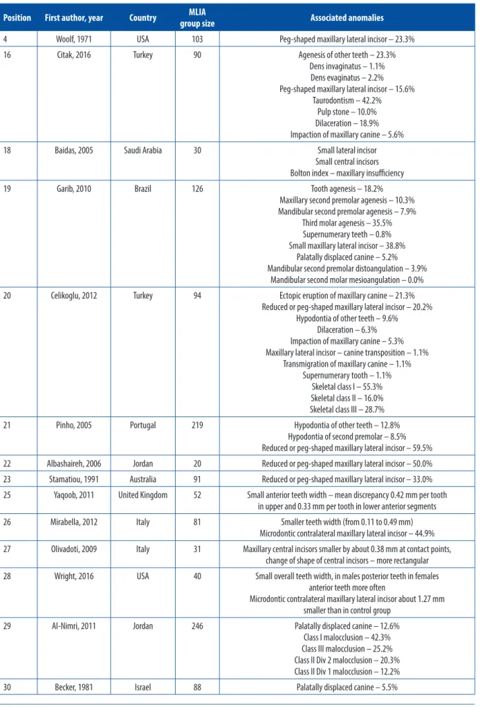
TABLE 1. Coexistence of agenesis of the maxillary lateral incisors with other dental anomalies based on a systematic review of the literature
Position First author, year Country group sizeMLIA Associated anomalies
4 Woolf, 1971 USA 103 Peg-shaped maxillary lateral incisor – 23.3% 16 Citak, 2016 Turkey 90 Agenesis of other teeth – 23.3%
Dens invaginatus – 1.1% Dens evaginatus – 2.2% Peg-shaped maxillary lateral incisor – 15.6%
Taurodontism – 42.2% Pulp stone – 10.0% Dilaceration – 18.9% Impaction of maxillary canine – 5.6% 18 Baidas, 2005 Saudi Arabia 30 Small lateral incisor
Small central incisors Bolton index – maxillary insufficiency 19 Garib, 2010 Brazil 126 Tooth agenesis – 18.2%
Maxillary second premolar agenesis – 10.3% Mandibular second premolar agenesis – 7.9%
Third molar agenesis – 35.5% Supernumerary teeth – 0.8% Small maxillary lateral incisor – 38.8%
Palatally displaced canine – 5.2% Mandibular second premolar distoangulation – 3.9%
Mandibular second molar mesioangulation – 0.0% 20 Celikoglu, 2012 Turkey 94 Ectopic eruption of maxillary canine – 21.3%
Reduced or peg-shaped maxillary lateral incisor – 20.2% Hypodontia of other teeth – 9.6%
Dilaceration – 6.3% Impaction of maxillary canine – 5.3% Maxillary lateral incisor – canine transposition – 1.1%
Transmigration of maxillary canine – 1.1% Supernumerary tooth – 1.1%
Skeletal class I – 55.3% Skeletal class II – 16.0% Skeletal class III – 28.7% 21 Pinho, 2005 Portugal 219 Hypodontia of other teeth – 12.8%
Hypodontia of second premolar – 8.5% Reduced or peg-shaped maxillary lateral incisor – 59.5% 22 Albashaireh, 2006 Jordan 20 Reduced or peg-shaped maxillary lateral incisor – 50.0% 23 Stamatiou, 1991 Australia 91 Reduced or peg-shaped maxillary lateral incisor – 33.0% 25 Yaqoob, 2011 United Kingdom 52 Small anterior teeth width – mean discrepancy 0.42 mm per tooth
in upper and 0.33 mm per tooth in lower anterior segments 26 Mirabella, 2012 Italy 81 Smaller teeth width (from 0.11 to 0.49 mm)
Microdontic contralateral maxillary lateral incisor – 44.9% 27 Olivadoti, 2009 Italy 31 Maxillary central incisors smaller by about 0.38 mm at contact points,
change of shape of central incisors – more rectangular 28 Wright, 2016 USA 40 Small overall teeth width, in males posterior teeth in females
anterior teeth more often
Microdontic contralateral maxillary lateral incisor about 1.27 mm smaller than in control group
29 Al-Nimri, 2011 Jordan 246 Palatally displaced canine – 12.6% Class I malocclusion – 42.3% Class III malocclusion – 25.2% Class II Div 2 malocclusion – 20.3% Class II Div 1 malocclusion – 12.2% 30 Becker, 1981 Israel 88 Palatally displaced canine – 5.5%
Position First author, year Country group sizeMLIA Associated anomalies
31 Pinho, 2011 Portugal 147 Deviation of the maxillary dental midline Molar and canine class II relationship
33 Woodworth, 1985 USA 43 Craniofacial deviations from normal: smaller maxillary length, smaller mandibular length, smaller anterior cranial base, nasal bone,
smaller vertical dimensions (anterior and posterior), smaller mandibular plane angle, 10o greater nasolabial angle
34 Pinho, 2011 Portugal 147 Shortening of the maxilla Reduced anterior facial height 35 Bassiouny, 2016 Saudi Arabia 52 Significant tendency for skeletal class III
Maxillary hypoplasia/retrognathia
TABLE 1. (Continued)
incisor occurs in 33% of patients with unilateral form of MLIA. Researchers highlight the genetic basis of this condition. Also, Pinho et al. [21] demonstrated a reduc tion in the dimensions of the lateral incisor of the oppo site side in 59.5% of patients. The Woolf [4] study showed that in 23.3% of patients with unilateral MLIA there is a conical shaped contralateral lateral incisor. In the study of Garib et al. [19] with unilateral agenesis of the max illary lateral incisor, the size of the crown of the incisor of the opposite side is decreased (38.8%). This result was very high compared to the reference studies carried out in 1998 by Bacetti [24], which estimated the incidence of this defect in the general population at 4.7%. Baidas and Hasim [18], when measuring the width of indi vidual teeth and analysis of the Bolton anterior index, found that in patients with unilateral MLIA, the width of the lateral incisor of the opposite side is 1 mm lower than the standard (5.5 mm, with the standard of 6.5 mm accepted by researchers). Maxillary central inci sors showed a reduction in width. Analysis of the Bolton anterior index showed the reduction of dental materi al, both in the case of bilateral and unilateral form
of MLIA. Also, a study conducted by Yaqoob et al. [25]
confirmed the reduction in the overall tooth size in pa tients with bilateral agenesis of the maxillary lateral in cisors compared to the control group, which comprised fully dented patients. The difference was noticeable in both the upper and lower dental arch. Researchers com pared the width of six front teeth. The mean values re sulting from the measurements of the maxillary teeth width in MLIA patients revealed a reduction in the di mensions of each tooth by an average of 0.42 mm, and in the case of mandibular teeth they were smaller by an average of 0.33 mm. Similar results, however, con sidering the width of 12 teeth in both the maxilla and the mandible, were obtained by Mirabella et al. [26]. Their study showed that the width of permanent teeth in patients with both unilateral and bilateral hypodon tia of the maxillary lateral incisors is on average 0.11 to 0.49 mm per tooth lower than in the control group with full permanent dentition. Only the maxillary first molars
did not show a reduced size. There was no difference in crown width compared to the control group in patients with bilateral and unilateral hypodontia of the maxillary lateral incisors. Researchers also evaluated the prev alence of the microdontic opposite side lateral inci sor in patients with unilateral MLIA. The frequency of the abovementioned anomaly was 44.8%. Mirabella et al. [26] also underlined the fact of the MLIA genet ic background. According to the researchers, the gene, causing hypodontia of the maxillary lateral incisors, reduces the total dimensions of the teeth. The authors emphasized how important from a clinical point of view the research they conducted is. The front teeth, due to the reduced size, will often require aesthetic restoration to ensure efficient occlusion and good aesthetics, both in the opening of space for the future prosthetic or im plant prosthetic restoration as well as in the orthodontic closure of the spaces. Another important clinical study is the work of Olivadoti et al. [27]. They assessed the shape and width of maxillary central incisors at the contact points and in the 1/3 gingival. The obtained results in dicated a reduction in the dimensions of these teeth in patients with MLIA at the level of contact points by an average of 0.38 mm, in the absence of a significant reduction in the width in 1/3 gingival. This indicated a change in the shape of the central incisors to more rectangular ones. Researchers emphasize the need for aesthetic correction of the shape of these teeth in pa tients with MLIA. Wright et al. [28] measured the width of the teeth in 40 patients with unilateral and bilateral hypodontia of the lateral incisors. The results indicated a reduction in the dimensions of the teeth. In men with MLIA, the lateral teeth were 0.28 mm to 0.78 mm smaller than the control group, and women showed a reduction in the dimensions of the anterior teeth by 0.22 mm to 0.42 mm. In cases of unilateral MLIA, the contralateral incisor showed an average width of 5.39 mm, compared with the average width of the maxillary lateral incisors in the control group of 6.66 mm, which gave a difference of 1.27 mm. Cooccurrence of the conical tooth was also more frequently observed.
Garib et al. [19] found an increased frequency of palatally displaced canines (PDC) in patients with hypo dontia of the lateral incisors in maxilla at 5.2%, which was statistically significant. Citak et al. [16] did not re port a higher incidence of impacted canines in patients with MLIA (5.6%). The results obtained in both studies are similar to each other, and the difference in their in terpretation may be caused by differences in the values in the reference data and the size of the studied groups. In the Garib et al. [19] trial, the incorrect positioning of other permanent teeth was also assessed: distoan gulation of second premolars in the mandible (3.9% in patients with MLIA, and only 0.2% in the reference study of the general population) and mesial inclina tion of the second molar in the mandible (no statisti cally significant differences). AlNimri and Bsoul [29] found a positive correlation between the occurrence of palatally impacted canines and the agenesis of the lat eral incisors in the maxilla. The study group consisted of 246 patients with single or bilateral maxillary later al incisor agenesis. In 12.6% of patients PDC coexisted with maxillary lateral incisor agenesis. However, there was no greater risk of occurrence of palatally impacted canines in patients with bilateral hypodontia of the lat eral incisors compared to the unilateral form. According to the researchers, the high percentage of coexistence of both disorders may confirm the guidance theory, ac cording to which the distal part of the root of the max illary lateral incisor leads the canine bud in the right direction during its long eruption path. The study is in line with the results obtained by Becker et al. [30], who also found a positive correlation between PDC and MLIA (5.5% of 88 patients with PDC showed agenesis of the maxillary lateral incisors). Celikoglu et al. [20], investigating the cooccurrence of dental anomalies with hypodontia of the maxillary lateral incisors, ob served that in a group of 94 patients with MLIA, 66% of patients suffer from structural defects, size, number or tooth position disorders. Ectopic canine eruption oc curred in 21.3% of patients with MLIA. There were 5.3% of patients with canine impaction, but the result was not statistically significant. Transmigration of the maxillary canines occurred only in 1.1% of patients.
Citak et al. [16] stated that the frequency of MLIA coexistence with the presence of dens invaginatus and evaginatus is respectively 1.1 and 2.2%. This result was not statistically significant and did not differ from the general population. Also, the incidence of pulp stones was not greater in patients with maxillary lateral incisor agenesis. However, there was a statistically significant increased frequency of taurodontic teeth in patients with MLIA and it was 42.2%. Dilaceration of the root was also more common (18.9%) than in the general pop ulation (9.5%). Celikoglu et al. [20] studies have shown that in patients with MLIA, the coexistence of root di laceration occurs at the level of 6.3%, but this result was not statistically significant for the population of patients
with full dentition. The abovementioned coincidence of the shape disorder of the lateral incisor of the oppo site side in patients with unilateral form of MLIA, often being at the same time a form of the abnormal shape of this tooth (conical tooth), has been underlined by re searchers in several works [4, 16, 22, 23]. This anomaly occurred in 15.6% up to 23.3% of patients with unilater al agenesis of the maxillary lateral incisor.
AlNimri and Bsoul [29] stated that among patients with hypodontia of maxillary lateral incisors, the most common was class I malocclusion (42.3%), the second was class III malocclusion (25.2%), then class II sub group 2 malocclusion (20.3%). The most rarely ob served was class II subgroup 1 malocclusion (12.2%). Pinho and Lemons [31] observed that in patients with unilateral and bilateral agenesis of maxillary lateral incisors, the deviation of the upper dental midline is common (in cases of unilateral agenesis, to the affected side) and the distal relationship is most often found on the molars and canines. A study by Celikoglu et al. [20] in which skeletal class was assessed in patients with aplasia of the maxillary lateral incisors showed that in MLIA patients skeletal class I was most common (55.3%), followed by skeletal class III (28.7%), and the least frequent skeletal class II (16%). There was also an increased incidence of skeletal class III in patients with MLIA with reference to the general population. In patients with agenesis, skeletal class III occurred in as many as 28.7%, and in the general population in
11.5% [20, 32]. Woodworth et al. [33], comparing ceph
alograms of patients with hypodontia of the maxillary lateral incisor with the norm, found that in patients with MLIA there occurs reduced length of the max illa and mandible, reduced anterior cranial base, and shortened nasal bone. There was also a decrease in the vertical relationship, both anterior and posterior face height, and reduction in the mandibular plane an gle. Analysis of soft tissues showed that in patients with MLIA the value of the nasolabial angle was on average 10° lower than the recognized norm. Researchers found that the results obtained gave a general picture very similar to those of cleft patients and this may indicate a weaker development of the craniofacial structures in these patients. Pinho et al. [34] also evaluated the effects of maxillary lateral incisor agenesis on skull structures in their studies. Their results confirmed the results ob
tained by Woodworth et al. [33]. The length of the max
illa was observed to be reduced (the measurement was made between the ANSPNS points) and the facial height also was reduced. However, MLIA did not cor relate with the change in the inclination of the palatal plane relative to the Frankfurt plane. A study by Bas
siouny et al. [35] showed that patients with congeni
tal lack of maxillary lateral incisors have a significant tendency to skeletal class III. The researchers attribute these results to the occurrence of maxillary hypoplasia (retrognathia) in these patients.
CONCLUSIONS
Based on information obtained from medical da tabases and library resources, it can be concluded that hypo dontia of the maxillary lateral incisors is a condition predisposing to the cooccurrence of other disorders: size, structure, position and quantity of other perma nent teeth. The distal molar and canine relationship was the most common. Studies carried out so far in Europe and worldwide indicate the genetic basis of the prob lem. Coexistence of the agenesis of permanent teeth, especially the maxillary lateral incisors, was observed in patients with ectopic canine eruption [29, 36]. The frequent lack of maxillary lateral incisors in patients with ectopic eruption, palatally displaced or impacted canines, confirms both guidance and genetic theories, which are cited as potential explanations of this con dition [30, 37]. From a clinical point of view, it is ex tremely important that patients with earlydetected maxillary lateral incisor agenesis should be able to check both the radiological and clinical process of maxil lary canines’ eruption, as it can be stated on the ba sis of the analysis of the referenced data that they are patients at risk of palatal retention of these teeth. In such cases, it may be necessary to conduct interceptive treatment involving the extraction of the deciduous maxillary canines at the appropriate stage of develop ment [3840]. Based on the systematic review of the lit erature it can be concluded that patients with maxillary lateral incisor agenesis also have a decrease in mesio distal crown dimensions of permanent teeth, especially the anterior teeth, and the crowns of the maxillary cen tral incisors most often have a rectangular shape. For this reason, very often the anterior teeth of patients with MLIA will require an aesthetic shape correction and in crease in width. The analysis of the conducted studies also showed that in patients with MLIA, skeletal class III was more frequent than in the general population, shorten ing the length of the maxilla, and a decrease in the ver tical relationship was also noted. Our own study [41] confirmed that the congenital lack of maxil lary lateral incisors is one of the symptoms indicating a maxillary deficiency syndrome. So far, no studies showing the fre quency of coexistence of the disorders discussed above have been carried out in patients with MLIA in the Polish population.
CONFLICT OF INTEREST
The authors declare no potential conflicts of interest with respect to the research, authorship, and/or publica tion of this article.
References
1. Schalk van der Weide Y, PrahlAndersen B, Bosman F. Tooth for mation in patients with oligodontia. Angle Orthod 1993; 63: 3137.
2. Polder BJ, Van’t Hof MA, Van der Linden FP, KuijpersJagtman AM. A metaanalysis of the prevalence of dental agenesis of permanent teeth. Community Dent Oral Epid 2004; 32: 217226.
3. Parkin N, Elcock C, Smith RN, et al. The aetiology of hypodontia: the prevalence, severity and location of hypodontia within fami lies. Arch Oral Biol 2009; 54: S52S56.
4. Woolf CM. Missing maxillary lateral incisors: a genetic study. Am J Hum Genet 1971; 23: 289.
5. Mostowska A, Biedziak B, Zadurska M, et al. WNT10A coding variants and maxillary lateral incisor agenesis with associated dental anomalies. Eur J Oral Sci 2015; 123: 18.
6. De Coster PJ, Marks LA, Martens LC, Huysseune A. Dental agenesis: genetic and clinical perspectives. J Oral Pathol Med 2009; 38: 117. 7. Kjær I, Kocsis G, Nodal M, Christensen LR. Aetiological aspects
of mandibular tooth agenesis – focusing on the role of nerve, oral mucosa, and supporting tissues. Eur J Orthod 1994; 16: 371375. 8. Kjær I. Neuroosteology. Crit Rev Oral Biol Med 1998; 9: 224244. 9. DaugaardJensen J, Nodal M, Skovgaard LT, Kjær I. Comparison
of the pattern of agenesis in the primary and permanent denti tions in a population characterized by agenesis in the primary dentition. Int J Paediatr Dent 1997; 7: 143148.
10. Goodman JR, Jones SP, Hobkirk JA, King PA. Hypodontia: clinical features and the management of mild to moderate hypodontia. Dent Update 1994; 21: 381384.
11. Larmour CJ, Mossey PA, Thind BS, et al. Hypodontia – a retrospec tive review of prevalence and etiology. Part I. Quintessence Int 2005; 36: 263270.
12. Garn SM, Lewis AB: The relationship between third molar agene sis and reduction in tooth number. Angle Orthod 1962; 32: 1418. 13. Garib DG, Peck S, Gomes SC. Increased occurrence of dental
anomalies associated with secondpremolar agenesis. Angle Orthod 2009; 79: 436441.
14. Rakhshan V. Congenitally missing teeth (hypodontia): a review of the literature concerning the etiology, prevalence, risk factors, patterns and treatment. Dent Res J (Isfahan) 2015; 12: 113. 15. Nieminen P. Genetic basis of tooth agenesis. J Exp Zool Part B Mol
Dev Evol 2009; 312B: 320342.
16. Citak M, Cakici EB, Benkli YA, et al. Dental anomalies in an or thodontic patient population with maxillary lateral incisor agene sis. Dent Press J Orthod 2016; 21: 98102.
17. Symons AL, Stritzel F, Stamation J. Anomalies associated with hypo dontia of the permanent lateral incisor and second premolar. J Clin Pediatr Dent 1993; 17: 109111.
18. Baidas L, Hashim H. An anterior tooth size comparison in uni lateral and bilateral congenitally absent maxillary lateral incisors. J Contemp Dent Pract 2005; 6: 5663.
19. Garib DG, Alencar BM, Lauris JR, Baccetti T. Agenesis of maxil lary lateral incisors and associated dental anomalies. Am J Orthod Dentofacial Orthop 2010; 137: 732.e16.
20. Celikoglu M, Kamak H, Yildirim H, Ceylan I. Investigation of the maxillary lateral incisor agenesis and associated dental anomalies in an orthodontic patient population. Med Oral Patol Oral Cir Bucal 2012; 17: e10681073.
21. Pinho T, Tavares P, Maciel P, Pollmann C. Developmental absence of maxillary lateral incisors in the Portuguese population. Eur J Orthod 2005; 27: 443449.
22. Albashaireh ZS, Khader YS. The prevalence and pattern of hypo dontia of the permanent teeth and crown size and shape deformity affecting upper lateral incisors in a sample of Jordanian dental pa tients. Community Dent Health 2006; 23: 239243.
23. Stamatiou J, Symons AL. Agenesis of the permanent lateral inci sor: distribution, number and sites. J Clin Pediatr Dent 1991; 15: 244246.
24. Baccetti T. A controlled study of associated dental anomalies. Angle Orthod 1998; 68: 267274.
25. Yaqoob O, DiBiase AT, Garvey T, Fleming PS. Relationship be tween bilateral congenital absence of maxillary lateral incisors and anterior tooth width. Am J Orthod Dentofacial Orthop 2011; 139: e229e233.
26. Mirabella AD, Kokich VG, Rosa M. Analysis of crown widths in subjects with congenitally missing maxillary lateral incisors. Eur J Orthod 2011; 34: 783787.
27. Olivadoti A, Doldo T, Treccani M. Morphodimensional analysis of the maxillary central incisor clinical crown in cases of congen itally missing upper lateral incisors. Prog Orthod 2009; 10: 1219. 28. Wright J, Bosio JA, Chou JC, Jiang SS. Maxillary lateral incisor
agenesis and its relationship to overall tooth size. J Prosthet Dent 2016; 115: 209214.
29. AlNimri KS, Bsoul E. Maxillary palatal canine impaction dis placement in subjects with congenitally missing maxillary lateral incisors. Am J Orthod Dentofacial Orthop 2011; 140: 8186. 30. Becker A, Smith P, Behar R. The incidence of anomalous maxil
lary lateral incisors in relation to palatallydisplaced cuspids. Angle Orthod 1981; 51: 2429.
31. Pinho T, Lemos C. Dental repercussions of maxillary lateral inci sor agenesis. Eur J Orthod 2011; 34: 698703.
32. Sayin M, Türkkahraman H. Malocclusion and crowding in an or thodontically referred Turkish population. Angle Orthod 2004; 74: 635639.
33. Woodworth DA, Sinclair PM, Alexander RG. Bilateral congenital absence of maxillary lateral incisors: a craniofacial and dental cast analysis. Am J Orthod 1985; 87: 280293.
34. Pinho T, Pollmann C, CalheirosLobo MJ, et al. Craniofacial reper cussions in maxillary lateral incisors agenesis. Int Orthod 2011; 9: 274285.
35. Bassiouny DS, Afify AR, Baeshen HA, et al. Prevalence of maxil lary lateral incisor agenesis and associated skeletal characteristics in an orthodontic patient population. Acta Odontol Scand 2016; 74: 456459.
36. Jang E, Lee K, An S, et al. Retrospective study of association be tween displacement of maxillary canine and tooth agenesis. J Clin Ped Dent 2015; 39: 488492.
37. Sambataro S, Baccetti T, Franchi L, Antonini F. Early predictive variables for upper canine impaction as derived from posteroan terior cephalograms. Angle Orthod 2005; 75: 2834.
38. Ericson S, Kurol J. Early treatment of palatally erupting maxil lary canines by extraction of the primary canines. Eur J Orthod 1988; 10: 283295.
39. Naoumova J, Kurol J, Kjellberg H. Extraction of the deciduous canine as an interceptive treatment in children with palatal dis placed canines – part I: shall we extract the deciduous canine or not?. Eur J Orthod 2015; 37: 209218.
40. Naoumova J, Kürol J, Kjellberg H. Extraction of the deciduous canine as an interceptive treatment in children with palatally dis placed canines – part II: possible predictors of success and cutoff points for a spontaneous eruption. Eur J Orthod 2015; 37: 219229. 41. Williams S, Loster JE, Loster BW. The relationship between maxil lary dental and occlusal anomalies: Evidence of a “Maxillary Defi ciency Syndrome”. Aust Orthod J 2018; 34: 212224.