Self-related processing in the sexual domain:
A parametric event-related fMRI study reveals neural
activity in ventral cortical midline structures
Alexander HeinzelUniversity of Du¨ sseldorf at the Research Centre Juelich, Du¨ sseldorf, Germany
Martin Walter, Felix Schneider, Michael Rotte, Christian Matthiae, Claus Tempelmann, Hans-Jochen Heinze, Bernhard Bogerts, and Georg Northoff
University of Magdeburg, Magdeburg, Germany
Self-related processing, reflecting the evaluation of environmental signals with regard to personal relevance, is fundamental for decision-making and subsequent behavioral responses. While self-related processing has already been investigated in several domains, one important domain, the sexual domain, has been spared so far.
Recent imaging studies suggest that self-related processing in different domains involves common regions in medial orbitofrontal and prefrontal cortex, the so-called ventral cortical midline structures (CMS). However, the same regions have also been implicated in sexual arousal, especially with regard to emotional processing in sexual arousal. Therefore it remains unclear whether this involvement of ventral cortical midline regions reflects emotional processing in sexual arousal or associated self-relatedness. We here report data from a parametric event-related fMRI study that investigated the neural correlates of self-related processing in sexual arousal, using erotic pictures from the International Affective Picture System. It was found that self-related activity associated with sexual arousal showed neural activity in ventral CMS regions such as the venteromedial prefrontal cortex (VMPFC) and the perigenual anterior cingulate cortex (PACC), while self-related activity not associated with sexual arousal showed neural activity in the dorsomedial prefrontal cortex (DMPFC). Our study indicates that self-relatedness may be considered a crucial component in sexual arousal that is mediated by neural activity in ventral cortical midline structures.
INTRODUCTION
The question of the self has intrigued researchers from various disciplines for centuries. The self represents a crucial point with regard to many different types of psychological and sociological processes. It contains the core of the identity of a human being, which is experienced as stable throughout lifetime and which also forms the
origin and the ultimate starting point of any social interaction. Recently, the theoretical concept of the self has been operationalized by referring to so-called ‘‘self-related processing.’’ Self-related processing concerns stimuli that are experienced as strongly related to one’s own person. They have also been described as ‘‘self-referential’’ or ‘‘self-relevant’’(Kelley, Macrae, Wyland, Caglar, Inati, & Heatherton, 2002; Northoff & Bermpohl,
Correspondence should be addressed to: Georg Northoff, Department of Psychiatry, University of Magdeburg, Leipziger Strasse 44, D-39120 Magdeburg, Germany. E-mail: [email protected]
The study was supported by a Doctoral student grant from the German Research Foundation to MW and by grants from the German Research Foundation (DFG, 304/4/1 to G. N.; SFB 426) to GN.
#2006 Psychology Press Ltd
2004; Phan, Taylor, Welsh, Ho, Britton, & Liber-zon, 2004). It is important to note that the self-relevance of a stimulus is not an intrinsic property of the stimulus, but determined by the experien-cing subject.
There is a growing body of literature indicating
common regions for self-related processing
throughout the different domains. Investigation of self-related processing in various domains, such as, for example, the verbal domain (Johnson, Baxter, Wilder, Pipe, Heiserman, & Prigatano, 2002; Kelley et al., 2002; Kjaer, Nowak, & Lou,
2002), memory domain (Macrae, Moran,
Heatherton, Banfield, & Kelley, 2004), the spatial domain (Maguire, Burgess, & O’Keefe, 1999; Maguire, Frith, Burgess, Donnett, & O’Keefe, 1998; Vogeley & Fink, 2003) and the emotional domain (Phan et al., 2004), consistently show involvement of medial cortical regions, the so-called ‘‘cortical midline structures’’ (CMS; North-off & Bermpohl, 2004). The cortical midline structures include an anterior and a posterior division. The anterior region includes ventral regions such as the medial orbitofrontal cortex (MOFC), the ventromedial prefrontal cortex (VMPFC) and the sub- and perigenual parts of the anterior cingulate cortex (PACC), whereas the dorsal region of the CMS includes the supragenual parts of the anterior cingulate cortex (SACC) and the dorsomedial prefrontal cortex (DMPFC). The posterior part of the CMS con-tains the posterior cingulate cortex (PCC), the medial parietal cortex (MPC), and the retro-splenial cortex (RSC).
The ventral division of the anterior CMS appears to have a major role in a number of different domains of self-related processing. In a recent study using visual emotional stimuli, Phan et al. (2004) found that the volitional act of appraising the extent of personal association specifically engaged VMPFC and PACC. Kelley et al. (2002) investigated a trait adjective judg-ment task comparing self-, other-, and case-referential adjectives. They demonstrated that the VMPFC and the DMPFC were selectively engaged in the self-related condition. Employing auditorily delivered statements, Johnson et al. (2002) compared judgments about one’s own abilities, traits and attitudes (such as ‘‘I can be trusted’’) to a semantic judgment task. The self-related condition was equally associated with activation in VMPFC and DMPFC, relative to the control condition. Another mode of stimula-tion was applied by Kjaer et al. (2002). Instead of
relying on sensory presentation of verbal items, they asked the subjects to mentally induce thoughts reflecting on one’s own personality traits and physical appearance. Self-related conditions (personality traits, physical appearance) induced activation predominately in VMPFC and PACC when compared to non-self-related conditions (i.e., thoughts about a famous person, the Danish queen). Finally, self-related processing in other domains like spatial stimuli (Vogeley, May, Ritzel, Falkai, Zilles, & Fink, 2004), autobiographical stimuli (Fossati et al., 2003, 2004; Lou et al., 2004; Macrae et al., 2004), and facial stimuli (Fossati et al., 2003, 2004; Kircher et al., 2000, 2001; Lou et al., 2004; Macrae et al., 2004; Platek, Keenan, Gallup, & Mohamed, 2004) confirm these find-ings.
While self-related processing has already been investigated in several domains, one important domain, the sexual domain, has not been exten-sively researched. Therefore in this paper we aim to investigate self-related processing in the sexual domain. The processing of erotic stimuli inducing sexual arousal may be considered a paradigmatic case of self-related processing. For example, the same stimulus may sexually arouse two different persons completely differently, i.e., one may be strongly aroused whereas the other may remain indifferent. Therefore, self-relatedness of the sexual stimulus may be considered a crucial variable in eliciting sexual arousal and the asso-ciated behavior.
What are the neural correlates of sexual arousal? Several imaging studies induced sexual arousal by showing erotic film excerpts or pic-tures. (Beauregard, Levesque, & Bourgouin, 2001; Ferretti et al., 2005; Karama et al., 2002; Park, Seo, Kang, Ryu, Kim, & Jeong, 2001; Stoleru et al., 1999). Among various other re-gions, these studies show involvement of the ventral and dorsal anterior CMS in sexual arou-sal. Based on the imaging of emotions (Phan, Wager, Taylor, & Liberzon, 2002), regions like the MOFC, the VMPFC, the DMPFC, and the PACC are supposed to be associated with the emotional component of sexual arousal as distinguished from its cognitive, motivational, and physiological components (Ferretti et al., 2005; Karama et al., 2002). However, as reported above, the same regions have also been implicated in self-related
processing. Accordingly, it remains unclear
whether neural activity in the ventral CMS reflects self-related processing of either sexual arousal or of associated emotional processing.
Based on prior findings of the involvement of especially ventral anterior CMS in self-related-ness, we hypothesized that self-relatedness of sexual arousal is associated with neural activity in CMS regions like the MOFC, the VMPFC, the DMPFC, and the PACC.
In order to differentiate self-related processing in sexual arousal and emotional processing, the factor self-relatedness needs to be varied across sexual and emotional stimuli with the factor
emotion (emotional valence and intensity)
remaining unaltered. We therefore presented participants with erotic-emotional and non-ero-tic-emotional and neutral stimuli from the Inter-national Affective Picture system (IAPS) (Center for the Study of Emotion and Attention, 1999). Furthermore, to avoid cognitive task components such as discrimination and decision-making, subjects only viewed the IAPS-pictures in the scanner without distinguishing between
self-and non-self-related stimuli. Subject-specific
evaluation of self-relatedness was obtained in a post-scanning session using a visual analogue scale. This allowed us to generate parametric correlation maps of self-relatedness independent of cognitive task components.
METHODS
Participants
We investigated 13 healthy male participants
(age: 38.79/6.7, M9/SD) without any known
psychiatric, neurological, or medical disease. All were right-handed as assessed by the Edinburgh Inventory for Handedness (Oldfield, 1971). After detailed explanation of the study design and potential risks all subjects gave written informed consent. The study was approved by the institu-tional review board of the Otto-von-Guericke University of Magdeburg.
Paradigm
Participants were asked to view photographs taken from the International Affective Picture System (IAPS). According to IAPS standard
valence, negative (valence: 1/3), neutral
(va-lence: 4/6), and positive (valence: 7/9) pictures
were presented for a duration of 5 s. Picture sets were counterbalanced across subjects as well as within each subject according to the three
cate-gories erotic-emotional (including positive and negative), non-erotic-emotional (including posi-tive and negaposi-tive), and neutral (including both erotic and non-erotic scenes). Predominantly erotic-emotional pictures were those where a body was presented and clearly visible with a strong erotic touch. For example, a naked woman showing her breast was considered a predomi-nantly erotic-emotional picture. In contrast, for example, a baby’s face including its dressed upper body was considered a non-erotic-emotional pic-ture (Figure 1). The strong erotic touch with sexual involvement was considered the main criterion for distinguishing between predomi-nantly erotic and non-erotic-emotional stimuli.
Erotic and non-erotic-emotional pictures were matched with respect to valence, and arousal. The
mean IAPS values (valence/arousal, Ms9/SD)
were 5.27/5.29 (9/2.26/0.92) for erotic-emotional
pictures, 4.96/5.84 (9/2.46/0.84) for
non-erotic-emotional pictures, and 4.96/3.21 (9/0.35/0.93)
for neutral pictures.
All three, erotic and non-erotic-emotional and neutral pictures were presented in a randomized order across all eight runs. Subjects were in-structed to view the pictures passively without making any efforts (rate, evaluate, or make a judgment about the content or the effects on the subjects). During IAPS picture viewing, an arbi-trary button press without any judgment was required to control for movement effects and to assure a constant level of attention during picture viewing. Reaction times from picture onset to button press were measured. At the same time of scanning, subjects were not aware of any post-scanning ratings.
Half of the presented pictures were started with
an expectancy period of 4/6 s (4.0, 4.5, 5.0, 5.5,
6.0 s) indicating the type of the subsequently presented picture. Mean durations of preceding expectancy and its variance were balanced for the main picture categories (erotic-emotional, non erotic but emotional and neutral) as well for their subcategories (expected/unexpected and positive/ negative). The expectancy period was indicated by presentation of a white arrow on a dark back-ground. A downward arrow indicated erotic-emotional expectancy, an upward arrow indicated non-erotic-emotional expectancy, and a rightward arrow indicated neutral expectancy (Figure 1).
After the picture presentation a fixation cross
(intertrial interval) was presented for 8/10 s
(8.0, 8.5, 9.0, 9.5, 10.0 s). This allowed the subjects to recover from emotional stimulation and, in
addition, served as a baseline condition (Stark & Squire, 2001). The baseline duration was ran-domly varied accounting for variable stimulus onset asynchrony. A total of 256 trials was presented in eight runs. Prior to the experimental session, subjects were familiarized with the para-digm by completing a test run with 32 trials
During fMRI, pictures were projected auto-matically via a computer and a forward projection system on a screen placed at the end of the subject’s gurney. Subjects lay supine in the scanner and viewed the screen through a mirror positioned on the head coil. Subjects were asked to keep their eyes open and fixate the middle of the screen in front of them. They were asked not to move finger, head or body during picture presentation and viewing with the exception of the button press for the response.
Behavior
Behavioral monitoring. We measured reaction times, which were defined as the time between the onset of the picture screen (IAPS photo-graph) and the subsequent button press. Reaction times were calculated in an ANOVA with three factors (erotic and non-erotic and neutral IAPS pictures) with each on two levels (pictures with and without preceding expectancy period).
Aver-age reaction times were compared using
Kruskal/Wallis Test.
Rating of pictures was conducted outside the scanner after the fMRI session. The same pictures as in fMRI were presented in a new and randomized order. Unlike in fMRI, each picture was now followed by a task period that consisted of ratings of self-relatedness, emotional intensity, and valence. All three responses were given using a visual analogue scale (see Figure 1). Self-relatedness was assessed using the question: ‘‘How much do I personally associate with or relate to this picture?’’ (translated from German)
and ranged from 1 (low personal association) to 9
(high personal association). Valence was assessed using the question: ‘‘How unpleasant/pleasant is that picture?’’ and ranged on a continuum from 1 (negative) to 9 (positive). Emotional intensity was assessed using the question: ‘‘How intense is this picture?’’ and ranged on a continuum from 1
(low) to 9 (high) (Figure 1B). We were aware that
emotional responses might attenuate when pic-tures were seen for a second time. However, this habituation effect applied equally to all picture conditions and was not expected to affect the differences between conditions (Anderson et al., 2003).
Analysis of behavioral data. We calculated the mean average ratings of self-relatedness (and the
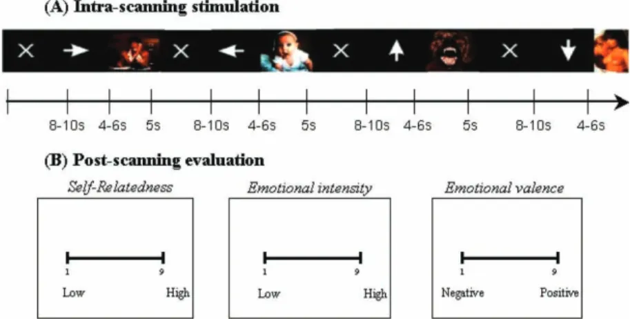
Figure 1. Schematic presentation of fMRI paradigm for IAPS picture viewing and post-scanning picture evaluation. (A) During scanning subjects were instructed to view pictures without making any decision about the nature of the picture (except for an arbitrary button click). Erotic- and non-erotic-emotional pictures as well as neutral pictures were presented. Presentation of pictures (duration: 5 s) was preceded by the respective expectancy period, erotic- and non-erotic-emotional, neutral, (variable duration: 4.0, 4.5, 5.0, 5.5, 6.0 s) as indicated by arrows in different directions. Baseline of variable duration (8.0, 8.5, 9.0, 9.5, 10.0 s) followed after each picture and was presented by a white fixation cross. (B) Post-scanning evaluation concerned exactly the same pictures presented in fMRI including some novel distraction pictures. Subjects had to evaluate self-relatedness, emotional intensity, and emotional valence on a visual-analogue scale ranging from 1 (low) to 9 (high). All three ratings (self-relatedness, valence, intensity) were presented in randomized order. As soon as the subject responded on the computer screen and made his mark on the scale, the next scale appeared. The paradigm was presented in post-scanning evaluation in the same way (time duration, expectancy, etc.) as during fMRI scanning though the order of the pictures was different, i.e., randomized.
other two ratings, valence and intensity) for each picture category (erotic, emotional, neutral).
Ratings of emotional intensity and valence were statistically compared with those provided
by the IAPS manual using at-test; no significant
differences were observed.
Imaging
Scanning procedures. Data acquisition was conducted on a 1.5 Tesla General Electric Sigma scanner using a standard headcoil. Imaging pro-cedures included collection of: (a) structural high resolution images (rf-spoiled GRASS sequence 60 slices sagittal, 2.8 mm thickness); (b) T1 weighted anatomic images coplanar with the functional images (23 slices, aligned to the plane connecting the anterior and posterior commissure axis covering the whole head in oblique axial orientation); (c) inversion recovery T1 weighted echo planar images coplanar with the functional images; and (d) echo planar functional images sensitive to BOLD contrast (402 sequential acquisitions, 23 slices with 3.125 mm in-plane resolution, 5 mm thickness, 1 mm gap; T2* weighted gradient echo sequence: TR 2 s, TE 40 ms). The first seven images were discarded due to T1 saturation effects. Subjects were positioned in the scanner and the head was immobilized with foam pieces and a Velcro strip around the fore-head. By means of a mounted mirror on the headcoil a screen was visible, on which stimuli were projected using a LCD projector.
Image analysis. Image processing and statistical analyses were carried out using MATLAB 6.5.1 (The Mathworks Inc., Natick, MA, USA) and SPM2 (Statistical parametric mapping software, SPM; Wellcome Department of Imaging Neu-roscience, London, UK; http://www.fil.ion.ucl.a-c.uk; Friston, Holmes, Worsley, Frith, Frackowiak, & Poline, 1995). The procedure is summarized below; cited papers provide the complete math-ematical detail. After discharge of the first seven
volumes to allow T1 stabilization, 3160 (8/395)
volume images were realigned to the first image to correct for head movement between scans, mean-adjusted by proportional scaling, resliced and normalized into standard stereotactic space (resulting in an isotrophic 3 mm resolution). Subjects showing translational head movement of more than 2 mm or rotational movement of more than 1 degree were excluded. We excluded two subjects (from the initial 15 subjects) for this
reason from further analysis. The space of the template image used in SPM for image normal-ization is based upon the Talairach system but derived from 152 brains at the Montreal Neuro-logical Institute (MNI; see SPM templates man-ual for details). This spatial transformation includes both linear and nonlinear dimensions and uses a nonlinear sampling algorithm (Friston et al., 1995). Data were thereafter expressed in terms of standard stereotactic co-ordinates in the x, y and z-axes. Transformed functional data sets from each subject were smoothed with a Gaussian kernel of 8 mm (full-width half-maximum) for the group analysis to meet the statistical require-ments of the general linear model and to com-pensate for normal variation in individual brain size, shape, and sulcal/gyral anatomy across sub-jects.
Subject-specific low frequency drifts in signal were removed by a high-pass filter of 128 s. For each subject we defined a design matrix modeling viewing of erotic-emotional IAPS pictures as well as viewing of erotic-emotional and non-erotic non-emotional IAPS pictures as separate events. Besides the three experimental conditions. In addition, we included the baseline as separate event into our design matrix. We modeled the variable duration (8.0 s, 8.5 s, 9.0 s, 9.5 s, 10.0 s) of the baseline making explicit use of variable intervals in the data analysis (Sakai & Passing-ham, 2003). After estimation of all model para-meters, specific effects were tested by applying appropriate linear contrasts to the parameter
estimates for each condition, resulting in a t
-statistic for each voxel.
Regions of interest. According to the a priori hypotheses we defined the ventral cortical mid-line structures including MOFC, VMPFC, PACC and DMPFC as regions of interest. The MOFC includes the gyrus rectus and the medial half of the orbital gyri, whereas the VMPFC contains the ventral half of the medial prefrontal surface including the frontal pole. The PACC covers the anterior part of the cingulate gyrus, which arches around the genu and the body of the corpus callosum. The ventral section of the PACC includes a subgenual and a pregenual part and is anatomically and functionally closely connected to the MOFC and the VMPFC, the dorsal section of the PACC is separated from the DMPFC by the cingulate sulcus. The DMPFC covers the dorsal section of the prefrontal cortex, including the middle parts of the superior and medial
frontal gyrus. It borders dorsally with the frontal eye field and ventrally with the VMPFC and the MOFC. (Northoff & Bermpohl, 2004)
In the interpretation of our results only activa-tions within these regions where considered, which additionally survived a global height
threshold of pB/.001 uncorrected for multiple
comparisons and an extent threshold of k/10
voxels. (See EditorialNature Neuroscience, 2001).
Statistical analysis. To elucidate the general effects of self-related processing in IAPS picture viewing we applied subject-specific regression analysis. To that end subjective ratings of self-relatedness for each picture as well as the baseline conditions were included as parametric regressors in the design matrix for each subject (Anderson et al., 2003; Goldin, Hutcherson, Ochsner, Glover, Gabrieli, & Gross, 2005; Heinzel et al., 2005). A random effects model was generated allowing inference to the general population.
In order to identify the specific activation of self-related processing in the sexual domain a separate correlation map for erotic stimuli was calculated and compared to the baseline.
To distinguish between effects of self-related-ness in erotic and non-erotic-emotional stimuli, a correlation map for non-erotic-emotional pictures was calculated. In order to elucidate specific effects we performed exclusive masking analysis. In contrast to subtraction analysis the results of masking are not influenced by deactivations, i.e., a strong deactivation in the control condition may not lead to ‘‘pseudo-activation’’ in the experimental condition by using masking. This can be of particular importance when using emotional activation as a control condition, since it has been shown that emotional processing is often characterized by deactivations (Heinzel et al., 2005).
Exclusive masking was applied to the condi-tion of interest (i.e., self-relatedness of erotic-emotional pictures) to eliminate all voxels associated and correlated with self-relatedness of non-erotic-emotional pictures.
We also performed the same analysis for self-relatedness of non-erotic-emotional pictures exclusively masking them with the map for self-relatedness of erotic-emotional pictures. The
significance level of the masks was set at pB/.05
uncorrected whereas the threshold for the main
contrast to pB/.001 uncorrected.
RESULTS
Behavioral data
Reaction times (from appearance of picture to arbitrary button press) in the scanner for all
pictures was M/860 ms, SD/9/270 ms. We
observed a significant difference (pB/.05)
be-tween erotic-emotional pictures (M/880 ms,
SD/9/260 ms) and non-erotic-emotional
pic-tures (M/860 ms, SD/9/280 ms). In contrast,
there was no significant difference in reaction
times between all expected pictures (M/859 ms,
SD/9/264 ms) and all unexpected/ambiguous
pictures (M/860 ms, SD/9/260 ms); the same
was true for sexual, emotional and neutral expected and unexpected pictures.
Subjective evaluation of self-relatedness of stimuli revealed the following results. Mean average ratings of self-relatedness for all pictures
were M/3.75, SD/9/1.08, for erotic-emotional
pictures M/5.23, SD/9/1.48, for
non-erotic-emotional pictures M/4.34, SD/9/1.64, and
for neutral picturesM/2.03,SD/9/0.82.
fMRI data
We first investigated the effects all pictures above baseline with the subjective ratings of self-relat-edness relying on subject-specific regression analysis. Regions significantly positively correlat-ing with the degree of self-relatedness included anterior medial cortical regions such as MOFC
(x/0, y/57, z/0; z-value/3.54) and (x/3,
y/51, z/24;z-value/3.70) as well as DMPFC
(x/3, y/54, z/39, z-value/3.34). We also
observed neural activity in the posterior cingulate cortex bordering the retrosplenium and
precu-neus (x/3,y//57,z/15;z-value/3.64) right
lateral parietal cortex (x/48, y//66, z/15;
z-value/4.09), left lateral parietal cortex (x/
/45,y//75,z/15,z-value/4.00), left lateral
premotor cortex (x//42, y//9, z/57,
z-value/3.91), dorsomedial thalamus bordering
the nucleus accumbens and caudatum (x//3,
y//9, z/6; z-value/3.65) and the midbrain/
tectum (x//3,y//24,z/0;z-value/3.56).
In the second analysis we tested for neural activation that specifically correlates with self-relatedness in erotic-emotional picture processing against baseline. Significant correlations within our regions of interest were found in the VMPFC
and the PACC. Furthermore, we found significant correlations in the right amygdala, the lateral prefrontal cortex and the midbrain (Table 1).
In order to distinguish self-relatedness of erotic- and non-erotic-emotional stimuli, we ob-tained separate correlation maps of self-related-ness for both stimuli and masked them with each other. This allowed the detection of those self-related signal changes specifically associated with erotic and non-erotic stimuli respectively.
Self-relatedness of erotic-emotional stimuli as exclusively masked with those of non-erotic-emotional stimuli revealed significant signal
changes in the VMPFC (x//9, y/45, z/12,
z-value/3.78), the PACC (x/9, y/42, z/6,
z-value/3.90), and the midbrain (x/6,y/9,z/
/12, z-value/4.00). These regions, therefore,
should be considered specific for self-relatedness of sexual arousal (Figure 2, upper part).
The converse analysis, self-relatedness of non-erotic-emotional stimuli as exclusively masked with those of erotic-emotional stimuli showed
signal changes in the DMPFC (x//3, y/30,
z/60,z-value/4.30) and the left lateral
premo-tor cortex (x//39, y//27, z/60, z-value/
4.04). These regions appear specific for self-relatedness of emotional processing (Figure 2, lower part).
DISCUSSION
We investigated the neural correlates of self-relatedness in the sexual domain distinguished from associated emotional processing. The fMRI data revealed neural activation that specifically correlates with self-relatedness in erotic picture processing. Significant correlations were found within the VMPFC and the PACC. Exclusive
masking analysis showed these regions to be specifically involved in self-related processing in the sexual domain rather than in the emotional domain, which instead revealed the DMPFC. Taken together, these findings lend support to our hypothesis of the crucial involvement of the ventral CMS in self-related processing during sexual arousal and its distinction from self-related processing in the emotional domain. The ventral CMS may therefore be considered a neural correlate of sexual self-related processing.
Our results are in accordance with studies investigating the neural correlates of sexual arousal that also found neural activation in VMPFC and PACC (Beauregard et al., 2001; Ferretti et al., 2005; Karama et al., 2002; Park et al., 2001; Stoleru et al., 1999). This complex neural network has been supposed to involve different components of sexual arousal, such as cognitive, emotional, motivational and physiolo-gical (Stoleru et al., 1999). According to this view the cognitive component is related to the evalua-tion of a stimulus as a sexual incentive. The emotional component involves the hedonic qual-ity of sexual arousal, the motivational component refers to the process that direct behavior to a sexual goal and the physiological component concerns the autonomic and endocrinological responses (Karama et al., 2002). Karma et al. (2002) attributed the neural activation in VMPFC and PACC to the emotional component of sexual arousal when comparing erotic film excerpts to neutral films in men and women. A similar view has been expressed by Ferretti et al. (2005) who suggest that PACC and the prefrontal cortex contribute to evaluate the emotional information associated with sexual arousal.
Our results suggest that neural activity in the VMPFC and the PACC may rather be associated
TABLE 1
Summary of brain regions with significant signal changes in subject-specific regression analysis of erotic pictures versus baseline
Co-ordinates
Region x y Z z-values
VMPFC /9 45 12 3.78
Right PACC 12 41 /2 3.93
Left precentral gyrus /27 /20 70 4.04
Right amygdala 18 /4 17 3.85
Right amygdala 27 /7 17 3.57
Left lateral premotor cortex /42 /9 57 3.91
Midbrain 6 /9 /12 3.64
Note: All foci were identified with a global height threshold ofpB/.001 uncorrected for multiple comparisons and an extent
with self-related processing during sexual arousal than with emotional processing. Since neither Karama et al. (2002) nor others explicitly inves-tigated self-relatedness of sexual stimuli, they were unable to distinguish between emotional and self-related processing because both types of processing are high in the experimental condition and low in the control condition. The same problem arises in other studies using similar stimuli. Arnow et al. (2002) used equally erotic film sequences compared to video material with sports or relaxing segments. The same holds for Ferretti et al. (2005), who applied neutral and erotic films as well as erotic and sport pictures.
Though these studies allow for the identification of the general neural network implicated in sexual arousal and potentially in associated emo-tional processing, they do not allow us to distin-guish the latter component from self-relatedness. Since our study matched for emotional valence and intensity across erotic-emotional and non-erotic-emotional pictures, we were specifically able to account for self-relatedness of sexual arousal as distinguished from self-relatedness of emotional processing. The results of our study suggest that in addition to the emotional, cogni-tive, motivational, and physiological component, a further component, self-relatedness, needs to be
Figure 2. Parametric correlation maps of self-relatedness for erotic- and non-erotic-emotional pictures. (A) The signal changes in the contrast all erotic-emotional pictures above baseline were correlated with post-scanning evaluation of self-relatedness (included as regressor in the design matrix). This contrast is exclusively masked with the contrast non-erotic-emotional pictures above baseline correlated with post-scanning evaluation of self-relatedness. Subject-specific regression analysis of self-relatedness was done atpB/
.001 uncorrected with extent thresholdk/10 voxels. The sagittal image depicts the right hemisphere. Note that self-relatedness for
erotic-emotional pictures predominantly correlates with signal changes in ventral cortical and subcortical midline regions. (B) The signal changes in the contrast all non-erotic-emotional pictures above baseline were correlated with post-scanning evaluation of self-relatedness (included as regressor in the design matrix). This contrast is exclusively masked with the contrast, erotic-emotional pictures above baseline correlated with post-scanning evaluation of relatedness. Subject-specific regression analysis of self-relatedness was done atpB/.001 uncorrected with extent thresholdk/10 voxels. The sagittal image depicts the right hemisphere.
Note that self-relatedness for non-erotic-emotional pictures predominantly correlates with signal changes in dorsal cortical midline region. Abbreviations: L/left; VMPFC/ventromedial prefrontal cortex; DMPFC/dorsomedial prefrontal cortex; MB/
considered in sexual arousal. A sexual stimulus may have no arousing effects as long as the
person cannot relate it to her/himself*/thus it is
self-related processing that may transform a mere sexual stimulus into a sexually arousing stimulus. A number of studies investigating emotions have reported activation in the VMPFC and the PACC (see Phan et al., 2002, for an overview). However, the classical attribution of emotional processing to the prefrontal medial cortex and the anterior cingulate is challenged by recent studies on self-related processing. Phan et al. (2004) investigated the degrees of self-relatedness during emotional processing. In this study, participants had to appraise the extent of personal association of emotionally salient pictures during fMRI. The fMRI results were correlated with subjective ratings (after fMRI) appraising the degree of self-relatedness of the picture content in a visual analogue scale. Regions associated with self-relatedness included the medial prefrontal cortex and the anterior cingulate (other regions acti-vated in relation to self-relatedness included the insula and the nucleus accumbens). It was found that the more self-related the picture content was appraised, the more activation was observed in these regions. In a related study (Fossati et al., 2003) subjects had to judge whether emotional, i.e., positive and negative personality trait adjec-tives described themselves properly. For control, subjects were asked if the adjectives described generally desirable traits. The medial prefrontal cortex and the anterior cingulate were specifically activated during self-related evaluation of words irrespective of their emotional valence. Ochsner et al. (2004) compared self-relevance of visually presented negative emotional pictures (self-focus) with alternative meanings for pictured actions and their situational contexts (situation-focus). They observed increased recruitment of the PPACC/SPACC in the self-focus and of right and left LPFC in the situation-focus. This is in line with other studies (Gusnard, Akbudak, Shul-man, & Raichle, 2001; Gusnard & Raichle, 2001), where attention to self-referent emotional condi-tions induced neural activity in PPACC/SPACC, VMPFC, and DMPFC when compared to exter-nally cued attention.
These results confirm, what we found for the sexual domain. It is suggested that the ventral cortical midline structures are rather related to self-related processing than to emotional proces-sing.
Our results contribute to these findings by showing that self-related processing in a hitherto uninvestigated domain, the sexual domain, also recruits ventral CMS as it has been observed during self-related processing in other domains. This suggests that at least in the ventral CMS self-related processing may be domain independent. Whether this indicates domain specificity or differences in some other, as yet unexplored, psychological factors may be the subject of future studies.
It could be argued that our erotic stimuli differed from non-erotic-emotional stimuli in dimensions other than sexual intensity. However, our post hoc ratings indicate that emotional intensity and the emotional valence were equal. Furthermore, the post hoc ratings demonstrate that the erotic-emotional pictures were consid-ered as more self-related compared to the non-erotic-emotional pictures underlining the signifi-cance of self-relatedness in erotic picture proces-sing. However, future studies might want to employ more sensitive manipulation checks.
Ventral cortical midline regions have been shown to be involved in functions other than self-related processing. For example, functions like reward (Rolls, Tovee, & Panzeri, 1999; Schultz, Tremblay, & Hollerman, 2003), mentaliz-ing (Mitchell, Banaji, & Macrae, 2005), theory of mind (Frith & Frith, 2003), moral judgments (Greene, Sommerville, Nystrom, Darley, & Co-hen, 2001), and person-related processing (Gilli-han & Farah, 2005) have all been shown to involve anterior cortical midline regions. How-ever, the exact relationship of self-related proces-sing and these other types of procesproces-sing remains unclear. It may be assumed that self-related processing is strongly implicated or even presup-posed in reward- or person-related processing. Future studies directly comparing self-related processing and reward- or person-related proces-sing are necessary to address this issue.
Another cognitive function to consider is attention, which could confound signal changes during self-related processing. Self-related pro-cessing may, for example, attract more attention compared to non-self-related processing. Signal changes associated with self-related processing might thus be at least partly traced back to attention effects rather than to self-related pro-cessing per se. Since we did not control for the influence of attention separate from self-related processing we cannot exclude the possibility that the activation may partly be confounded by
attention effects. It has been reported that the DMPFC is involved in mediating attentional modulation of self-related stimuli (Gusnard et al., 2001, Phan et al., 2004). However, investiga-tions explicitly testing the direct interference between attention and self-related stimuli remain to be reported.
Furthermore, picture viewing during scanning as well as post hoc evaluation of self-relatedness might include some implicit cognitive component that could possibly confound signal changes (Cunningham, Raye, & Johnson, 2005; Hutcher-son, Goldin, Ochsner, Gabrieli, Barrett, & Gross, 2005) supposedly associated with self-relatedness. Furthermore, it should be considered that post-scanning evaluation could principally contain some confounding cognitive function (repetition and memory effects, etc.). Therefore, our results need support from studies controlling for such cognitive confounds.
Manuscript received 18 December 2005 Manuscript accepted 27 February 2006
REFERENCES
Anderson, A. K., Christoff, K., Stappen, I., Panitz, D., Ghahremani, D. G., Glover, G., et al. (2003). Dissociated neural representations of intensity and valence in human olfaction.Nature Neuroscience,6, 196/202.
Arnow, B. A., Desmond, J. E., Banner, L. L., Glover, G. H., Solomon, A., Polan, M. L., et al. (2002). Brain activation and sexual arousal in healthy, heterosex-ual males.Brain,125, 1014/1023.
Beauregard, M., Levesque, J., & Bourgouin, P. (2001). Neural correlates of conscious self-regulation of emotion.The Journal of Neuroscience,21, RC165. Center for the Study of Emotion and Attention. (1999).
International affective picture system. Gainesville, FL: University of Florida.
Cunningham, W. A., Raye, C. L., & Johnson, M. K. (2005). Neural correlates of evaluation associated with promotion and prevention regulatory focus. Cognitive, Affective & Behavioral Neuroscience, 5, 202/211.
Editorial (2001). Analyzing functional imaging studies. Nature Neuroscience,4, 333.
Ferretti, A., Caulo, M., Del, G. C., Di, M. R., Merla, A., Montorsi, F., et al. (2005). Dynamics of male sexual arousal: Distinct components of brain activation revealed by fMRI.Neuroimage,26, 1086/1096. Fossati, P., Hevenor, S. J., Graham, S. J., Grady, C.,
Keightley, M. L., Craik, F., et al. (2003). In search of the emotional self: An fMRI study using positive and negative emotional words.American Journal of Psychiatry,160, 1938/1945.
Fossati, P., Hevenor, S. J., Lepage, M., Graham, S. J., Grady, C., Keightley, M. L., et al. (2004). Distributed
self in episodic memory: Neural correlates of successful retrieval of self-encoded positive and negative personality traits. Neuroimage, 22, 1596/ 1604.
Friston, K. J., Holmes, A. P., Worsley, K. J., Frith, C. D., Frackowiak, R. S. J., & Poline, J. P. (1995). Statistical parametric maps in functional imaging: A general linear approach. Human Brain Mapping, 2, 189/ 210.
Frith, U., & Frith, C. D. (2003). Development and neurophysiology of mentalizing. Philosophical Transactions of the Royal Society of London: B. Biological Sciences,358, 459/473.
Gillihan, S. J., & Farah, M. J. (2005). Is self special? A critical review of evidence from experimental psy-chology and cognitive neuroscience. Psychological Bulletin,131, 76/97.
Goldin, P. R., Hutcherson, C. A., Ochsner, K. N., Glover, G. H., Gabrieli, J. D., & Gross, J. J. (2005). The neural bases of amusement and sadness: A comparison of block contrast and subject-specific emotion intensity regression approaches. Neuro-image,27, 26/36.
Greene, J. D., Sommerville, R. B., Nystrom, L. E., Darley, J. M., & Cohen, J. D. (2001). An fMRI investigation of emotional engagement in moral judgment.Science,293, 2105/2108.
Gusnard, D. A., Akbudak, E., Shulman, G. L., & Raichle, M. E. (2001). Medial prefrontal cortex and self-referential mental activity: Relation to a default mode of brain function. Proceedings of the National Academy of Sciences of the USA,98, 4259/ 4264.
Gusnard, D. A., & Raichle, M. E. (2001). Searching for a baseline: Functional imaging and the resting human brain. Nature Reviews: Neuroscience, 2, 685/694.
Heinzel, A., Bermpohl, F., Niese, R., Pfennig, A., Pascual-Leone, A., Schlaug, G., et al. (2005). How do we modulate our emotions? Parametric fMRI reveals cortical midline structures as regions speci-fically involved in the processing of emotional valences.Brain Research: Cognitive Brain Research, 25, 348/358.
Hutcherson, C. A., Goldin, P. R., Ochsner, K. N., Gabrieli, J. D., Barrett, L. F., & Gross, J. J. (2005). Attention and emotion: Does rating emotion alter neural responses to amusing and sad films? Neuro-image,27, 656/668.
Johnson, S. C., Baxter, L. C., Wilder, L. S., Pipe, J. G., Heiserman, J. E., & Prigatano, G. P. (2002). Neural correlates of self-reflection.Brain,125, 1808/1814. Karama, S., Lecours, A. R., Leroux, J. M., Bourgouin,
P., Beaudoin, G., Joubert, S., et al. (2002). Areas of brain activation in males and females during viewing of erotic film excerpts.Human Brain Mapping, 16, 1/13.
Kelley, W. M., Macrae, C. N., Wyland, C. L., Caglar, S., Inati, S., & Heatherton, T. F. (2002). Finding the self? An event-related fMRI study. Journal of Cognitive Neuroscience,14, 785/794.
Kircher, T. T., Senior, C., Phillips, M. L., Benson, P. J., Bullmore, E. T., Brammer, M., et al. (2000). Towards a functional neuroanatomy of self processing:
Effects of faces and words.Brain Research: Cogni-tive Brain Research,10, 133/144.
Kircher, T. T., Senior, C., Phillips, M. L., Rabe-Hesketh, S., Benson, P. J., Bullmore, E. T., et al. (2001). Recognizing one’s own face.Cognition,78, B1/B15. Kjaer, T. W., Nowak, M., & Lou, H. C. (2002). Reflective self-awareness and conscious states: PET evidence for a common midline parietofrontal core.Neuroimage,17, 1080/1086.
Lou, H. C., Luber, B., Crupain, M., Keenan, J. P., Nowak, M., Kjaer, T. W., et al. (2004). Parietal cortex and representation of the mental Self. Pro-ceedings of the National Academy of Sciences of the USA,101, 6827/6832.
Macrae, C. N., Moran, J. M., Heatherton, T. F., Banfield, J. F., & Kelley, W. M. (2004). Medial prefrontal activity predicts memory for self. Cere-bral Cortex,14, 647/654.
Maguire, E. A., Burgess, N., & O’Keefe, J. (1999). Human spatial navigation: Cognitive maps, sexual dimorphism, and neural substrates.Current Opinion in Neurobiology,9, 171/177.
Maguire, E. A., Frith, C. D., Burgess, N., Donnett, J. G., & O’Keefe, J. (1998). Knowing where things are parahippocampal involvement in encoding object locations in virtual large-scale space. Journal of Cognitive Neuroscience,10, 61/76.
Mitchell, J. P., Banaji, M. R., & Macrae, C. N. (2005). General and specific contributions of the medial prefrontal cortex to knowledge about mental states. Neuroimage,28, 757/762.
Northoff, G., & Bermpohl, F. (2004). Cortical midline structures and the self.Trends in Cognitive Science, 8, 102/107.
Ochsner, K. N., Ray, R. D., Cooper, J. C., Robertson, E. R., Chopra, S., Gabrieli, J. D., et al. (2004). For better or for worse: Neural systems supporting the cognitive down- and up-regulation of negative emotion.Neuroimage,23, 483/499.
Oldfield, R. C. (1971). The assessment and analysis of handedness: The Edinburgh inventory. Neuropsy-chologia,9, 97/113.
Park, K., Seo, J. J., Kang, H. K., Ryu, S. B., Kim, H. J., & Jeong, G. W. (2001). A new potential of blood
oxygenation level dependent (BOLD) functional MRI for evaluating cerebral centers of penile erection. International Journal of Impotence Re-search,13, 73/81.
Phan, K. L., Taylor, S. F., Welsh, R. C., Ho, S. H., Britton, J. C., & Liberzon, I. (2004). Neural corre-lates of individual ratings of emotional salience: A trial-related fMRI study.Neuroimage,21, 768/780. Phan, K. L., Wager, T., Taylor, S. F., & Liberzon, I. (2002). Functional neuroanatomy of emotion: A meta-analysis of emotion activation studies in PET and fMRI.Neuroimage,16, 331/348.
Platek, S. M., Keenan, J. P., Gallup, G. G., Jr., & Mohamed, F. B. (2004). Where am I? The neurolo-gical correlates of self and other. Brain Research: Cognitive Brain Research,19, 114/122.
Rolls, E. T., Tovee, M. J., & Panzeri, S. (1999). The neurophysiology of backward visual masking: Information analysis. Journal of Cognitive Neuro-science,11, 300/311.
Sakai, K., & Passingham, R. E. (2003). Prefrontal interactions reflect future task operations. Nature Neuroscience,6, 75/81.
Schultz, W., Tremblay, L., & Hollerman, J. R. (2003). Changes in behavior-related neuronal activity in the striatum during learning.Trends in Neuroscience,26, 321/328.
Stark, C. E., & Squire, L. R. (2001). When zero is not zero: The problem of ambiguous baseline conditions in fMRI. Proceedings of the National Academy of Sciences of the USA,98, 12760/12766.
Stoleru, S., Gregoire, M. C., Gerard, D., Decety, J., Lafarge, E., Cinotti, L., et al. (1999). Neuroanato-mical correlates of visually evoked sexual arousal in human males.Archives of Sexual Behavior,28, 1/21. Vogeley, K., & Fink, G. R. (2003). Neural correlates of the first-person-perspective. Trends in Cognitive Science,7, 38/42.
Vogeley, K., May, M., Ritzl, A., Falkai, P., Zilles, K., & Fink, G. R. (2004). Neural correlates of first-person perspective as one constituent of human self-con-sciousness. Journal of Cognitive Neuroscience, 16, 817/827.