5. Research Proposal A. Specific aims:
Rationale: While managing a tooth with failed root canal treatment, clinicians are often faced with the dilemma of whether to retain the tooth or extract the tooth followed by placing an implant. Guidelines are lacking to assist in this clinical decision making process. To select the optimal treatment plan for each patient, it is critical to compare the outcome of these treatment modalities. Evidence presented in the existing literature does not allow the comparative analysis between these treatment options due to the lack of a standardized assessment system and large variations in treatment procedures. Endodontic retreatment and implant-supported prostheses have fundamental differences that make it difficult to establish a standardized assessment system to evaluate the physiological outcome of the treatments, such as the presence of symptoms and the radiographic appearance. Compromised oral health and dental treatments considerably affect various aspects of the quality of life including function, esthetics, and psychological behavior. The impact of different treatment modalities on these aspects of the quality of life is a critical measurement of treatment outcome. It is feasible to apply a standardized quality of life measurement system to both endodontic retreatment and implant-supported prostheses to allow the comparison of the treatment outcome.
The objective of this proposed study is to compare the outcome of endodontic retreatment and single implant-supported prostheses on quality of life issues including (1) masticatory function; (2) patient perceived oral health; and (3) patient satisfaction. Such comparison is critical for clinical decision making in order to select the most appropriate treatment option for the patient. Information gathered will assist in establishing guidelines to most effectively restore the function and esthetics of the dentition and improve the quality of life while providing an evidence-based approach in clinical dentistry. The working hypothesis of the study is that endodontic retreatment allows for more rapid and higher level of functional recovery and better improvement in the quality of life compared to single implant-supported prostheses. A prospective cohort study is designed to address the following three specific aims:
Specific aims 1: To compare the improvement in masticatory function following endodontic retreatment and single implant restoration. Objective chewing performance and bite force will be measured at different post-treatment time points and compared to a pre-treatment baseline; subjective self-evaluation of masticatory ability will be determined using a questionnaire; association between objective and subjective evaluation will also be tested.
Specific aim 2: To compare the improvement in patient-perceived oral health following endodontic retreatment and single implant restoration using a modified version of Oral Health Impact Profile (OHIP)[1, 2].
Specific aim 3: To compare patient satisfaction towards endodontic retreatment and single implant restoration through the use of a questionnaire.
B. Background and significance The dilemma
Maintaining and restoring the natural dentition is the ultimate goal of dental health care. Dentition with disrupted integrity significantly compromises the individual’s dental health as well as general well-being and quality of life. Partially dentate people with tooth loss feel less confident, enjoy food less, and suffer more from social anxiety [3, 4]. Approximately 50 percent of the adult population suffers from missing teeth [5]. Caries and subsequent pulpal and periradicular disease is one of the leading causes of tooth loss. Natural teeth compromised with pulpal and periradicular disease can be predictably maintained with endodontic treatment followed by the placement of coronal restoration. However, the value of natural dentition has been challenged with the advent of osseous integrated dental implants. Both endodontic treatment and implant-supported restoration are viable treatment options. They both enjoy a high clinical success rate and a favorable long-term outcome. When restoring an extensively damaged dentition, the practitioner is often faced by the dilemma of whether to retain a tooth or extract a tooth followed by placing a prosthesis.
Retaining an endodontically involved tooth usually requires non-surgical root canal treatment (NSRCT) followed by placing a permanent coronal restoration. NSRCT has a high success rate with a reported survival rate of greater than 97% [6, 7]. The main advantages of maintaining a natural tooth include preservation of dentition integrity, crestal bone level, and the ability to sense and adapt to occlusal force[8, 9]. When original NSRCT fails, either due to incomplete cleaning and shaping or an inadequate seal, then root canal retreatment or periradicular surgery is indicated. The prognosis generally becomes less favorable with repeated procedures [10-13]. The success rate for retreatment ranges from 40-100% [12, 13]. The presence of periradicular lesion appears to be the most important factor in determining the outcome of retreatment. Teeth with periradicular lesion have significantly lower success rate compared to teeth without periradicular lesions. A study observed 103 cases over four year follow- up period shows 97% of teeth without periradicular lesion healed after non-surgical retreatment while only 86% healed in teeth with periradicular periodontitis [14]. Alteration in root canal anatomy during the initial treatment also plays a major role in determining the outcome. With the advance of new instruments such as NiTi rotary instruments, ultrasonic instruments, micro-surgery instruments, and new materials such as Mine ral Trioxide Aggregates (MTA) and other bio- inductive materials, positive outcomes are expected to improve [15].
Osseointegrated implants have emerged as an exciting alternative to conventional prostheses such as removable partial dentur se (RPD) and fixed partial dentures (FPD). Reported survival rates for implants are greater than 95% [16, 17]. One of the advantages offered by an implant over conventional RPD and FPD is that implants do not require the modification of tooth structure adjacent to the tooth to be replaced. Implants are also claimed to be mechanically stronger than endodontically treated teeth [18]. On the other hand, stringent selection criteria are applied to implant patients. Systemic
conditions and poor local bone quality often preclude implant placement [19]. The requirement for diligent oral hygiene care is essential following implant placement [20]. In addition, when an implant fails, few alternatives other than extraction of the implant are available. The extraction of an implant often results in bony defects [21]. Implant-supported prostheses are also associated with a significant number of mechanical complications such as fracture, loosening of the abutment screw, or debonding. It has been shown that the occurrence of these complications can be as high as 17% during the first five years of service [16]. Implant placement and restoration are technique-sensitive and require a coordinated team approach. Traditionally, implant placement is staged with a waiting time between 6 and 8 months before the impla nt can be restored to allow healing [22]. New implant technologies consisting of new materials and surface modifications allow better osseous integration. New materials and augmentation techniques are allowing more predictable immediate implant placement.
When treating a tooth with failed NSRCT, the decision making process is critical because of the serious consequence of losing a tooth. However, guidelines are lacking for treatment planning in such cases. Outcome studies in endodontic treatment and implant restorations vary tremendously in study design, evaluation criteria, patient population and the follow-up period resulting in the wide range of success rates reported in the literature. This makes it impossible to appropriately make any comparative analysis between the two treatment modalities.
Endodontic retreatment versus implant
The choice between retaining a tooth with endodontic retreatment and extraction followed by implant placement is under intense debate as exemplified in recent editorials published by experts from both fields [23, 24] . The rationale for extracting an endodontically involved tooth and replacing it with an implant is primarily based on the belief that endodontic treatment, especially retreatment, is less predictable, weakens the tooth, and is not cost-effective [18]. This belief is not founded on sound scientific evidence.
Unfortunately, the increasing popularity of implant placement is not entirely science-driven. The massive marketing campaign conducted by implant companies significantly shifts the public perception towards implants among both the general public as well as dental practitioners. The financial interest in performing implant procedures also inadvertently impacts clinical decision making. Our mission as dental health care professionals is to offer patients the treatment modality that provides optimal function, esthetics, and longevity, without increasing the burden of health care expenses on society. Overall, both endodontic retreatment and single implant restoration offer high success rates and predictability. Each of these treatment options has its own advantages and limitations. Significant technical advances have occurred within each field since the major studies of prognosis were conducted. How these advances have affected the treatment outcomes is yet to be investigated. It is critical to develop a novel
comprehensive outcome assessment system that allows comparison of the two treatment options when the best possible approach is used.
Outcome assessment
Comprehensive measurement of dental treatment outcome should encompass four dimensions [25]: (1) The physical/physiological dimensions including the presence of pathosis, symptoms, and function; (2) patient-perceived aesthetics, level of oral health and satisfaction; (3) cost of the treatment; and (4) the longevity/survival of the treatment. Traditional outcome systems used in both the endodontic and implant literature have focused on the physiological aspects of treatment outcome. The presence of clinical signs and symptoms and radiographic appearance have been the primary criteria used to judge treatment outcome [26]. It is often not feasible to apply the same clinical and radiographic criteria to different treatment disciplines, such as root canal retreatment and implant restorations, to allow comparative analysis. In addition, psychological and quality of life issues have been largely ignored from outcome studies, especially in the endodontic literature. Compromised oral health has a considerable impact on various aspects of quality of life such as function, esthetics, and psychological state. In addition, since the majority of dental treatments such as root canal treatment and implant restoration are elective, subjective assessment by the patient or patient-perceived benefits often play a critical role in the ultimate decision making process. Therefore, it is critical to consider quality of life criteria and patient satisfaction when establishing an outcome assessment system to compare two dental treatment modalities.
Masticatory function
Masticatory function is an important aspect of the quality of life which is affected by the condition of the dentition. The amount of total digestion is directly related to the quality of mastication. Compromised mastication can affect the type of food people choose to eat, and the health of the gastrointestinal system, potentially leading to malnutrition. Chewing efficiency is influenced by a number of factors such as age, gender, and the number of occluding teeth [27, 28]. Partially dentate people have reduced masticatory function compared to people with full natural dentition due to reduced occlusal contact area. The loss of molars significantly reduced chewing performance and also results in less positive feelings concerning an individual’s ability to chew [29].
Molars are the tooth type most frequently affected by caries and they are the most common tooth type receiving endodontic treatment and implant restorations. The loss of mandibular first molar significantly affects the masticatory efficiency which can be partially restored by a RPD or a FPD [30]. Endodontically treated teeth and osseointegrated implants have fundamental differences in their mechanisms of support. Endodontically treated teeth retain the natural periodontal ligament, which allows physiological movement, and teeth can respond and adapt to functional occlusal forces. Implants on the other hand have direct contact with bone, and therefore no physiological movement is possible at the implant/bone interface. Due to the lack of the PDL’s
cushioning effect and the lack of an ability to sense overloading, most implant treatment protocols recommend restoring posterior single- unit implants slightly out of occlusion. Failure to do so may lead to implant overload and the loss of osseointegration [31, 32]. On the other hand with endodontic treatment, physiological tooth movement allows maximum occlusal contact during chewing. Therefore it is hypothesized that endodontically treated natural teeth provide more effective occlusal contact during masticatory function compared to implant-supported restorations, leading to more efficient mastication. The ability to sense occlusal load and food texture provided through the macha noreceptors in the PDL may also lead to better enjoyment of chewing. Quality of life instrument
The impact of dental treatment on patient satisfaction and oral health-related quality of life (OHQOL) can be determined by various assessments using established questionnaires. OHQOL is a multidimensional concept that captures how oral health and dental treatment affect the person’s ability to function (chewing, speech), psychological states, social factors, and pain/discomfort related to dental conditions.
Among the available assessment instruments, the Oral Health Impact Profile (OHIP) is the most widely used and accepted. This instrument was introduced by Slade and Spencer in 1994 [1]. Many validated adaptations of OHIP are available today. The majority of these instruments have been used to study various treatments in edentulous patients [33, 34]. There is little evidence on how other common prosthodontic treatments affect OHQOL, such as endodontic treatment followed by single crown placement or single implant supported restorations [35]. A few shortened OHIP versions with fewer questions were designed to collect data from specific patient populations. Dugas at al. introduced a modified version of OHIP in 2002 to measure the impact of endodontic treatment on the quality of life [2]. This questionnaire used 17 questions that are related to endodontic disease and treatment. These questions were designed to gather information on functional limitation, physical pain, psychological discomfort, physical disability, psychological disability, social disability, and handicap. However, some important elements of the quality of life, such as esthetics, masticatory function, and the relationship between economic issues of dental health, are not captured in this instrument. This instrument was used retrospectively on a group of patients who had received endodontic treatments. It has never been applied to other prosthetic situations where a single tooth is affected, such as single crown or single-implant supported prosthesis. To compare endodontic treatment with other alternative prosthetic treatment options, it is necessary to formulate a new instrument, which includes questions relevant to critical aspects of the qua lity of life and can be potentially impacted by these treatments.
In summary, faced by the challenge of whether to retain a tooth with failed NSRCT through endodontic retreatment or to extract a tooth followed by implant restoration, it is necessary to compare different treatment modalities using a novel outcome assessment system that incorporates function, quality of life, and patient satisfaction criteria. Prospective studies are needed to determine the feasibility and validity of this outcome assessment system.
C. Preliminary Studies
A preliminary retrospective study was performed to compare patient satisfaction towards endodontic treatment and single implant restorations. Patients who had received endodontic treatment and single implant supported restorations in the mandibular molar regions during the past two years, were identified through the electronic record system at Baylor College of Dentistry. 254 surveys were sent out. Survey questions included level of satisfaction with the cost, duration of the treatment, appearance, and the ability to eat after treatment (Appendix 1). 53 responses were received from endodontic patients and 36 responses were received from implant patients. Patient responses were recorded using the Lickert scale from 1 to 5 with 5 being very satisfied and 1 being very unsatisfied. Treatment records of responding patients were reviewed to record the duration of the treatment, number of visits, treatment protocol, post-op intervention, and cost. Patient survey results were evaluated using Pearson chi square analysis to determine difference in the response to each question between the groups. Results showed that time to function was significantly longer in implant patients compared to endodontically treated patients. Implant also required more post-op interventions. Endodontically treated patients were significantly more likely to report satisfaction with treatment cost (p<0.05) and less likely to report dissatisfaction regarding treatment duration, as compared to implant patients (p<0.05). In summary, implant treatment requires more time and intervention to achieve function compared to endodontic treatment. This delay may cause significant dissatisfaction among patients.
D. Methods and Analyses 1. Study Design
This study is designed to be a prospective cohort study to compare endodontic retreatment and single implant-supported restoration regarding their impact on quality of life issues. Three different aspects of the quality of life including masticatory function, patient-perceived oral health, and patient satisfaction will be evaluated.
2. Study organization
A research team responsible for carrying out this proposed study is composed of the following six investigators: (A) J. He, DMD, PhD, an endodontist/scientist with extensive training in conducting basic science research and clinical research; (B) P.H. Bushcang, PhD, an established authority on craniofacial growth and the evaluation of treatment effects, with considerable expertise in masticatory function and clinical research; (C) U. Frohberg, DMD, MD, an established authority on the practice and education in implantology; (D) E. Kontogiorgos, DDS, a graduate student in the Department of Prosthodontics and a PhD candidate in the Department of Biomedical Sciences; (E) L. Hynan, PhD, a biostatistician from the Department of Clinical Sciences (Biostatistics) at the UT Southwestern Medical Center; (G) M. Packer, MD, an established authority on clinical trials and PI of a K12 grant involving University of Texas Southwestern Medical Center and Baylor College of Dentistry. This study results from an ongoing collaboration between the two institutions to develop an infrastructure for clinical research in the North
Texas area (K12 grant funded, CTSA U 54 125 scored that will be funded in the fall of 2007).
This team will convene on a regular basis throughout the course of the project. Regular meetings will be held to monitor progress, uncover problems, ensure proper recruitment and retention of patients, ensure proper treatment protocol, address potential adverse events, and review data management.
3. Patient Population
Patients with failed previous RCT meeting the selection criteria will receive either root canal retreatment or extraction followed by implant placement. Best treatment approach will be adopted for each patient within each treatment strategy. The mandibular first molar region is chosen because of the high prevalence of endodontic disease in this area; less potential complications with implant placement; and the fact tha t the integrity of these teeth has a critical impact on the overall masticatory function and the quality of life. The sample population will consist of a total of 200 patients with 100 in each treatment group (please refer to section 6c. Data analysis – sample size determination for details). Patients will be recruited during screenings at the Graduate Endodontic Clinic and the Oral and Maxillofacial Surgery Clinic at Baylor College of Dentistry (please see section 4). Patients referred to the Gradua te Endodontic Clinic that meet the selection criteria will be presented with the option to participate in the study. These patients will be enrolled in the endodontic retreatment group. Patients present to the Oral and Maxillofacial Surgery Clinic with failed previous NSRCT requiring single implant placement who meet the selection criteria will be given the option to be enrolled in the implant treatment group upon obtaining informed consent. The selection criteria include: Inclusion criteria:
§ Age: 20-65
§ Both men and women will be included
§ Systemic conditions: generally healthy, ASA I or II
§ Dental conditions: Prosthodontic Diagnostic Index (PDI) class I (Appendix 2) § Tooth conditions: mandibular first molars; opposing teeth are natural teeth
• For patients receiving root canal retreatment, the tooth has had previous NSRCT and a periradicular diagnosis of “chronic periradicular periodontitis”; retreatment is indicated due to persistent infection; the tooth is restorable, but significant tooth structural loss is present that requires full crown coverage.
• For patients receiving implant treatment, the tooth has had previous NSRCT and a periradicular diagnosis of “chronic periradicular periodontitis”; root canal retreatment is not possible or the patient declines retreatment; there is sufficient bone support and the local anatomy allows implant placement.
§ Uncontrolled systemic disease that compromises the immune system of the patient. Examples: Diabetes, AIDS, cancer patients on chemotherapy, etc.
§ Smokers
§ Pregnant women
§ For patients receiving root canal retreatment, the tooth has a vertical fracture, internal or external resorption, or poor periodontal prognosis.
§ For patients receiving implant treatment, there is local anatomical condition or occlusal condition that does not allow implant placement.
§ Patients receiving bisphosphonate treatment
4. Patient recruitment plan
Baylor College of Dentistry has a large patient pool available from which to screen and recruit study subjects. There are approximately 103,500 patient care visits each year completed at Baylor College of Dentistry. The Graduate Endodontic Clinic screens and treats an average of 40-50 patients per week. More than 50% of the cases treated in this clinic involve mandibular molars, and approximately one third of the cases are retreatment cases. The Oral and Maxillofacial Surgery Clinic placed over 800 implants in the year of 2005. Over 25% of these cases are single implants in the mandibular first molar region.
Initial screening will include a comprehensive oral evaluation and a full mouth radiographic survey. Patients meeting the inclusion criteria will be presented with the option of participating in the study. Informed consent will be obtained from participating patients. Due to the high percentage of qualifying patients in our existing patient pool, it is expected that the recruitment can be completed within 18 months of the study’s starting date. Recruitment rate will be closely monitored by the research team.
5. Intervention and Methods a. Root canal retreatment:
Patients referred to the Graduate Endodontic Clinic will be screened. Mandibular first molars with a diagnosis of “previously treated and chronic periradicular periodontitis” will receive a customized treatment plan according to the individual clinical condition. The optimal treatment plan will be adopted. Root canal retreatment will be completed in two visits. All endodontic treatment will be performed by second-year residents in the Graduate Endodontic Clinic under the supervision of the Principle Investigator. A detailed treatment protocol is listed in Table 1.
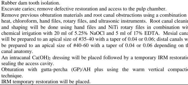
Table 1. Root canal retreatment protocol
Local anesthesia with inferior alveolar block using 2% Lodocaine with 1:100,000 epinephrine; supplemental anesthesia with intraligamental injection or intraosseous injection if necessary.
Rubber dam tooth isolation.
Excavate caries; remove defective restoration and access to the pulp chamber.
Remove previous obturation materials and root canal obstructions using a combination of heat, chloroform, hand files, rotary files, and ultrasonic instruments. Root canal cleaning and shaping will be done using hand files and NiTi rotary files in combination with chemical irrigation with 20 ml of 5.25% NaOCl and 5 ml of 17% EDTA. Mesial canals will be prepared to an apical size of #35-40 with a taper of 0.04 or 0.06; distal canals will be prepared to an apical size of #40-60 with a taper of 0.04 or 0.06 depending on the canal anatomy.
An intracanal Ca(OH)2 dressing will be placed followed by a temporary IRM restoration
sealing the access cavity.
Obturation with gutta-percha (GP)/AH plus using the warm vertical compaction technique.
IRM temporary restoration will be placed.
Restoration of endodontically treated teeth will be done within two weeks following RCT. A detailed treatment protocol is listed in Table 2.
Table 2. Treatment protocol for coronal restoration
Under rubber dam isolation, GP will be removed 2-3 mm into the root canal orifices. Following acid etch and bond, a composite core build- up with Ti-Core or Core-paste will be placed and cured.
The tooth will be prepared to receive a metal ceramic crown.
Final impression using a Poly Vinyl Siloxane material (PVS) will be obtained and sent to laboratory for crown fabrication.
Crown delivery and occlusal adjustment. b. Implant treatment
Patients referred to the Oral Maxillofacial Surgery Clinic for implant treatment will be screened. A panoramic radiograph will be used for initial treatment planning. Cases meeting the study inclusion criteria will be assessed to determine an individualized treatment plans. A staged implant treatment strategy will be adopted. Cone-beam computed tomography (CT) will be performed in selected cased to determine the location of manibular canal to guide surgical implant placement. The type of implant used will be NobelReplace Straight 5.0 mm (Nobel Biocare). A detailed treatment protocol is listed in Table 3.
Table 3. Implant treatment protocol -staged approach
Tooth extraction under local anesthesia, minor socket preservation may be performed. The extraction site will be allowed to heal for 4-6 months.
Preliminary impressions using irreversible hydrocolloid material (Alginate) will be obtained for fabrication of the radiographic index and surgical guide.
create implant receiving site with a series of drills under coolant, check position and orientation with radiographs, place implant, place healing cap, reposition flap, suture, and final radiograph.
Allow site to heal for 4-6 months.
Final implant level impressions using PVS will be obtained and sent to laboratory for abutment selection and metal ceramic crown fabrication.
Crown delivery and occlusal adjustment.
c. Endodontic treatment follow-up
All patients will be recalled at 6, 12, and 24 months following the completion of the root canal retreatment to evaluate treatment outcome. During these appointments, standard intra- and extra-oral exams will be performed to evaluate the health of periradicular tissue related to the tooth treated. Digital periapical radiographs will be taken with a positioning device to monitor the healing of the periradicular lesion. This device is composed of an XCP beam-guiding device and a film holder stabilized by a polyvinyl siloxane impression material [36]. This device will be used to take the post-operative and all the follow- up radiographs.
Based on clinical findings and radiographic presentation, treatment outcome will be categorized into “healed,” “non-healed,” and “healing” according to the following definitions recommended by the AAE/AAEF:
§ Healed—Functional*, asymptomatic teeth with no or minimal radiographic periradicular pathosis.
§ Non-healed—Nonfunctional, symptomatic teeth with or without radiographic periradicular pathosis.
§ Healing—Teeth with periradicular pathosis, which are asymptomatic and functional, or teeth with or without radiographic periradicular pathosis, which are symptomatic but whose intended function is not altered.
* Functional—A treated tooth or root that is serving its intended purpose in the dentition. If the tooth is determined to be “non- healed” at the 12 month-recall, the patient will be presented with the options of non-surgical root canal retreatment or endodontic surgery depending on the individual situation.
d. Implant follow-up
Patient receive implant treatment will be recalled at 1 week, 1, 3, 6, 12, 18, and 24 months after initial loading. Clinical assessment will be performed and radiographs will be taken to evaluate the outcome of the treatment. Oral hygiene instructions will also be given at these appointments.
Complications identified during the follow-up will be recorded and managed according to the individual situation.
e. Masticatory functional analyses
Analyses of masticatory function will be used to determine the level of masticatory functio n regained at various time points following treatment. Masticatory performance will be evaluated objectively using an artificial food (CutterSil) [37], and subjectively using a questionnaire.
Objective masticatory function
Chewing performance is defined as the particle size distribution of food particles after a given number of chewing strokes [38]. Each patient will be instructed to chew 3 of the standardized quarter-tablets of CutterSil (Heraeus Kulze, Inc, South Bend, Indiana). At the end of the 20th cycle, patients will be instructed to expectorate the sample into a plastic filter and rinse with water until all particles are removed. This process will be repeated 5 times for each patient. Samples will be dried and separated using a series of sieves. The content of each sieve will be weighed. Median particle size and broadness of particle distribution will be determined from cumulative weight percentages [37, 39]. The degree of fragmentation of the food (chewing performance) is given by the median particle size, X50, which is the aperture of a theoretical sieve through which 50% of the weight of the comminuted food could pass [40].
Bite force
Unilateral bite force will be measured with a modified miniature strain- gauge bite- force transducer described by Fontijin- Tekamp et al. [38]. Subjects will be asked to bite at a level they consider to be equivalent to the force they normally use to bite, and at a level with maximum force. The measurement unit for bite force will be Newton (N). The point of measurement will be between the treated molar and the apposing teeth. The same tooth on the contra lateral side will also be measured and will serve as a control. Measurements will be repeated three times. Group means and standard deviations will be calculated.
Subjective evaluation of masticatory ability
A questionnaire will be used to evaluate the patients’ perceived masticatory ability [39]. Questions are listed in Table 4.
Table 4. Subjective masticatory ability questionnaire
Are you ordinarily, or would you be able to chew or bite fresh carrot or celery sticks? Are you ordinarily, or would you be able to chew or bite fresh lettuce or spinach?
Are you ordinarily, or would you be able to chew or bite steaks, chops or firm meat, e.g. beef jerky?
Are you ordinarily, or would you be able to chew or bite boiled peas, carrots, or green or yellow beans?
Are you ordinarily, or would you be able to chew or bite a whole fresh apple without cutting?
Each subject will be asked to indicate the response on a visual analog scale (VAS) 150 mm long (delimited by “not” and “very”) located below each question. The summative score will be used for group comparison.
All masticatory functional measurements will be performed before treatment, 1 week, 3 months, 6 months, 1 year, and 2 years following the placement of the final coronal restoration.
f. Quality of life measurement
A modified version of the Oral Health Impact Profile (OHIP) will be adopted to be used as the instrument to measure patient-perceived oral health [2]. This version of OHIP was modified by Dugas et al. (2002). Reliability and validity have been established. Responses to the questions will be measured using a five-point Likert scale. Improvement on OHQOL can be assessed by following each item with a question “after the treatment, has this improved, worsened, or stayed same?” The improvement score will be calculated for each individual item. An improvement score of 1 will be assigned to each subject if each item that exists before and improves after. The scores will be summed for each subject. The overall improvement will also be determined based on a derived summative score. Items in this questionnaire are listed in Table 5.
Table 5. Conceptual dimensions and quality of life items (adapted from Dugas et al.[2])
Conceptual dimension Item
Functional limitation Have you had trouble pronouncing words because of your teeth or mouth?
Have you felt that your sense of taste has worsened because of your teeth or mouth?
Physical pain Have you had painful aching in your mouth?
Have you found it uncomfortable to eat any foods because of your teeth or mouth?
Have you had to alter the temperature of the foods that you eat because of your teeth or mouth?
Psychological discomfort Have you been self-conscious because of your teeth or mouth?
Have you felt tense because of your teeth or mouth? Physical disability Has your diet been unsatisfactory because of your teeth
or mouth?
Have you had to interrupt meals because of your teeth or mouth?
or mouth?
Have you found it difficult to fall asleep because of your teeth or mouth?
Have you ever been awakened by problems with your teeth or mouth?
Have you been embarrassed because of your teeth or mouth?
Social disability Have you been irritable with other people because of your teeth or mouth?
Have you had difficulty doing your usual jobs because of problems with your teeth or mouth?
Handicap Have you felt that life in general was less satisfying because of your teeth or mouth?
Have you been totally unable to function because of your teeth or mouth?
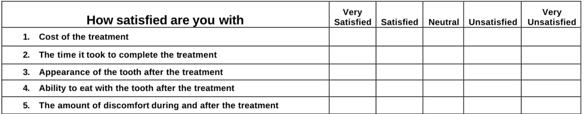
g. Patient satisfaction measurement
Patient satisfaction towards root canal retreatment or implant treatment will be evaluated using a questionnaire (Table 6). This questionnaire is comprised of 5 questions addressing the cost of the treatment; the time it took to complete the treatment; discomfort associated with the treatment; and esthetics and function following the treatment. Responses are scored from 1 to 5 with 5 being very satisfied and 1 being very unsatisfied.
h. Management of adverse events
During root canal retreatment, in case of adverse events, such as perforation, root fracture, inability to remove canal blockage, the tooth will be managed with the best possible treatment plan. Periapical surgery may be indicated in some cases. An extraction may be necessary followed by implant placement. Data of these patients will be analyzed as “intention to treat”. In the implant group, if the implant fails, it will be managed with the best treatment plan determined by the oral surgeon and the prosthodontist. Extraction of failed implant followed by bone grafting and new implant placement may be necessary. These patients will also be analyzed as “intention to treat”.
Table 6. Patient satisfaction questionnaire
How satisfied are you with
Satisfied Satisfied Neutral Unsatisfied VeryVery Unsatisfied 1. Cost of the treatment
2. The time it took to complete the treatment 3. Appearance of the tooth after the treatment 4. Ability to eat with the tooth after the treatment
6. Data and Statistical analyses a. Overall summary
This study is a prospective cohort study to compare treatment outcome of non-surgical root canal retreatment and single implant-supported prosthesis using various quality-of-life criteria. All patients will have a mandibular first molar with failed NSRCT requiring either endodontic retreatment or extraction followed by implant placement. Pre-treatment masticatory function level will be established in both groups. Changes in the masticatory function, patient-perceived oral health will be determined 1 week, 3 months, 6 months, 12 months, and 24 months following treatment. Patient satisfaction towards different treatment modalities will also be evaluated.
b. Data Collection Management
Overview: All data will be entered and stored at Baylor College of Dentistry. The statistical analyses will be performed by Dr. Linda Hyman, the study statistician in the Department of Clinical Sciences (Biostatistics) at the University of Texas Southwestern (UTSW) Medical Center. Baylor College of Dentistry has partnered with UTSW Medical Center Department of Clinical Science in a funded K12 and soon-to-be funded U54 grant. Baylor College of Dentistry has been assigned statistical and other support by the Department of Clinical Science. A CD with coded patient data base (no identifiable private patient information) will be used to transfer data from Baylor College of Dentistry to UTSW for analyses.
Data collection: Data collection forms will be organized for each patient visit. Each form has space for the following identifiers: patient number, patient initials, treatment type, and visit number. Treatment/measurement performed will be recorded on the form. When a visit is complete, the Research Coordinator will check each form for accuracy and completeness. After the forms have been reviewed for clinical features and checked for completeness, they will be logged into the database and update a forms checklist for each patient. After logging, the forms will be entered into an Access database, producing primary and secondary data files.
The electronic data entry program will be designed to allow only codes listed on the form and values in the expected format to enter the database. Missing or empty fields must be entered with a code, such as ‘-9’. From the database, reports will be developed to list completion status, exits, and forms in the database. The database will provide the basis for queries for data integrity and subsequent statistical analysis.
Confidentiality Issues: Confidentiality of patient data is an important consideration. The biostatistician will not receive the patient’s name or any identifier such as medical record number or social security number. Copies of the data forms will be stored in locked file cabinet in the office of the Principle Investigator.
Hardware, Software, Security: The software platform for the study database will be Microsoft® Access 2000. Programs and data will reside on a file server at Baylor College of Dentistry. The server is located in a room with restricted access. In addition to passwords necessary to log into the server and receive access to the database directory, security features of Access limit entry to the database to only specific users via password. The file server will be backed up on a daily basis to an external hard drive. The study database will be backed up to zip disk or CD periodically. The server is protected with an Uninterruptible Power Supply (UPS).
c. Plan for analysis of data
Sample Size and Power Analysis: To compare the two treatment groups (non-surgical root canal retreatment versus single implant-supported prosthesis), a minimum of 70 patients in each group are needed to detect a .25 effect size difference for the between subjects factor (treatment) resulting in a type II error rate of 15.3% (power of 84.7%) and a .30 effect size for the within subjects factor (measurements at baseline, and 1 week, 3 months, 6 months, and 1 year post treatment) resulting in a type II error rate of 17.7% (power of 82.3%) using a mixed effects two-factor analysis of variance and a type I error rate (a) of 0.05.. Assuming a 30% drop out rate, 100 patients for each group (for a total of 200 patients) will be recruited for this study.
Summary descriptive statistics: For normally distributed, continuous data, means and standard deviations will be used to describe the demographic and clinical characteristics of the patient population participating in the study. Groups will be compared using Student’s t-test. Medians and semi- interquartile ranges will be used to describe characteristics of the patient population when the data are continuous, non- normally distributed and Mann Whitney test will be used for comparison of groups. For categorical or dichotomous variables, proportions will be reported and Chi Square or Fisher’s Exact Test will be performed for group comparisons.
Description of analysis for primary hypothesis: The primary hypothesis of the study is that endodontic treatment allows better functional recovery compared to single implant-supported prostheses. Chewing performance will be measured to reflect masticatory function. A 2 factor mixed models analysis of variance (ANOVA) will be used to examine the outcome variable chewing performance. The between factors will be the two treatment groups (endodontic retreatement versus implant), while the within factor will be the time measurements were made (baseline, 1 week, 3 months, 6 months, 12 months, and 24 months after treatment has been completed).
Description of analysis for secondary hypotheses: The secondary hypotheses that endodontic treatment provides better treatment results include:
Patient-perceived oral health evaluated by OHIP. Seventeen quality of life items each item having a five-point Likert response scale will be summed to create a total score. The total score and each item score will be analyzed using a 2 factor mixed models ANOVA with 1 between group (treatment) and 1 within group (5 time points).
Improvement in unilateral bite force. The bite force between the mandibular first molar receiving treatment and the opposing teeth will be measured with a difference in force between these teeth calculated. A 2-factor, mixed models ANOVA will be used to compare the treatment group (two between groups) across time (5 time points).
Patient satisfaction toward treatment. Patient responses to the satisfaction survey will be summed into a total score. This total score will be analyzed using a 2- factor mixed models ANOVA with one between factor (treatment group) and one within factor (measurement time).
General Statistical Issues:
a) SPSS V15 and SAS V9 will be used to analyze these data.
b) All tests, unless otherwise noted, will be performed using p < 0.05.
c) Treatment of missing data – The following procedures will be used to account for the effects of missing data on the analysis.
1. No variables will be included in the analyses that have more than 10% of the values missing. Variables with values missing 10% of the time or more do not occur by chance, and there are systematic causes for missing data at frequencies at this level. Analysis strategies include performing the analysis using the following three strategies:
2. Use of PROC MIXED. In MANOVA, ANCOVA, or ANOVA models, missing data have historically caused serious statistical analysis problems. Missing data, when using a mixed models approach (as PROC MIXED in SAS), are not as serious a problem. In a mixed models approach the patient is considered randomly chosen from a larger group of subjects. These models have been found tolerant of missing data as long as the missing data are random.
The results of these two strategies will be compared with alternative
interpretations considered. Data for missing values will also be analyzed in an intent-to-treat fashion with results “carried forward” from the previous time. Further, an end-point analysis will involve an analysis of change-scores from first to last (regardless of the follow-up day) will be included for interpretation of the intent-to-treat analysis.
d) Check of assumptions – For all analyses performed, each statistical test will be checked to verify that the assumptions for each are satisfied and, if not,
appropriate transformations (e.g., log transformations for counts, arcsine square root transformations for proportions, etc.) will be performed before analysis. Non-parametric analyses will be used where possible.
e) Type I error rates - We propose using multivariate analysis of variance, wherever feasible, rather than individual analysis of covariance adjusting for Type I error rates using Bonferroni correction.
E. Time-line for research
a. Study preparation, patient recruitment, initial screening and record: month 1-18
b. Root canal treatment and coronal restoration: month 19-20 c. Implant placement and coronal restoration: month 19-36
d. Recall visits month 19-60 (1 week, 3 months, 6 months, 12 months and 24 months following treatment completion)
i. Masticatory functional analyses (1 week, 3 months, 6 months, 12 months and 24 months)
ii. Quality of life measurement (1 week, 3 months, 6 months, 12 months and 24 months)
iii. Patient satisfaction measurement (3 months following treatment completion)
e. Data analyses: month 43-48
F. Letters of approval a. IRB letter
d. Informed consent form
BAYLOR COLLEGE OF DENTISTRY
Dallas, TexasPARTICIPATION EXPLANATION AND CONSENT FORM
PROJECT TITLE: A comparative outcome analysis of endodontic retreatment and single implant -supported restoration
Sponsor: American Association of Endodontists Foundation (AAEF)
INVESTIGATORS: Jianing He DMD, PhD; Uwe Frohberg, MD, DDS; Peter Buschang, MA, PhD; Elias Kontogiorgos, DDS
TELEPHONE NUMBER/24 HOUR EMERGENCY NUMBER: 214-828-8473/emergency number: 972-841-1685
Introduction
This is a research study. Before agreeing to participate in this study, it is
important that you read and understand what is involved in participating. This process is called “informed consent.” This consent form provides detailed information about this study and describes the purpose, procedure, benefits, and risks of the study as well as the other options that are available to you. This consent form may contain words that you do not understand. Please ask the study staff to explain any information that you do not clearly understand. Discuss participation with your family and friends. Do not sign this consent form unless you have received answers to all your questions. At the end of this process, you will be given a copy of the form to keep.
Why is this study being done?
The purpose of this study is to compare the outcome of endodontic retreatment and single implant-supported restorations regarding their impact on various aspects of the quality of life including the ability to chew, patient perceived oral health, and patient satisfaction. Results from this study will help to establish guidelines for clinical decision making to choose the most appropriate treatment modality for the patients to allow quicker recovery of function and better improvement of the quality of life.
You are being asked to participate in this research study because the treatment outcome of the two procedures needs to be evaluated clinically in patients. You meet the inclusion criteria and qualify as a candidate for the study.
All procedures involved in the study are well established standard treatment procedures. All instruments and materials used in the study are FDA approved for clinical use.
How many people will take part in the study?
A total of 200 patients will participate in the study with 100 patients in each group. What is involved in the study?
Inclusion criteria: § Age : 20-65
§ Both men and women will be included
§ Systemic conditions: generally healthy, ASA I or II
§ Dental conditions: Prosthodontic Diagnostic Index (PDI) class I (Appendix 2) § Tooth conditions: mandibular first molars; opposing teeth are natural teeth
• For patients receiving root canal retreatment, the tooth has had previous NSRCT and a periradicular diagnosis of “chronic periradicular periodontitis”; retreatment is indicated due to persistent infection; the tooth is restorable, but significant tooth structural loss is present that requires full crown coverage.
• For patients receiving implant treatment, the tooth has had previous NSRCT and a periradicular diagnosis of “chronic periradicular periodontitis”; root canal retreatment is not possible or the patient denies retreatment; there is sufficient bone support and the local anatomy allows implant placement.
Exclusion criteria:
§ Uncontrolled systemic disease that compromises the immune system of the patient. Examples: Diabetes, AIDS, cancer patients on chemotherapy, etc.
§ Smokers
§ Pregnant women
§ For patients receiving root canal retreatment, the tooth has a vertical fracture, internal or external resorption, or poor periodontal prognosis.
§ For patients receiving implant treatment, there is local anatomical condition or occlusal condition that does not allow implant placement.
§ Patients receiving bisphosphonate treatment
If you choose to have a root canal retreatment done, the treatment will be performed by second- year endodontic residents under faculty supervision. Root canal retreatment involves removing decay and previous root filling, recleaning and filling the root canal space. A crown will be placed over the tooth after the root canal treatment is completed. If you choose to have the tooth extracted and an implant placed, the treatment will be performed by a faculty member of the Department of Oral and Maxillofacial surgery. After the tooth is extracted, you will need to wait 4-6 months for the bone to heal. A titanium dental implant will
then be placed into the jaw bone of the site with the missing tooth. A crown supported by the implant will be placed after another 4-6 months to allow the implant to heal. All the crown will be prepared by a resident prosthodontist.
Patients will be reminded to return after 1 week, 3 months, 6 months, 1 year and 2 years for a short follow-up visit. At each visit, you will be asked to chew on an artificial silicon food to measure your chewing ability. You will also be asked to bite on a piece of plastic-covered metal transducer to measure your bite force. A questionnaire with 5 simple questions will be given to you to determine your perceived chewing ability. At the 3 month follow-up visit, you will be presented with two other questionnaires. One of them will have 5 questions to measure your satisfaction of the treatment; the other will have 17 questions to determine the effect of treatment on your quality of life.
How long will I be in the study?
The root canal retreatment and the crown will be completed in 4 weeks. Extraction followed by implant placement and crown can take up to 8 to 12 months. You will need to return for follow-up at 1 week, 3 months, 6 months, 1 year and 2 years following the treatment.
What are the risks of the study?
Both root canal retreatment and implant placement are well established standard clinical practice. Potential risks are within the scope of the standard treatments. For root canal treatment, potential risks include pain and swelling, perforation or fracture of the tooth. For the implant procedure, potential risks include pain, swelling, infection, temporary or permanent numbness of the lower jaw, fracture of the porcelain, loosening of the screw, and the loss of the implant.
Are there benefits to taking part in the study?
Yes. Participating patients will receive a reimbursement of 25% of the standard
treatment fee upon the completion of the study. Patient will be compensated with 10% of the total reimbursement at each of the first four follow-up visits; the remaining 60% of reimbursement will be paid upon the conclusion of the 2 year follow-up.
What other options are there?
If you choose not to participate in the study, you can receive either a root canal treatment, extraction, extraction followed by a fixed prosthesis, extraction followed by implant-supported prosthesis, extraction followed by a removable prosthesis, or no treatment.
Protected Health Information is any personal health information through which you can be identified. A decision to participate in this research means that you agree to the use and disclosure of your health information for the purposes explained in this consent form. The research team may use the following sources of health information: X-rays, medical and dental histories, treatment procedures, clinical exam findings, filled questionnaires, etc. Information from these sources will be recorded into a separate research file with the following identifiers: A random 3-digit number will be assigned to each patient; only the principle investigator will have access to the document that links the code to patient chart number. Study records that identify you will be kept confidential as required by law. The research team will use this information until the end of the research study (March 2010). At that point the investigator will remove the identifiers from your information, making it impossible to link you to the study.
Baylor College of Dentistry will make every effort to protect your information in accordance wit h State and Federal laws. The Notice of Privacy Practices (a separate document) describes the procedures BCD follows to protect your health information. You are required to sign an acknowledgment of receipt of this document. If you have not already done so, the research team will make a copy of the Notice of Privacy Practices available for your review and signature of acknowledgment of receipt. Your identity will not be revealed in any publication that may result from this study. Videos and
photographs which may reveal your identity and likeness will not be used without your separate, written authorization.
What are the costs?
There is no additional cost in participating in the study. The patient will receive a reimbursement of 25% of the standard treatment cost upon the completion of the study. What if I am injured or become ill while participating in this study?
We will make every effort to prevent injury that could result from this research. Compensation for injuries incurred as a result of participating in this research is not available. The investigator(s) is prepared to advise you about medical or dental treatment in case of adverse effects related to this study, which you should report to him/her
promptly at the phone number at the beginning of this form.
What are my rights as a participant?
Your participation in this research is entirely voluntary. You may refuse to participate or may quit participating in this study at any time without affecting your status at Baylor (as a student, faculty member or staff member) or your future care at Baylor College of Dentistry. If you withdraw from the study, no new data about you will be collected for study purposes unless these data concern an adverse event (a bad effect) related to the study. If such an adverse event occurs, we may need to review your entire medical file. You may withdraw your authorization for us to use all identifiable data (other than data
needed to report an adverse event or to keep track of your withdrawal) that have already been collected, but you must do this in writing. The researchers may remove you from the study at any time if this should become necessary, after explaining the reason(s) for doing so.
Whom do I call if I have questions or problems?
1. To report a research-related injury, you should contact Dr. Jianing He, (214) 828-8473
2. For questions about the research study specifically, you should contact Dr. Jianing He, (214) 828-8473
3. If you require information about your rights as a research subject, you may contact Dr. Brendan S. Wong, Chairman of the Institutional Review Board of Baylor College of Dentistry, (214) 828-8323.
Disclosure Statement(s)
You should also be aware that some insurance companies and government health care programs may limit their payment for procedures that are experimental.
Confirmation of Consent:
You are making a decision whether or not to participate in this study. You should not sign until you understand all the information presented in the previous pages and until all of your questions about the research have been answered to your satisfaction. Your signature indicates that you have decided to participate, having read (or been read) the information provided above. More specifically:
1. I clearly understand that this is an experimental procedure. ( ) Yes ( ) No
2. I clearly understand the risks associated with participation in this study. ( ) Yes ( ) No 3. I clearly understand the length of time during which I will be
participating in this study. ( ) Yes ( ) No
4. I clearly understand the purpose and anticipated outcomes of this study. ( ) Yes ( ) No 5. I clearly understand that my participation in this study is voluntary. ( ) Yes ( ) No 6. I clearly understand that my participation in this study does not
affect my legal rights. ( ) Yes ( ) No
7. I certify that I am 18 years of age or older. ( ) Yes ( ) No
Print Name of Subject Date of Birth
Signature of Subject Date
Name of Person Obtaining Consent (Print) Date
Signature of Principal Investigator Date
YOU WILL BE GIVEN A SIGNED COPY OF THIS CONSENT DOCUMENT TO KEEP.
6. References:
[1] Slade GD, Spencer AJ. Development and evaluation of the Oral Health Impact Profile. Community Dent Health. 1994 Mar;11(1):3-11.
[2] Dugas NN, Lawrence HP, Teplitsky P, Friedman S. Quality of life and satisfaction outcomes of endodontic treatment. Journal of endodontics. 2002 Dec;28(12):819-27.
[3] Davis DM, Fiske J, Scott B, Radford DR. The emotional effects of tooth loss in a group of partially dentate people: a quantitative study. Eur J Prosthodont Restor Dent. 2001 Jun;9(2):53-7.
[4] Fiske J, Davis DM, Leung KC, McMillan AS, Scott BJ. The emotional effects of tooth loss in partially dentate people attending prosthodontic clinics in dental schools in England, Scotland and Hong Kong: a preliminary investigation. Int Dent J. 2001 Dec;51(6):457-62.
[5] Behavioral Risk Factor Surveillance System (BRFSS): Centers for Disease Control and Prevention; 1999.
[6] Dammaschke T, Steven D, Kaup M, Ott KH. Long-term survival of root-canal-treated teeth: a retrospective study over 10 years. J Endod. 2003 Oct;29(10):638-43. [7] Alley BS, Kitchens GG, Alley LW, Eleazer PD. A comparison of survival of teeth following endodontic treatment performed by general dentists or by specialists. Oral Surg Oral Med Oral Pathol Oral Radiol Endod. 2004 Jul;98(1):115-8.
[8] Cho GC. Evidence-based approach for treatment planning options for the extensively damaged dentition. J Calif Dent Assoc. 2004 Dec;32(12):983-90.
[9] Cohn SA. Treatment choices for negative outcomes with non-surgical root canal treatment: non-surgical retreatment vs. surgical retreatment vs. implant. Endodontic Topics. 2005;11:4-24.
[10] Wang Q, Cheung GS, Ng RP. Survival of surgical endodontic treatment performed in a dental teaching hospital: a cohort study. Int Endod J. 2004 Nov;37(11):764-75.
[11] Zuolo ML, Ferreira MO, Gutmann JL. Prognosis in periradicular surgery: a clinical prospective study. Int Endod J. 2000 Mar;33(2):91-8.
[12] Fristad I, Molven O, Halse A. Nonsurgically retreated root filled teeth--radiographic findings after 20-27 years. Int Endod J. 2004 Jan;37(1):12-8.
[13] Gorni FG, Gagliani MM. The outcome of endodontic retreatment: a 2-yr follow-up. J Endod. 2004 Jan;30(1):1-4.
[14] Farzaneh M, Abitbol S, Lawrence HP, Friedman S. Treatment outcome in endodontics-the Toronto Study. Phase II: initial treatment. J Endod. 2004 May;30(5):302-9.
[15] Kim S. Modern endodontic practice: instruments and techniques. Dent Clin North Am. 2004 Jan;48(1):1-9.
[16] Creugers NH, Kreulen CM, Snoek PA, de Kanter RJ. A systematic review of single-tooth restorations supported by implants. Journal of dentistry. 2000 May;28(4):209-17.
[17] Scheller H, Urgell JP, Kultje C, Klineberg I, Goldberg PV, Stevenson-Moore P, et al. A 5- year multicenter study on implant-supported single crown restorations. Int J Oral Maxillofac Implants. 1998 Mar-Apr;13(2):212-8.
[18] Ruskin JD, Morton D, Karayazgan B, Amir J. Failed root canals: the case for extraction and immediate implant placement. J Oral Maxillofac Surg. 2005 Jun;63(6):829-31.
[19] Wood MR, Vermilyea SG. A review of selected dental literature on evidence-based treatment planning for dental implants: report of the Committee on Research in Fixed Prosthodontics of the Academy of Fixed Prosthodontics. J Prosthet Dent. 2004 Nov;92(5):447-62.
[20] Chang M, Wennstrom JL, Odman P, Andersson B. Implant supported single-tooth replacements compared to contralateral natural teeth. Crown and soft tissue dimensions. Clinical oral implants research. 1999 Jun;10(3):185-94.
[21] Watson CJ, Tinsley D, Sharma S. Implant complications and failures: the single-tooth restoration. Dent Update. 2000 Jan-Feb;27(1):35-8, 40, 2.
[22] Meffert RM. Issues related to single-tooth implants. J Am Dent Assoc. 1997 Oct;128(10):1383-90.
[23] Trope M. Implant or root canal therapy: an endodontist's view. J Esthet Restor Dent. 2005;17(3):139-40.
[24] Felton DA. Implant or root canal therapy: a prosthodontist's view. J Esthet Restor Dent. 2005;17(4):197-9.
[25] Bader JD, Shugars DA. Variation, treatment outcomes, and practice guidelines in dental practice. J Dent Educ. 1995 Jan;59(1):61-95.
[26] Friedman S, Mor C. The success of endodontic therapy--healing and functionality. J Calif Dent Assoc. 2004 Jun;32(6):493-503.
[27] Akeel R, Nilner M, Nilner K. Masticatory efficiency in individuals with natural dentition. Swed Dent J. 1992;16(5):191-8.
[28] Helkimo E, Carlsson GE, Helkimo M. Chewing efficiency and state of dentition. A methodologic study. Acta Odontol Scand. 1978;36(1):33-41.
[29] Oosterhaven SP, Westert GP, Schaub RM, van der Bilt A. Social and psychologic implications of missing teeth for chewing ability. Community Dent Oral Epidemiol. 1988 Apr;16(2):79-82.
[30] Nagasawa T, Tsuru H. A comparative evaluation of masticatory efficiency of fixed and removable restorations replacing mandibular first molars. J Prosthet Dent. 1973 Sep;30(3):263-73.
[31] Mayer TM, Hawley CE, Gunsolley JC, Feldman S. The single-tooth implant: a viable alternative for single-tooth replacement. Journal of periodontology. 2002 Jul;73(7):687-93.
[32] Esposito M, Hirsch JM, Lekholm U, Thomsen P. Biological factors contributing to failures of osseointegrated oral implants. (II). Etiopathogenesis. European journal of oral sciences. 1998 Jun;106(3):721-64.
[33] Strassburger C, Heydecke G, Kerschbaum T. Influence of prosthetic and implant therapy on satisfaction and quality of life: a systematic literature review. Part 1--Characteristics of the studies. Int J Prosthodont. 2004 Jan-Feb;17(1):83-93.
[34] Strassburger C, Kerschbaum T, Heydecke G. Influence of implant and conventional prostheses on satisfaction and quality of life: A literature review. Part 2:
Qualitative analysis and evaluation of the studies. Int J Prosthodont. 2006 Jul-Aug;19(4):339-48.
[35] Sonoyama W, Kuboki T, Okamoto S, Suzuki H, Arakawa H, Kanyama M, et al. Quality of life assessment in patients with implant-supported and resin-bonded fixed prosthesis for bounded edentulous spaces. Clin Oral Implants Res. 2002 Aug;13(4):359-64.
[36] Trope M, Delano EO, Orstavik D. Endodontic treatment of teeth with apical periodontitis: single vs. multivisit treatment. Journal of endodontics. 1999 May;25(5):345-50.
[37] Albert TE, Buschang PH, Throckmorton GS. Masticatory performance: a protocol for standardized production of an artificial test food. J Oral Rehabil. 2003 Jul;30(7):720-2.
[38] Fontijn-Tekamp FA, Slagter AP, Van Der Bilt A, Van THMA, Witter DJ, Kalk W, et al. Biting and chewing in overdentures, full dentures, and natural dentitions. J Dent Res. 2000 Jul;79(7):1519-24.
[39] English JD, Buschang PH, Throckmorton GS. Does malocclusion affect masticatory performance? Angle Orthod. 2002 Feb;72(1):21-7.
[40] van Kampen FM, van der Bilt A, Cune MS, Fontijn-Tekamp FA, Bosman F. Masticatory function with implant-supported overdentures. J Dent Res. 2004 Sep;83(9):708-11.