Mode of Action of Chronotropic
Agents in Cardiac Purkinje Fibers
Does Epinephrine Act
by Directly Modifying the
External Surface Charge?
RICHARD W. TSIENFrom the Department of Physiology, Yale University School of Medicine, New Haven, Connecticut 06510
ABSTRACT Hauswirth et al. (1968) proposed that epinephrine acts on i, by adding its own positive charge to the external membrane surface near the iK, channel. This hypothesis was tested by using noncationic compounds, theophyl-line and R07-2956, which mimicked epinephrine's effects on pacemaker activity and on ix,. In maximally effective doses, theophylline or R07-2956 occluded the effect of epinephrine, indicating a shared final common mechanism. Since theo-phylline and R07-2956 are noncationic at pH 7.4, the common mechanism cannot be a direct change in external surface charge. On the contrary, epineph-rine does not interfere with the voltage shift produced by La+ + +, which is thought to modify the external surface charge. The results argue against the original hypothesis but leave open the possibility that an alteration in internal surface charge generates the observed voltage shift. The potency of theophylline and R07-2956 as phosphodiesterase inhibitors suggests that the final common mechanism begins with the elevation of intracellular cyclic AMP, leading to a saturable process which limits the voltage shift's magnitude. This hypothesis is used to generate dose-response curves describing the combined effects of epi-nephrine and theophylline, and these are compared with experimental data.
INTRODUCTION
The ionic basis of epinephrine's acceleratory effect in cardiac Purkinje fibers has been examined by means of voltage clamp experiments (Hauswirth et al., 1968; Tsien 1974). Epinephrine acts rather selectively in modifying the prop-erties of the pacemaker potassium current, ix,, whose time dependence under-lies the diastolic pacemaker (Noble and Tsien, 1968). The drug favors a more rapid and more complete deactivation of i,, at any given potential in the
THE JOURNAL OF GENERAL PHYSIOLOGY ' VOLUME 64, 1974 pages 320-342 320
R. W. TSIEN Action of Chronorropic Agents in Cardiac Purkinje Fibers
diastolic voltage range, and thus promotes spontaneous activity. This effect may be described to a first approximation as a "voltage shift," a displacement of the voltage-dependent kinetic parameters toward less negative potentials.
The epinephrine-produced voltage shift bears a strong qualitative resem-blance to the effect of elevating the calcium concentration in the bathing fluid. In Purkinje fibers, as in other excitable tissues, calcium and various other cations produce voltage shifts in the kinetic variables (e.g. Weidmann, 1955; Frankenhaeuser and Hodgkin, 1957; Frankenhaeuser, 1957; Jenerick, 1959). In the specific case of iK,, increasing [Ca]0 fourfold shifts the activation curve by about 8 mV (Noble and Tsien, unpublished; Brown, 1973). The similarity of effects, and the fact that epinephrine is positively charged, led Hauswirth et al. (1968) to suggest that epinephrine had a "calcium-like" mechanism of action. Thus, following a suggestion of A. F. Huxley (Frankenhaeuser and Hodgkin, 1957), it was hypothesized that epinephrine directly altered the ex-ternal surface charge and local electric field near the iK, channel. The change in the electric field would bias the activation process toward the deactivated state, so that a stronger depolarization would be required to activate i,. The extreme sensitivity of i, to epinephrine, and the insensitivity of iN, (Traut-wein and Schmidt, 1960) could be accounted for if the catecholamine pos-sessed a high affinity for a specific site on or near the i, channel.
What criteria can be used to test the idea of a directly effective alteration of the external surface charge? The effects of changes in surface charge have been considered in various theoretical treatments (e.g. Gilbert and Ehren-stein, 1969; Brown, 1974). Specific locations for the added charge are assumed in each case, but the models do not help in settling the question of its location. Any scheme (say, positive charge on the outside membrane surface) can be reformulated for the opposite case (negative charge on the inside surface), and still give rise to an appropriate voltage shift. Thus, the existing theory neither supports nor rules out the external surface charge hypothesis.
Another possible approach is to study the hormone effect as a function of pH, to see if the effect declines as the drug molecules are neutralized in alka-line solutions. Unfortunately, this type of experiment is rather impractical since in catecholamines the pK.'s are near 10. In any case, the results would be ambiguous; they might show that the drug acted in the cationic form, but this would not settle the question of whether the cationic form directly altered
the electric field sensed by the iK, channel.
This paper reports the results of a less direct approach to the hypothesis, which nevertheless provides evidence concerning its validity. I have studied the effects of two chronotropic agents, theophylline and R07-2956, which are often employed as inhibitors of intracellular phosphodiesterase. These com-pounds are interesting in the present context because it turns out that they have epinephrine-like actions on iK,, despite the fact that the molecules are
THE JOURNAL OF GENERAL PHYSIOLOGY VOLUME 64 1974 tually uncharged at physiological pH. Studies of the effects of theophylline and R07-2956, individually, and in combination with epinephrine, demonstrate that these agents share a final common mode of action with the hormone. Since theophylline and R07-2956 are not cations, it is concluded that the final action cannot be a simple and direct modification of the external surface charge, as originally hypothesized by Hauswirth et al. (1968). To replace the original hypothesis, it is proposed that the final common mode of action of epi-nephrine and phosphodiesterase inhibitors involves intracellular cyclic AMP, and that cyclic AMP in some way mediates the effect on i 2. The dose
de-pendence of the epinephrine effect is reinterpreted in terms of a quantitative
scheme involving cyclic AMP. Some aspects of this work have already ap-peared in a preliminary communication to the British Physiological Society (Tsien, 1973 b).
METHODS
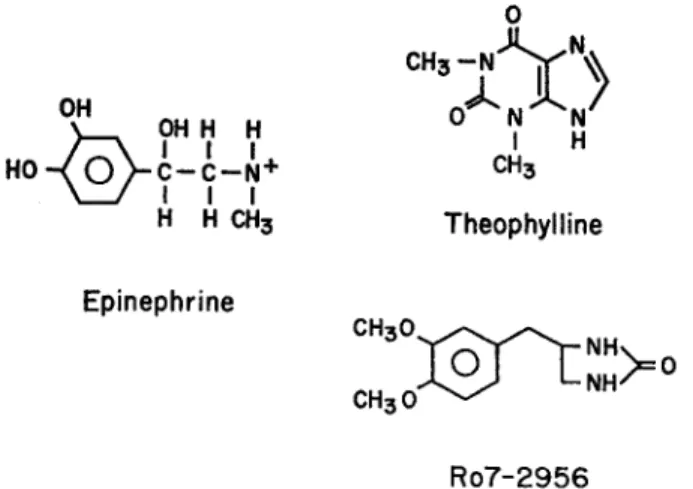
The experiments in this paper compare the action of three chronotropic agents whose chemical formulae are shown in Fig. 1. Epinephrine bears a single positive charge on the amine group at the pH used in these experiments (7.2-7.4). The action of epi-nephrine has been related to the effects of two other compounds, theophylline and R07-2956, also indicated in Fig. 1. Theophylline is a methylxanthine and at physio-logical pH the concentration of the cationic form is negligible (pK = 0.9). Some 5 % is present as anion (see Hardman, 1962). This compound has been widely used in bio-chemical studies as an inhibitor of phosphodiesterase and it is known to have inotropic effects in heart (e.g. Rall and West, 1963; Blinks et al., 1972). Finally, R07-2956 (4-(3,4-dimethoxybenzyl)-2-imidazolidinone) is also a potent phosphodiesterase in-hibitor (Sheppard and Wiggan, 1970) but its chemical structure is distinct from the
0 CH3 -OH 0 N N .~ OH H H N HO-(0 C--C-- N + CH3 I I I H H CH3 Theophylline Epinephrine CH30 NH Ro7-2956
FIGURE . The structure of the chronotropic agents used in the experiments. At physio-logical pH epinephrine bears a single positive charge, but theophylline and R07-2956 are noncationic.
R. W. TSIEN Action of Chronotropic Agents in Cardiac Purkinje Fibes
methylxanthines. Unlike theophylline or caffeine, R07-2956 does not appear to cause the release of calcium from intracellular stores, judging from its ineffectiveness as a modifier of mechanical activity in single frog skeletal muscle fibers (H. Oetliker and Tsien, unpublished), even in maximal concentrations (approx. 2 mM). R07-2956 is
also uncharged at physiological pH.
The compounds were obtained from the following sources: epinephrine bitartrate (Sigma Chemical Co., St. Louis, Mo.), theophylline (Sigma) and R07-2956
(Hoff-man-LaRoche, Nutley, N. J.). Theophylline- and R07-2956-containing solutions were made up from concentrated stock solutions which were stored in a refrigerator for pro-longed periods without loss of activity. Epinephrine solutions were freshly made up during the course of the experiments and contained 4 X 10- 5M Na2EDTA to retard
oxidation. The experimental methods were similar to those used in the previous paper (Tsien, 1974).
RESULTS
The electrophysiological effects of both theophylline and R07-2956 are very similar to the actions of epinephrine. Fig. 2 shows a pen recording of the
trans-membrane potential in a calf Purkinje fiber preparation and the effect of
R07-2956 (44 ,uM). The upper panel shows stimulated action potentials on a
I min
mV
-100
is
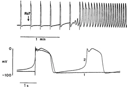
FIGURE 2. Effect of R07-2956 on electrical activity. Transmembrane potential is re-corded by a chart recorder on a slow time base (upper panel) and on an expanded time base (lower panel). The upper panel initially shows externally stimulated action poten-tials (4/min). At the arrow the superfusing solution was changed to one containing R07-2956 (44 M). The diastolic depolarization progressively increases and full-blown spontaneous activity soon follows. The lower panel compares the activity before (trace 1) and 3 min after (trace 2) beginning the exposure to R07-2956. Apart from the appearance of spontaneous activity, there is an elevation of the plateau level and a decrease in action potential duration. Preparation 53-5 in 1.8 mM [Ca]o.
THE JOURNAL OF GENERAL PHYSIOLOGY VOLUME 64 1974
slow time base before drug exposure. There was some diastolic depolarization, but no spontaneous activity; this behavior is typical in 4 mM [K],. The solu-tion was changed from normal Tyrode's to drug-containing solusolu-tion at the time marked by the arrow; after some delay (due in part to dead space in the superfusion system) the magnitude of the diastolic depolarization progressively increased and soon led to full-blown pacemaker activity. This pattern of be-havior was also observed in each of seven preparations which were exposed to theophylline (0.2 mM or greater) and was very similar to the chronotropic effect of epinephrine.
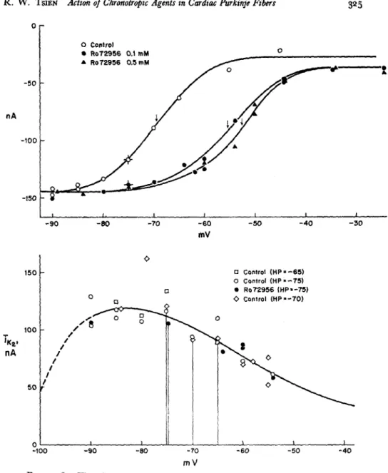
The changes in the action potential and pacemaker potential are more clearly seen in the lower panel of Fig. 2, which shows photographically super-imposed recordings on a faster time base from the same experiment. Trace 1 is the externally stimulated action potential under control conditions, and trace 2 is the spontaneous action potential during exposure to R07-2956. Be-sides the obvious increase in the steepness of the diastolic depolarization, there are changes in the configuration of the plateau as well. These also resemble the effects produced by epinephrine (Tsien et al., 1972), but they will not be considered in this paper since they do not involve the pacemaker K current. The observation that R07-2956 and theophylline produce epinephrine-like effects on electrical activity leads naturally to the question of whether these compounds mimic epinephrine at the level of individual ionic currents. Volt-age clamp experiments were carried out to test the effect of these Volt-agents on the pacemaker potassium current, i,2, which underlies the pacemaker depolariza-tion. Fig. 3 (top panel) shows that R07-2956 produces a voltage shift in the activation curve of i,,. A concentration of 500 M is only slightly more effec-tive than 100 jiM R07-2956, suggesting that the maximal voltage shift was at-tained. The magnitude of the maximal shift, 17 mV, corresponds quite well to the average maximal shift produced by epinephrine (Tsien, 1974). The thresh-old for detectable activation of the excitatory sodium current was not affected; the level of background current (when iK2 is fully deactivated) is also
un-changed.
The lower panel of Fig. 3 shows that the fully activated current-voltage relationship of iK is unaltered by R07-2956. The absence of a change in the rectifying characteristic is similar to the lack of change seen with epinephrine.
It is clear, therefore, that the chronotropic action of R07-2956 is confined to an alteration in the kinetics of i,,; in this specificity of action, R07-2956 strongly resembles epinephrine.
The effects of theophylline on i,, were also studied under voltage clamp conditions. As in the case of the other chronotropic agents, the activation curve for i,K is shifted toward less negative potentials by about 20 mV (Fig. 4, lower panel). The effect of theophylline on the rate of change of the pacemaker current is indicated in the upper panel. The left-hand limb of the rate constant
R. W. TSEN Action of Chronotropic Agents in Cardiac Purkinje Fibers -50 nA -100 -150 325 mV 150 - a Control (HP -65) o Control (HP -75) O · Ro72956 (HP'-75) 0 a O 'Control (HP -70) -loo -90 -0 - 0 - -K1 00 nA 50 50 -100 -90 -80 -70 -60 -50 -40 mV
FIGURE 3. The effect of R07-2956 on the pacemaker potassium current, ik The steady-state activation curve (top panel) is shifted toward less negative potentials by the drug (arrows denote midpoint voltage, V). The fully activated current voltage relationship, iK, (lower panel) appears unchanged. See Fig. 7 of previous paper for further description of this type of data.
curve is displaced to a much smaller degree than the right-hand limb. In this deviation from simple voltage shift behavior, the effects of theophylline and epinephrine are quite similar (see Table I, Tsien, 1974). On the other hand, the activation curve is not steepened in this case, although in some experiments steepening was observed.
THE JOURNAL OF GENERAL PHYSIOLOGY VOLUME 64 1974 3 .~ 2 sec-0 3 4 1.0 4 S.0.5 0.5 _ Ij 2 IO-7A Theophylline 0.5 mM I 5 0 -100 -80 -60 -40 -20 mV
FIGURE 4. The effect of theophylline on the kinetics of iK2. (Top) Rate constant data in the presence of 0.5 mM theophylline, and in a control run after washing off the drug. Open symbols show direct measurements of the exponential time constant of decaying ix2 current (as in Fig. 3, previous paper). Filled symbols are obtained by fitting the enve-lope of tails after pulses to the indicated test potentials (see Fig. 5, previous paper). Smooth curves drawn by eye through points. (Bottom) The steady-state activation curve in the presence of theophylline, and in the subsequent control run. The vertical scales on the right-hand side give the magnitude of the tail currents measured at the holding po-tential (-80 mV) in the respective solutions. The left-hand scale gives the normalized degree of activation. The data are presented in this form to allow direct comparison with Figs. 2 and 3 of Hauswirth et al. (1968). Experiment 52-1, 1.8 mM [Ca]o.
Do the Actions of These Chronotropic Agents Coincide?
The epinephrine-like effects of theophylline and R07-2956 might be rather trivial if they took place (a) through the release of endogenous catecholamines in fine nerve endings, or (b) through the direct stimulation of the fl-adrenergic receptor. These possibilities can be ruled out by my observation that the chronotropic effects of theophylline (two experiments) or R07-2956 (two
R. W. TSIEN Action of Chronotropic Agents in Cardiac Purkinje Fibers
periments) cannot be prevented by the presence of 10-6 M propranolol, even though this treatment does block the effect of epinephrine (Davis and Temte, 1968; Tsien, 1974). It is evident, therefore, that the modes of action of theoph-ylline or R07-2956 do not completely coincide with that of epinephrine.
A Final Common Mechanism?
The differential effect of propranolol does not preclude the possibility that the various chronotropic agents share a common mechanism at some later stage. This question was examined by studying the effects of theophylline and R07-2956 in combination with epinephrine.
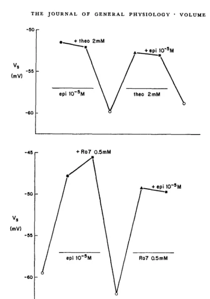
Fig. 5 (top) plots the midpoint voltage (V.) of the pacemaker potassium current during successive exposures to different drug-containing solutions. Large concentrations of epinephrine (10-5 M) and theophylline (2 mM) were employed individually and in combination. The plot shows that, in the presence of 10-5 M epinephrine, 2 mM theophylline produced no additional shift in V. Removal of both compounds abolished the shift. Subsequent addition of theophylline (2 mM) alone restored the full magnitude of voltage shift. Now, the addition of epinephrine (10-6 M) produced no additional shift. Finally, removal of both compounds gave a nearly complete return to the unshifted state. Fig. 5 (bottom) shows a similar result using epinephrine and R07-2956.
This type of experiment can be summarized by the statement that epineph-rine and theophylline (or R07-2956) are mutually occlusive. Each compound is individually capable of eliciting the maximal voltage shift. The result indi-cates that epinephrine does, in fact, share some final common mechanism with the noncationic compounds, and that each compound is able to saturate this mechanism.
Stated diagramatically,
propranolol epinephrine //
voltage shift acceleratory / in i activation effect theophylline
or R07-2956
The diagram leaves open the question of whether the final common mecha-nism consists of a single step or a number of steps. But the mere existence of a final common mechanism is significant, since it argues against the hypothe-sis of Hauswirth et al. (1968).
Additive Effects of Epinephrine and La+++ on i,,
A possible objection to the previous experiment is that the extent of the voltage shift is restricted under all circumstances by some factor which has
THE JOURNAL OF GENERAL PHYSIOLOGY VOLUME 64 · I974 -50 Vs (mV) -60 -50 Vs (mV) -55 -60 + theo 2mM A\ + epi 10-5M epi 10 5M \ theo 2mM epi 10-5M Ro7 0.5mM
FIGURE 5. Effects of chronotropic agents, individually and in combination. The mid-point voltage of the steady-state activation curve (V8) is indicated by the vertical axis. Each point corresponds to a separate determination of Vs during maintained exposure to a drug solution, or in the absence of chronotropic agents (open symbols). Consecutive determinations are plotted from left to right. (Top) Mutually occlusive effects of epineph-rine (10-5M) and theophylline (2 mM). Preparation 79-4 in 1.8 mM [Ca]o. (Bottom) Mutually occlusive effects of epinephrine (10- 5M) and R07-2956 (0.5 mM). Preparation 105-2 in 5.4 mM [Ca],. D600 (5 X 10- 7g/ml) was used to inhibit contractions in all solutions except the initial control. Propranolol (10-6 M) was present in the second con-trol run, and hastened the removal of the previous epinephrine effect.
little to do with the mode of action of the individual compounds. This criti-cism can be overcome by showing that some other agents can produce a voltage shift over and above the effect of epinephrine.
If epinephrine does not act by a direct modification of the external surface
charge near the i channel, it should not interfere with the voltage shift produced by multivalent cations. These are thought to act, at least in part,
328 MA r
w
it. -45 _ _ _R. W. TSIEN Action of Chronotropic Agents in Cardiac Pukinje Fibers
by modifying the external surface charge (Frankenhaeuser and Hodgkin, 1957; Gilbert and Ehrenstein, 1969). Among multivalent cations, the most obvious choice would be calcium ion, since its voltage shift-producing be-havior is well known, and because increases in [Ca], do shift the activation curve of iK, (Noble and Tsien, unpublished; Brown, 1973). There are special difficulties with using calcium, however, since the combination of epinephrine and a high calcium concentration gives rise to vigorous contractions which often dislodge the microelectrodes. To avoid these problems it is convenient to use another multivalent cation, La+++. Mole-for-mole, lanthanum is much more potent than calcium in producing voltage shifts in Hodgkin-Huxley-type processes. Its mechanism of action is thought to be a direct binding to the external membrane surface as originally proposed for calcium (Franken-haeuser and Hodgkin, 1957), and like calcium, lanthanum shifts both iK, and
iNs in Purkinje fibers. Unlike calcium, however, La+++ does not promote
vigorous contractile activity. On the contrary, it suppresses the twitch in Purkinje fibers (probably through its interference with the inward move-ment of calcium ions via the secondary inward current).
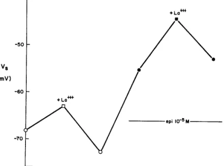
Fig. 6 shows an experiment where the effect of La+++ was determined in the absence, and then in the presence of epinephrine. As in the previous figure, the ordinate plots V,, the midpoint of the iK, activation curve. In the absence of the catecholamine, exposure to La+++ gives a 7.5-mV shift (deter-mined using the mean of the control V, values before and after La+++
expo-vs
(mV)
FIGURE 6. The effect of LaCI3 (0.5 mM) in the absence (open symbols) and presence (filled symbols) of epinephrine. Voltage shifts are plotted as vertical displacements. Ex-periment 94-1 in 5.4 mM [Ca]o, with D600 (5 X 10- 7g/ml) in La+++-free solutions to inhibit contractions.
THE JOURNAL OF GENERAL PHYSIOLOGY VOLUME 64 · 1974
sure). The preparation was then superfused with solution containing a maxi-mally effective dose of epinephrine (10-6 M), which produced a typical voltage shift. In the maintained presence of epinephrine, the preparation was then reexposed to 0.5 mM La+++. In this case a 9.8-mV shift was produced. The experiment shows that the La+ ++ voltage shift is not occluded by the presence of epinephrine. The result stands in obvious contrast with the mutual occlusion observed between epinephrine and theophylline, or epineph-rine and R07-2956.
The additive effects of epinephrine and La+++ provide further evidence on the mechanism of the hormone action. Suppose that La+++ and epinephrine each produced voltage shifts by putting their own charge near the i,2 channel.
Even if there were separate binding sites for each cation, some interaction between their effects would be expected, since each cation must be close enough to the activation process to give substantial voltage shifts individually. According to this argument, the presence of epinephrine should reduce the local [La+++ ] near the channel, and, therefore, the amount of La+++ bound (the applied dose of La+ ++ is far from giving a saturated effect). The effect of epinephrine on [La+++]it. can be roughly estimated if the La+ ++ bind-ing site experiences a surface potential change as large as the net voltage shift (A-V). Then, [La+++]i'/[La+++]°i t ° = exp(-zFAV8/RT). For
z = +3 and V, = 17 mV (as in Fig. 6), the Boltzmann factor is exp(-2) = 0.135. Thus, a large reduction in the amount of bound La+++ would have been expected if epinephrine had, in fact, put its own charge near the chan-nel. The experiment shows that the effect of La+++ is not diminished, and argues against the idea that epinephrine modifies the external surface charge in the immediate vicinity of the channel.
An Alternative Hypothesis for the Action of Epinephrine
The epinephrine-like action of theophylline and R07-2956 comes as no great surprise from the biochemical point of view. Both of these compounds are potent inhibitors of 3',5'-phosphodiesterase, the intracellular enzyme which hydrolyzes adenosine 3' ,5'-monophosphate (cyclic AMP). The conventional interpretation of their effects (see, e.g. Robison et al., 1971) explains their effects through the elevation of intracellular cyclic AMP levels. To state the
hypothesis diagramatically:
propranolol rate of
epinephrine // , cAMP
formation elevated saturable voltage [cAMP] t step(s) shift rate of j
theophylline - * cAMP
breakdown
330
R. W. TSIEN Action of Chronotropic Agents in Cardiac Purkinje Fibers
The hypothesis proposes that the final common mechanism begins with an increase in the intracellular level of cyclic AMP, which eventually leads to the shift in the voltage dependence of iK,. The mutual occlusion of epineph-rine and theophylline is accounted for by the existence of some saturable process downstream to the elevated [cAMP]i. The model presumes that either epinephrine, theophylline, or R07-2956 can elevate [cAMP], beyond the level required to saturate the hypothetical process, so that each of these
agents is individually capable of evoking the maximal effect.
Synergistic Effects of Eiinephrine and Theophylline
In the above experiments epinephrine and theophylline were used in maxi-mally effective concentrations. At low concentrations, however, the model gives rise to the expectation that epinephrine and theophylline will act syner-gistically in elevating [cAMP]i and in producing an accompanying voltage shift. This intuitive expectation is supported by the Appendix, which puts the cyclic AMP scheme in quantitative terms.
Fig. 7 shows the combined results from seven preparations. In three ex-periments, the normalized dose-response curve to epinephrine was obtained in the presence of a submaximal dose of theophylline (10-4 M). These results
(open symbols) are compared with results in the absence of theophylline (filled symbols), replotted from Fig. 9 of the previous paper. By itself, 10-4M theophylline produces a voltage shift (points on ordinate) that is about one-third of the maximal effect. The addition of epinephrine (in the maintained presence of theophylline) leads to a further voltage shift (points at 3 X 10⁻⁸ M). Increasing the epinephrine concentration to 10- 7M gives an essentially maximal effect (compare open symbols at 10-' M and 10-6 M). Thus, the dose-response curve for epinephrine is modified by the presence of theophyl-line.
The experimental points in Fig. 7 have been fitted by quantitative predic-tions arising from the cyclic AMP hypothesis (see Appendix). The curve fitting was carried out in the following way. First, the theophylline-free data were fitted by eye to the theoretical control curve (see Fig. 9 C) which holds for the normal degree of phosphodiesterase activity. The fit is made by speci-fying that half-occupation of the -receptor occurs at an epinephrine con-centration of 1.88 X 10⁻⁷M (vertical arrow).
Thus 1/K, = 1.88 X 10⁻⁷ M, and the normalized abscissa in Fig. 9 C can
be reexpressed in terms of actual epinephrine concentrations. The next step is to fit the data obtained in the presence of 10-4 M theophylline. This was
done by choosing a value of k2 (= 0.625) and the corresponding curve in
Fig. 9 C. The chosen value corresponds to reduction of phosphodiesterase activity to / normal. It is evident in Fig. 7 that the adjustment of the single parameter, k2, gives a fairly good fit to the data points, both in the absence
of epinephrine, and in the presence of the range of epinephrine concentrations. 331
THE JOURNAL OF GENERAL PHYSIOLOGY VOLUME 64 I1974 1.0 a, a c ID mo To ' 0.5 G) N a E 0 zo z u 1 10 10-I 1O t0- 1o0-[epinephrine], M
FIGURE 7. Epinephrine dose-response curves in the absence and presence of
theophyl-line. The ordinate gives the normalized voltage shift, AV8/AV,; the abscissa plots the
epinephrine concentration on a log scale. The different symbols signify seven individual preparations. Open symbols are results in the presence of 10- 4M theophylline, filled symbols in the absence of theophylline. [Ca]o = 5.4 mM throughout. The smooth curves are generated by the model described in the Appendix (for further description see text). In this respect, the experimental results are in reasonable agreement with the predictions of the cyclic AMP model.
DISCUSSION
Explanations of Epinephrine's Action
The original aim of the experiments was to test the most simple explanation for epinephrine's ability to shift the activation curve for i, along the voltage axis. As Hauswirth et al. (1968) proposed, the voltage shift could be accounted for if epinephrine added its own positive charge to the outside of the mem-brane near the i, channel. This mechanism of action can be effectively ruled out by two types of experiment.
The first experiment compares the action of epinephrine with the effect of theophylline or R07-2956. In maximally effective concentrations, these agents occlude epinephrine's action indicating that a final common mode of action is shared. Since theophylline or R07-2956 are not cations at pH 7.4 this finding is not compatible with the original hypothesis. The second experi-ment studies the action of epinephrine in combination with an agent which
is thought to modify external surface charge: lanthanum ion. In this case
the voltage shift produced by La+ + + simply adds to the maximal epinephrine voltage shift: no occlusion occurs. This result is also contradictory to the expectations of the external surface charge hypothesis, which predicts partial occlusion of the La+++ effect.
It appears that the direct external surface charge explanation, for all its 332
R. W. TSIEN Action of Chronotropic Agents in Cardiac Purkinje Fibers
simplicity, must be abandoned. This removes the necessity for explaining the fact that -blockers such as propranolol or pronethalol do not produce voltage shifts (Hauswirth et al., 1968; Tsien, 1974). From the point of view of the original hypothesis, their lack of effect as voltage shifters was awkward since they are positively charged molecules that presumably compete for
the same receptor that binds epinephrine.
FURTHER CONCLUSIONS The experiments show that the initial effects of theophylline or R07-2956 are propranolol insensitive, and, therefore, not identical with the initial action of epinephrine. Since the final action of epi-nephrine does coincide with that of the other compounds, it follows that there are at least two steps in epinephrine's mechanism of action (see diagram on p. 327). If it is accepted that a sequence of steps takes place, the results do not argue against the idea that epinephrine's initial action does, in fact, in-volve a change in external surface charge at some distance from the iK, channel. It seems entirely possible that a change in surface potential plays some part in the interaction of epinephrine with the t-adrenergic receptor. There is no evidence that I know of which suggests that catecholamines act in anything but the cationic form.
Possibility of Internal Surface Charge Mechanism
The results in this paper leave open the possibility that a change in the internal surface charge gives rise to the voltage shift. Such an explanation retains the electrostatic character of the original scheme: as in the presumed action of calcium (Frankenhaeuser and Hodgkin, 1957), no change is re-quired in the gating reaction itself. Whether outside or inside, the postulated changes in surface charge would merely alter the electric field that is sensed by the gating reaction.
How might the internal surface charge be altered? One hypothesis is that epinephrine brings about a specific and direct phosphorylation of the inside membrane surface near the i, channel (Tsien, 1973 a). Another explanation is that epinephrine leads to an increased rate of calcium uptake by the sarco-plasmic reticulum, a fall in the free intracellular calcium concentration and a
removal of calcium from specific binding sites near the i,, channel
(McNaugh-ton and Noble, 1973). Both of these schemes attempt to account for the voltage shift by the net removal of positive charge from the inside of the mem-brane: it follows that the electrostatic effects of the proposed mechanisms would be indistinguishable. It will be interesting to see if either of these internal surface charge hypotheses survive the test of further experiments.
Possible Involvement of Cyclic AMP
A common feature of the two internal surface charge hypotheses is their reliance on cyclic AMP as a mediator of the effect of epinephrine (see Tsien,
THE JOURNAL OF GENERAL PHYSIOLOGY VOLUME 64 · 1974 1973 a; McNaughton and Noble, 1973). This raises the more general issue of the possible role of cyclic AMP as a mediator of the iK, voltage shift. A variety of experimental evidence is relevant to this question and will be briefly mentioned here.
Until recently there has been no biochemical data on cyclic AMP levels in Purkinje fibers. Danilo et al. (1974) have shown that epinephrine elevates cyclic AMP levels in intact Purkinje fibers from dog heart, as well as in-creases adenylate cyclase activity in fiber homogenates (Danilo et al., 1973). The observed increase falls short of the fivefold elevation found in perfused rat hearts (Robison et al., 1965; Cheung and Williamson, 1965) but there is
considerable variability both in the basal cyclic AMP level and the percentage increase with exposure to epinephrine. The results of Danilo et al. (1973, 1974) raise the possibility of quantitative revision of parameter values used in the prediction of dose-response curves (Appendix). However, their findings
are generally consistent with the cyclic AMP hypothesis.
If cyclic AMP mediates the effect of epinephrine, it is expected that similar action should be produced by other agents which elevate cyclic AMP levels. Compounds which inhibit 3' ,5'-phosphodiesterase are known to have epi-nephrine-like effects in many tissues, including heart, and it is generally pre-sumed (although not proven) that these agents act by elevating cyclic AMP. The present results show that two chemically unrelated phosphodiesterase inhibitors, theophylline and R07-2956, each mimic the action of epinephrine on the pacemaker potential, and on the underlying slow current changes. The effect of these compounds on ix, is certainly consistent with the cyclic AMP hypothesis.
Exposure of cells to butyryl derivatives of cyclic AMP is another procedure which has often succeeded in giving epinephrine-like effects in intact tissues, although the mechanism of this action is not clear (see Robison et al., 1971). Borasio and Vassalle (1971) studied the effect of N6, 2'-O-dibutyryl cyclic AMP (DBcAMP) on automaticity in dog Purkinje preparations. These workers found effects which could be attributed to indirect actions (e.g. release of catecholamines from fine nerve terminals) but no direct effect of DBcAMP itself. I have confirmed this negative result, using N6-monobutyryl cyclic AMP (MBcAMP) as well as DBcAMP, in concentrations up to 2 mM. The insensitivity of the pacemaker (and also i,) to cyclic AMP derivatives is particularly puzzling since, in the same preparations, dramatic changes in the plateau currents are observed at considerably lower doses of MBcAMP (Tsien et al., 1972). It may yet be possible to reconcile the apparent ineffec-tiveness of cyclic AMP derivatives with the cyclic AMP hypothesis. For ex-ample, it is conceivable that DBcAMP and MBcAMP have limited access to the site of action within the cell. Until some explanation is established, it seems premature to fully accept the idea that cyclic AMP mediates the action of epinephrine on i, although most of the evidence favors this hypothesis.
R. W. TSIEN Action of Chronotropic Agents in Cardiac Purkinje Fibers
In this light the present scheme (Appendix) is offered as a tentative frame-work for studying the dose-dependent effects of chronotropic agents.
LOCATION OF THE SATURABLE PROCESS
A key feature of the proposed
scheme (Appendix) is that the magnitude of voltage shift is limited by the saturation of some process downstream to the elevation in cyclic AMP; the location of the saturable process is important in explaining the mutual occlusion of epinephrine and theophylline. If the scheme is correct, it follows that the concentration of cyclic AMP should continue to increase beyond the point at which the voltage shift attains its maximum value. This expecta-tion is supported by the results in Fig. 9 of the previous paper (Tsien, 1974). The maximal voltage shift of i, is reached at 10-6 M epinephrine, whereas the enhancement of the secondary inward current is well short of maximal at that point. Since there is rather clear evidence to indicate that the effect of epinephrine on the secondary inward current is cyclic AMP mediated (Car-meliet and Vereecke, 1969; Tsien et al., 1972) it appears that the cyclic AMP level must still be increasing at an epinephrine concentration of 10-6 M. NATURE OF THE SATURABLE PROCESSThe scheme presented in the
Appendix does not specify the details of the "enzyme system" which incorpo-rates the saturable process. The only requirement is that the end effect, the voltage shift, should saturate rather abruptly with increasing levels of cyclic AMP. Of the many possibilities, two biochemical mechanisms may be men-tioned since both of these are linked to the previous discussion of internal surface charge. In one hypothesis (Tsien, 1973 a), cyclic AMP would activate a protein kinase which brings about the phosphorylation of the inside of the membrane near the channel. This mechanism is relatively direct but it would be difficult to study experimentally. The other hypothesis (McNaughton and Noble, 1973) proposes the removal of Ca++ from specific binding sites near the channel, through stimulation of calcium uptake by the sarcoplasmic reticulum (SR). This idea also depends upon a cyclic AMP-activated protein kinase; cyclic AMP increases calcium uptake by SR microsomes (Entman, et al., 1969) by promoting phosphorylation of SR membrane by protein kinase (Kirchberger et al., 1972). The calcium removal hypothesis calls for more steps than the other mechanism, but it lends itself readily to experi-mental test by procedures which alter [Ca] i (see McNaughton and Noble, 1973).On the Dose Dependence of Chronotropic Effects
on the Pacemaker K Current
APPENDIX
The concentration-dependent effects of chronotropic agents on i,, are in-terpreted in terms of a simple reaction scheme (Fig. 8). It is proposed that
THE JOURNAL OF GENERAL PHYSIOLOGY VOLUME 64 - 1974
ATP
Catecholamine + AC - AC* - k1 unshifted Vs
[E]
cyclic AMP + system >
[A]
Phosphodiesterase + PDE- - PDE k2 shifted Vs
inhibitor IT]
5'AMP
FIGURE 8. A simple scheme for describing the combined effects of epinephrine and theophylline. Two-way arrows (-) signify reversible reactions, while broad arrows indi-cate enzymatic control of a reaction. The nature of the enzyme system is not specified
(see Discussion) and may consist of a number of steps.
the actions of catecholamines (e.g. epinephrine) and phosphodiesterase in-hibitors (e.g. theophylline) are mediated by elevations in the intracellular level of cyclic AMP. The increase in [cAMP]i in turn gives rise to the volt-age shift, via a saturable process or processes (labeled "enzyme system" in Fig. 8). From this scheme a family of theoretical dose-response curves is de-rived. The rationale and background to this scheme is described elsewhere; this appendix merely puts the model in quantitative terms.
It is assumed that catecholamines combine with the -adrenergic receptor in a one-for-one fashion, converting the associated enzyme adenylate cyclase (AC) to an activated form (AC*). The proportion of activated adenylate cyclase is then
AC*/(AC + AC*) = 1/(1 + E-'), (1)
where the normalized catecholamine concentration is E =
[catecholamine]-Kr, and K, [M- 1] is the binding constant of the catecholamine for the -re-ceptor. The associated enzyme, adenylate cyclase, catalyzes the reaction ATP -- cyclic AMP + pyrophosphate, with rate coefficient k. Using nor-malized units, the activity of adenylate cyclase in unstimulated form (100% AC) is called 1, and the fully stimulated activity (100% AC*) is called F.
Then, the rate coefficient kl will be given by
k = 1 + (F- 1)/(1 + E-'). (2)
In other words, as [catecholamine] increases, k increases from 1 to F along a hyperbolic curve. F is assigned the value 5 on the basis of biochemical studies of cyclic AMP levels in perfused rat heart (Robison et al., 1965; Cheung and Williamson, 1965). For simplicity it is assumed (cf. Cheung and
R. W. TSIEN Action of Chronotropic Agents in Cardiac Purkinje Fibers
Williamson, 1965) that elevations of cyclic AMP are steadily maintained in the continued presence of catecholamine.
In the scheme phosphodiesterase inhibitors act through their effect on k2, the rate coefficient of cyclic AMP breakdown. The inhibitor is assumed to combine with phosphodiesterase, converting it from a more active form (PDE*) to a less active form (PDE). If the uninhibited activity (100% PDE*) is called 1, the presence of inhibitor reduces k2 to some value less
than 1. This behavior will be treated more quantitatively at the end of the Appendix.
The steady-state level of cyclic AMP [A] is given by
[A] = k[ATP]/k2. (3)
The intracellular concentration of ATP is normally controlled within narrow limits by metabolic processes and may be normalized for convenience. Then,
[A] = [I + (F - 1)/(1 + E-')]/k2. (4)
This equation states a very simple synergism in the action of catecholamines and phosphodiesterase inhibitors. In the absence of inhibitor (k2 = 1), [A]
rises hyperbolically from 1 to F (Fig. 9 a). The maintained presence of fixed amount of inhibitor gives rise to a multiplicative increase in [A]. This is shown in Fig. 9 a by the curve labeled k2 = 0.625 (phosphodiesterase activity
reduced to Y normal). The cyclic AMP concentration is elevated by a factor of 1.6, regardless of the concentration of catecholamine.
Prediction of the Dose-Response Curves
The next step is to relate the level of cyclic AMP, [A], to the eventual voltage shift. There is evidence (see Discussion) that the cyclic AMP level continues to rise under conditions where the voltage shift has already reached maximum value. This indicates that a saturating process or processes links the rise in
[A] and the eventual effect. Without specifying the details of the process or
processes, it is useful to put the present scheme into quantitative form and to generate a set of theoretically predicted dose-response curves.
Fig. 9 b shows the saturating relationship which was used. It is assumed that the maximal voltage shift is produced by something less than a fivefold increase in [A]. (The saturating level is shown as a dashed line in 9 a and b.) This assumption is justified by the knowledge that the maximal effect on i,2 can be evoked by exposure to epinephrine alone. The form of the saturating relationship is a quarter sine wave. This waveform was chosen partly for convenience of computation, and partly because the sine wave gives linear behavior for small A, but an appropriately abrupt saturation as A increases.
(A rectangular hyperbola does not saturate abruptly enough.) The results
®
E (normalized catecholamine concentration)
1.0 0 0 a 0 0 z 0 1 2 3 4 -F-5 6 7 8
A (normalized cyclic AMP level)
I I I I I I I I 8 0 6 45 0 U 0 e" 3 E ° 2 1 0 0, E0 °o. 0 z
E (normalized catecholamine concentration)
FIGURE 9 100 /m I I I v
R. W. TIEN Action of Chronotropic Agents in Cardiac Purkinje Fibers
would not be significantly changed by using another function with similar properties, or by using a lower saturating level of [A].
A family of predicted dose-response curves is shown in Fig. 9 c. The curves labeled 1.0 and 0.625 can be obtained by combining Fig. 9 a and b. These were used in fitting the experimental data in Fig. 7. The other curves were produced using other values for k2as indicated. The results show that
inhibi-tion of phosphodiesterase alters the dose dependence to the catecholamine. As the degree of inhibition increases, the normalized dose-response curve is shifted upward and to the left. The presence of phosphodiesterase inhibitor lowers the concentration of catecholamine that is required to evoke the maximal voltage shift. As the degree of inhibition increases, a point is reached
(k2 < 2/3 7r) where the voltage shift is maximal, regardless of the
concentra-tion of catecholamine. This corresponds to the experimental observaconcentra-tion that theophylline in concentrations above 5 X 10⁻⁴ M elicits the maximal voltage shift (Fig. 4) and occludes the effect of epinephrine (Fig. 5).
The form of the predicted dose-response curves is not very critically de-pendent on the parameter values used to generate Fig. 9 c. Let As.t be the level of [A] required to give full effect (A.,t = 3r/2 in Fig. 9). Both F and
A.,t can each be adjusted over a range of values, without qualitatively
chang-ing the model behavior. The only important requirement is that F
2
Ast.This point may be illustrated by letting k increase linearly with E (this is equivalent to letting F become very large or making A.st small). In this case the saturation of the dose-response curve arises entirely from the effect of
[A] on the enzyme system. The resulting dose-response curves (not shown
here) are rather similar to those in Fig. 9 c and would also provide an ade-quate fit to the experimental data in Fig. 7. One important conclusion from this analysis is that the value of K, used in fitting the experimental data is dependent upon other assumptions, and may or may not reflect the real binding constant of epinephrine for the P-receptor.
FIGURE 9. The generation of dose-response curves from the cyclic AMP scheme in Fig. 8. 9 a shows the theoretical relationship between catecholamine concentration and the steady level of cyclic AMP. 9 b shows the presumed saturating relationship between the cyclic AMP level and the displacement of V,; for A < 3r/2, voltage displacement cc sin (A/3). Note that the ordinate is labeled so that "zero" voltage shift occurs at the resting cyclic AMP level (A = 1). 9 c shows the normalized dose-response curves which result from combining relationships of the type shown in 9 a and b.
THE JOURNAL OF GENERAL PHYSIOLOGY VOLUME 64 · 1974
Dose-Dependent Effects of Phosphodiesterase Inhibitors
Experiments studying the concentration-dependent effects of phosphodies-terase inhibitors were not presented in this paper. However, it still may be useful to give corresponding theoretical predictions from the present scheme. Let us assume that inhibitor combines with PDE* in a one-for-one manner, converting it to PDE. Then, the fraction of inhibited phosphodiesterase is given by the binding equation,
PDE/(PDE + PDE*) = 1/(1 + T-1') (5)
where the normalized concentration T = [inhibitor] Kr, and K is the binding constant of phosphodiesterase for the inhibitor. If the uninhibited activity (100% PDE*) is called 1 (as above), and the maximally inhibited activity (100% PDE) is G- ,
k2 = 1 + (G-' - 1)/(1 + T-'). (6)
Then from Eq. 3,
[A] = k/k2 = F + E-) / (G + T-1) (7 This equation emphasizes the similarity between the effects of E and T.
In the absence of phosphodiesterase inhibitor (T = 0), increasing E causes
[A] to rise hyperbolically from 1 to F (Fig. 9 a). Analogously, in the absence
of catecholamine (E = 0), increasing T causes [A] to rise hyperbolically from 1 to G (not shown). If G were equal to F, the theoretical dose-response curve for theophylline (for T = 0) would have exactly the same form as the epi-nephrine dose-response curve (for E = 0, Fig. 9 c). Preliminary results sug-gest that such a relationship may apply to the voltage shift produced by theo-phylline; further experiments will be particularly interesting when a value for G becomes available from biochemical studies.
I am grateful to Roger Tsien and W. K. Chandler for their constructive criticisms of the manuscript, and Patricia Parillo for patient secretarial help.
This research was supported by U. S. Public Health Service Grant 5 R01 HL 13306 (Heart and Lung Institute).
Received for publication 14 February 1974. REFERENCES
BLINKS, J. R., C. B. OLSON, B. R. JEWELL, and P. BRAVENY. 1972. Influence of caffeine and other methylxanthines on mechanical properties of isolated mammalian heart muscle. Circ. Res.
30:367.
BORASIO, P. G., and M. VASSALLE. 1971. Actions of cyclic AMP on K transport and automaticity in cardiac Purkinje fibres. Fed. Proc. 30:2641. (Abstr.).
R. W. TSIEN Action of Chronotropic Agents in Cardiac Purkinje Fibers
BROWN, R. H., JR. 1973. The characterization and functional significance of surface negative charges in cardiac muscle. Ph.D. Thesis. Oxford University, Oxford, England.
BROWN, R. H., JR. 1974. Membrane surface charge: discrete and uniform modelling. Prog. Biophys. Mol. Biol. In press.
CARMELIET, E., and J. VEREECKE. 1969. Adrenaline and the plateau phase of the cardiac action potential. Pfluegers Archiv. Gesamte Physiol. Menschen Tiere. 313:303.
CHEUNG, W. Y., and J. R. WILLIAMSON. 1965. Kinetics of cyclic adenosine monophosphate changes in rat heart following epinephrine administration. Nature (Lond.). 207:79.
DANILO, P., Y. VULLIEMOZ, M. VEROSKY, and M. R. ROSEN. 1973. Activation o cardiac Purkinje fiber adenyl cyclase by 0-adrenergic amines. Fed. Proc. 32:773. (Abstr.).
DANILO, P., Y. VULLIEMOZ, M. VEROSKY, M. R. ROSEN, and B. F. HOFFMAN. 1974. Epinephrine-induced automaticity and cAMP concentration in canine cardiac Purkinje fiber bundles. Fed. Proc. 33:1518. (Abstr.).
DAVIS, L. D., and J. TEMTE. 1968. Effects of propranolol on the transmembrane potentials of ventricular muscle and Purkinje fibers of the dog. Circ. Res. 22:661.
ENTMAN, M. L., G. S. LEVEY, and S. E. EPSTEIN. 1969. Mechanism of action of epinephrine and glucagon on the canine heart. Evidence for increase in sarcotubular calcium stores mediated by cyclic 3'5' AMP. Circ. Res. 25:429.
FRANKENHAEUSER, B. 1957. The effect of calcium on the myelinated nerve fibre. J. Physiol. (Lond.). 137:245.
FRANKENHAEUSER, B., and A. L. HODGKIN. 1957. The action of calcium on the electrical proper-ties of squid axons. J. Physiol. (Lond.). 137:217.
GILBERT, D. L., and G. EHRENSTEIN. 1969. Effect of divalent cations on potassium conductance of squid axons: determination of surface charge. Biophys. J. 9:447.
HARDMAN, H. F. 1962. Molecular form of theophylline responsible for positive chronotropic activity. Circ. Res. 10:598.
HAUSWIRTH, O., D. NOBLE, and R. W. TsIEN. 1968. Adrenaline: mechanism of action on the pacemaker potential in cardiac Purkinje fibres. Science (Wash. D. C.). 162:916.
JENERICK, H. 1959. The control of membrane ionic currents by the membrane potential of muscle. J. Gen. Physiol. 42:923.
KIRCHBERGER, M. A., M. TADA, D. I. REPKE, and A. M. KATZ. 1972. Cyclic adenosine 3',5'-monophosphate-dependent protein kinase stimulation of calcium uptake by canine cardiac
microsomes. J. Mol. Cell. Cardiol. 4:673.
McNAUGHTON, P. A., and D. NOBLE. 1973. The role of intracellular calcium ion concentration in mediating the adrenaline-induced acceleration of the cardiac pacemaker potential. J. Physiol. (Lond.). 234:53.
NOBLE, D., and R. W. TSIEN. 1968. The kinetics and rectifier properties of the slow potassium current in cardiac Purkinje fibres. J. Physiol. (Lond.). 195:185.
RALL, T. W., and T. C. WEST. 1963. The potentiation of cardiac inotropic responses to norepi-nephrine by theophylline. J. Pharmacol. Exp. Ther. 139:269.
ROBISON, G. A., R. W. BUTCHER, I. YE, H. E. MORGAN, and E. W. SUTHERLAND. 1965. The effects of epinephrine on adenosine 3'5'-monophosphate levels in the isolated perfused rat heart. Mol. Pharmacol. 1:168.
ROBISON, G. A., R. W. BUTCHER, and E. W. SUTHERLAND. 1971. Cyclic AMP. Academic Press, Inc., New York.
SHEPPARD, H., and G. WIGGAN. 1970. Analogues of (4-(3, 4-dimethoxybenzyl)-2-imizolidinone as potent inhibitors of rat erythrocyte adenosine cyclic 3', 5'-phosphate phosphodiesterase. Mol. Pharmacol. 7:111.
TRAUTWEIN, W., and R. F. SCHMIDT. 1960. Zur Membranwirkung des Adrenalins an der Herzmuskelfaser. Pfluegers Archiv. Gesamte Physiol. Menschen Tiere. 271:715.
TSIEN, R. W. 1973 a. Control of conductance in cardiac membrane permeability. Neurosci. Res. Program Bull. 11:204.
TSIEN, R. W. 1973 b. Does adrenaline act by directly modifying the external membrane surface charge of cardiac Purkinje fibres? J. Physiol. (Lond.). 234:37.
342 THE JOURNAL OF GENERAL PHYSIOLOGY VOLUME 64 · I974
TSIEN, R. W. 1974. Effects of epinephrine on the pacemaker potassium current of cardiac Purkinje fibers. J. Gen. Physiol. 64:293.
TSIEN, R. W., W. R. GILES, and P. GREENGARD. 1972. Cyclic AMP mediates the action of epi-nephrine on the action potential plateau of cardiac Purkinje fibres. Nat. New Biol. 240:181. WEIDMANN, S. 1955. Effects of calcium ions and local anesthetics on electrical properties of