INVESTIGATION OF THE METHYLATION STATUS OF FGF2 GENE IN PERIODONTAL TISSUES AS DETERMINANTS OF HEALING RESPONSE AFTER
IMPLANT SURGERY
Travis Joe Whitley
A thesis submitted to the faculty of the University of North Carolina at Chapel Hill in partial fulfillment of the requirements for the degree of Master of Science in the
Department of Periodontology, School of Dentistry.
Chapel Hill 2016
iii ABSTRACT
Travis Joe Whitley: INVESTIGATION OF THE METHYLATION STATUS OF FGF2 GENE IN PERIODONTAL TISSUES AS DETERMINANTS OF HEALING
RESPONSE AFTER IMPLANT SURGERY (Under the direction of Silvana Barros)
Periodontal wound healing is a complex process involving a series of interactions of growth factors, including FGF-2. Previous studies have suggested that the FGF2 gene may be down-regulated in diseased periodontal tissues. Aberrant gene expression in smokers and diabetics has also been reported. The aims of this study were to determine the association of FGF2 methylation level with wound healing after implant surgery and the association of smoking and diabetes with FGF2 methylation. Thirty-four included subjects were distributed into control, smoking and diabetic groups. During the implant surgery, gingival tissue biopsies were collected for real-time PCR analysis. This study demonstrated that FGF-2hyper-methylation and associated gene down-regulation were associated with smoking status and diabetes.
iv
ACKNOWLEDGEMENTS
v
TABLE OF CONTENTS
LIST OF TABLES ... vii
LIST OF FIGURES ... viii
INTRODUCTION AND BACKGROUND ...1
Osseointegration and Dental Implant Wound Healing ...1
Role of FGF-2 and Bone Healing ...5
DNA Methylation – An Overview ...6
Smoking and Its Effect on the Periodontium ...8
DNA Methylation and Smoking ...12
Diabetes and Its Effect on the Periodontium ...14
DNA Methylation and Type II Diabetes ...19
AIMS ...21
HYPOTHESES ...22
MATERIALS AND METHODS ...23
Study Population ...23
Gingival Tissue Biopsies, Gingival Crevicular Fluid (GCF) Collection and Implant Stability Quotient (ISQ)Measurements...24
RNA Isolation and Quantitative Real-Time Reverse Transcription PCR...25
DNA Isolation, Selective Digestion and Quantitative Real-Time PCR...25
vi
RESULTS ...28
Participant Recruitment Process ...28
Study Population Demographics ...30
Selected Implant Sites ...32
Differential DNA Methylation of FGF2 Gene in Smokers and Diabetic Participants ...34
Differential mRNA Expression of FGF2 Gene in Smokers and Diabetic Participants ...36
APPENDIX I ...47
vii
LIST OF TABLES
Table 1. Proposed Mechanisms for the Negative Periodontal Effects of Smoking...10
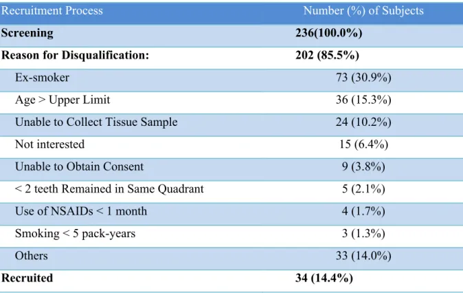
Table 2. Participant Recruitment Process ...29
Table 3. Study Participant Demographics ...31
Table 4. Implant Sites of Study Participants ...33
viii
LIST OF FIGURES
Figure 1. The DNA Methylation Process ...7 Figure 2. Possible Linkage between Diabetes and Periodontal Disease Severity ...18 Figure 3. Methylation Level of FGF2 Gene in Smokers or Diabetics in Comparison to
Control Subjects. ...35 Figure 4. Messenger Level of FGF2 Gene in Smokers or Diabetics in Comparison to
1
INTRODUCTION AND BACKGROUND Osseointegration and Dental Implant Wound Healing
Wound healing involves a series of complex events that are coordinated and sequential. Many cells, signaling pathways, and growth factors play integral roles in this process with precise coordination. In implant bone healing, these processes lead to osseointegration; the direct structural and function connection between ordered, living bone and the surface of a load-carrying implant (Brånemark 1983). Animal and human studies have demonstrated the timeline of events that occur following surgical placement of a dental implant (Berglundh 2003; Lang 2011). Four phases are seen in the implant healing process that correlates with similar soft tissue healing events: hemostasis,
2
the recruitment of cells, formation of new extracellular matrix, and angiogenesis to the implant surface.
Fibroblast Growth Factor -2 (FGF-2) and Wound Healing
Fibroblast growth factor (FGF) is protein involved with many aspects of wound healing. FGF was isolated completely in 1974 from bovine pituitary glands
(Gospodarowicz 1974). FGF exists in two different isometric points, 1 and 2. While one was known as the acidic FGF (aFGF) or FGF1, the other became known as the basic FGF (bFGF) or FGF-2 (Bohlen et al. 1984). The entire nucleotide sequence of human FGF-2 was isolated two years later (Abraham et al. 1986).
As a member of the heparin-binding growth factor family, FGF-2 is found in various body tissues and exerts a range of biological effects on cells from different embryological origins. It acts as a potent angiogenic molecule in vivo and in vitro, aiding in wound healing and facilitating events necessary for re-vascularization. During this process, it causes proliferation of macrophages and other cell types involved in repair (Bikfalvi et al. 1997, Lee et al. 2010).
3
(Murakami et al. 1999). During the bone remodeling phase, it can stimulate migration of mesenchymal progenitor cells which ultimately differentiate into osteoblasts and
cementoblasts (Murakami et al. 1999).
Several animal (Murakami et al. 1999, Murakami et al. 2003, Takayama et al. 2001) and human (Kitamura et al. 2008, Kitamura et al. 2011, Kitamura et al. 2015) studies have shown promising results in the use of FGF-2 for periodontal regeneration. In 1999, Murakami surgically created 2- and 3-wall intrabony defects along with Class II furcation defects in beagle and primate models. Six to eight weeks following surgical access and topical application of recombinant FGF-2, significantly greater cementum, periodontal ligament and bone formation was seen in the FGF-2 sites compared to control sites. Histological evidence of regeneration was also portrayed in this study. Epithelial down growth, ankylosis or root resorption were not observed in FGF-treated sites (Murakami et al. 1999). In 2001, another animal study was conducted by Takayama on non-human primates. Surgically-created Class II furcation defects were treated with either 0.1% human recombinant FGF-2, 0.4% rhFGF-2, carrier alone or no treatment. Their results demonstrated a significant increase in periodontal regeneration clinically and
histologically with the higher dose (0.4%) of rhFGF-2 compared to other treatment groups (Takayama et al. 2001). In 2003, Murakami surgically created Class II furcation defects in beagle dogs and treated with rhFGF-2 compared with control sites.
4
In 2008, a Phase II randomized, controlled, double-blind clinical trial was
conducted by Kitamura at 13 study facilities with 74 subjects. These subjects presented with 2- or 3-wall vertical bone defects and randomly assigned to four treatment groups: carrier alone, carrier + 0.03% FGF-2, carrier + 0.1% FGF-2, or carrier + 0.3% FGF-2. The results demonstrated a significantly greater rate of increase in alveolar bone height between the group with 0.3% FGF-2 compared to the carrier alone group at 36 weeks. No differences were noted for clinical attachment level or alveolar bone gains between groups (Kitamura et al. 2008). Three years later, the group performed a subsequent larger clinical trial which involved 253 patients. In this study, 200µL of 0%, 0.2%, 0.3% or 0.4% FGF-2 was administered to 2- or 3-walled bony defects at the time of a modified Widman flap surgery. All FGF-2 sites demonstrated significant higher percentage of bone fill at 36 weeks (Kitamura et al. 2011).
In 2015, Kitamura again published results on two randomized, placebo-controlled clinical trials comparing rhbFGF-2 and enamel matrix derivative (EMD) in periodontal regeneration for infrabony defects. The first study included 323 subjects who randomly received 0.3% FGF-2 or placebo during flap surgery. At 36 weeks, a significantly greater percentage of bone fill was seen for the FGF-2 group vs placebo (37% vs 21.5%). The second study included 264 subjects randomly assigned to receive either 0.3% FGF-2, EMD, or flap alone for treatment of infrabony defects. At 36 weeks, linear alveolar bone growth for the FGF-2 and EMD groups were 1.93 and 1.36 mm, respectively, which was statistically significantly different.
5
Recently, Bizenjima published an animal study on the healing of surgical periodontal defects in diabetic rats treated with FGF-2. The group created defects on maxillary first molars, which were treated with either a control carrier or FGF-2. Histological and radiographic analyses at 2 and 4 weeks showed a significant increase in bone volume and trabecular patterns for the FGF-2 group vs the control group (Bizenjima, 2015).
Role of FGF-2 and Bone Healing
While FGF-2 is well known for its role in angiogenesis, several studies have also identified a role for it in osteogenic pathways (Marie, 2003). The growth and function of mesenchymal stem cells from bone marrow seems to be stimulated by FGF-2 (Murikami 2011). FGF-2 plays a key role in not only cell recruitment and proliferation in the area of injury, but differentiation of osteoblastic cell lineages to induce bone formation. FGF-2 null mice have been show to demonstrate low bone mass and decreased bone formation as they age (Montero 2000, Behr 2010). Evidence reveals that FGF-2 has a significant impact in bone formation and remodeling by upregulation of the RANK – RANKL pathway (Fei, 2013). FGF-2 has also been shown to have positive effects on bone fracture healing. Animal and human studies have demonstrated that with increasing doses of FGF-2 applied to long bone fractures, an increase in osteocytes, bone mineral density, and bone repair is seen (Kawaguchi 1994, Furuya 2014, Tanaka, 2012). Mutations in FGF receptors have been associated with a number of craniofacial disorders, including achondroplasia and craniosynostosis. These processes are important events in the healing after dental implant placement and can greatly impact osseointegration. Recently, a beagle dog model was used to study the effect of FGF-2 application on implant
bone-6
to-implant contact (BIC) at 4, 8, and 12 weeks. The study demonstrated that FGF-2 application around dental implants significantly increased the bone area and BIC at 4 weeks compared to control implants. Implant stability quotient (ISQ) measurements were also obtained with significantly higher values for the FGF-2 group at 4, 8, and 12 weeks compared to controls (Nagayasu-Tanaka 2016).
DNA Methylation – An Overview
The term “epigenetics” was first used by Conrad H. Waddington, a developmental biologist and evolutionist, in the middle of the twentieth century to describe heritable changes in patterns of gene expression without changes in the DNA sequence (Barros & Offenbacher 2009). He stressed that phenotype was a product of an interaction between the organism’s genetic sequence and its environment. Epigenetics refers to events which chemically modify certain DNA regions, leading to alteration of the chromatin and ultimately silencing or activation of a gene (Gomez et al. 2009). Various mechanisms of epigenetic alterations were discovered in the past half century including DNA
methylation, post-translational histone modifications, or chromatin structural changes. Among them, DNA methylation is the most extensively studied mechanism in mammals (Barros & Offenbacher 2009).
DNA methylation occurs when a methyl group binds to a cytosine residue within the cytosine-guanine dinucleotide (CpG) regions (Barros & Offenbacher 2009, Gomez et al. 2009, Barros & Offenbacher 2014). Methylated CpG regions interfere with the access of transcription factors to the promoter region, thereby silencing the gene (Barros & Offenbacher 2009 and 2014, Wilson 2008) (Figure 1). This DNA methylation
7
During corneal wound healing, hyper-methylation of the maspin gene promoter causes down-regulation of maspin synthesis in corneal stromal cells, enabling these cells to undergo phenotypic changes to fibroblasts and myofibroblasts (Horswill et al. 2008). In the periodontal ligament of aged individuals, the decreased expression of collagen α1(1) gene is associated with elevated levels of methylation at most CpG sites in the proximal and distal regions of the promoter (Ohi et al. 2006).
Figure 1. The DNA Methylation Process (adapted from Barros & Offenbacher 2009 and reproduced with permission).
In addition to physiological responses, DNA methylation has also been demonstrated to play a role in pathological cellular processes. Alteration in the
methylation status at the promoter region of tumor suppressor genes was demonstrated to have an association with cancer (Wilson 2008). Persistent periodontal inflammation can also cause DNA methylation, which inactivates suppressors of cytokine signaling, resulting in exaggerated cytokine production. This lack of suppression renders an
8
periodontitis (Gomez et al. 2009). In our laboratory, we discovered that chronic
periodontal disease is associated with increased DNA methylation of the prostaglandin-endoperoxide synthase-2 (PTGS2 or COX2) promoter, resulting in dampening of COX2 expression. The methylation level of the PTGS2 promoter from diseased gingival
biopsies demonstrated a 5.06-fold increase when compared with non-diseased samples (p = 0.03) (Zhang et al. 2010).
Although DNA methylation has been identified as important components in various physiological and pathological processes, its role in periodontal wound healing has not yet been investigated.
Smoking and Its Effect on the Periodontium
According to the National Health Interview Survey performed by the U.S. Census Bureau in 2014, 17% of the US adult population was current smokers (CDC 2015). The detrimental effects of smoking on systemic health are widely known. Smoking is a major risk factor for heart attacks, strokes, chronic obstructive pulmonary diseases, emphysema and cancer.
The adverse effects exerted by smoking on periodontal health have been demonstrated by compelling evidence. Locally, smoking causes a reduction in the gingival blood flow, thereby reducing the number of circulating leukocytes and the amount of oxygen reaching the gingiva (Giannopoulou et al. 1999). Systemically,
smoking reduces serum immunoglobulin G production (Johnson & Guthmiller 2007) and inhibits chemotactic and phagocytic activities of peripheral blood neutrophils
9
Nicotine, a cytotoxic and vasoactive substance present in cigarette smoke, contributes to most of the negative effects caused by smoking. In addition to those already mentioned, it has been shown to inhibit the proliferation and attachment of PDL fibroblasts, as well as alkaline phosphatase production in cultures (Giannopoulou et al. 1999). Reduced collagen production and increased collagenase activity in gingival fibroblast cultures exposed to nicotine has been demonstrated (Tipton & Dabbous 1995). The production of superoxide anion and hydrogen peroxide by neutrophils is inhibited by smoking. Oxygen uptake by neutrophils and release of superoxide and IL-1β by
10
Table 1. Proposed Mechanisms for the Negative Periodontal Effects of Smoking (adapted from Johnson & Guthmiller 2007 and reproduced with permission)
Proposed Mechanisms for the Negative Periodontal Effects of Smoking
- Decreased immunoglobulin G2 production - Chronic reduction in blood flow and vascularity
- Increased prevalence of potential periodontal pathogens - Shift in neutrophil function towards destructive activities - Negative effects on cytokine and growth factor production
- Inhibition of fibroblast growth, attachment and collagen production
11
Smoking also has a negative impact on outcomes of periodontal therapy. A meta-analysis evaluated the effect of smoking on non-surgical therapy and found that probing depth reduction was significantly reduced in smokers compared with non-smokers (Labriola et al. 2005). A review of various forms of periodontal surgical therapies indicated that smokers exhibited only 50-75% as much improvement in probing depth and attachment level as non-smokers (Johnson & Guthmiller 2007). These sub-optimal therapeutic outcomes are likely attributable to the compromised healing response seen in smokers.
Smoking can increase the risk of failure or complications in implant therapy. Based on a review article, smokers were found to have at least twice the implant failure rate compared to non-smokers. The survival rates were summarized to be 80-100% and 93-98% for smokers and non-smokers respectively (Johnson & Guthmiller 2007). However, majority of these studies were retrospective in nature. In 2007, a meta-analysis evaluating the effect of smoking on implant failure showed a significantly enhanced risk for implant failure among smokers with an overall odds ratio of 2.25 (Strietzel, 2007). Another review in 2009 showed a 3.6 to 4.6 increased risk of peri-implantitis in smokers compared to non-smokers (Heitz-Mayfield, 2009).
12
that a benefit to smoking cessation was seen and outcomes following treatment in
patients who stopped smoking were improved. In a systematic review, Fiorini et al, found that in patients who quit smoking, less progression of clinical attachment loss, a decrease in radiographic bone loss and periodontal probing depths could be seen during the course of periodontal treatment and maintenance (Fiorini, 2014).
Some studies have demonstrated a relationship between smoking cessation and dental implant failures. A significantly greater number of implant failures were seen in smokers than in non-smokers (11.3% vs 4.8%) in a study of over 2,000 Brånemark implants over a 6-year period (Bain, 1993). A follow-up study of a cessation protocol where patients quit smoking 1 week prior and 8 weeks following dental implant placement was also completed by the same author. Bain demonstrated a significantly lower failure rate for those who followed the protocol compared to those who continued to smoke (11.8% vs 38.5%) (Bain, 1996). It is important to note that the implants used in the two previous studies were machined surface implants. Several studies have
demonstrated no significant difference between implant failures in smokers vs non-smokers when roughened surface implants are placed (Kumar, 2002; Balshe, 2008; Chambrone, 2014); however, cigarette smoke and its components negatively impact wound healing, which can affect outcomes of periodontal and dental implant therapy and cessation should be discussed and implemented when possible.
DNA Methylation and Smoking
13
Most of these studies demonstrated a positive correlation between cigarette smoking and abnormal promoter methylation patterns. Recently, Zhang performed a meta-analysis on the association between smoking and p16INK4α methylation in surgically resected tumor tissues from non-small cell lung carcinoma (NSCLC) patients. Nineteen cross-sectional studies were evaluated. The group found an odd ratio of 2.25 (CI = 1.81 – 2.80) of p16INK4α methylation in NSCLC patients with smoking habits compared to those who did not smoke. This positive association was even stronger in an Asian population (Zhang et al. 2011). In a separate study, Murphy investigated the relationship between in utero exposure to cigarette smoking, DNA methylation and birth weight of infants in 418 pregnant women. Results showed that approximately 20% of smoking-related low birth weight in male infants was mediated by DNA methylation at the insulin-like growth factor-2 differentially methylated region (Murphy et al. 2011).
14
smoking and methylation status of the gene of interest. The inconsistent results may be attributed to the lower smoking doses of these subjects. More investigations regarding the contribution of smoking to epigenetic modification in periodontal tissues are needed. Diabetes and Its Effect on the Periodontium
Diabetes is a common metabolic disease that affects over 29 million Americans and 415 million adults worldwide (ADA, 2014). Because of disturbances in insulin
production and/or insulin resistance of cells and tissues, abnormal metabolism of sugar, fat and protein are seen in diabetics (Southerland 2006). The condition is associated with an increased incidence of hypertension, obesity, stroke, heart failure, blindness, limb amputations, end-stage renal disease, birth complications and sexual dysfunction (Winer & Sowers 2004). In 2015, the American Diabetes Association reported 12% of global health expenditure ($673 billion) is spent on diabetes. It was also projected that by 2040, 1 in 10 people (642 million) will have diabetes (American Diabetic Association 2014).
15
type 1 and type 2 diabetic mice. The group noted the diabetic mice had less epithelial wound coverage, less new connective tissue formation and reduced fibroblast density five days after the surgery (Desta 2010). These studies demonstrated the ability of diabetes to compromise periodontal wound healing.
Diabetes is a well-known risk factor for periodontal disease. In 1990, Nelson investigated in the rate of periodontal disease in 2273 Pima Indians with non-insulin dependent diabetes mellitus (NIDDM) and found it to be 2.6 times higher than those without (Nelson 1990). In 1991, Emrich also assessed the relationship between diabetes mellitus and oral health status in 1342 Pima Indians. Their results indicated that Type 2 diabetics had an odds ratio of 2.81 for attachment loss and an odds ratio of 3.43 for bone loss when compared to non-diabetic subjects (Emrich 1991). In 1998, Taylor studied data obtained on the oral health status of 362 Gila River Indians and found the NIDDM subjects were at 4.23 times higher risk of alveolar bone loss (Taylor 1998).
16
gingival index, probing depth and clinical attachment loss (Khader 2006). The
relationship between diabetes mellitus and destructive periodontal disease was further strengthened when Chavarry performed a systematic review in 2009. Among the 49 cross-sectional studies included in the review, 27 detected more periodontal diseases in diabetic subjects compared with non-diabetic subjects. A significant association was detected in clinical attachment level and periodontal pocket depth for type 2 diabetic subjects. The group concluded that type 2 diabetes should be considered as a risk factor of periodontitis (Chávarry 2009). Recently, Morita showed that the risk of developing periodontal disease was associated with HbA1c level. As the level of HbA1c increased, the risk of periodontitis also increased. This again confirmed the effects diabetes can exert on the periodontium (Morita 2012).
17
Abnormal growth and impaired regeneration of vessels have been demonstrated in the periodontium of diabetic subjects. AGEs have been shown to accumulate on the walls of blood vessels (Wautier 1998). This ultimately impairs tissue perfusion and wound healing. When AGEs form on collagen in bone, altered osteoblastic differentiation (McCarthy 2001) and diminished bone healing can be observed (Santana2003).
While most culture studies demonstrated bacterial compositions in periodontally diseased sites in both diabetic and non-diabetic patients were similar, alterations in immune and inflammatory responses of the host were believed to be the major cause of increased extent and severity of periodontal diseases in diabetic patients (Mealey 2006). Neutrophil adherence, chemotaxis and phagocytosis have been shown to be defective, resulting in decreased bacterial killing and increased periodontal destruction
(Manouchehr-Pour 1981, McMullen 1981). On the other hand, monocytes and
18
Figure 2. Possible Linkage between Diabetes and Periodontal Disease Severity (adapted from Southerland et al. 2006 and reproduced with permission).
19 DNA Methylation and Type II Diabetes
Currently, no investigations have been initiated on the impact of diabetes on the DNA methylation status of the periodontal tissues. Only limited studies that have examined the relationship among epigenetic status in target tissues, insulin production and development of type 2 diabetes exist. One study reported that DNA methylation in the promoter of transcriptional co-activator, PRARGC1A, was elevated in pancreatic islets from patients with type 2 diabetes compared with healthy subjects. PRARGC1A expression was shown to correlate inversely with the degree of DNA methylation, and thus was reduced in pancreatic islets of diabetic patients (Ling et al. 2008). Another study examined the role of DNA methylation in the regulation of mouse and human insulin gene expression. The CpG sites in both mouse Ins2 and human INS promoters were found to be hypomethylated in healthy pancreatic beta cells. When these CpG regions were methylated, insulin promoter activity was suppressed by almost 90% (Kuroda et al. 2009). Review articles on epigenetics in diabetic kidney disease also highlight the
implications of DNA methylation in development of diabetes, fibroblast proliferation and fibrosis in injured kidneys, and vascular complications of diabetic patients (Ling & Groop 2009, Reddy & Natarajan 2011). Recently in 2014, Lou et. al, demonstrated that the expression of proinflammatory cytokines such as IL-1β, IL-6, and TNF-α in diabetic rat aortas was significantly higher compared to control rats (Lou, 2014). Many studies have demonstrated a hyperinflammatory state in diabetics that contributes to decreased wound healing and other complications.
20
21 AIMS
The overall goal of this study is to identify alterations of methylation status of the FGF2 gene and how this methylation affects its expression after implant surgeries in healthy individuals, smokers and diabetics.
The specific aims include:
1. To determine the methylation status of the FGF2 gene and gene expression during wound healing after implant placement.
22
HYPOTHESES
23
MATERIALS AND METHODS Study Population
This study was approved by the Institutional Review Board at the University of North Carolina at Chapel Hill. Subjects of age 18 – 70 years were recruited into this case-control study (Appendix I. Supplementary Materials and Methods). The subjects were placed into one of three groups according to their medical condition. Group 1 (control) consisted of non-smokers and non-diabetics. Group 2 (smoking category) consisted of smokers who were not diabetic. Group 3 (diabetic category) consisted of diabetics who were not smokers. The smoking group could be current smokers or former smokers who had quit smoking within the last 10 years. The target number of recruitment was 20 for the control group, 12 for the smoking group and 12 for the diabetic group. All subjects received at least one dental implant placement within the study time frame. Implants were placed as one-stage, either on edentulous ridges or in extraction sockets. Recruited
subjects must have probing depth ≤ 4 mm or 5mm without bleeding on probing for all teeth in the same quadrant of implant placement. Individuals were excluded from the study if they had chronic disease with oral manifestations (e.g. Crohn’s disease, lupus erythematosus, Behcet’s disease, etc), exhibited gross oral pathology, have used antibiotics or NSAIDs within 2 weeks of the screening examination, have undergone chronic treatment (i.e. two weeks or more) with any medication known to affect
24
that are known to affect the periodontal status, had active infectious diseases such as hepatitis A – E, HIV or tuberculosis, and were known to be pregnant or breastfeeding Gingival Tissue Biopsies, Gingival Crevicular Fluid (GCF) Collection and Implant
Stability Quotient (ISQ) Measurements
For each enrolled subject, a screening periodontal examination was performed within two weeks of the implant surgery or on the day of implant placement. Plaque index (PI), gingival index (GI), probing depth (PD), percentage of bleeding on probing (BOP %) and clinical attachment level (CAL) were recorded for all teeth in the same quadrant of implant placement. Glycosylated hemoglobin (HbA1c) levels of the diabetic study subjects were also measured by a DCA 2000®+ Analyzer (Bayer Inc., Toronto, ON, Canada). On the day of surgery and prior to implant placement, 8 GCF samples were collected from the mesiolingual, mesiobuccal, distolingual and distobuccal surfaces of the two teeth that are in the closest proximity to the implant site. GCF samples were collected using filter paper strips (Pro Flow, Inc., Amityville, NY). During the implant surgery of each subject, a gingival biopsy was obtained over the implant site using a 4mm punch biopsy blade or a scalpel blade and divided in half. One half of the sample was to be used for DNA methylation analysis, the other for mRNA gene expression analysis. Biopsy samples for mRNA expression were immediately placed in RNAlater® (Applied Biosystems / Ambion Inc., Austin, TX) overnight at 4°C and stored at -80°C 24 hours later. Samples for DNA methylation were immediately stored at -80°C. One sample for mRNA expression from the diabetic group was lost during lab processing.
25
(WHI) as indicators of degree of soft tissue healing were recorded for the implant sites, and GI and PI were scored for all teeth in the same quadrant of implant placement during these three visits. Degree of osseointegration in terms of implant stability was measured using Osstell ISQ instrument (Osstell Inc., Linthicum, MD) at 4 weeks and 6 weeks after implant surgeries. WHI measurements, ISQ data, and GCF samples were collected but due the purpose of this paper, the findings will not be presented. This data will be used for future analyses.
RNA Isolation and Quantitative Real-Time Reverse Transcription PCR
RNA was isolated from the collected gingival tissue samples using a TissueLyser LT (Qiagen, Valencia, CA) and a RNeasy Mini Kit (Qiagen, Valencia, CA). The RNA was quantified using the NanoDrop (Thermo Scientific, Wilmington, DE). For each sample, a volume of 300 ng of RNA was used to generate complementary DNA (cDNA) through reverse transcription reactions using the SuperScript® VILOTM cDNA Synthesis Kit (InvitrogenTM, Life TechnologiesTM, Grand Island, NY). Quantitative real-time PCR (qRT-PCR) was performed with 2µL of synthesized cDNA, 7.5µL of Taqman® Universal PCR Master Mix, .75µL of Taqman® Gene Expression Assay Mix for FGF2 gene and 5.5µL of ddH2O in a 7000 Sequence Detection System (ABI Prism, Applied Biosystems, Carlsbad, CA). mRNA expression level was normalized using GAPDH as a housekeeping gene.
DNA Isolation, Selective Digestion and Quantitative Real-Time PCR
26
335452). DNA was aliquoted into four equal portions. Each portion was subjected to a different reaction: Mock digest (no digestion), methylation sensitive digest (digestion of unmethylated and partially methylated DNA only), methylation dependent digest (digestion of methylated DNA only), and double digest (digestion of both methylated and unmethylated DNA). DNA digestion was allowed to take place at 37°C for 6 hours to overnight. Following the digestion inactivate the enzymes at 65 ºC for 20 minutes. Digested DNA was then quantified by qPCR using EpiTect Methyl II PCR Primer Assay for Human FGF-2 (CpG Island ID: 111236, Cat#: EPHS111236-1A). qRT-PCR was performed using 12.5µL of RT SYBR Green qPCR Master Mix, 1µL of PCR primer mix for FGF2 gene (Catalogue no. MePH06727-1A, SABiosciences, Frederick, MD), 1µL of each digested DNA and 10.5µL of ddH2O in a 7000 Sequence Detection System (ABI Prism, Applied Biosystems, Carlsbad, CA). Data analysis was done using integrated Excel-based templates provided by the manufacturer, which provided gene methylation status as percentage unmethylated (UM) and percentage methylated (M) fraction of input DNA. Unmethylated (UM) represents the fraction of input genomic DNA no methylated CpG sites in the amplified region of a gene. Methylated (M) represents the fraction of input genomic DNA containing two or more methylated CpG sites in the targeted region of a gene. A relative methylation fold change to controls was reported.
Statistical Analysis
27
28 RESULTS Participant Recruitment Process
29 Table 2. Participant Recruitment Process
Recruitment Process Number (%) of Subjects
Screening 236(100.0%)
Reason for Disqualification: 202 (85.5%)
Ex-smoker 73 (30.9%)
Age > Upper Limit 36 (15.3%)
Unable to Collect Tissue Sample 24 (10.2%)
Not interested 15 (6.4%)
Unable to Obtain Consent 9 (3.8%)
< 2 teeth Remained in Same Quadrant 5 (2.1%)
Use of NSAIDs < 1 month 4 (1.7%)
Smoking < 5 pack-years 3 (1.3%)
Others 33 (14.0%)
30 Study Population Demographics
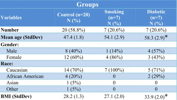
The 34 recruited participants included 20 control subjects, 7 smokers and 7 diabetics. The mean participant age varied from 47.9 to 58.9 years with a standard deviation ranged from 1.8 to 2.9 (Table 3). Majority of the participants were Caucasians, and more female participants were recruited in the control and smoking groups. BMI was recorded for all participants with the mean values for groups ranging from 28.3 to 33.9 kg/m2. Statistical significance was not found with gender and race among the three study groups; however, the mean age (p=0.003) and BMI (p=0.03) for the diabetic group were statistically significantly higher than the control group (Table 3).
31
Table 3. Study Participant Demographics (Total n = 34)
Groups
Variables Control (n=20) N (%) Smoking (n=7) N (%)
Diabetic (n=7) N (%)
Number 20 (58.8%) 7 (20.6%) 7 (20.6%)
Mean age (StdDev) 47.4 (1.8) 54.1 (2.9) 58.3 (2.9)* Gender:
Male 8 (40%) 1 (14%) 4 (57%)
Female 12 (60%) 4 (86%) 3 (43%)
Race:
Caucasian 14 (70%) 7 (100%) 5 (71%)
African American 4 (20%) 0 2 (29%)
Asian 1 (5%) 0 0
Other 1 (5%) 0 0
BMI (StdDev) 28.2 (1.3) 27.1 (2.0) 33.9 (2.0)*
32 Selected Implant Sites
33
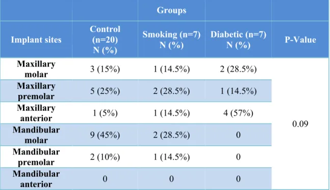
Table 4. Implant Sites of Study Participants (Total n = 34)
Groups
Implant sites
Control (n=20) N (%)
Smoking (n=7) N (%)
Diabetic (n=7)
N (%) P-Value
Maxillary
molar 3 (15%) 1 (14.5%) 2 (28.5%)
0.09 Maxillary
premolar 5 (25%) 2 (28.5%) 1 (14.5%) Maxillary
anterior 1 (5%) 1 (14.5%) 4 (57%)
Mandibular
molar 9 (45%) 2 (28.5%) 0
Mandibular
premolar 2 (10%) 1 (14.5%) 0
Mandibular
34
Differential DNA Methylation of FGF2 Gene in Smokers and Diabetic Participants
A comparison among the control subjects, smokers and diabetic participants indicated increased methylation of FGF2 gene was present in the smoking and diabetic groups (Figure 3). The percentages of hyper-methylation in the gingival tissues of the control, diabetic and smoking groups were 6.29±3.85, 8.92±5.64 and 16.84±5.64
respectively. The p-values were 0.70 and 0.34 for the gingival samples from diabetics and smokers respectively when compared to the control samples. This indicated the
35
Figure 3. Methylation Level of FGF2 Gene in Smokers or Diabetics in Comparison to Control Subjects.
0 5 10 15 20 25
Control Smoking Diabetic
%
of
H
yp
er
m
et
h
yl
at
ion
*p-value for smoking group= 0.34 *p-value for diabetic group= 0.70
Figure 3
36
Differential mRNA Expression of FGF2 Gene in Smokers and Diabetic Participants
37
Figure 4. Messenger Level of FGF2 Gene in Smokers or Diabetics in Comparison to Control Subjects
0 0.2 0.4 0.6 0.8 1 1.2
Control Smoking Diabetic
M
ean
F
ol
d
C
h
an
ge
Groups Figure 4
*p-value < 0.05
38 DISCUSSION
In this study, hyper-methylation of FGF2 gene was consistently observed in the gingiva from smoking and diabetic subjects relative to the gingiva from healthy subjects. The level of mRNA expression of the FGF2 gene is down-regulated in gingiva from smokers and diabetics. Overall, differential methylation patterns of the FGF2 gene among gingiva from healthy individuals, smokers and diabetics were associated with levels of FGF2 gene expression.
Limited literature relating smoking and diabetes with DNA methylation of other genes could be identified. Regarding smoking, majority of studies focused on bronchial or lung tissues in lung cancer patients (Belinsky et al. 2002, Soria et al. 2002, Divine et al. 2005, Liu et al. 2007, Han et al. 2009, Tekpli et al. 2011). Most studies demonstrated a positive correlation between cigarette smoking and abnormal methylation patterns. In a meta-analysis performed by Zhang on the association between smoking and p16INK4α methylation in tumor cells from lung carcinoma patients, more than a 2-fold increase of p16INK4α methylation in NSCLC patients with smoking habits was noted. The group later found the association to be stronger in an Asian population (Zhang et al. 2011). Lin demonstrated nicotine-derived nitrosamine ketone, a cigarette smoke carcinogen, was capable of influencing the expression of DNA methyltransferase 1 (DNMT1) and
39
lung tumor tissues (Lin et al. 2010). However, in two studies performed by Oliveira (Oliveira et al. 2009) De Oliveira et al. 2011) regarding the impact of smoking on the DNA methylation status of IL-8, TLR2 and TLR4 gene promoters in the periodontal tissues, both failed to demonstrate a correlation between smoking and methylation of these gene promoters. Smoking dose likely had a significant role in the inconsistent results. In the lung cancer study, smokers were defined as subjects who demonstrated at least a 5 pack-year history, but 1.25 pack-years was the threshold in Oliveira’s studies.
40
in wound healing (Singh, 2015). In general, existing evidence supports findings from our current study, that smoking and diabetes are associated with hyper-methylation pattern of FGF2 gene.
A statistically significant association was not detected for DNA methylation of FGF2 gene in smokers and diabetics compared to control subjects; however, the smoking group demonstrated almost two times greater % methylation than the diabetic group. This can partly be explained by the severity of the systemic condition of subjects included. Subjects in the smoking group were mostly heavy smokers, with two being former smokers who had quit within the last 10 years. While the dose-response relationship exerted by smoking on periodontal health has been well documented in literature (Bergstrom et al. 1991, Machuca et al. 2000, Haffajee & Socransky 2001, Bergstrom 2004, , Natto et al. 2005, Rosa et al. 2008), the degree of DNA hyper-methylation in the gingival tissues can be expected to be higher than subjects who have never smoked. The mean DNA methylation and gene expression could have been affected, but in a more significant way due to smokers having increased smoking duration, amount, and current smoking status. On the other hand, the diabetic subjects had HbA1C levels of 7.0% or less, indicating that their diabetic condition was controlled. Diabetic patients who are controlled have been shown to exhibit healthier periodontal status than those with poor glycemic control (Taylor 2001). It is reasonable to observe the increased methylation pattern of FGF2 gene in the gingiva of smokers, even more so than diabetics. Although the methylation results did not reach statistical significance, a pattern was observed.
41
SABiosciences Corporation, Frederick, MD). Unlike the bisulfate-based methods, this method detects remaining input DNA after cleavage with a methylation-sensitive and/or a methylation-dependent restriction enzyme. These enzymes can cleave a minimum of three unmethylated CpG sites and three methylated CpG sites respectively in the promoter region. Following digestion, the remaining DNA fragments of FGF2 gene are quantified by real-time PCR using primers that bind to the promoter region of interest. The exact number of CpG sites cleaved by each enzyme, the CpG locations on the FGF2 gene and the sequence of the primers are proprietary to the manufactory company, and thus they could not be obtained for result evaluation.
FGF-2 can up-regulate many genes and affect various downstream reactions, but the exact mechanisms by which the FGF2 gene is regulated remain unclear. No studies at the present have investigated how smoking can affect RNA expression of FGF2 gene during wound healing. Only one study could be identified to support the inhibitory effect that smoking has on the activity of FGF-2 protein. Pauwels and his group (Pauwels et al. 2010) evaluated whether cigarette smoke triggered pulmonary and systemic pentraxin 3 (PTX3) expression in vivo in a murine model. PTX3, a soluble pattern recognition receptor, is produced in response to pro-inflammatory cytokines IL-1β and TNF-α and mediates angiogenesis by influencing FGF-2 activity. They detected elevated levels of PTX3 and reduced mRNA expression of FGF2 at 4 weeks and 24 weeks after exposure to cigarette smoke. These findings could be related to binding of FGF-2 protein by the N-terminal domain of PTX3, which inhibited the angiogenic activity of FGF-2.
42
RNA expression of FGF2 was reduced in diabetic mice compared with normal mice during the period of wound re-epithelialization and granulation tissue formation. They stated this observation could be resulted from delayed infiltration of acute inflammatory cells into the wound area, impaired ability for diabetic macrophages to synthesize FGF-2, and reduced level of serum components in diabetic mice to induce FGF2 expression. Lu and his group (Lu et al. 2008) found a circulating electronegative low density lipoprotein (LDL) named L5 in type 2 diabetics diabetes. L5 induced endothelial cell apoptosis by suppressing FGF2 expression. FGF2 expression exerted most of its effects by activating the phosphatidylinositol 3-kinase (PI3K)-Akt pathway. Once activated by FGF2, the phosphorylated Akt activated endothelial nitric oxide synthase, which is essential for survival of endothelial cell. Lu suggested FGF-2 was auto-regulated by Akt activation through a positive feedback loop. They isolated L5 from type 2 diabetic patients, and found L5 caused reduction in Akt phosphorylation causing a decrease in the transcription of the FGF2 gene. He concluded that L5 could potentially impair FGF-2 dependent re-endothelialization and could play a role in diabetes-associated wound impairment and atherosclerosis.
FGF-2 plays an important role in bone formation, differentiation and metabolism (Marie 2003, Kawaguchi et al. 2000). It is produced by osteoprogenitor cells and mature osteoblasts and accumulates in bone extra-cellular matrix. Being a potent mitogen for mesenchymal cells, it directs these cells to commit to the osteoblastic lineage
43
signaling pathways involving the expression of transcription factors such as Runx2 and AP-1 that activate downstream osteoblast differentiation genes (Marie 2003). Coffin (Coffin et al. 1995) discovered that over-expressing FGF-2 in mice induced abnormal long bone formation, and Montero (Montero et al. 2000) later noted bone formation was inhibited by deactivating FGF-2.
Using Fgf2-null mice, Fei and his group (Fei et al. 2011) demonstrated a role of FGF2 in osteoblastogenesis through Wnt signaling. Wnts are a family of 19 glycoproteins secreted in mammals, and were well demonstrated to positively regulate osteoblast
differentiation and bone formation (Krishnan et al. 2006). The group found that mRNA and protein expression for Wnt10b and β-catenin decreased significantly in Fgf2-null mice, and the addition of exogenous FGF-2 promoted β-catenin accumulation in the nucleus of bone marrow stromal cells while also increasing their mineralization.
The effects of FGF-2 can be enhanced with bone morphogenetic protein-2 (BMP-2). Rat marrow-derived mesenchymal stem cells exposed with FGF-2 and BMP-2
44
osteoprecursor cells, influencing osteoblast differentiation (Park 2011). In addition to BMP-2, a review written by Marie in 2003 also provided evidence to support interaction of FGF-2 with other biological factors, such as cAMP and TGF-β to enhance
osteogenesis (Marie 2003).
The ability of FGF-2 to regulate bone remodeling is supported by studies that demonstrated bone resorption caused by FGF-2 stimulation in cell cultures (Kawaguchi et al. 2000, Simmons & Raisz 1991, Kawaguchi et al. 1995, Chikazu et al. 2001). In these studies, FGF-2 was shown to modulate osteoclast differentiation through various
mechanisms. The indirect mechanism involved FGF-2 activating cyclo-oxygenase 2 (COX2) and producing prostaglandin, leading to the induction of the receptor activator of nuclear factor κB ligand (RANK-L) in osteoblasts (Kawaguchi et al. 2000, Kawaguchi et al. 1995). This effect was abolished by NSAIDs (Kawaguchi et al. 2000). On the other hand, the direct mechanisms involved FGF-2 binding to FGF receptor type 1, a tyrosine kinase receptor on osteoclasts and their precursors, to induce bone resorption and prevent differentiation (Kawaguchi et al. 2000, Chikazu et al. 2001) (Chikazu et al. 2001). FGF-2 has been shown to play a key role in affecting molecular interactions and intracellular mechanisms in osteoblasts to induce osteoblastic differentiation and enhance
osteogenesis.
Several limitations can be identified in the study. 1) The principle one is the small sample sizes in the smoking and diabetic groups. The target number of recruitment was 20 for the control group, 12 for the smoking group and 12 for the diabetic group;
45
DNA methylation. 3) Smoking history and dosage were self-reported, and HbA1c was not evaluated in the other two non-diabetic groups. This led to reporting biases and potential for ex-smokers, current smokers and undiagnosed diabetics to be recruited into the wrong group. 4) Subjects in the smoking and diabetic groups were not stratified according to their smoking doses or HbA1c levels. Evaluation of dose-response relationships was not possible. This dosage could have impacted the amount of DNA methylation seen in each sample. 5) Bone structure and implant size nor surface were not controlled. Although all implant surfaces were roughened, a difference in surface
modification could play a role in recruitment of growth factors, migrations of cells, and the rate of implant healing and osseointegration. Future investigations should 1) increase the sample sizes, 2) increase number of follow-up visits to assess wound healing more closely, 3) address causality and dose response of smoking and control of diabetes with DNA methylation, 4) specify the bone quality of the patients, implant type and implant size to be included in the study, and 5) evaluate DNA methylation, mRNA expression and protein expression of other biological factors such as platelet-derived growth factor, transforming growth factor-β or interleukin-1β.
In conclusion, association patterns of differential methylation of FGF2 gene in smokers and diabetics were identified in this study. Hyper-methylation levels of FGF2 gene coincided with down-regulation of its mRNA expression. Systemic and
46
47 APPENDIX I
SUPPLEMENTARY MATERIALS AND METHODS
Institutional Review Board, University of North Carolina at Chapel Hill IRB Study Number 10-1184
1. Clinical Study Design
This is a longitudinal case-control study of 6-8 weeks in duration. Clinical parameters and biological samples were collected from healthy subjects, smokers and diabetics during the course of implant therapy. The study was performed in a single masked or blinded manner. All laboratory assessments were performed without knowledge of the subjects’ type of treatment and health condition.
2. Human Subjects
self-48
reported. The level of control of the diabetic condition was confirmed by HbA1c test within 3 months prior to screening. Screening and follow-up examinations were
performed in the General Oral Health Center in the School of Dentistry. Implant surgery was performed in the Graduate Periodontology clinic.
The inclusion criteria of study participation included: (1) males or females between the age of 18 and 70 years (inclusive), (2) must be able and willing to follow study procedures and instructions, (3) must have read, understood and signed an informed consent form, (4) must be in good general health, (5) must have one or more implant placements as their future treatment needs, (6) must fit into one of the study groups as listed previously, and (7) must have probing depth ≤ 4 mm or 5mm without bleeding on probing for all teeth at the same quadrant of implant placement as determined by two calibrated examiners .
Individuals were not enrolled into the study if they presented with the following exclusion criteria: (1) chronic disease with oral manifestations (e.g. Crohn’s disease, lupus erythematosus, Behcet’s disease), (2) gross oral pathology, (3) use of either antibiotics or NSAIDs within 2 weeks prior to screening examination, (4) chronic treatment (i.e. two weeks or more) with any medication known to affect periodontal status (e.g. phenytoin, calcium, antagonists, cyclosporin, warfarin) within 1 month prior to screening examination, (5) systemic conditions, except smoking and diabetes, that were known to affect the periodontal status, (6) active infectious diseases such as hepatitis, HIV or tuberculosis, and (7) known to be pregnant or breastfeeding.
49
implant site for the entire duration of the study. Subjects not meeting this criterion would be withdrawn. Infection around an implant would be reported as an adverse event and subject would be followed until it was resolved.
3. Clinical Study Method
The following procedures were performed on study participants during a total of 5 visits within a study period of 6-8 weeks.
Visit 1 (≤ 2 weeks before implant surgery):
• Study personnel obtained a signed written informed consent from the subject. • Study personnel obtained medical history and demographics related to the patient. • Study personnel performed HbA1C test on all subjects in the diabetic group (if a test
result within past 3 months was not available). A drop of blood was obtained by finger prick and was loaded into a DCA 2000 HbA1C reagent cartridge, which was then processed immediately by a DCA 2000®+ Analyzer (Bayer Inc., Toronto, ON, Canada) for HbA1C level.
• Patient vital signs (blood pressure and pulse), height and weight were recorded. • A dental examiner performed a general oral examination for presence of any
abnormalities.
• A dental examiner scored all teeth in the same quadrant of implant placement for plaque index, gingival index, probing depth, percent bleeding on probing and clinical attachment level.
Visit 2 (Implant surgery):
50
• A dental examiner performed a general oral examination for presence of any abnormalities.
• The dental examiner collected 8 GCF samples from the mesiolingual, mesiobuccal, distolingual and distobuccal surfaces of the two teeth that were in the closest proximity to the implant site. Samples were collected using filter paper strips (Pro Flow, Inc., Amityville, NY).
• The dental examiner collected 1 biopsy at the site of implant placement for quantitative PCR, DNA methylation sequence analysis and differential methylation analysis. The biopsy sample was obtained with a 4mm punch biopsy blade or a scalpel blade. It was removed in the same way as how discarded tissues will be removed. It was removed at the beginning of the surgery as the clinician exposed the underlying bone at the implant site, or before suturing when the clinician reduced redundant tissues to achieve better flap closure.
Visits 3-5 (2 weeks ± 2 days, 4 weeks ± 2 days, 6 weeks ± 2 days after surgery): • Study personnel updated medical history and obtained vital signs.
• A dental examiner performed a general oral examination for presence of any abnormalities.
• A dental examiner scored all teeth in the same quadrant of implant placement for plaque index and gingival index.
• The dental examiner recorded wound healing indices (WHI) as indicators of degree of soft tissue healing for the implant site.
51
• The dental examiner collected 8 GCF samples in the same areas and using the same method as indicated previously.
Table 5. Schedule of Procedures by Visit
Week
Visit 1 -2 to -1
Visit 2 0 Visit 3 2 Visit 4 4 Visit 5 6 Procedure:
Informed Consent X
Medical/Dental History X X X X X
Demographics X
Vital Signs X X X X X
HbA1C Test (for diabetic group
only) X
Oral Examination X X X X X
PI, GI X X X X
PD, BOP, CAL X
WHI X X X
Implant Stability Measurement X X
Biopsy Sample X
GCF Sample X X X X
Saliva Sample X X X X
Adverse Experience X X X X X
52 4. Clinical Periodontal Assessments
Clinical examiners were calibrated prior to commencement of the study. Clinical parameters assessed in the periodontal examination included plaque index, gingival index, probing depth, bleeding on probing, and clinical attachment level. Clinical parameters were measured using a manual University of North Carolina (UNC-15) periodontal probe. These parameters were measured at six periodontal sites per tooth (i.e., mesiobuccal, buccal, distobuccal, mesiolingual, lingual, and distolingual) and at all teeth in the same quadrant where implant placement is desired (including the third molar).
Silness and Löe Plaque Index (PI) (SILNESS & LOE 1964):
PI was defined as the relative amount of supragingival plaque and was recorded on an ordinal scale of 0-3.
0- No plaque in the gingival area.
1- A film of plaque adhering to the free gingival margin and the adjacent tooth. The plaque may be recognized only by running a probe across the tooth surface
2- Moderate accumulation of soft deposits within the gingival pocket and on the gingival margin and/or adjacent tooth surface, which can be seen by the naked eye.
3- Abundance of soft matter within the gingival pocket and/or on the gingival margin and the adjacent tooth surface.
Löe and Silness Gingival Index (GI) (LOE & SILNESS 1963):
53 0- Normal gingiva.
1- Mild inflammation (slight change in color, slight edema); no bleeding on palpation (i.e., sulcular sweep).
2- Moderate inflammation (redness, edema, glazing); bleeding on palpation (i.e., sulcular sweep).
4- Severe inflammation (i.e., marked redness, edema); ulceration, tendency to spontaneous bleeding.
Probing depth (PD):
PD was defined as the linear distance from the gingival margin (GM) to base of the pocket. If a PD reading fell between two millimeter readings, the rule was to round down and the lower of the two readings was recorded.
Bleeding on Probing (BOP):
BOP was defined as the presence or absence of bleeding to manual probing and was recorded as a dichotomous variable.
0- No bleeding within 10 seconds after probing. 1- Bleeding within 10 seconds after probing. Clinical attachment level (CAL):
CAL was defined as the linear distance from the cemento-enamel junction (CEJ) to base of the pocket. If a CAL reading fell between two millimeter readings, the rule was to round down and the lower of the two readings was recorded.
Wound healing indices (WHI):
54 Degree of Edema
0- No gingival edema. 1- Mild gingival edema. 2- Moderate gingival edema. 3- Severe gingival edema. Degree of Erythema
0- No erythema. 1- Mild erythema. 2- Moderate erythema. 3- Severe erythema. Degree of Pain on Palpation
0- No pain. 1- Mild pain. 2- Moderate pain. 3- Severe pain. Degree of Flap Closure
0- No fibrin line or folding line in the incision area. 1- Fine fibrin line or folding line in the incision area. 2- Fibrin clot in the incision area.
3- Partial or complete necrosis of flap. Implant Stability:
55
56
REFERENCES
(U.S.) C. f. D. C. (2009) State-specific smoking-attributable mortality and years of potential life lost--United States, 2000-2004. MMWR.Morbidity and mortality weekly report 58, 29-33.
Abraham J. A., Whang J. L., Tumolo A., Mergia A., Friedman J., Gospodarowicz D. & Fiddes J. C. (1986) Human basic fibroblast growth factor: nucleotide sequence and genomic organization. The EMBO journal 5, 2523-2528.
American Diabetes Association (2014). National Diabetes Statistics Report. June 10 2014. Diabetes.org
Association A. (2008) Economic costs of diabetes in the U.S. In 2007. Diabetes care 31, 596-615.
Aykol G., Baser U., Maden I., Kazak Z., Onan U., Tanrikulu-Kucuk S., Ademoglu E., Issever H. & Yalcin F. (2011) The Effect of Low-Level Laser Therapy as an Adjunct to Non-Surgical Periodontal Treatment. Journal of periodontology 82, 481.
Bain C. A., Moy P. K. (1993) The Association Between the Failure of Dental Implants and Cigarette Smoking. Int J Oral Maxillofac Implants 1993; 8: 609-615.
Bain C. A. (1996) Smoking and Implant Failure - Benefits of a Smoking Cessation Protocol. Int J Oral Maxillofac Implants 1996; 11: 756-759.
Balshe A. A., Eckert S. E. (2008). The Effects of Smoking on the Survival of Smooth- and Rough-Surface Dental Implants. Int J Oral Maxillofac Implants 2008; 23: 1117-1122. Barros S. P. & Offenbacher S. (2009) Epigenetics: connecting environment and genotype to phenotype and disease. Journal of dental research 88, 400-408. doi:
10.1177/0022034509335868
Barros S. P. & Offenbacher S. (2014) Modifiable risk factors in periodontal disease: Epigenetic regulation of gene expression in the inflammatory response. Periodontology 2000, Vol. 64, 2014, 95-110.
Behr B, Panetta NJ, Longaker MT, Quarto N. Different endogenous threshold levels of Fibroblast Growth Factor-ligands determine the healing potential of frontal and parietal bones. Bone 2010; 47(2): 281-94.
57
Berglundh T, Abrahamsson I, Lang NP, Lindhe J. De Novo alveolar bone formation adjacent to endosseous implants. Clin Oral Implants Res 2003; 14: 251-262.
Bergstrom J. (2004) Influence of tobacco smoking on periodontal bone height. Long-term observations and a hypothesis. Journal of clinical periodontology 31, 260-266. doi: 10.1111/j.1600-051X.2004.00475.x
Bergstrom J., Eliasson S. & Preber H. (1991) Cigarette smoking and periodontal bone loss. Journal of periodontology 62, 242-246.
Bikfalvi A., Klein S., Pintucci G. & Rifkin D. B. (1997) Biological roles of fibroblast growth factor-2. Endocrine reviews 18, 26-45.
Bizenjima T., Seshima F., Ishizuka Y., Takeuchi T, Kinumatsu T., Saito A. Fibroblast growth factor-2 promots healing of surgically created periodontal defects in rats with early, streptozotocin-induced diabetes via increasing cell proliferation and regulating angiogenesis. J Clin Periodontol 2015; 42: 62-71.
Bohlen P., Baird A., Esch F., Ling N. & Gospodarowicz D. (1984) Isolation and partial molecular characterization of pituitary fibroblast growth factor. Proceedings of the National Academy of Sciences of the United States of America 81, 5364-5368.
Brånemark PI. Osseointegration and its experimental studies. J Prosthet Dent 1983; 50: 399-410.
Center for Disease Control and Prevention (2015). Summary Health Statistics Tables for US Adults: National Health Interview Survey, 2014.
Chambrone L., Preshaw P.M., Rosa E.F., Heasman P.A., Romito G.A., Pannuti C. M., Tu Y. K. (2013) Effects of Smoking Cessation on the Outcomes of Non-Surgical Periodontal Therapy: A Systematic Review and Individual Patient Data Meta-Analysis. J Clin
Periodontol 2013; 40: 607-615. doi: 10.111/jcpe.12106
Chambrone L, Preshaw P.M., Ferreira J.D., Rodrigues F.A., Cassoni A, Shibli J.A. Effects of Tobbaco Smoking on the Survival Rate of Dental Implants Placed in Areas of Maxillary Sinus Floor Augmentation: A Systmatic Review. Clin Oral Impl Res 2014; 25:408-416.
58
: the official journal of the American Society for Bone and Mineral Research 16, 2074-2081. doi: 10.1359/jbmr.2001.16.11.2074
Coffin J. D., Florkiewicz R. Z., Neumann J., Mort-Hopkins T., Dorn G. W.,2nd,
Lightfoot P., German R., Howles P. N., Kier A. & O'Toole B. A. (1995) Abnormal bone growth and selective translational regulation in basic fibroblast growth factor (FGF-2) transgenic mice. Molecular biology of the cell 6, 1861-1873.
de Morais,Juliana Aparecida Najarro Dearo. (2009) Effect of diabetes mellitus and insulin therapy on bone density around osseointegrated dental implants: a digital subtraction radiography study in rats. Clinical oral implants research 20, 796-801.
De Oliveira N. F., Andia D. C., Planello A. C., Pasetto S., Marques M. R., Nociti F. H.,Jr, Line S. R. & De Souza A. P. (2011) TLR2 and TLR4 gene promoter methylation status during chronic periodontitis. Journal of clinical periodontology 38, 975-983. doi: 10.1111/j.1600-051X.2011.01765.x; 10.1111/j.1600-051X.2011.01765.x
Desta T. (2010) Altered fibroblast proliferation and apoptosis in diabetic gingival wounds. Journal of dental research 89, 609-14.
Devlin H. (1996) Healing of tooth extraction sockets in experimental diabetes mellitus. Journal of oral and maxillofacial surgery 54, 1087-91.
Divine K. K., Pulling L. C., Marron-Terada P. G., Liechty K. C., Kang T., Schwartz A. G., Bocklage T. J., Coons T. A., Gilliland F. D. & Belinsky S. A. (2005) Multiplicity of abnormal promoter methylation in lung adenocarcinomas from smokers and never smokers. International journal of cancer.Journal international du cancer 114, 400-405. doi: 10.1002/ijc.20761
Emrich L. J. (1991) Periodontal disease in non-insulin-dependent diabetes mellitus. Journal of periodontology (1970) 62, 123-31.
Fei Y., Xiao L., Doetschman T., Coffin D. J. & Hurley M. M. (2011) Fibroblast growth factor 2 stimulation of osteoblast differentiation and bone formation is mediated by modulation of the Wnt signaling pathway. The Journal of biological chemistry 286, 40575-40583. doi: 10.1074/jbc.M111.274910
Fei Y, Gronowicz G, Hurley M. Fibroblast Growth Factor-2, Bone Homeostasis and Fracture Repair. Current Pharmceutical Design, 2013, 19, 3354-3363.
Fiorini T, Musskopf M. L., Opperman R. V., Susin C. (2014) Is There a Positive Effect of Smoking Cessation on Periodontal Health? A Systematic Review. J Periodontology 2014; 85: 83-91.
59
Giannopoulou C., Geinoz A. & Cimasoni G. (1999) Effects of nicotine on periodontal ligament fibroblasts in vitro. Journal of clinical periodontology 26, 49-55.
Glickman I. (1967) Postsurgical periodontal healing in alloxan diabetes. Journal of periodontology (1970) 38, 93-9.
Gomez R. S., Dutra W. O. & Moreira P. R. (2009) Epigenetics and periodontal disease: future perspectives. Inflammation research : official journal of the European Histamine Research Society ...[et al.] 58, 625-629. doi: 10.1007/s00011-009-0041-7
Gooch H. L. (2000) Alterations of cartilage and collagen expression during fracture healing in experimental diabetes. Connective tissue research 41, 81-91.
Gospodarowicz D. (1974) Localisation of a fibroblast growth factor and its effect alone and with hydrocortisone on 3T3 cell growth. Nature 249, 123-127.
Haffajee A. D. & Socransky S. S. (2001) Relationship of cigarette smoking to attachment level profiles. Journal of clinical periodontology 28, 283-295.
Han W., Wang T., Reilly A. A., Keller S. M. & Spivack S. D. (2009) Gene promoter methylation assayed in exhaled breath, with differences in smokers and lung cancer patients. Respiratory research 10, 86. doi: 10.1186/1465-9921-10-86
Hanada K., Dennis J. E. & Caplan A. I. (1997) Stimulatory effects of basic fibroblast growth factor and bone morphogenetic protein-2 on osteogenic differentiation of rat bone marrow-derived mesenchymal stem cells. Journal of bone and mineral research : the official journal of the American Society for Bone and Mineral Research 12, 1606-1614. doi: 10.1359/jbmr.1997.12.10.1606
Heitz-Mayfield L. JA, Huynh-Ba G. (2009) History of Treated Periodontitis and Smoking as Risks for Implant Therapy. Journal of Oral and Maxillofacial Implants 24 (suppl): 39-68.
Henemyre C. L., Scales D. K., Hokett S. D., Cuenin M. F., Peacock M. E., Parker M. H., Brewer P. D. & Chuang A. H. (2003) Nicotine stimulates osteoclast resorption in a porcine marrow cell model. Journal of periodontology 74, 1440-1446. doi:
10.1902/jop.2003.74.10.1440
60
Johnson G. K. & Guthmiller J. M. (2007) The impact of cigarette smoking on periodontal disease and treatment. Periodontology 2000 44, 178-194. doi:
10.1111/j.1600-0757.2007.00212.x
Kaigler D., Cirelli J. A. & Giannobile W. V. (2006) Growth factor delivery for oral and periodontal tissue engineering. Expert opinion on drug delivery 3, 647-662. doi:
10.1517/17425247.3.5.647
Kawaguchi H, Kurokawa T, Hanada K, Hiyama Y, Tamura M, Ogata E, Matsumoto T. Endocrinology 135 (1994) 774-781.
Kawaguchi H., Chikazu D., Nakamura K., Kumegawa M. & Hakeda Y. (2000) Direct and indirect actions of fibroblast growth factor 2 on osteoclastic bone resorption in cultures. Journal of bone and mineral research : the official journal of the American Society for Bone and Mineral Research 15, 466-473. doi: 10.1359/jbmr.2000.15.3.466 Kawaguchi H., Pilbeam C. C., Gronowicz G., Abreu C., Fletcher B. S., Herschman H. R., Raisz L. G. & Hurley M. M. (1995) Transcriptional induction of prostaglandin G/H synthase-2 by basic fibroblast growth factor. The Journal of clinical investigation 96, 923-930. doi: 10.1172/JCI118140
Khader Y. S. (2006) Periodontal status of diabetics compared with nondiabetics: a meta-analysis. Journal of diabetes and its complications 20, 59-68.
Kitamura M., Akamatsu M., Machigashira M., Hara Y., Sakagami R., Hirofuji T., Hamachi T., Maeda K., Yokota M., Kido J., Nagata T., Kurihara H., Takashiba S., Sibutani T., Fukuda M., Noguchi T., Yamazaki K., Yoshie H., Ioroi K., Arai T.,
Nakagawa T., Ito K., Oda S., Izumi Y., Ogata Y., Yamada S., Shimauchi H., Kunimatsu K., Kawanami M., Fujii T., Furuichi Y., Furuuchi T., Sasano T., Imai E., Omae M., Yamada S., Watanuki M. & Murakami S. (2011) FGF-2 stimulates periodontal
regeneration: results of a multi-center randomized clinical trial. Journal of dental research 90, 35-40. doi: 10.1177/0022034510384616
Kitamura M., Nakashima K., Kowashi Y., Fujii T., Shimauchi H., Sasano T., Furuuchi T., Fukuda M., Noguchi T., Shibutani T., Iwayama Y., Takashiba S., Kurihara H.,
Ninomiya M., Kido J., Nagata T., Hamachi T., Maeda K., Hara Y., Izumi Y., Hirofuji T., Imai E., Omae M., Watanuki M. & Murakami S. (2008) Periodontal tissue regeneration using fibroblast growth factor-2: randomized controlled phase II clinical trial. PloS one 3, e2611. doi: 10.1371/journal.pone.0002611
Krishnan V., Bryant H. U. & Macdougald O. A. (2006) Regulation of bone mass by Wnt signaling. The Journal of clinical investigation 116, 1202-1209. doi: 10.1172/JCI28551 Kumar A, Jaffin R. A., Berman C (2002). The Effect of Smoking on Achieving