MODULATING INNATE IMMUNITY IMPROVES OUTCOMES IN ACUTE LUNG INJURY
Julia Louise Malik Dunn
A dissertation submitted to the faculty at the University of North Carolina at Chapel Hill in
partial fulfillment of the requirements for the degree of Doctor of Philosophy in the Department
of Microbiology & Immunology in the School of Medicine.
Chapel Hill
2016
Approved by:
Bruce A. Cairns
Robert Maile
Tom Kawula
Claire Doerschuk
© 2016
ABSTRACT
Julia Louise Malik Dunn: Modulating Innate Immunity Improves Outcomes in Acute Lung
Injury
(Under the Direction of Bruce A. Cairns)
Pulmonary inflammation following traumatic injury disrupts the lung architecture, leading to
impaired oxygen exchange and rendering patients susceptible to life threatening bacterial
infections. Innate immunity is central to the resolution of infection; however, excessive immune
responses cause unresolved inflammation that exacerbate tissue damage. We hypothesize that
partially attenuating recruitment of innate immune cells without impairing their function in situ
will improve outcomes for patients diagnosed with acute lung injury (ALI) following burns and
smoke inhalation by restoring homeostasis. Using mouse models of injury, we characterized the
innate immune response in the lung following burns and smoke inhalation to establish
mechanistic relationships that drive inflammation and to test therapeutic interventions. We show
that significant neutrophil recruitment to the lung following burn injury is driven by damage
associated molecular patterns (DAMPs); however, this recruitment does not result in improved
bacterial clearance following pulmonary infection. To explore the factors that drive inflammation
in a clinically relevant direct lung injury model, we developed and validated a novel murine
model of acute smoke inhalation. This model mimics granulocyte recruitment,
oxide synthase is required for upregulation of interleukin-10 (IL-10), monocyte chemotactic
protein 1, and hyaluronic acid (HA) following inhalational injury, but not for bacterial clearance.
Thus, we propose that the relationship between elevated IL-10 and the onset of bacterial
ACKNOWLEDGEMENTS
In 2006, Deb Siwik inspired me to aspire to a career in academic research, and that
inspiration has helped me to overcome obstacles and push through hard times ever since.
When my time as a graduate student almost came to an untimely end, countless friends,
colleagues, and mentors came to my rescue. Kevin Blauth and Danny Bruce never stopped
believing in me. Neither did Bob Bourret or Bill Goldman, who were the greatest advocates I
could have ever hoped for, and who have continued to support me ever since. I will always be
grateful to Rob Maile and Bruce Cairns, who decided to take a chance on me and to offer me
their uncompromising support professionally and personally. Many thanks to my mentees, Karli,
Kelly, Janice, Marci, Amber, and Maddy –their growth and success motivate me to overcome the
long odds on the road to professorship. Last but not least, nobody could ask for a more
supportive friend and colleague than Laurel Kartchner.
TABLE OF CONTENTS
LIST OF TABLES ...x
LIST OF FIGURES ... xi
LIST OF ABBREVIATIONS ... xiii
CHAPTER 1: INNATE IMMUNITY IS CRITICAL FOR SURVIVAL
FOLLOWING TRAUMA ...1
Section 1: Introduction ...1
Section 2: Inflammation drives outcomes in trauma patients ...2
Section 3: The lung is a site of immunopathology and infection
that hinders patient outcomes...4
Section 4: Immune function in the lung is altered by injury...6
Section 5: Optimized innate immune function would improve patient outcomes ...13
Section 6: Experimental approach to study lung injury in mice ...13
CHAPTER 2: DIRECT DETECTION OF BLOOD NITRIC OXIDE REVEALS A
BURN-DEPENDENT DECREASE OF NITRIC OXIDE IN RESPONSE TO
PSEUDOMONAS AERUGINOSA INFECTION. ...25
Section 1: Summary ...25
Section 2: Introduction ...26
Section 3: Methods ...27
Section 4: Results ...28
Section 5: Discussion ...30
CHAPTER 3: MAMMALIAN TARGET OF RAPAMYCIN REGULATES
A HYPER-RESPONSIVE STATE IN PULMONARY NEUTROPHILS
LATE AFTER BURN INJURY ...40
Section 2: Introduction ...40
Section 3: Methods ...42
Section 4: Results & Discussion ...44
CHAPTER 4: DAMPS IN CIRCULATION FOLLOWING TRAUMA PROMOTE
INFLAMMATION IN THE LUNG ...58
Section 1: Summary ...58
Section 2: Introduction ...59
Section 3: Methods ...60
Section 4: Results ...62
Section 5: Discussion ...64
CHAPTER 5: CXCL1 DRIVES NEUTROPHIL RECRUITMENT AND ONSET OF ALI
FOLLOWING ACUTE WOOD SMOKE INHALATION ...74
Section 1: Summary ...74
Section 2: Introduction ...75
Section 3: Methods ...77
Section 4: Results ...81
Section 5: Discussion ...85
CHAPTER 6: NOX2 ACTIVITY PROMOTES RESOLUTION OF
INFLAMMATION AFTER ACUTE WOOD SMOKE INHALATION ...100
Section 1: Summary ...100
Section 2: Introduction ...100
Section 3: Methods ...103
Section 4: Results ...105
Section 5: Discussion ...107
CHAPTER 7: CONCLUDING REMARKS ...109
LIST OF TABLES
LIST OF FIGURES
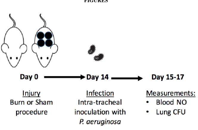
Figure 2.1: Experimental design for burn injury, infection, and blood NO analysis ...32
Figure 2.2: Nitric oxide (NO) levels are elevated following infection without
prior injury in a dose-dependent fashion ...33
Figure 2.3: Relative to sham mice, burn injury causes decreased blood NO concentrations
and increased pulmonary bacterial load following a 14 d post-burn infection ...34
Figure 2.4: Inoculation dose impacts blood NO concentration in burn mice
following infection ...35
Figure 3.1: Neutrophils are recruited to the lung following burn injury ...48
Figure 3.2: Burn injury impacts reactive oxygen and nitrogen species
production in neutrophils ...49
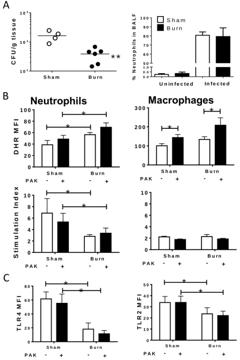
Figure 3.3: Changes in immune function, but not recruitment, lead to improved bacterial
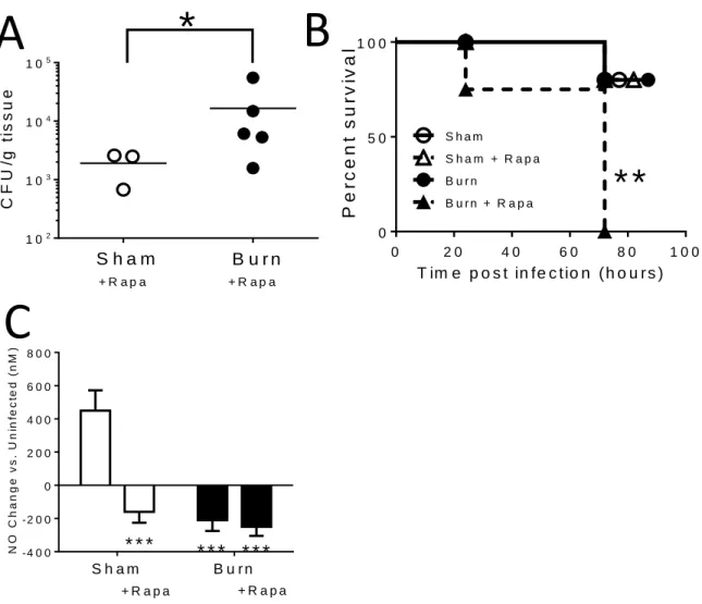
clearance in the lung following burn injury ...50
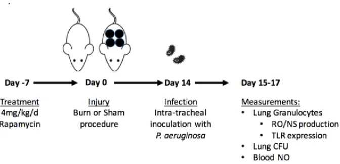
Figure 3.4: Experimental design for rapamycin administration, burn injury, infection,
and analysis. ...51
Figure 3.5: mTOR activity is required for survival and efficient bacterial clearance
in burned mice following infection ...52
Figure 3.6: mTOR inhibition impacts innate immune function following
burn injury and infection ...53
Figure 4.1: Experimental design for intravenous DAMP administration ...67
Figure 4.2: Hyaluronic acid administration leads to mild pulmonary inflammation
regardless of administration route ...68
Figure 4.3: MTD administration after burn injury leads to pulmonary inflammation
marked by neutrophil recruitment to the airway ...69
Figure 4.4: mtDNA administration after burn injury does not induce
pulmonary inflammation ...70
Figure 4.5: Coadministration of HA and mtDNA after burn injury leads to
Figure 5.3: Histological evidence of lung injury following acute smoke exposure ...91
Figure 5.4: Temporal regulation of IL-10 and DAMP release following smoke inhalation ...92
Figure 5.5: Pulmonary infection subsequent to systemic infection
following smoke inhalation...93
Figure 5.6: iNOS deficiency prevents cytokine production but not inflammation
after smoke inhalation ...94
Figure 5.7: CXCL1 drives neutrophilia following smoke exposure ...95
Figure 5.8: Early inhibition of neutrophil migration protects against
infection and HA release ...96
Figure 6.1: NOX2 deficiency amplifies inflammation after acute smoke inhalation ...109
Figure 6.2: NOX2 drives cytokine production but not DAMP release
after smoke inhalation ...110
Figure 6.3: HIF1
stabilization attenuates smoke-dependent inflammation
LIST OF ABBREVIATIONS
ALI Acute lung injuryARDS Acute respiratory distress syndrome BALF Broncho-alveolar lavage fluid
CARS Compensatory anti-inflammatory response syndrome CXCL1 Chemokine (C-X-C motif) ligand 1
CXCL2 Chemokine (C-X-C motif) ligand 2 CXCR2 Chemokine (C-X-C motif) receptor 2 DAMP Damage-associated molecular pattern dsDNA Double stranded deoxyribonucleic acid HA Hyaluronic acid
HIF1 Hypoxia inducible factor 1 IB Inhibitor of NFB
IL Interleukin
LPS Lipopolysaccharide
MCP1 Monocyte chemoattractant protein 1 MIP2 Macrophage inflammatory protein 2 MMP Matrix metalloprotease
MMW Medium molecular weight mRNA messenger ribonucleic acid MTD Mitochondrial DAMPs
mtDNA Mitochondrial deoxyribonucleic acid NFB Nuclear factor B
NO Nitric oxide
NOX2 Nicotinamide adenine dinucleotide phosphate (NADPH) oxidase 2 PPAR Peroxisome proliferator-activated receptor
PS Phosphatidylserine RNS Reactive nitrogen species ROS Reactive oxygen species
RONS Reactive oxygen and nitrogen species SDF Stromal-derived factor
SIRS Systemic inflammatory response syndrome SOD Superoxide dismutase
TLR Toll-like receptor
CHAPTER 1: INNATE IMMUNITY IS CRITICAL FOR SURVIVAL FOLLOWING
TRAUMA
Introduction
Traumatic injury is the leading cause of death in Americans under 44 years of age, and poses a threat to human health that will never be eliminated as long as people continue to drive cars, live inside flammable1 structures, or go to war. Recovery from traumatic injury requires the body to accomplish two
objectives: first, to combat the threat of infection that results from damage to normal physiological barriers, and second, to replace injured tissue with new, healthy tissue. Both processes are orchestrated by the immune system and in most cases they occur sequentially. In the early stages of inflammation, immune cells are recruited to damaged tissue and infectious threats are neutralized. Resolution can then occur, during which phase the immune system clears dead or damaged cells and debris, promotes proliferation of local stromal cells, and restructures the extracellular matrix. Failure to initiate the initial inflammatory response can lead to infectious complications. Failure to progress to the resolution phase leads to scar tissue, fibrosis, and may increase the extent of tissue damage. Immune activity that causes tissue damage or interferes with organ function is referred to as immunopathology, which can be associated with a hyperactive inflammatory phase or persistent inflammation that fails to resolve. A well-orchestrated innate immune response is a critical determinant of patient outcomes following traumatic injury, and presents a constellation of potential therapeutic targets.
Our central hypothesis is that some aspects of the innate immune response following trauma are beneficial and assist in efficient progress from the inflammatory to the resolution phase during recovery,
while aberrant responses either fail to clear infection or interfere with resolution. Our objective is to identify opportunities for intervention by examining the relationship between inflammation and outcomes in patients and animal models of injury. This study outlines research aimed at understanding how the lung is impacted by burn and inhalational injuries, and how immune activity can be modulated to promote successful transition from inflammation to resolution.
Inflammation drives outcome in trauma patients.
For many years it was thought that traumatic injury resulted in a biphasic inflammatory response that has been extensively characterized following sepsis. This paradigm suggests an initial systemic inflammatory response syndrome (SIRS) marked by pro-inflammatory cytokines and a hyperactive immune state. The SIRS phase was thought to be followed by a compensatory anti-inflammatory response syndrome (CARS), in which the anti-inflammatory cytokine profile limits tissue damage due to immunopathology. Novel methodologies of the ‘-omics’ age have enabled a more nuanced analysis, which suggests that pro- and anti-inflammatory genes are expressed simultaneously after trauma (1-6).
Traumatic injury causes differential expression of >80% of genes in the human genome within 28 days (1). Remarkably, the type of injury (i.e. blunt trauma vs. burn) is of only marginal importance to the genetic changes that occur, indicating that the physiological responses to these injuries are more alike than they are different. Despite the presence of anti-inflammatory cytokines during the early phase and pro-inflammatory cytokines during the late phase, which contradict the straightforward SIRS/CARS paradigm, it remains true that patients are at the greatest risk of developing organ failure secondary to shock early after injury and of succumbing to fatal opportunistic infections late after injury (4, 7, 8).
Changes in serum levels of cytokines, chemokines, and immunogenic growth factors persist for years after burn injury, especially in pediatric patients (9). Because of the prolonged inflammatory and hypermetabolic state that follows burn injury, patients experience many physiological problems
(9-11). Interference with aberrant inflammatory processes after trauma is therefore likely to improve long-term prognosis for trauma patients by promoting a prompt and complete return to homeostasis.
Excessive inflammation leads to organ dysfunction. Damaged tissue and the tissue-resident
immune cells release a battery of signals into circulation that impact tissues beyond the initial damage site. Increased systemic circulation of damage-associated molecular patterns (DAMPs) as well as cytokines, chemokines and growth factors in serum have been observed following burn injury in both humans and animal models (12-15). Inflammatory signals from the burn wound exit the site of damage through venous circulation and cause inflammation in secondary organs, with the heart and lungs being chief collateral targets because they receive the greatest concentration of inflammatory factors in the blood (16, 17).
Presence of a burn wound increases sensitivity to inflammatory and infectious signals such as C5a and LPS, and this sensitivity disrupts normal organ function by promoting edema and congestion of microvasculature. Attenuating inflammation after trauma in animal models by depleting complement or neutrophils either prior to or shortly after burn injury significantly reduces –or eliminates entirely –the collateral damage to distal organs (18, 19). These results have failed to translate to the clinic because they do not account for the opportunistic infections that would swamp patients if their innate immune
functions were so completely abolished. They do, however, illustrate the power of immune modulation in alleviating organ dysfunction after injury.
Aberrant inflammatory response leads to infectious complications. Anti-inflammatory signals
present in patient biofluids, specifically serum and broncho-alveolar lavage fluid (BALF), are predictive of outcomes in patients diagnosed with burns and inhalational injury (3, 12, 20). Interleukin 10 (IL-10) is a hallmark anti-inflammatory cytokine and its elevation in patients with inhalational injury predicts the onset of infection (20). In a murine model of burn injury, an abundance of innate immune cells
Moreover, administration of flagellin stimulated innate immune activation through toll-like receptor (TLR) ligation, reversed the anti-inflammatory immune polarization following burn injury, and improved bacterial clearance (21). These data illustrate two points. First, that despite immune hyperactivity sufficient to cause organ damage, the immune system struggles to clear even minimally invasive
pathogens such as Pseudomonas aeruginosa. Second, that it is possible to modulate the immune response to improve detection and clearance of certain pathogens.
The lung is a key site of immunopathology and infection that hinders patient outcomes.
In the latter half of the 20th century significant gains were made in improving survival andprognosis in burn patients through improved wound excision and grafting strategies and resuscitation techniques (22, 23). Whereas burn wound infections had long been the main challenge to patient recovery, this clinical threat has been overtaken by bacterial pneumonia, which today accounts for the majority of burn unit fatalities (8).
The lung presents a two-fold challenge after trauma (24, 25). First, immune infiltration disrupts gas exchange and can lead to acute lung injury (ALI) or acute respiratory distress syndrome (ARDS) (26). Second, despite significant immune cell recruitment, immunological surveillance fails to control bacteria and patients often develop life-threatening bacterial pneumonia (8). The leading cause of pneumonia in burn patients is P. aeruginosa, which is an opportunistic bacterium that is rapidly cleared by neutrophils in healthy individuals (8, 27).
ALI follows trauma. ALI is defined in humans as the presence of bilateral infiltrates to the lung
of the alveolar/capillary barrier and damage to the airway epithelium either in the alveoli or in larger airways.
Mouse models of ALI mirror the complexity and non-uniformity of the human disease,
comprising a full spectrum of stimuli and resulting in a wide array of outcomes (29-31). Measurement of ALI is somewhat different in mice compared to humans in large part because few laboratories have access to the equipment required to measure oxygen saturation in the blood or to observe lung infiltrates in real-time (29). Small animal models of ALI are therefore defined by the extent to which they fulfill each of four broadly defined categories: perturbation of the alveolar/capillary barrier, evidence of an
inflammatory response, histological evidence of tissue damage, and disrupted lung function.
A direct pulmonary insult is not required for progression to ALI. Inflammatory signals from an
injury that are released into the blood invariably make their way via venous circulation to the heart before being pumped into pulmonary circulation. The lung is therefore a target for organ damage subsequent to injury because it receives 100% of cardiac output, therefore it sees more highly concentrated
inflammatory mediators than other organs such as the liver or kidney. C5a release after burn injury causes an increase in vascular ICAM-1 expression in pulmonary microvasculature (19), which causes neutrophil accumulation in the capillaries and contributes to vascular permeability (18). Inhibiting complement activation or depleting complement factors prevented sequestration of neutrophils in pulmonary microvasculature after burn injury, however because sequestration occurs within minutes of the injury it is only possible to inhibit it through prophylactic treatment, which is impractical for the treatment of burn injuries outside of the laboratory (18, 19).
alveoli, or both. In summary, release of inflammatory mediators from damaged tissue precipitates complement activation, resulting in changes to the pulmonary microvasculature and accumulation of activated neutrophils that degranulate and cause tissue damage that culminates in ALI.
Immune function in the lung is altered by injury.
Inhalational injury presents a significant clinical challenge. Inhalational injury is a common
comorbidity observed in burn patients. The impact of inhalational injury on the outcome of trauma is significant, as it increases mortality and, in survivors, leads to a three-fold increase in the duration of hospitalization and a substantial increase in healthcare costs (32). Despite its frequency, inhalational injury is not a straightforward diagnosis because disease presentation depends on the smoke source, which varies from structure to structure, as well as concentration and duration of exposure (20, 33, 34). Additionally, the rate and severity of inhalational injuries are increasing as structures transition to synthetic building materials, because many synthetics burn faster and release more airborne toxics than wood or stone structures (24).
Due to the significant cost of treating inhalational injuries without targeted therapies, it is imperative that ongoing research attempt to address this patient population. Optimal immune function in the lung plays a pivotal role in the two primary challenges that face patients, specifically combatting infection and healing damaged tissue. We therefore propose that it is imperative to characterize the immune response in the lung following injury, specifically burns and inhalational injury, and to explore treatments to modulate that response.
literature surrounding immune function in various models of ALI, and have divided the analysis into six categories: transcription factors, proteases, reactive oxygen and nitrogen species (RONS), toll-like receptors, cytokines, and chemokines.
Transcription factors. Nuclear Factor (NF)-B signaling in both epithelial cells and myeloid cells
in the lung play a significant role in the etiology of ALI. Introduction of a viral vector to promote expression of IB attenuated NFB signaling in epithelial cells in a lipopolysaccharide (LPS) model of ALI (35). This attenuated neutrophil recruitment resulted in decreased IL-6 expression and protein leakage in the BALF (35). Additionally, low concentrations of nitric oxide (NO) promote NFB activation, which promotes iNOS expression to generate a positive feedback loop that increases pro-inflammatory cytokine and chemokine production (46).
PPAR is required for the maturation of anti-inflammatory “alternately” activated macrophages, which drive resolution and tissue repair (47). In a mouse model of asthma, agonists of peroxisome proliferator-activated receptor (PPAR)- and - attenuated inflammation by reducing the recruitment of neutrophils and eosinophils to the airway (48).
Proteases. Matrix metalloproteinase 8 (MMP8), also referred to as neutrophil collagenase, is an
enzyme released by activated neutrophils upon extravasation from the vasculature to render the
extracellular matrix more permissive to cellular migration and tissue remodeling. MMP8 is upregulated in peripheral leukocytes from trauma patients within 12h of injury (1). Furthermore, MMP8-deficient mice were partially protected from injury in a ventilator-induced lung injury (VILI) model, and MMP8 inhibitors exhibited a similar protective effect in VILI and a burn-blast injury (31, 49, 50).
IL-8 potency, which promotes neutrophil recruitment/degranulation and therefore MMP9 activity, results in compounded inflammation. The ability of MMP9 to promote runaway inflammation may be why MMP9 deficiency was found to be protective against septic shock in an endotoxemia model (51).
Reactive oxygen and nitrogen species. Reactive oxygen species (ROS) and reactive nitrogen
species (RNS) are highly reactive small molecules that serve a variety of effector functions. Under physiological conditions, reactive oxygen and nitrogen species (RONS) are potent signaling molecules that can act in an autocrine or paracrine manner and are especially important in regulating vascular tone to promote healthy blood pressure. When activated, phagocytic cells pump ROS into phagosomes in order to degrade microbial pathogens and scavenged debris (52). ROS are potent antimicrobial effector molecules that can cause unwanted tissue damage when they are produced in excess, as is the case in the lung following injury (18, 36, 37). ROS and RNS react with one another to generate peroxynitrite, which is an especially potent oxidizing agent (53).
ROS in the lung after injury can be produced by multiple processes. In addition to xanthine oxidase, which becomes activated following damage to the airway epithelium and produces ROS, infiltrating phagocytes express NADPH oxidase 2 (NOX2), a potent source of both intra- and
extracellular ROS (19, 52). NOX2 exists as an inactive complex in the plasma membrane that becomes part of the phagosome when the membrane folds during phagocytosis (52). Upon activation, the NOX2 machinery fully assembles on the cytoplasmic side of the membrane and results in an electron transport chain that generates ROS inside the phagosome (52). In some activated cells, the NOX2 machinery assembles at the cytoplasmic membrane, resulting in generation of extracellular ROS (52).
cells to cope with the elevated burden of ROS (2). The relative success of antioxidants derived from Vitamin E, most notably γ-tocopherol, in attenuating ALI after smoke inhalation further implicates ROS in immunopathology (24). Similarly, administration of a peroxynitrite scavenger in a mouse model of cotton smoke inhalation with burn injury inhibited protein oxidation and resulted in a concomitant increase in IL-10 production as well as decreased damage to lung architecture (37). Extracellular ROS leads to oxidation of proteins and lipids, which can alter or abrogate their function and may render them immunostimulatory. Oxidized lipids are detected by TLR4 to promote inflammation (38).
There are several established mechanisms by which ROS production promotes resolution of inflammation by decreaseing neutrophil recruitment, promoting neutrophil apoptosis, and increasing efferocytosis. Leukotriene B4 (LTB4), a small molecule with potent activity as a neutrophil
chemoattractant, is inactivated in the presence of ROS, potentially reflective of a self-limiting immune mechanism (54). NOX2-dependent ROS production in neutrophils is critical for upregulation of modified phosphatidyl serine (Lyso-PS) on the cell surface, which leads to clearance of local neutrophils (55). In a model of peritonitis using mice lacking the main transmembrane subunit of the NOX2 machinery, gp91phox, local neutrophils were not cleared and recruitment continued, resulting in sustained inflammation (55).
The role of neutrophil derived ROS in limiting inflammation has been similarly suggested by a murine model of experimental colitis, which is marked by recruitment of activated neutrophils (56). In this model, depletion of local oxygen by neutrophils generating ROS resulted in stabilization of hypoxia-inducible factor 1 (HIF1) in stromal cells, which promoted resolution of inflammation and a return to tissue homeostasis. This mechanism was disrupted when colitis was induced in mice deficient for NOX2 activity, leading to increased tissue damage that was partially reversible through pharmacological
A hallmark RNS nitric oxide (NO) is a key regulator of vascular dynamics and, though interactions with the vascular endothelium, inflammation. NO is produced by nitric oxide synthases (NOS) of which there are three known isoforms: endogenous (eNOS), inducible (iNOS), and neuronal (nNOS). iNOS is expressed by activated leukocytes, especially myeloid cells and granulocytes, and NO derived from iNOS is associated with changes in cytokine and chemokine production and signaling as well as leukocyte migration and apoptosis (46, 53, 57-59). NO regulates iNOS expression, promotes leukocyte adhesion to –and rolling on –the vascular endithelium, modulates chemokine production (including MCP1 and MIP2) and activity (IL-8), and promotes neutrophil degranulation and ROS production (46, 53). In certain cell types high concentrations of NO promote apoptosis; however, NO differentially impacts macrophage survival and function to allow macrophages to persist during inflammation, when NO concentrations are elevated, and to apoptose during resolution when NO concentrations abate (59). NO has been used as a highly sensitive real time biomarker of inflammation and immune competence, and can be measured directly in exhaled gases or blood (30, 60).
The signals that induce iNOS expression and activity are incompletely understood, in part because the impact of certain stimuli appears to depend largely on concentration. Under resting
conditions, epithelial cells and alveolar macrophages express Arg2, which counteracts iNOS expression and activity; however, iNOS is expressed in these cell types following sepsis (61). Upregulation of iNOS expression and production of NO are hallmarks of acute lung injury in a variety of models, including smoke inhalation (40, 57, 61, 62). Increased iNOS expression and activity in the airway were observed in a mouse model of cotton smoke inhalation with and without burn injury (40). Moreover, NO derived from iNOS contributed to cardiac dysfunction following smoke inhalation and burn injury in sheep (41).
Toll-like receptors. Toll-like receptors (TLR) recognize conserved molecular patterns associated
resolution of ALI. On one hand, detection of oxidized lipids and signaling via TLR-TRIF-NFkB in myeloid cells promotes ALI in an acid aspiration model (38). On the other hand, deficient TLR signaling adversely effects survival in a Pseudomonas-induced model of ALI (42).
Changes in expression of TLR and sensitivity to TLR ligation on immune cells following burn injury have been described previously in both humans and mice (1, 21, 65, 66). Administration of flagellin, a TLR5 ligand, following burn injury improved clearance of P. aeruginosa in mice (21, 67). In another study neutrophil recruitment to the lung after bacterial challenge was positively correlated with bacterial clearance and survival, in connection with TLR4 signaling (42).
Cytokine production and secretion. Interleukin (IL)-6 has been extensively described for its role
in the acute pro-inflammatory response following burn and inhalational injury (38, 68-70). IL-6 concentration is tightly correlated with tissue damage, persistent inflammation, and the onset of fibrosis (71). Most evidence suggests that IL-6 may be more relevant in burn and combined injuries as opposed to inhalational injuries, alone. IL-6 signaling is upregulated in trauma patients (1), however it was not found to be elevated in BALF from patients with inhalational injury, alone (4). More sensitive methods do point to some role of IL-6 in pulmonary inflammation. Patient BAL-derived leukocytes produced IL-6 following ex vivo LPS stimulation (4), and IL-6 mRNA was detected in lung tissue within 24 h of injury and infection in an ovine model of cotton smoke exposure (45).
efficacious in reducing tissue damage due to IL-10 production by macrophages upon subsequent challenge (43). IL-10+IL-12- neutrophils that appear following burn injury have been implicated in
susceptibility to infection, however they can be eliminated by treatment with a TLR ligand, leading to improved response to bacterial challenge (21).
Chemokine production and secretion. IL-8, also known as CXCL8 in humans, is a chemokine
produced by granylocytes and epithelial cells that acts on neutrophils and other granulocytes to promote migration and phagocytosis. IL-8 is elevated in burn patients, even more in severe burns than mild injuries (72). Furthermore, while levels of IL-8 dropped off following injury without infectious
complications, sepsis resulted in a second peak in IL-8 (73). IL-8 was elevated in patients diagnosed with inhalational injury, with higher concentrations observed in survivors compared to non-survivors (4).
Mice have two homologues of IL-8; CXCL1, also known as KC, and CXCL2, also known as macrophage inhibitory protein 2 (MIP2). In an animal model, inhibition of CXCL1 secretion into the serum following burn led to a decrease in several markers of lung injury (74). It has been shown that diesel exhaust particles exacerbate asthma symptoms in mice by upregulating expression of CXCL1 and CXCL2, and that inhibition of these signals suppressed pulmonary inflammation and improved outcomes (75). Both CXCL1 and CXCL2 are recognized by CXCR2, and blockade of either the ligands or receptor significantly attenuated pulmonary damage in a murine model of ventilator-induced lung injury (VILI) (44). Moreover, CXCR2 agonists have been found to be relevant in several models of direct lung injury including acid aspiration, VILI, and LPS instillation (31).
and stromal-derived factor (SDF)1α, which are elevated in patient BALF, are agonists of CXCR4 and are thought to have a protective, anti-inflammatory effect (78).
Optimized innate immune function would improve patient outcomes.
Correct resolution of inflammation requires the coordination of tissue resident macrophages and recruited immune cells. Neutrophils are typically the first cells recruited, and once activated they phagocytose bacteria, release neutrophil extracellular traps to kill extracellular pathogens, and modulate the response of other immune cells. Macrophage numbers do not change between the acute
(inflammatory) phase and resolution of ARDS, while a significant decrease is observed in neutrophil numbers (2). During resolution, neutrophils initiate apoptosis and signal to macrophages to phagocytose and degrade the dying cells through a process known as efferocytosis. Clearance of apoptotic neutrophils causes macrophages to adopt an anti-inflammatory phenotype to suppress further immune cell recruitment and repair local tissue that may have been damaged by the initial insult, ensuing infection,
immunopathology, or any combination thereof.
In modulating the immune response to promote resolution without impairing antimicrobial function, it is important to visualize a return to homeostasis rather than “skewing” towards either a pro- or anti-inflammatory response. Suppressing immunopathology will not benefit patients if it renders them immunologically paralyzed so that they succumb to infection. Conversely, prompting phagocytes to clear bacteria and debris must be achieved without causing collateral damage and thereby creating more debris, potentially leading to lung failure.
Experimental Approach to Study Lung Injury in Mice
production, leukocyte circulation and activation (79). Nevertheless, model selection and design must be approached with care to ensure that results of mouse studies are translationally relevant. Existing studies of acute lung injury, particularly subsequent to smoke inhalation, exhibit disagreement between clinical data and animal data (24). The majority of research into acute inhalational injury has relied on cotton smoke inhalation in either rats, mice, or sheep (36, 37, 40, 41, 45, 80, 81). It has been shown, however, that there are differences between injury induced by cotton smoke, wood smoke, and plastic smoke (81). Because the majority of acute inhalational injuries that result in hospitalization do not result from the combustion of pure cotton toweling, as is used in the above animal studies, we propose that a novel model is needed to study inhalational injury.
There is a diverse and extensive array of animal models of ALI –for this reason the American Thoracic Society produced a document outlining standards, characteristics, and measurements that should be present in any model (29). We used these guidelines to design an appropriate animal model and to define parameters that would be used to determine success of any therapeutic intervention.
We started by approaching the impact of systemic inflammation subsequent to cutaneous burn injury on the immunological environment of the lung. Because infections after burn injury pose the greatest clinical threat late after injury, we conducted initial studies two weeks after burn injury. Chapter 2 describes how systemic immune dysfunction following burn injury correlates with bacterial clearance in the lung. In chapter 3 we investigated metabolic control of innate immunity in the lung and identified a distinction between neutrophil recruitment and neutrophil RONS production in bacterial clearance. These studies highlight changes in innate immune function after burn injury that impact immunological
surveillance in the lung.
direct lung insult with ALI that develops following inhalational injury. We therefore chose to utilize both direct and an indirect injury models in mice. In chapter 4 we describe the impact of systemic damage on pulmonary inflammation by measuring the ability of intravenous DAMPs to induce ALI. We show that damage from a 20% total body surface area (TBSA) burn, alone, did not induce ALI, but supplementation with exogenous DAMPs promoted leakage of serum proteins and recruitment of neutrophils to the airway. Observation of these phenotypes without either a pathogenic signal or a direct insult to the lung is a novel finding and represents a meaningful step in the study of ALI in trauma patients who are not diagnosed with inhalational injury.
TABLES
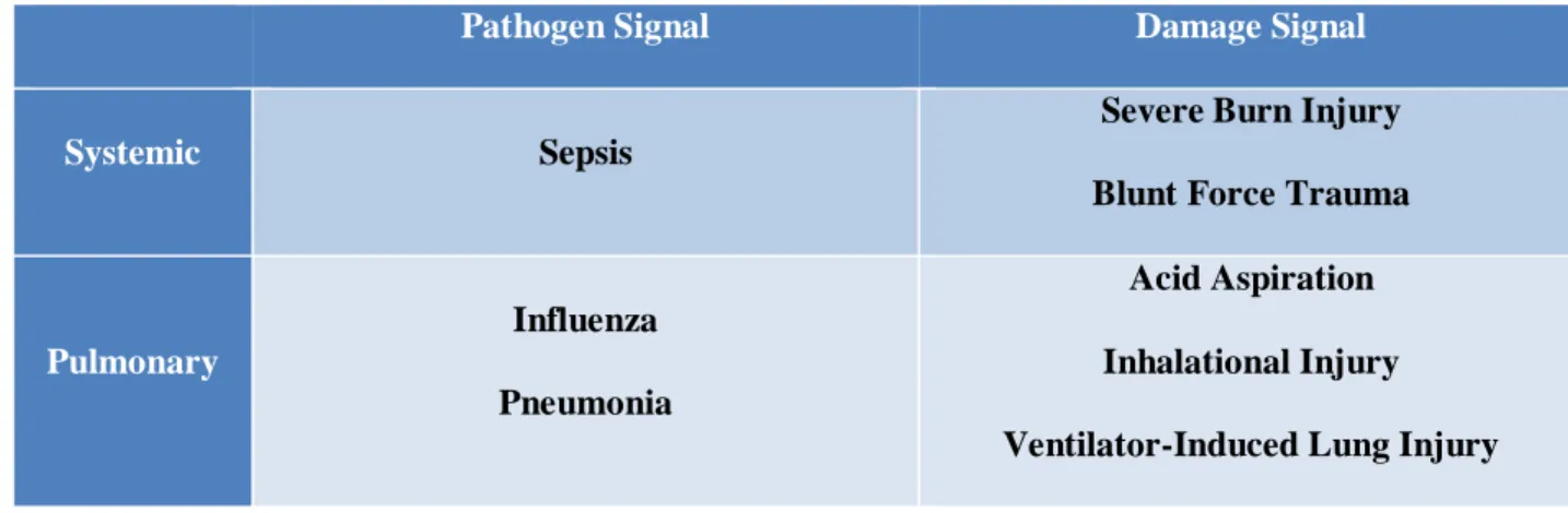
Table 1.1: Challenges that lead to Acute Lung Injury (ALI).
Pathogen Signal Damage Signal
Systemic Sepsis
Severe Burn Injury
Blunt Force Trauma
Pulmonary
Influenza
Pneumonia
Acid Aspiration
Inhalational Injury
REFERENCES
1. Xiao W, Mindrinos MN, Seok J, Cuschieri J, Cuenca AG, Gao H, Hayden DL, Hennessy L, Moore EE, Minei JP, Bankey PE, Johnson JL, Sperry J, Nathens AB, Billiar TR, West MA, Brownstein BH, Mason PH, Baker HV, Finnerty CC, Jeschke MG, Lopez MC, Klein MB, Gamelli RL, Gibran NS, Arnoldo B, Xu W, Zhang Y, Calvano SE, McDonald-Smith GP, Schoenfeld DA, Storey JD, Cobb JP, Warren HS, Moldawer LL, Herndon DN, Lowry SF, Maier RV, Davis RW, Tompkins RG, Inflammation, Host Response to Injury Large-Scale Collaborative Research P. A genomic storm in critically injured humans. J Exp Med. 2011;208(13):2581-90. doi: 10.1084/jem.20111354. PubMed PMID: 22110166; PMCID: PMC3244029.
2. Dong H, Li J, Lv Y, Zhou Y, Wang G, Hu S, He X, Yang P, Zhou Z, Xiang X, Wang CY. Comparative analysis of the alveolar macrophage proteome in ALI/ARDS patients between the exudative phase and recovery phase. BMC Immunol. 2013;14:25. doi: 10.1186/1471-2172-14-25. PubMed PMID: 23773529; PMCID: PMC3727986.
3. Finnerty CC, Jeschke MG, Herndon DN, Gamelli R, Gibran N, Klein M, Silver G, Arnoldo B, Remick D, Tompkins RG, Investigators of the I, the Host Response Glue G. Temporal cytokine profiles in severely burned patients: a comparison of adults and children. Mol Med. 2008;14(9-10):553-60. doi: 10.2119/2007-00132.Finnerty. PubMed PMID: 18548133; PMCID: PMC2424320.
4. Davis CS, Albright JM, Carter SR, Ramirez L, Kim H, Gamelli RL, Kovacs EJ. Early pulmonary immune hyporesponsiveness is associated with mortality after burn and smoke inhalation injury. J Burn Care Res. 2012;33(1):26-35. doi: 10.1097/BCR.0b013e318234d903. PubMed PMID: 21979852; PMCID: PMC3253958.
5. Liu T, Qian WJ, Gritsenko MA, Xiao W, Moldawer LL, Kaushal A, Monroe ME, Varnum SM, Moore RJ, Purvine SO, Maier RV, Davis RW, Tompkins RG, Camp DG, 2nd, Smith RD, Inflammation, the Host Response to Injury Large Scale Collaborative Research P. High dynamic range characterization of the trauma patient plasma proteome. Mol Cell Proteomics. 2006;5(10):1899-913. doi: 10.1074/mcp.M600068-MCP200. PubMed PMID: 16684767; PMCID: PMC1783978. 6. Sweeney TE, Shidham A, Wong HR, Khatri P. A comprehensive time-course-based multicohort
analysis of sepsis and sterile inflammation reveals a robust diagnostic gene set. Sci Transl Med. 2015;7(287):287ra71. doi: 10.1126/scitranslmed.aaa5993. PubMed PMID: 25972003; PMCID: PMC4734362.
7. Mayhall CG. The epidemiology of burn wound infections: then and now. Clin Infect Dis. 2003;37(4):543-50. doi: 10.1086/376993. PubMed PMID: 12905139.
8. Wurtz R, Karajovic M, Dacumos E, Jovanovic B, Hanumadass M. Nosocomial infections in a burn intensive care unit. Burns. 1995;21(3):181-4. PubMed PMID: 7794498.
10. Branski LK, Herndon DN, Barrow RE, Kulp GA, Klein GL, Suman OE, Przkora R, Meyer W, 3rd, Huang T, Lee JO, Chinkes DL, Mlcak RP, Jeschke MG. Randomized controlled trial to determine the efficacy of long-term growth hormone treatment in severely burned children. Ann Surg. 2009;250(4):514-23. doi: 10.1097/SLA.0b013e3181b8f9ca. PubMed PMID: 19734776; PMCID: PMC3970433.
11. Williams FN, Jeschke MG, Chinkes DL, Suman OE, Branski LK, Herndon DN. Modulation of the hypermetabolic response to trauma: temperature, nutrition, and drugs. J Am Coll Surg. 2009;208(4):489-502. doi: 10.1016/j.jamcollsurg.2009.01.022. PubMed PMID: 19476781; PMCID: PMC3775552.
12. Maile R, Jones S, Pan Y, Zhou H, Jaspers I, Peden DB, Cairns BA, Noah TL. Association between early airway damage-associated molecular patterns and subsequent bacterial infection in patients with inhalational and burn injury. Am J Physiol Lung Cell Mol Physiol. 2015;308(9):L855-60. doi: 10.1152/ajplung.00321.2014. PubMed PMID: 25770180; PMCID: PMC4421787.
13. Zhang Q, Raoof M, Chen Y, Sumi Y, Sursal T, Junger W, Brohi K, Itagaki K, Hauser CJ. Circulating mitochondrial DAMPs cause inflammatory responses to injury. Nature. 2010;464(7285):104-7. doi: 10.1038/nature08780. PubMed PMID: 20203610; PMCID: PMC2843437.
14. Onarheim H, Reed RK, Laurent TC. Elevated hyaluronan blood concentrations in severely burned patients. Scand J Clin Lab Invest. 1991;51(8):693-7. PubMed PMID: 1806984.
15. Simmons JD, Lee YL, Mulekar S, Kuck JL, Brevard SB, Gonzalez RP, Gillespie MN, Richards WO. Elevated levels of plasma mitochondrial DNA DAMPs are linked to clinical outcome in severely injured human subjects. Ann Surg. 2013;258(4):591-6; discussion 6-8. doi: 10.1097/SLA.0b013e3182a4ea46. PubMed PMID: 23979273; PMCID: PMC3935616.
16. Hultman CS, Cairns BA, deSerres S, Frelinger JA, Meyer AA. Early, complete burn wound excision partially restores cytotoxic T lymphocyte function. Surgery. 1995;118(2):421-9; discussion 9-30. PubMed PMID: 7638760.
17. Ipaktchi K, Mattar A, Niederbichler AD, Hoesel LM, Vollmannshauser S, Hemmila MR, Su GL, Remick DG, Wang SC, Arbabi S. Attenuating burn wound inflammatory signaling reduces systemic inflammation and acute lung injury. J Immunol. 2006;177(11):8065-71. PubMed PMID: 17114480.
18. Till GO, Beauchamp C, Menapace D, Tourtellotte W, Jr., Kunkel R, Johnson KJ, Ward PA. Oxygen radical dependent lung damage following thermal injury of rat skin. J Trauma. 1983;23(4):269-77. PubMed PMID: 6842628.
19. Schmid E, Piccolo MT, Friedl HP, Warner RL, Mulligan MS, Hugli TE, Till GO, Ward PA. Requirement for C5a in lung vascular injury following thermal trauma to rat skin. Shock. 1997;8(2):119-24. PubMed PMID: 9261902.
2013;8(5):e64250. doi: 10.1371/journal.pone.0064250. PubMed PMID: 23691180; PMCID: PMC3656836.
21. Neely CJ, Kartchner LB, Mendoza AE, Linz BM, Frelinger JA, Wolfgang MC, Maile R, Cairns BA. Flagellin treatment prevents increased susceptibility to systemic bacterial infection after injury by inhibiting anti-inflammatory IL-10+ IL-12- neutrophil polarization. PLoS One. 2014;9(1):e85623. doi: 10.1371/journal.pone.0085623. PubMed PMID: 24454904; PMCID: PMC3893295.
22. Tompkins RG, Hilton JF, Burke JF, Schoenfeld DA, Hegarty MT, Bondoc CC, Quinby WC, Jr., Behringer GE, Ackroyd FW. Increased survival after massive thermal injuries in adults: preliminary report using artificial skin. Crit Care Med. 1989;17(8):734-40. PubMed PMID: 2546715.
23. Ryan CM, Schoenfeld DA, Thorpe WP, Sheridan RL, Cassem EH, Tompkins RG. Objective estimates of the probability of death from burn injuries. N Engl J Med. 1998;338(6):362-6. doi: 10.1056/NEJM199802053380604. PubMed PMID: 9449729.
24. Rehberg S, Maybauer MO, Enkhbaatar P, Maybauer DM, Yamamoto Y, Traber DL. Pathophysiology, management and treatment of smoke inhalation injury. Expert Rev Respir Med. 2009;3(3):283-97. doi: 10.1586/ERS.09.21. PubMed PMID: 20161170; PMCID: PMC2722076.
25. Johnson ER, Matthay MA. Acute lung injury: epidemiology, pathogenesis, and treatment. J Aerosol Med Pulm Drug Deliv. 2010;23(4):243-52. doi: 10.1089/jamp.2009.0775. PubMed PMID: 20073554; PMCID: PMC3133560.
26. Aeffner F, Davis IC. Respiratory syncytial virus reverses airway hyperresponsiveness to methacholine in ovalbumin-sensitized mice. PLoS One. 2012;7(10):e46660. doi: 10.1371/journal.pone.0046660. PubMed PMID: 23056391; PMCID: PMC3462783.
27. Driscoll JA, Brody SL, Kollef MH. The epidemiology, pathogenesis and treatment of Pseudomonas aeruginosa infections. Drugs. 2007;67(3):351-68. PubMed PMID: 17335295. 28. Matthay MA, Ware LB, Zimmerman GA. The acute respiratory distress syndrome. J Clin Invest.
2012;122(8):2731-40. doi: 10.1172/JCI60331. PubMed PMID: 22850883; PMCID: PMC3408735. 29. Matute-Bello G, Downey G, Moore BB, Groshong SD, Matthay MA, Slutsky AS, Kuebler WM,
Acute Lung Injury in Animals Study G. An official American Thoracic Society workshop report: features and measurements of experimental acute lung injury in animals. Am J Respir Cell Mol Biol. 2011;44(5):725-38. doi: 10.1165/rcmb.2009-0210ST. PubMed PMID: 21531958.
30. Liu F, Li W, Pauluhn J, Trubel H, Wang C. Rat models of acute lung injury: exhaled nitric oxide as a sensitive, noninvasive real-time biomarker of prognosis and efficacy of intervention. Toxicology. 2013;310:104-14. doi: 10.1016/j.tox.2013.05.016. PubMed PMID: 23770417.
32. Ahn CS, Maitz PK. The true cost of burn. Burns. 2012;38(7):967-74. doi: 10.1016/j.burns.2012.05.016. PubMed PMID: 22795515.
33. Mosier MJ, Pham TN, Park DR, Simmons J, Klein MB, Gibran NS. Predictive value of bronchoscopy in assessing the severity of inhalation injury. J Burn Care Res. 2012;33(1):65-73. doi: 10.1097/BCR.0b013e318234d92f. PubMed PMID: 21941194.
34. Walker J. Diagnosis and management of patients with hypercalcaemia. Nurs Older People. 2015;27(4):22-6. doi: 10.7748/nop.27.4.22.e685. PubMed PMID: 25924757.
35. MacLoughlin RJ, Higgins BD, Devaney J, O'Toole D, Laffey JG, O'Brien T. Aerosol-mediated delivery of AAV2/6-IkappaBalpha attenuates lipopolysaccharide-induced acute lung injury in rats. Hum Gene Ther. 2015;26(1):36-46. doi: 10.1089/hum.2014.053. PubMed PMID: 25382145; PMCID: PMC4302961.
36. Lange M, Szabo C, Traber DL, Horvath E, Hamahata A, Nakano Y, Traber LD, Cox RA, Schmalstieg FC, Herndon DN, Enkhbaatar P. Time profile of oxidative stress and neutrophil activation in ovine acute lung injury and sepsis. Shock. 2012;37(5):468-72. doi: 10.1097/SHK.0b013e31824b1793. PubMed PMID: 22266977; PMCID: PMC4646062.
37. Esechie A, Kiss L, Olah G, Horvath EM, Hawkins H, Szabo C, Traber DL. Protective effect of hydrogen sulfide in a murine model of acute lung injury induced by combined burn and smoke inhalation. Clin Sci (Lond). 2008;115(3):91-7. doi: 10.1042/CS20080021. PubMed PMID: 18315525.
38. Imai Y, Kuba K, Neely GG, Yaghubian-Malhami R, Perkmann T, van Loo G, Ermolaeva M, Veldhuizen R, Leung YH, Wang H, Liu H, Sun Y, Pasparakis M, Kopf M, Mech C, Bavari S, Peiris JS, Slutsky AS, Akira S, Hultqvist M, Holmdahl R, Nicholls J, Jiang C, Binder CJ, Penninger JM. Identification of oxidative stress and Toll-like receptor 4 signaling as a key pathway of acute lung injury. Cell. 2008;133(2):235-49. doi: 10.1016/j.cell.2008.02.043. PubMed PMID: 18423196. 39. Liu B, Luo XJ, Yang ZB, Zhang JJ, Li TB, Zhang XJ, Ma QL, Zhang GG, Hu CP, Peng J. Inhibition of
NOX/VPO1 pathway and inflammatory reaction by trimethoxystilbene in prevention of cardiovascular remodeling in hypoxia-induced pulmonary hypertensive rats. J Cardiovasc Pharmacol. 2014;63(6):567-76. doi: 10.1097/FJC.0000000000000082. PubMed PMID: 24492474. 40. Mizutani A, Enkhbaatar P, Esechie A, Traber LD, Cox RA, Hawkins HK, Deyo DJ, Murakami K,
Noguchi T, Traber DL. Pulmonary changes in a mouse model of combined burn and smoke inhalation-induced injury. J Appl Physiol (1985). 2008;105(2):678-84. doi: 10.1152/japplphysiol.00232.2007. PubMed PMID: 18436699.
41. Soejima K, Traber LD, Schmalstieg FC, Hawkins H, Jodoin JM, Szabo C, Szabo E, Virag L, Salzman A, Traber DL. Role of nitric oxide in vascular permeability after combined burns and smoke inhalation injury. Am J Respir Crit Care Med. 2001;163(3 Pt 1):745-52. doi: 10.1164/ajrccm.163.3.9912052. PubMed PMID: 11254534.
type III secretory toxins. Respir Res. 2004;5:1. doi: 10.1186/1465-9921-5-1. PubMed PMID: 15040820; PMCID: PMC389879.
43. Aggarwal NR, Tsushima K, Eto Y, Tripathi A, Mandke P, Mock JR, Garibaldi BT, Singer BD, Sidhaye VK, Horton MR, King LS, D'Alessio FR. Immunological priming requires regulatory T cells and IL-10-producing macrophages to accelerate resolution from severe lung inflammation. J Immunol. 2014;192(9):4453-64. doi: 10.4049/jimmunol.1400146. PubMed PMID: 24688024; PMCID: PMC4001810.
44. Belperio JA, Keane MP, Burdick MD, Londhe V, Xue YY, Li K, Phillips RJ, Strieter RM. Critical role for CXCR2 and CXCR2 ligands during the pathogenesis of ventilator-induced lung injury. J Clin Invest. 2002;110(11):1703-16. doi: 10.1172/JCI15849. PubMed PMID: 12464676; PMCID: PMC151632.
45. Lange M, Hamahata A, Traber DL, Connelly R, Nakano Y, Traber LD, Schmalstieg FC, Herndon DN, Enkhbaatar P. Pulmonary microvascular hyperpermeability and expression of vascular endothelial growth factor in smoke inhalation- and pneumonia-induced acute lung injury. Burns. 2012;38(7):1072-8. doi: 10.1016/j.burns.2012.02.019. PubMed PMID: 22647495; PMCID: PMC3893691.
46. Bogdan C. Nitric oxide and the immune response. Nat Immunol. 2001;2(10):907-16. doi: 10.1038/ni1001-907. PubMed PMID: 11577346.
47. Odegaard JI, Ricardo-Gonzalez RR, Goforth MH, Morel CR, Subramanian V, Mukundan L, Red Eagle A, Vats D, Brombacher F, Ferrante AW, Chawla A. Macrophage-specific PPARgamma controls alternative activation and improves insulin resistance. Nature. 2007;447(7148):1116-20. doi: 10.1038/nature05894. PubMed PMID: 17515919; PMCID: PMC2587297.
48. Trifilieff A, Bench A, Hanley M, Bayley D, Campbell E, Whittaker P. PPAR-alpha and -gamma but not -delta agonists inhibit airway inflammation in a murine model of asthma: in vitro evidence for an NF-kappaB-independent effect. Br J Pharmacol. 2003;139(1):163-71. doi: 10.1038/sj.bjp.0705232. PubMed PMID: 12746235; PMCID: PMC1573830.
49. Albaiceta GM, Gutierrez-Fernandez A, Garcia-Prieto E, Puente XS, Parra D, Astudillo A, Campestre C, Cabrera S, Gonzalez-Lopez A, Fueyo A, Taboada F, Lopez-Otin C. Absence or inhibition of matrix metalloproteinase-8 decreases ventilator-induced lung injury. Am J Respir Cell Mol Biol. 2010;43(5):555-63. doi: 10.1165/rcmb.2009-0034OC. PubMed PMID: 19995943. 50. Chai JK, Cai JH, Deng HP, Zou XF, Liu W, Hu QG, Shen CA, Yin HN, Zhang XB, Chi YF, Ma L, Feng R.
Role of neutrophil elastase in lung injury induced by burn-blast combined injury in rats. Burns. 2013;39(4):745-53. doi: 10.1016/j.burns.2012.08.005. PubMed PMID: 22999209.
51. Dubois B, Starckx S, Pagenstecher A, Oord J, Arnold B, Opdenakker G. Gelatinase B deficiency protects against endotoxin shock. Eur J Immunol. 2002;32(8):2163-71. doi: 10.1002/1521-4141(200208)32:8<2163::AID-IMMU2163>3.0.CO;2-Q. PubMed PMID: 12209628.
53. Armstrong R. The physiological role and pharmacological potential of nitric oxide in neutrophil activation. Int Immunopharmacol. 2001;1(8):1501-12. PubMed PMID: 11515815.
54. Sumimoto H, Minakami S. Oxidation of 20-hydroxyleukotriene B4 to 20-carboxyleukotriene B4 by human neutrophil microsomes. Role of aldehyde dehydrogenase and leukotriene B4 omega-hydroxylase (cytochrome P-450LTB omega) in leukotriene B4 omega-oxidation. J Biol Chem. 1990;265(8):4348-53. PubMed PMID: 2155225.
55. Frasch SC, Fernandez-Boyanapalli RF, Berry KA, Murphy RC, Leslie CC, Nick JA, Henson PM, Bratton DL. Neutrophils regulate tissue Neutrophilia in inflammation via the oxidant-modified lipid lysophosphatidylserine. J Biol Chem. 2013;288(7):4583-93. doi: 10.1074/jbc.M112.438507. PubMed PMID: 23293064; PMCID: PMC3576064.
56. Campbell EL, Bruyninckx WJ, Kelly CJ, Glover LE, McNamee EN, Bowers BE, Bayless AJ, Scully M, Saeedi BJ, Golden-Mason L, Ehrentraut SF, Curtis VF, Burgess A, Garvey JF, Sorensen A, Nemenoff R, Jedlicka P, Taylor CT, Kominsky DJ, Colgan SP. Transmigrating neutrophils shape the mucosal microenvironment through localized oxygen depletion to influence resolution of inflammation. Immunity. 2014;40(1):66-77. doi: 10.1016/j.immuni.2013.11.020. PubMed PMID: 24412613; PMCID: PMC3951457.
57. Speyer CL, Neff TA, Warner RL, Guo RF, Sarma JV, Riedemann NC, Murphy ME, Murphy HS, Ward PA. Regulatory effects of iNOS on acute lung inflammatory responses in mice. Am J Pathol. 2003;163(6):2319-28. doi: 10.1016/S0002-9440(10)63588-2. PubMed PMID: 14633605; PMCID: PMC1892362.
58. Trifilieff A, Fujitani Y, Mentz F, Dugas B, Fuentes M, Bertrand C. Inducible Nitric Oxide Synthase Inhibitors Suppress Airway Inflammation in Mice Through Down-Regulation of Chemokine Expression. The Journal of Immunology. 2000;165(3):1526-33. doi: 10.4049/jimmunol.165.3.1526.
59. Bosca L, Zeini M, Traves PG, Hortelano S. Nitric oxide and cell viability in inflammatory cells: a role for NO in macrophage function and fate. Toxicology. 2005;208(2):249-58. doi: 10.1016/j.tox.2004.11.035. PubMed PMID: 15691589.
60. Dunn JL, Hunter RA, Gast K, Maile R, Cairns BA, Schoenfisch MH. Direct detection of blood nitric oxide reveals a burn-dependent decrease of nitric oxide in response to Pseudomonas aeruginosa infection. Burns. 2016;42(7):1522-7. doi: 10.1016/j.burns.2016.05.005. PubMed PMID: 27268107; PMCID: PMC5056119.
61. Carraway MS, Piantadosi CA, Jenkinson CP, Huang Y-CT. Differential Expression of Arginase and iNOS in the Lung in Sepsis. Experimental Lung Research. 2009;24(3):253-68. doi: 10.3109/01902149809041533.
63. Hoshino K, Takeuchi O, Kawai T, Sanjo H, Ogawa T, Takeda Y, Takeda K, Akira S. Cutting edge: Toll-like receptor 4 (TLR4)-deficient mice are hyporesponsive to lipopolysaccharide: evidence for TLR4 as the Lps gene product. J Immunol. 1999;162(7):3749-52. PubMed PMID: 10201887. 64. Black KE, Collins SL, Hagan RS, Hamblin MJ, Chan-Li Y, Hallowell RW, Powell JD, Horton MR.
Hyaluronan fragments induce IFNbeta via a novel TLR4-TRIF-TBK1-IRF3-dependent pathway. J Inflamm (Lond). 2013;10(1):23. doi: 10.1186/1476-9255-10-23. PubMed PMID: 23721397; PMCID: PMC3682892.
65. Cairns B, Maile R, Barnes CM, Frelinger JA, Meyer AA. Increased Toll-like receptor 4 expression on T cells may be a mechanism for enhanced T cell response late after burn injury. J Trauma. 2006;61(2):293-8; discussion 8-9. doi: 10.1097/01.ta.0000228969.46633.bb. PubMed PMID: 16917441.
66. Cairns BA, Barnes CM, Mlot S, Meyer AA, Maile R. Toll-like receptor 2 and 4 ligation results in complex altered cytokine profiles early and late after burn injury. J Trauma. 2008;64(4):1069-77; discussion 77-8. doi: 10.1097/TA.0b013e318166b7d9. PubMed PMID: 18404077.
67. Hayashi F, Smith KD, Ozinsky A, Hawn TR, Yi EC, Goodlett DR, Eng JK, Akira S, Underhill DM, Aderem A. The innate immune response to bacterial flagellin is mediated by Toll-like receptor 5. Nature. 2001;410(6832):1099-103. doi: 10.1038/35074106. PubMed PMID: 11323673.
68. Zahs A, Bird MD, Ramirez L, Choudhry MA, Kovacs EJ. Anti-IL-6 antibody treatment but not IL-6 knockout improves intestinal barrier function and reduces inflammation after binge ethanol exposure and burn injury. Shock. 2013;39(4):373-9. doi: 10.1097/SHK.0b013e318289d6c6. PubMed PMID: 23376955; PMCID: PMC3602394.
69. Faunce DE, Gregory MS, Kovacs EJ. Acute ethanol exposure prior to thermal injury results in decreased T-cell responses mediated in part by increased production of IL-6. Shock. 1998;10(2):135-40. PubMed PMID: 9721981.
70. Fontanilla CV, Faunce DE, Gregory MS, Messingham KA, Durbin EA, Duffner LA, Kovacs EJ. Anti-interleukin-6 antibody treatment restores cell-mediated immune function in mice with acute ethanol exposure before burn trauma. Alcohol Clin Exp Res. 2000;24(9):1392-9. PubMed PMID: 11003205.
71. Fielding CA, Jones GW, McLoughlin RM, McLeod L, Hammond VJ, Uceda J, Williams AS, Lambie M, Foster TL, Liao CT, Rice CM, Greenhill CJ, Colmont CS, Hams E, Coles B, Kift-Morgan A, Newton Z, Craig KJ, Williams JD, Williams GT, Davies SJ, Humphreys IR, O'Donnell VB, Taylor PR, Jenkins BJ, Topley N, Jones SA. Interleukin-6 signaling drives fibrosis in unresolved inflammation. Immunity. 2014;40(1):40-50. doi: 10.1016/j.immuni.2013.10.022. PubMed PMID: 24412616; PMCID: PMC3919204.
72. Vindenes H, Ulvestad E, Bjerknes R. Increased levels of circulating interleukin-8 in patients with large burns: relation to burn size and sepsis. J Trauma. 1995;39(4):635-40. PubMed PMID: 7473946.
74. Zheng H, Chen XL, Han ZX, Zhang Z, Wang SY, Xu QL. Ligustrazine attenuates acute lung injury after burn trauma. Burns. 2005;31(4):453-8. doi: 10.1016/j.burns.2004.10.023. PubMed PMID: 15896507.
75. Kim J, Natarajan S, Vaickus LJ, Bouchard JC, Beal D, Cruikshank WW, Remick DG. Diesel exhaust particulates exacerbate asthma-like inflammation by increasing CXC chemokines. Am J Pathol. 2011;179(6):2730-9. doi: 10.1016/j.ajpath.2011.08.008. PubMed PMID: 21967814; PMCID: PMC3260803.
76. Shallo H, Plackett TP, Heinrich SA, Kovacs EJ. Monocyte chemoattractant protein-1 (MCP-1) and macrophage infiltration into the skin after burn injury in aged mice. Burns. 2003;29(7):641-7. PubMed PMID: 14556721.
77. Furukawa K, Kobayashi M, Herndon DN, Pollard RB, Suzuki F. Appearance of monocyte chemoattractant protein 1 (MCP-1) early after thermal injury: role in the subsequent development of burn-associated type 2 T-cell responses. Ann Surg. 2002;236(1):112-9. PubMed PMID: 12131093; PMCID: PMC1422556.
78. Baker TA, Davis CS, Bach HHt, Romero J, Burnham EL, Kovacs EJ, Gamelli RL, Majetschak M. Ubiquitin and stromal cell-derived factor-1alpha in bronchoalveolar lavage fluid after burn and inhalation injury. J Burn Care Res. 2012;33(1):57-64. doi: 10.1097/BCR.0b013e31823dc559. PubMed PMID: 22105097; PMCID: PMC3626094.
79. Copeland S, Warren HS, Lowry SF, Calvano SE, Remick D, Inflammation, the Host Response to Injury I. Acute inflammatory response to endotoxin in mice and humans. Clin Diagn Lab Immunol. 2005;12(1):60-7. doi: 10.1128/CDLI.12.1.60-67.2005. PubMed PMID: 15642986; PMCID: PMC540200.
80. Zawacki BE, Jung RC, Joyce J, Rincon E. Smoke, burns, and the natural history of inhalation injury in fire victims: a correlation of experimental and clinical data. Ann Surg. 1977;185(1):100-10. PubMed PMID: 831629; PMCID: PMC1396243.
CHAPTER 2: DIRECT DETECTION OF BLOOD NITRIC OXIDE REVEALS A
BURN-DEPENDENT DECREASE OF NITRIC OXIDE IN RESPONSE TO PSEUDOMONAS
AERUGINOSA INFECTION
12Summary
Purpose: Burn injury is associated with severe immune dysfunction, including an anti-inflammatory state that occurs late after burn injury. While increased nitric oxide (NO) production is associated with severe infection and sepsis, the effect of burn trauma on these levels during a non-lethal infection remains unknown. We hypothesized that in a mouse model, 1) NO levels would be increased after nonlethal infection without trauma and 2) burn injury would lead to decreased NO production even during infection. Methods: Mice were infected via intra-tracheal inoculation with Pseudomonas
aeruginosa 14 d following a 20% total body surface area contact burn. At 48 h following infection, blood
was drawn to quantify NO concentrations using a microfluidic electrochemical sensor. Significant findings: In uninjured mice, infection caused a significant increase in blood NO levels. Increases in NO occurred in a dose-dependent response to the bacterial inoculum. Following burn injury, an identical infection did not elicit increases in NO. Conclusions: While increases in NO are expected over the course of an infection without prior trauma, burn injury and subsequent immune suppression decreases NO levels even in the presence of infection.
1 Adapted for this dissertation from Dunn JLM, Hunter RA, Gast K, Maile R, Cairns BA, and Schoenfish MH. Direct detection of blood nitric oxide reveals a burn-dependent decrease of nitric oxide in response to Pseudomonas aeruginosa infection. 2016. Burns 42(7):1522-1527.
Introduction
Physical trauma such as burn injury causes severe immune dysfunction, often resulting in infection, sepsis, multiple organ dysfunction, and death (1). Historically, this immune response has been characterized by an initial pro-inflammatory period and a subsequent anti-inflammatory phase (1, 2). These periods have been referred to as the systemic inflammatory response syndrome (SIRS), during which pro-inflammatory mediators (e.g., tumor necrosis factor, interleukin-6, interleukin-1) are released, and a compensatory anti-inflammatory response syndrome (CARS) thought to limit damage due to chronic inflammation (1-6). More recent studies have illustrated a more complex scenario, wherein pro- and anti-inflammatory cytokines may be secreted simultaneously, suggesting that the SIRS/CARS paradigm may be insufficient to characterize the immune response to trauma (3-5). Despite this paradigm shift, it remains true that late after trauma patients exhibit elevated susceptibility to nosocomial infection (6-8). Burn patients are especially prone to ventilator-associated pneumonia and wound infections (6, 9), and the leading cause of death following burn injury is related to infection and sepsis (6, 10). In particular, pulmonary infections by Gram-negative Pseudomonas aeruginosa are quite common (7). Novel methods are needed that will enable healthcare providers and researchers to monitor the immune response in real-time during trauma and infection.
Direct measurement of NO is likely to rapidly and sensitively detect changes in patient status; therefore, such measurements should be incorporated into clinically relevant models. Because of the availability of genetically modified mice, confirming the usefulness of NO as a readout in mouse models of trauma will enable elucidation of disease mechanisms that impact NO production. Recently, a microfluidic amperometric sensor was developed and used to directly measure increases in NO levels during a lethal murine model of sepsis (28).
We expect that NO levels may be decreased during an infection following burn injury. Because patients demonstrate susceptibility to infection late after trauma, we focused on NO levels in a nonlethal infection model at 14 days after burn injury. Quantifying changes in NO concentration may allow for the elucidation of immune dysfunction, indicated by either elevated levels during systemic infection or decreased levels during immune suppression.
Methods
Murine model of burn injury and infection. Nine week-old female C57BL/6 mice weighing 18-20
g underwent a 20% total body surface area (TBSA) burn injury as previously described (34). Briefly, mice were anesthetized with gaseous isofluorane, their dorsal flanks were shaved, and they received a subcutaneous injection of morphine sulphate prior to receiving a full-thickness burn with 4 applications of a copper rod heated in boiling water. Following burn injury, mice were resuscitated via an intraperitoneal injection of lactated Ringer’s solution. Throughout the experiment, mice were monitored and received morphine in their drinking water (0.02 mg mL-1; 4 mg kg-1 body weight per day) ad libitum. Sham (0%
TBSA) mice also underwent these treatments, except the application of the copper rod.
At 14 d following burn injury, pneumonia was induced via the intratracheal administration of 50
L Pseudomonas aeruginosa (PAK strain) following sedation with Avertin. Uninfected groups were
administered 50 L PBS with 1% protease peptone in the same manner. At 48 h following infection, ~300
was immediately injected into the microfluidic device and analyzed amperometrically to determine NO concentrations.
Microfluidic amperometric sensor for nitric oxide measurement. Devices were fabricated as
previously described (28). Working electrodes were 100 µm wide and consisted of 150 nm thick platinum with a 10 nm titanium seed layer, coated with a selective film (adhesion layer of 1% v/v (3-aminopropyl)triethoxysilane and a fluoroalkoxysilane xerogel) to impart selectivity to NO. The fluoroalkoxysilane membrane solution was prepared via the acid catalyzed hydrolysis and condensation of (heptadecafluoro-1,1,2,2-tetrahydrodecyl)trimethoxysilane and methyltrimethoxysilane as reported previously (35, 36). Reference electrodes consisted of a 10 nm titanium adhesion layer followed by a ~1 µm silver layer, chemically oxidized by reaction with 50 mM ferric chloride for 10 s to create a silver/silver chloride pseudo-reference/counter electrode. The ~90 µm microfluidic channel was formed using Kapton® tape.
The working and reference/counter electrodes of the microfluidic device were connected to a CH Instruments 1030A 8-channel potentiostat (Austin, TX). Prior to sample analysis, the device was polarized at +800 mV vs. the Ag/AgCl pseudo-reference/counter electrode for at least to 1 h in PBS. To calibrate the device, a saturated NO standard solution (prepared by purging deaerated PBS with NO gas for ~10 min to yield a 1.9 mM solution of NO) was diluted with PBS and introduced into the inlet reservoir.
Statistical analysis. Where appropriate, either an unpaired, two-tailed Student’s t-test or two-way
ANOVA with Bonfennori post-test was used to determine statistical significance between groups, with p
<0.05 considered to be significant.
Results
Nitric oxide levels are increased during pneumonia without prior burn injury. Blood samples
the microfluidic sensor (Figure 2.1). As shown in Figure 2.2A, blood NO levels were significantly increased in the infected versus uninfected mice at this time point (810 180 nM and 370 40 nM, respectively). As expected, these data suggest that activation of the immune response following infection leads to increased production of NO.
Increased infectious dose corresponds with elevated serum NO. Blood samples were drawn 24 h
and 72 h following infection with either 5 x 10⁵ or 5 x 10⁶ CFU of P. aeruginosa. As indicated in Figure
1B, blood NO levels were higher in uninjured mice exposed to a higher infectious dose at 24 h (3.0 0.2 µM vs. 6.2 0.7 µM) and 72 h (7.0 1.4 µM vs. 15.2 4.7 µM) following infection. At both high- and low-dose inocula, blood NO concentrations were higher at 72 h than at 24 h. We also harvested lungs and quantified bacterial load in these mice at 72 hours after infection. There was a significant difference in bacterial load between mice with high and low dose bacterial inoculum, with a corresponding significant difference in plasma NO levels (Figure 2.2B).
Burn injury inhibits nitric oxide release following infection. Blood samples from infected (5 x 10⁶
CFU) and uninfected mice 14 d after burn injury were analyzed using the electrochemical sensor. Concentrations of NO in blood of infected burned mice were not significantly elevated compared to uninfected burn mice (290 80 nM vs. 410 110 nM) (Figure 2). We also harvested lungs and quantified bacterial load in these mice at 72 hours after infection, and observed a significant reduction in pulmonary bacterial clearance in burn mice compared to sham mice and corresponding reduction in NO (Figure 2.3). These data indicate a late-stage effect of burn injury on immune function and NO production during an infection. Also of note, the NO concentrations observed in uninfected burn mice were equivalent to those in the uninfected sham mice.
We measured blood NO levels at 24 h and 72 h following a low-dose (5 x 10⁵ CFU) infection