Tailoring Materials for Biological Applications
Nathan Westcott
A dissertation submitted to the faculty of the University of North Carolina at Chapel Hill in partial fulfillment of the requirements for the degree of doctor of philosophy in the Department
of Chemistry.
Chapel Hill 2011
Approved by Advisor: Professor Muhammad Yousaf Chair: Professor Wei You Professor Nancy Allbritton Professor Marcy Waters Professor Mark Schoenfisch
Abstract
Nathan Westcott: Tailoring Materials for Biological Applications (Under the Direction of Muhammad Yousaf)
The ECM is a very complex, heterogeneous mixture of proteins, peptides, and
hormones, which has proven difficult to model in vitro. Currently, a number of model substrates
and systems have been developed utilizing polymers, layer-‐by-‐layer methods, and self-‐
assembled monolayers (SAMs). SAMs of alkanethiolates on gold in particular, have proven to
be useful model substrates with a number of key advantages; SAMs are chemically well defined,
synthetically flexible, conductive, compatible with live-‐cell high resolution fluorescence
microscopy techniques, can be patterned at the micro-‐ and nanoscale, and most importantly,
they can be made to resist non-‐specific protein adsorption. These advantages allow for
fabrication of complex, flexible substrates for studies of cell phenomena at the molecular level.
To tailor SAMs on gold with precise spatial control and quantification of ligand density,
smart SAM surfaces have been developed to immobilize a variety of ligands using the
hydroquinone. By installing the peptide ligand sequence RGD (an epitope for the ECM protein
fibronectin), cells have been biospecifially adhered to SAMs to study cell behavior based on
specific ligand-‐receptor interactions. During the course of my thesis, I have combined analytical
techniques with the unique capabilities of surface chemistry to study cell biology problems
regarding ligand-‐receptor and small molecule-‐protein interactions. I have fabricated flexible
biological substrates capable of binding different cellular ligands based on SAMs and hydrogels
In chapter 1, I review the relevant literature on SAMs and cell adhesion and migration.
In chapter 2 and 3, two methods combining microfluidics and SAMs are described. In chapter 4,
alcohol oxidation was used to functionalize simple SAMs. In chapter 5, this method was
extended to create protein affinity platforms. In chapter 6, cell adhesion was monitored at the
nanoscale using DPN and evaporative lithography. In chapter 7, hydrogels were created to
monitor cell adhesion in 3D. Chapter 8 is dissertation conclusions and future directions. These
interactions and substrates were characterized by a variety of techniques including XPS,
fluorescence microscopy, electrochemistry, and mass spectrometry.
Acknowledgements
I would like to acknowledge my advisor and mentor Muhammad Yousaf for the
guidance and help in growing as a scientist. I would also like to thank my family, friends, and
labmates for their support and advice during my graduate studies.
Table of Contents
List of Figures ……..……..……..……..……..……..……..………..……..……..……..…….……..……..…….viii
List of Tables and Schemes ………..……..……..……..………..……..……..……..…….……..……..……….xi
List of Abbreviations and Symbols ……..……..……..……..……..……..……..………..……..……..……..xii
Chapter I: Literature Review of Self-‐Assembled Monolayers on Gold and Cell-‐ECM Interactions.……..……..……..……..……..……..………..……..……..……..……….1
1.1 Introduction ……..……..……..……..……..……..……..………..……..……..……..………1
1.2 The Interaction of Cells and the Extracellular Matrix ……..……..……..………1
1.3 SAM Structure and Function ……..……..……..……..……..……..……..………..……….7
1.4 SAMs for Biology ……..……..……..……..……..……..……..………..……..……..……….14
1.5 Covalent and Non-‐Covalent SAM Modification ……..……..……..……..……….16
1.6 Patterning Planar SAMs ……..……..……..……..……..……..……..………..……..………….20
1.7 Non-‐biological Applications of SAMs ……..……..……..……..……..……..……..…….……25
1.8 Biological Applications of SAMs ……..……..……..……..……..……..……..………..26
1.9 Dissertation Goals and Organization ……..……..……..……..……..……..……..………….28
1.10 References……..……..……..……..……..……..……..………..……..……..……..……….29
Chapter II: Electrochemical Pattering of SAMs with Microfluidics ……..……..……..……..…………41
2.1 Introduction ……..……..……..……..……..……..……..…………..……..….……..……….41
2.2 Experimental …………...……..……..……..……..……..……..……..…………..……...42
2.3 Results and Discussions ……..……..……..……..……..……..……..………44
2.5 References……..……..……..……..……..……..……..………..……..……..……..………53
Chapter III: Chemically and Electrochemically Etched Gold Substrates for Cell Adhesion and Migration Studies ……..……..……..……..………..……..……..……..………..……..……..……….55
3.1 Introduction ……..……..……..……..……..……..……..…………..……..….……..………..55
3.2 Experimental …………...……..……..……..……..……..……..……..…………..……...57
3.3 Results and Discussions ……..……..……..……..……..……..……..………61
3.4 Conclusions ……..……..……..……..……..……..……..…………..……..….……..………..75
3.5 References……..……..……..……..……..……..……..………..……..……..……..………77
Chapter IV: Alcohol Oxidation of SAMs ……..……..……..……..………..……..……….…..………..80
4.1 Introduction ……..……..……..……..……..……..……..…………..……..….…….………..80
4.2 Experimental …………...……..……..……..……..……..……..……..……….…...82
4.3 Results and Discussions ……..……..……..……..……..……..……..………..88
4.4 Conclusions ……..……..……..……..……..……..……..…………..……..….……..………100
4.5 References……..……..……..……..……..……..……..………..……..……..……..……….102
Chapter V: SAMs as an Affinity Platform for Mass Spectrometry ……..……..……..……..………..104
5.1 Introduction ……..……..……..……..……..……..……..…………..……..….……..………104
5.2 Experimental …………...……..……..……..……..……..……..……..…………..……...106
5.3 Results and Discussions ……..……..……..……..……..……..……..……….108
5.4 Conclusions ……..……..……..……..……..……..……..…………..……..….……..………112
5.5 References ..…..……..……..……..……..……..……..………..……..……..……..………..114
Chapter VI: Controlling Cell Adhesion at the Nanoscale ……..……..……..……..………..…....116
6.1 Introduction ……..……..……..……..……..……..……..…………..……..….……..………116
6.2 Experimental …………...……..……..……..……..……..……..……..…………..……...119
6.3 Results and Discussions ……..……..……..……..……..……..……..……….122
6.5 References ……..……..……..……..……..……..……..………..……..……..……..……….137
Chapter VII: Chemically Dynamic Hydrogels for 3D Cell Culture ……..……..……..……..………..140
7.1 Introduction ……..……..……..……..……..……..……..…………..……..….……..………140
7.2 Experimental …………...……..……..……..……..……..……..……..…………..……...142
7.3 Results and Discussions ……..……..……..……..……..……..……..………..147
7.4 Conclusions ……..……..……..……..……..……..……..…………..……..….……..………153
7.5 References……..……..……..……..……..……..……..………..……..……..……..………..155
Chapter VIII: Dissertation Conclusions and Future Directions ……...……..………..……..……157
8.1 Dissertation Conclusions …..……..……..………..……..……..……..………..…….157
8.2 Future Directions ……..……..……..……..………..……..……..……..………158
List of Figures
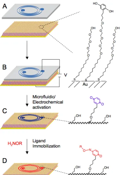
Figure 2.1 Synergistic strategy to selectively activate a SAM surface using microfluidic
networks and electrochemistry.……..……..……..……..……..……..……..………..……..……..…………46
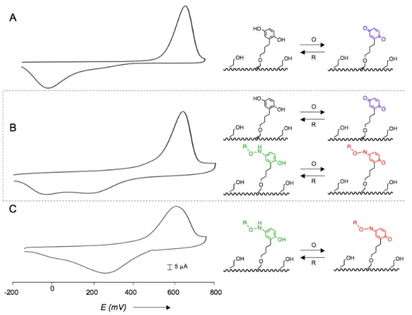
Figure 2.2 Electrochemical characterization of a 1:1 tetra(ethylene glycol):hydroquinone undecane thiol SAM surface undergoing microfluidic and electrochemical activation….……….49
Figure 2.3 A comparison of the original mask design for the microfluidic channel and the final ligand immobilization surface pattern to demonstrate mask fidelity…….………...………...50
Figure 2.4 A micrograph of Swiss albino 3T3 fibroblasts attached biospecifically to a
microfluidic/electrochemical activated surface presenting RGD immobilized ligands.…….…….51
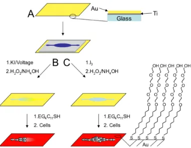
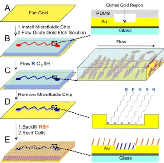
Figure 3.1 A scheme outlining the strategy to electrochemically and chemically generate gold/glass hybrid substrates for cell culture.….……….….……….….……….….……….….……….….…62
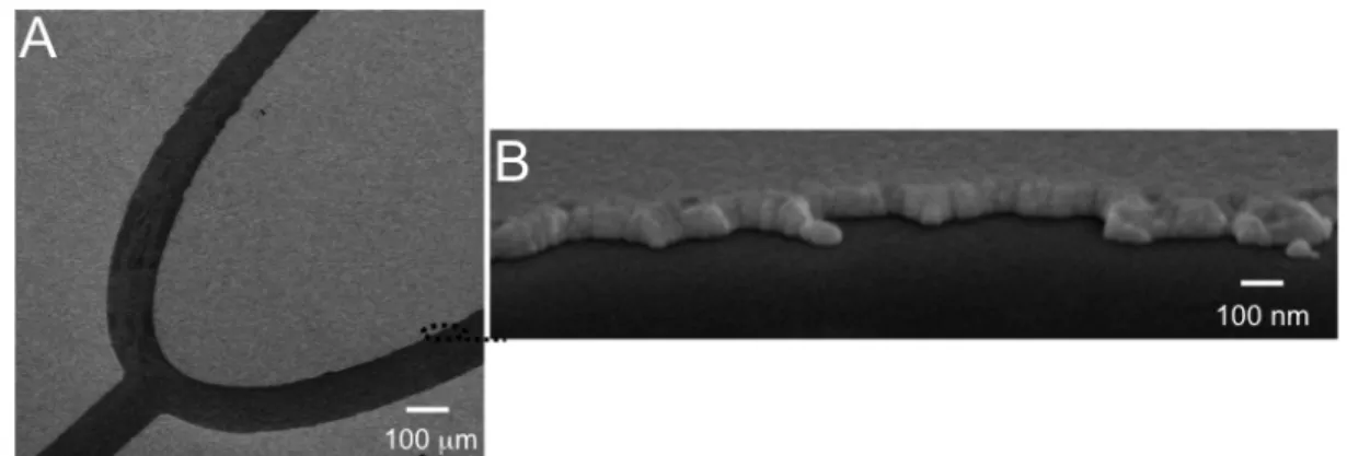
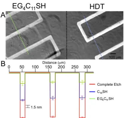
Figure 3.2 Etched gold surfaces imaged by scanning electron microscopy (SEM).….……….……64
Figure 3.3 Swiss Albino 3T3 mouse fibroblasts seeded on chemically etched gold
surfaces with subsequent installation of inert EG4C11SH SAMs …….….……….….……….….………64
Figure 3.4 The type of SAMs on gold influence the depth of chemical etch ….….……….….…….66
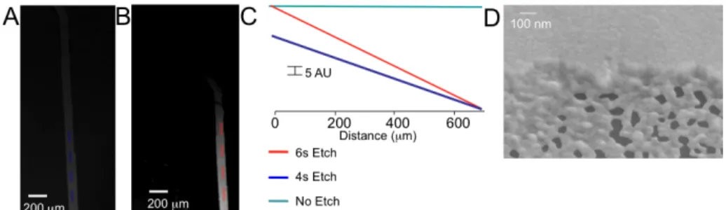
Figure 3.5 Images of partially etched and gradient etched gold surfaces. ….……….….……….….67
Figure 3.6 Controlled differential gradient slopes obtained with different
voltage application times. ….……….….……….….……….….……….….……….….……….….………68
Figure 3.7 A two-‐step process to generate partially
etched and patterned alkanethiol surfaces ………..….69
Figure 3.8 Cell attachment to microfluidic
generated partially etched and functionalized gold substrates …….….……….….……….….………70
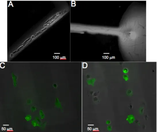
Figure 3.9 Cells seeded to an electrochemically etched glass/gold gradient surface. ….………..72
Figure 3.10 Electrochemical characterization of a chemoselective ligand
immobilization strategy on a patterned partially etched gold surface. ….……….….……….74
Figure 3.11 Multiwavelength time-‐lapse live-‐cell fluorescence microscopy of transfected Rat2 fibroblast cells undergoing directed migration on partially etched electroactive RGD presenting SAM gold surfaces. ….……….….……….….……….….……….….……….….……….….……..75
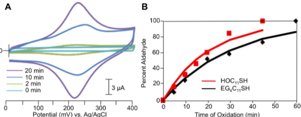
Figure 4.2 X-‐ray Photoelectron Spectroscopy (XPS) data of
ferrocene oxyamine immobilized to oxidized HOC11SH and EG4C11SH SAMs. ….……….….……..90
Figure 4.3 Characterization of aldehyde generation on EG4C11SH and HOC11SH surfaces. ….…92
Figure 4.4 Fluorescent micrographs of patterned ligands and cells from microfluidic oxidation of HOC11SH and EG4C11SH surfaces
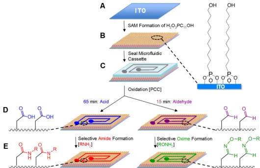
to generate aldehydes for chemoselective immobilization ………..92
Figure 4.5 . Schematic for the oxidative activation of H2O3PC11OH SAMs on ITO with controlled generation of aldehyde and carboxylic acid head-‐groups for subsequent
chemoselective ligation. ….……….….……….….……….….……….….……….….……….….……….….….94
Figure 4.6 Electrochemical characterization of ferrocene-‐oxyamine and dopamine
immobilized to aldehydes and acids generated on SAMs of H2O3PC11OH. ….……….….….….…..96
Figure 4.7 XPS characterization of oxime and amide bonds on ITO. Surfaces containing SAMs of H2O3PC11OH were oxidized with controlled generation of aldehyde or carboxylic acid head-‐ groups for subsequent chemoselective ligation, and XPS measurements were performed. …..97
Figure 4.8 Fluorescent micrographs of a mixed aldehyde and acid surface patterned by microfluidic oxidation followed by chemoselective oxime and amide immobilization.
Ligands were imaged directly on the surface. ….……….….……….….……….….……….….……….…98
Figure 4.9 Schematic for the oxidative activation of H2O3PC11OH SAMs on ITO for controlled generation of aldehyde, carboxylic acid, and a mixed surface of both aldehyde and acid head-‐groups for subsequent chemoselective ligation. ….……….….……….….………99
Figure 4.10 Fluorescent micrographs of an aldehyde and acid surface dually-‐patterned by microfluidic oxidation followed by chemoselective oxime and amide immobilization………….100
Figure 5.1 Outline of the affinity pulldown methodology. ….……….….……….….……….….…….109
Figure 5.2 Affinity pulldown of streptavidin. ….……….….……….….……….….……….….……….….110
Figure 5.3 Concanavilin A (ConA) affintiy pulldown. ….……….….……….….……….….……….……111
Figure 5.4 Affintiy pulldown of the FLAG antibody. ….……….….……….….……….….……….….…112
Figure 6.1 Schematic of the cell biochips and patterns used for evaluating cell behavior on
Dip-‐pen nanolithography (DPN) patterned peptide nanoarrays. ….……….….……….….…………123
Figure 6.2 Dip-‐pen nanolithography (DPN) pattern design and dimensions. ….……….….…….124
Figure 6.3 Schematic of the DPN methodology used to control cell polarity and cell division.125
Figure 6.4 Lateral force microscope (LFM) images of the patterns used to control cell
Figure 6.5 Cell images, polarity vectors, and division planes of cells adhered to the
corresponding nanopatterns. ….……….….……….….……….….……….….……….….……….….……..128
Figure 6.6 Cell images and polarity vectors of cells adhered to the corresponding
nanopatterns after cell division. ….……….….……….….……….….……….….……….….……….….…..130
Figure 6.7 Strategy to generate patterned hybrid nanohole self-‐assembled monolayer
surfaces for studies of cell adhesion and cell migration. ….……….….……….….……….….……….132
Figure 6.8 Environmental scanning electron microscopy (ESEM) of the hybrid nanohole
surfaces. ….……….….……….….……….….……….….……….….……….….……….….……….….…………132
Figure 6.9. Representative micrographs of fibroblast cells adhered to various hybrid
nanohole surfaces. ….……….….……….….……….….……….….……….….……….….……….….………..134
Figure 6.10. Comparison of cell migration rates on various surfaces. Swiss 3T3 fibroblast
migration on the nanohole surfaces. ….……….….……….….……….….……….….……….….…………135
Figure 7.1 Polymerization of the ketone containing hydrogels. ….……….….……….….……….…148
Figure 7.2 Flourescence Microscopy of functionalized Hydrogels. ….……….….……….….………149
Figure 7.3 Procedural Outline of Hydrogel Formation and Functionalization. ….……….….……150
Figure 7.4 3D cell culture in the hydrogels. ….……….….……….….……….….……….….……….….…151
Figure 7.5 Photopatterning hydrogels. ….……….….……….….……….….……….….……….….……..153
Figure 7.6 Confocal Microscopy of patterned cells within a dynamic hydrogel. ….……….….….154
List of Schemes and Tables
Scheme 4.1. Synthesis of 11-‐hydroxyundecylphosphonic acid. ….……….….……….….……….….88
Table 4.1. Contact angle data for the oxidation of alcohol to aldehyde for both HOC11SH and EG4C11SH SAMs. ….……….….……….….……….….……….….……….….……….….……….….………90
Table 4.2. Contact angle measurements of alcohol-‐, aldehyde-‐, and carboxylic
acid-‐terminated surface-‐groups on ITO. ….……….….……….….……….….……….….……….….……..90
Scheme 7.1. Synthesis of α-‐methacrylic -‐ω-‐acetoacetate poly (ethylene glycol). ….…………..146
Scheme 7.2 Synthesis of γ -‐3-‐(4,5-‐Dimethoxy-‐2-‐nitrophenyl)-‐2-‐butyl-‐l-‐aspartate. ….…………147
List of Symbols and Abbreviations
μm micrometer
AFM atomic force microscopy
CDCl3 deuterated chloroform
C16SH hexadecane thiol
d day(s)
Da dalton
DI deionized
DPN dip-‐pen nanolithography
ECM extracellular matrix
EG ethylene glycol
EG4C11SH tetra (ethylene glycol) undecane thiol
EtOH ethanol
h hours
HDT hexadecane thiol
HQEG4C11SH hydroquinone butyl tetra (ethylene glycol) undecane thiol
HQC11SH hydroquinone undecane thiol
mM millimolar
nm nanometer
PEG poly (ethylene glycol)
PDMS poly (dimethyl siloxane)
RGD arginine-‐glycine-‐aspartic acid
SAM self-‐assembled monolayer
SEM scanning electron microscopy
TEG tetra (ethylene glycol) undecane thiol
THF tetrahydrofuran
w/w weight/weight ratio
XPS x-‐ray photoelectron spectroscopy
PCC pyridinium chlorochromate
Chapter I: Literature Review of SAMs on Gold and Cell-‐ECM Interactions
1.1 Introduction
The majority of this thesis is concerned with smart surfaces that are generated with
self-‐assembled monolayers (SAMs) of alkanethiolates on gold. These surfaces respond to
external stimuli to switch from unreactive to reactive. To create these smart surfaces, I used
multiple molecule functionalities that respond to electrochemical, chemical, and light inputs.
These materials were used to study a variety of cell behaviors, such as cell polarity, division,
migration, and adhesion. In the following chapter, I will review the literature on the relevant cell
behaviors as well as SAMs.
1.2 The Interaction of Cells and the Extracellular Matrix
The extracellular matrix (ECM) is a complex mixture of proteins, signaling molecules,
and other soluble factors that provide a scaffold for cell adhesion and migration. The
constituent cells maintain the ECM, and fibroblasts in particular serve in this role. Fibroblasts
secrete proteins and enzymes that not only add to the matrix, but also destroy other proteins.
This constant remolding creates a dynamic environment wherein the cells are constantly
experiencing ligand conformational changes and orientations. In fact, these orientations affect
cell adhesion and migration rates. Typically, the ECM is composed of fibronectin, collagen,
structural regions.1-‐3 The first region is a condensed region and is adjacent to epithelial cells
with covering sheeting of muscle cells and the like. The second regions comprises the
interstitial matrix.4 Collagen can form many different structures depending on the structure of
the tissue and is typically synthesize by ECM caretaker cells, including fibroblasts and
osteoblasts.5 Laminin is another important component of the ECM and serves a major roll in
influencing cell behavior. Various laminins can promote stem cell differentiation, adhesion, and
migration.6-‐8 Additionally, these proteins appear to be crucial for cancer metastasis and viral
invasion.9 Other proteins also affect cell behavior, such as tenascins and proteoglycans.10-‐17 For
the ECM to dynamically remodel itself, it must first be broken down. This role is accomplished
by matrix metalloproteases (MMPs). MMPs are a subclass of metazinicin proteins that are zinc
dependent18-‐21 actively secreted by a host of cells, including fibroblasts and chondrocytes, and
involved in tissue remodeling during development, playing a large role in inflammation and
other disease states.
One of the most important ECM components is fibronectin, a proteoglycan. Fibronectin
exists in many different isoforms inside the body, including both an insoluble and soluble
form.22-‐24 The soluble form is found in blood plasma and plays a role in blood clotting and cell
adhesion. The insoluble form is primarily located in the ECM and is implicated in adhesion,
migration, and the maintenance of phenotype. The insoluble form is a multimer with a
molecular weight (MW) ranging from 200 to 250 kDa and a carbohydrate content of ~5%.
Fibronectin has ten domains that are composed of different repeating units, which are
recognized by a variety of cell types and proteins. Fibronectin’s capacity to bind many different
cell types and up to 20 different integrins mediates its influence on a wide variety of cell types
and tissues.25 Fibronectin is especially important for vertebrate development in which it is
be remodeled, unmasking specific domains makes it a dynamic protein capable of modulating
affinities and most likely, is the mechanism to modulate cell behavior.27 The two most
important cell-‐binding domains are the type III repeat found in the 10th domain (Arg-‐Gly-‐Asp
(RGD))and the type III repeat found in the 9th domain (Phe-‐His-‐Ser-‐Arg-‐Asn (PHSRN)).28 The
10th domain type III binds a host of integrins and mediates cell adhesion to the ECM, while the
9th domain type III repeat has been shown to act as a synergy peptide for integrin binding. For
some integrins, both domains are required for cell-‐ECM adhesion.29
Cells interact with constituent proteins of the ECM predominately through a set of
proteins called integrins. Integrins are heterodimeric proteins consisting of an alpha and beta
chain with one single membrane span.30,31 Twelve alpha and 24 beta chains combine to form up
to 24 known integrins that are found on all cell types.32 The majority of the proteins interact
with the external environment and are found outside the cell. Inside the cell, a small tail
projects into the cytoplasm, serving as an activation and attachment site for the actin
cytoskeleton.33 These attachments allow for the force to be transmitted across the plasma
membrane, which is crucial for cell migration and adhesion. In addition to serving as anchor
points to the ECM, integrins transmit information to the cell concerning the cell’s environment,
location, and adhesive state. These signals are integrated inside the cell and affect its adhesion,
migration, and death.34 Additionally, integrins can be activated internally through proteins,
such as talin, into a higher affinity state.35,36 Talin acts by binding the cytoplasmic tail of the
integrin, and induces a conformational change in the extracellular head portion.37 The
conformational change raises the affinity of the integrin towards various extracellular matrix
proteins. Proteins can also deactivate integrins. Typically, proteins such as Dok1 and ICAP1
competitively bind the cytoplasmic tail to displace talin, lowering the integrin affinity.38,39 As
information to the cell, integrins serve as conduits for information for the cell that allow it to
not only sense its environment, but also respond to it.
When integrins bind their environment, a protein cascade is initiated that leads to the
formation of focal adhesions.40-‐45 Currently, several subsets of focal adhesions exist. Focal
complexes are found at the periphery of a migrating cell.46 Focal adhesions are larger protein
complexes found at the center of cells and at the end of stress fibers in strongly adhered cells.47
Fibrillar adhesions are a subset of elongated focal adhesions containing tenesin.48 3D matrix
adhesions are attachments that have been described for fibroblasts adhered to various 3D
substrates.49-‐52 Various proteins that constitute focal adhesions have multiple interacting
proteins, which allow the cell to construct various focal adhesion types. Currently, it is not
known how matrix composition affects the protein make up of the focal adhesions. Two
important kinases found at focal adhesions are focal adhesion kinase (FAK) and Src. Each of
these proteins activates different proteins and modulates focal adhesion composition and
activity. FAK can bind the integrin tail directly to proteins, such as p130CAS and paxillin.53,54
Both of these proteins are phosphorylated upon binding, and even though FAK is an important
kinase, it also serves as an important scaffold protein for focal adhesions.55-‐57 In addition, FAK
acts as GTPase activator.58 Src also acts as a kinase.59,60 It activates upon binding to FAK and can
also bind the integrin cytoplasmic tail. Src is believed to help regulate focal adhesion turnover.61
Another protein that regulates focal adhesion turnover is PTP-‐TEST.62 This protein acts as a
dephosphorylating agent and helps regulate focal adhesion activity.63 Other important proteins
are p130CAS and paxillin.64 Both act as a scaffold proteins with many binding partners and
partner with other proteins to help regulate focal adhesion function and behavior in the cell.
Vinculin is another important protein66 and sequesters the Arp2/3 protein to the focal adhesions
to promote localized actin branching.67
Integrin-‐cell surface binding is not sufficient for integrin clustering, which leads to focal
adhesion formation.68 Instead, this process must be initiated through intracellular events.
Proteins Rho and R-‐Ras have been shown to mediate this process and thereby, control the
integrin activity.69,70 Initially, when a focal adhesion is formed at the leading edge of cells, it is
composed of mostly paxillin and al-‐actinin, and as the adhesion matures other proteins such as
vinculin, FAK, Src, and talin bind to the site.71,72 Next, zyxin and tensinin are recruited to act as
stabilize the adhesion,73 and finally in order for the cell to effectively migrate, focal adhesions
need to be turned over. It is believed that this process is mediated by FAK, Src, and Caplain,74-‐77
that help increase focal adhesion turnover rate in the cell when they are present. Another
possible regulating process of integrin function is binding forces. It is believed this process is
regulated through force-‐sensitive proteins, such as FAK, Src, and Talin.77-‐79
Rho and Rac are as the master control proteins of cell migration and adhesion. Theses
proteins are GTPases that regulate the formation of stress fibers, focal adhesions, and
membrane ruffling.80,81 Rho specifically modulates contractility, stress formation fibers, and
focal adhesion formation.82,83 Their activity is regulated by Rho kinases that phosphorylate the
protein. Rho upregulates MAPK kinase and myosin light chain phosphorylation to increase
contractility.84,85 Rho also downregulates cofilin, which slows actin polymerization.86 The sum
of these and other actions leads to the formation of stress fibers. Rho significantly upregulates
focal adhesion formation by inducing integrin clustering in fibroblasts.87 This effect is also
observed with Rac; focal complexes of high-‐affinity integrin complexes are formed. However,
Rac’s major function is the cell is to regulate membrane protrusions and ruffling, and its
Rac activates PI 5-‐kinase, Arp 2/3 complex, and PAK,89 all of which remove actin-‐capping
proteins and increase actin polymerization. Since Rho and Rac are GTPases, they are controlled
by both GAPs and GEFs that are specific to each protein.90,91 These controls are governed by
growth factors and other cell surface receptors.
A number of different cell surface receptor-‐ligand interactions have been investigated
to study the dynamic processes of cell adhesion and migration. Simple methods have used
ECM-‐derived protein fragments. These ligands are limited by the inability to control their
orientation and the difficulty of either generating or synthesizing such ligands. The discovery of
the minimal adhesion sequence in fibronectin, RGD, represents a more appealing alternative.92
Peptides can easily be readily synthesized or commercially purchased without the need to
modify proteins. Another peptide sequence that has been found to affect cell adhesion, PHSRN,
acts as a synergy peptide with RGD to enhances cell migration rate.93 These two sequences are
located in fibronectin, and their orientation has been shown to affect cell adhesion to its surface.
When introduced to various materials, these two peptide sequences have been shown to
promote cell adhesion and allow for the generation of materials for biological studies.94
Many factors determine not only how the ECM will respond to cell adhesion, but also
how the composition and orientation influence cell behavior. To isolate the myriad variables,
the ECM has been simplified with a number of different model systems. Polymers are one of
the most popular systems and allow for the flexibility in chemical composition, structure, and
various physical properties to modulate diverse aspects of the ECM.95 Layer-‐by-‐layer
depositions have been employed to create model systems simply.96 In general, the layers must
alternate electric charge or hydrophobicity and are not as compositionally variable as polymer
materials. In contrast, self-‐assembled monolayers are synthetically well defined, and by nature
with indium tin oxide (ITO), iron oxide, and glass and siloxanes coupled with glass, ITO, and iron
oxide. Perhaps the most well studied and most often used SAM systems are thiol SAMs on
noble metals.98 SAMs have been generated from silver, gold, and even nickel. However, the
most common of these metals is gold.
1.3 SAM Structure and Function
SAMs of alkanethiolates on gold are the most well studied system for a number of
reasons. Gold is easy to obtain in a variety of forms, including solids and colloids. Thin films can
be prepared using different deposition techniques, such as physical vapor deposition,
sputtering, and electrodeposition. Crystalline substrates are easily obtained with few defects
other relatively large areas. Single crystals are also available for spectroscopic studies. Gold is
also relatively easy to pattern. Photolithography, etching, and micromachining have been used
to pattern gold substrates. Gold is also one of the most stable metals under room temperature
and ambient air conditions and does not oxidize as easily as other noble metals. Thus, gold
does not have to be handled under UHV conditions. Thiols bind gold strongly and do not form
other side products. This high affinity allows the thiols to not only bind gold, but also to displace
other material adhered to the surface. Additionally, thin gold films are commonly used in
several spectroscopies such as surface plasmon resonance (SPR), reflection adsorption infrared
spectroscopy (RAIRS), and elipsometry making the transition to studying SAMs with these
techniques easier. Gold is inert to biological systems and does not poison cells like other metals.
Additionally, SAMs composed of olgio(ethylene glycol) form resistant surfaces to non-‐specific
protein and cell adsorption and adhesion. Other common metals used for thiol SAMs are silver
and nickel; however, these metals are not as well studied and are not compatible with cell
SAM Substrates can be formed from different systems. One of the most popular
methods has been thin-‐layer deposition on flat substrates, such as glass, silicon, quartz, and
mica. The gold is deposited by physical vapor deposition in two layers.99-‐102 A ~10-‐200 nm
chrome or titanium layer is first deposited on the substrate because gold does not directly
adhere to many typical flat substrates. Next, a 100-‐1000 nm gold layer is deposited. The metal
layers are polycrystalline in nature and can form grains and islands. These films have dominant
<111> texture at the exposed surface.103-‐106 Metal that is deposited on quartz and mica have a
stronger crystal structure than glass.107 Mica surfaces can provide grain structure of 1000 nm
with flat <111> terraces. Quartz can also provide structurally uniform gold surfaces by thermal
annealing. By starting with substrates of other dominant crystal structure such as <100>,
substrates have been fabricated with other crystal structures.108,109 Additionally, other
deposition methods, including underpotential and electroless, have been used to create
surfaces with alternate crystal structures.110
Many factors affect grain structure and size of the deposited surfaces. By raising the
temperature from 0 °C to 400 °C, the average gold grain size can be increased from 200 to 106
nm2. 111 Additionally, the angle of deposition and deposition rate can affect the grain size and
shape of the surface; the faster the deposition, the larger the grain size.112 To modify the grain
size after deposition, thermal annealing of the substrates has been used to enhance the overall
<111> structure of the surface.113,11 4
Film thickness is another important parameter in film deposition. Thin, translucent
films are useful for imaging applications, which make them desirable for biological studies.
However, various characteristics of these films must be controlled. For thinner (>15 nm) gold
layers, the adhesion layer can migrate to the surface.115 This process can be problematic for
may be induced. Thus, titanium is typically the adhesion layer used for biological substrates for
this reason. Additionally, thicker substrates are useful for spectroscopic and electrochemical
applications.
Even though planar SAM systems are well studied and characterized, SAMs can be
formed on other metal supports. Metal features have been machined using photolithography,
molding, or photolithography.1156,117 However, the substrates’ features introduce structural
defects into the metal substrate and therefore, the SAMs.118 Perhaps the second most popular
substrates after planar supports are colloid gold nanoparticles.119,120 These nanoparticles can be
synthesized easily utilizing the Brust synthesis and are spectroscopically well studied.121 SAMs
on colloid gold nanoparticles have been used for a variety of applications including
hypothermal cancer treatments and MRI contrast imaging agents. 122
Once the substrate has been fabricated, the SAM can be formed in a variety of ways.
The most common method is solution deposition.123 Gas phase adsorption is performed as well
but suffers from a lack of generality and does not produce a SAM with same level of packing as
solution phase adsorption. Typically, substrates are immersed in 1 mM ethanolic thiolate
solutions for 12-‐18 h. The SAMs are formed in two mechanistic steps. The thiolate molecule
adsorbs to the surface following a Langmuir isotherm. The adsorption takes place on the order
of milliseconds to seconds, followed by slow reorganization of the thiolate molecules into the
lowest energy configuration; this step occurs over hours. The reorganization maximizes the
thiolate packing density and minimizes the SAM defects. This final structure can be affected by
a number of factors, including solvent, temperature, immersion time, substrate cleanliness, and
alkane chain length.
Solvent effects SAM formation; however, how it effects SAM formation is not
toxicity.123 Generally, the choice of solvent can affect both the SAM formation rate and defect
quantity vs. ethanol. Generally, more polar solvents such as water form SAMs with fewer
defects than ethanol, which is most likely due to low alkanethiolate solubility.124-‐126 Also,
solvents like heptane and hexane have been shown to increase SAM formation rates.127,128
Increasing the temperature from 25 °C has been shown to increase adsorption kinetics, lower
the defect rate, and help remove adventitious materials that are bound to the gold
substrate.129,130 The higher temperature increases the crossing of certain activation barriers in
SAM formation, which has been shown to be most relevant to the first few minutes of
formation. As expected, concentration and immersion time are inversely related with lower
concentrations requiring longer immersion times.131 RAIRS studies have shown that the overall
structure of SAMs does not change after 18 h.123 However, electrochemistry and STM have
shown that SAMs do show a decrease in both pinhole and alkane conformation defects over the
order of 7-‐10 days. 132
Thiol solutions can contain common purities that affect SAM formation. Generally, it
has been found that solutions with <5% impurities can form SAMs.133 However, if the impurities
are less soluble than the alkanethiols, it can lead to physisorption of the material, which can
affect SAM formation. The cleanliness of the substrate is also important. Materials that are
physisorbed to the surface impede SAM adsorption and formation and can introduce defects to
the SAM surface. However, due to the strength of the thiol gold bond, most of the adventitious
materials can be competitively desorbed from the surface. This process slows down SAM
formation and some materials cannot be removed in the 12-‐18 h timeframe allowed for SAM
formation.123
Mixed SAMs have been used to install multiple functionalities on the surface. Typically
can be done after SAM formation. Varying ratios of two different thiols or asymmetric
disulfides in solution can be used to create SAMs with mixed functionalities. These SAMs are
especially useful for biology where only a small percentage of ligand is needed on the surface
for biorecognition and the rest can be another background alkanethiol. If solutions of
alkanethiols are used to create these SAMs, their molar ratio reflects but does not always equal
the surface ratio.134,135 Many factors influence the overall distribution including solvent,
alkanethiol structure, and adsorption time. Asymmetric disulfides have also been used with
some limitations.136 Most notably, the ratio is restricted to 1:1. However, over time the SAMs
will adopt the more energetically favorable state and may deviate from the initial 1:1 ratio.
Disulfides are also less soluble than their thiol counterparts and form SAMs with a greater
defect rate.137
The gold-‐thiol bond has been well studied both thermodynamically and
spectroscopically. To determine the gold-‐thiol binding energy, Dubois used temperature
programming to desorb thiols from gold and determined that it occurs dissociatively in the gas
phase. The barrier to recombinative dissociation is roughly 30 kcal/mol, which corresponds to
theoretical calculations.138,139 This value corresponds to a high degree of charge transfer from
the thiol to the gold. Scoles and coworkers verified these numbers by using different
experimental protocols.140 For liquid phase desorption, the barrier appears to be lower. SAMs
were desorbed while immersed in a solvent and it was found the barrier was approximately 20
kcal/mol.141 By using these numbers, combined with the segmental heat of interaction, heat of
dissolution, and heat immersion, the strength of the gold-‐thiol bond is believed to ~50
kcal/mol.138 One ambiguity remaining in the reaction is the final form of the hydrogen after
reductive elimination is weakly activated by gold. In solution, hydrogen could also be converted
to water with gold serving as the catalyst.142,143
Once the gold thiol bond has formed on the surface, the SAMs can adopt many
different configurations. The most accepted and common form the SAM adopts is the
√(3x3)R30° overlayer with the alkane chains adopting a c(4x2) superlattice.144-‐148 These
structures are not only driven by the gold-‐thiol bond formation, but van der Waals interactions
between alkane chains in the overlayer. Both the nearest neighbor distances and geometric
arrangement affect the structure of the alkane overlayer of SAMs.149 The interthiol distance
constrains ability of alkane chains to pack closely to maximize the van der Waals interactions.
For these reasons, the alkane chains are tilted 30° to the surface normal of the substrate to
maximize the van der Waals interaction between the chains. Each of the thiols is individually
bonded to the gold; neighboring thiols do not interact with each other. A majority of the SAM
structure studies are performed on substrates with dominant <111> structure. The few studies
with substrates of different crystallographic directions have shown the effect of the underlying
substrate on the final SAM structure. For <100> gold surfaces, the alkane chains adopted a
c(2x2) structure as opposed to the c(4x2) structure.150 For alkanethiolates comprising of
different alkane structures such as biphenyl and terpphenyl thiols, the overall SAM structure is
constrained by the steric hindrance of the chains. The steric hindrance can reduce the angle the
chains take to the surface normal.151,152 However, the underlying thiol structure appears to be
unaffected. Another factor in SAM structure is the odd-‐even affect for straight chain
alkanethiolates.153,154 Due to constant 30° of tilt found in these SAMs, it has been observed that
the even numbered alkanethiolates have a slightly lower free energy than their odd number
counterparts.155 Another factor affect SAM stability is the overall chain length, alkanes longer
Several factors affect the mechanism of SAM formation. An interplay exists between
the formation of the covalent gold-‐thiol bonds, hydrogen bonding, and the van der Waals
interaction between chains. The gold surface structure dictates available reaction sites for the
thiol to bind. These sites determine the structure of the thiol layer while the van der Waals and
hydrogen bonding of the alkane chains add an additional 1 kcal/mol of stabilization per
methylene unit.157 For gas phase reactions, several low coverage phases proceed the fully
formed SAM.141 The alkanethiols lie flat on the gold surface and as other alkanethiols pack onto
the substrates, and these alkanethiols eventually form a well packed SAM.143 The conversion
from the striped phase constitutes the slow step in SAM formation. In solution, the complexity
of the environment has stymied efforts to study the mechanism of assembly.143,158 Qualitatively,
the SAM formation follows a Langmuir isotherm with fast adsorption of thiols on solution and a
slow packing and order step.143 It is believed both gas and solution phases roughly follow the
same mechanism. However, this has not been demonstrated effectively.
Defects in SAMs can come from a number of sources including the surface cleanliness,
impurities, and substrate preparation. However, another major source of defects are the
dynamic nature of SAMs. In solutions, they are not simply the well-‐ordered substrates of theory.
The substrate of choice, polycrystalline gold, has many defects from intergrain boundaries,
faceting, and a varying density of atomic steps. These surface defects impact SAM formation
and can introduce structural defects into the SAM.140,159,160 Other types of defects are
introduced during the SAM induced rearrangement of the gold surface atoms.143,161 A clean gold
surface has a higher density of reactions sites than can be filled by available thiols. As the thiols
bind the gold, single atom vacancies form in the gold structure and cannot be concealed by the
SAM itself. Impurities, if they have a stronger affinity for the gold surface, can impact SAM
once the SAM is removed from its solution. By its nature, SAM formation is a thermodynamic
process in equilibrium between well-‐formed SAM and single alkanethiolates.162 Once the SAM
is removed from solution and used for other studies, the thiols start desorbing from the surface,
which can introduce defects.
Once SAMs are formed, they can be removed a using a number of different methods.
First, by applying a negative potential to the surface, the SAMs can be reductively desorbed
from the surface.163,164 The thiol leaves the surface negatively charged while the metal goes
from positively charged to neutral. The process is reversible. Once the potential is removed,
thiolates in solution can reabsorb to the surface. Another method to remove a SAM is through
a displacement exchange.165 By removing the SAM from its original alkanethiolate solution and
placing it in a new solution, the new alkanethiols will displace the original ones from the surface
over time. The rate typically depends on the inter chain attraction of the new alkanethiolate vs
the original one and the amount of defects found within the original SAM.166 The defects
present serve as nucleation point for new SAM formation and greatly speed exchange.167 SAMs
can also be photooxidized with UV light.168 When exposed, the thiols are oxidized to sulfonates,
which can be washed away with a polar solvent.169
1.4 SAMs for Biology
For biological applications, the most important alkanethiolates are terminated in olgio
(ethylene glycols).170 SAMs composed from these alkanethiolates resist non-‐specific adsorption
and adhesion of both proteins and cells. This feature makes SAMs a unique platform for biology.
Rather than studying whole proteins on ill-‐defined surfaces, specific ligands can be presented
against an inert background.171 SAMs of these molecules pack normally, however, the ethylene
glycol chains adopt a number of different conformations that provide the resistance to protein
amorphous phase leads to a liquid like phase on the surface. Above room temperature, other
phases such as an all trans conformer are visible with NEFAXS. It is believed these structures
resist proteins and cell adsorption for two reasons. These structures adsorb a significant
amount of water on the surface, which in turn must be removed before a protein can adsorb to
the surface. The water removal generates an enthalpic penalty for binding on the surface.
Second, protein bind restricts the possible conformers available to the ethylene glycol chains
and providing an entropic penalty to protein and cell binding. The combination of these two
penalties provides an energy barrier to protein binding that allows these SAMs to resist protein
binding for a finite time (~1-‐2 weeks).173,174
Several factors influence the ability of the SAMs resistance to non-‐specific binding of
cells and proteins. The substrate cleanliness and age have a strong effect. The substrate must
be clean and as free from defects as possible. The defects introduced from a poorly prepared or
poorly cleaned SAM generate binding sites for protein adsorption to the surface. If the
substrate is not freshly prepared, the adventitious materials and oxidation that take place on
the gold surface can introduce defects as well. Additionally, the ethylene glycol purity is of
utmost importance. For the SAM to resist protein and cell binding the most strongly, the thiol
must be >99% pure. If not, the impurities can introduce surface defect sites. In the same vein,
the solution must be freshly prepared, so no disulfides or other impurities are present in the
solution. The ethylene glycol chain length is important as well. For the SAMs to resist protein
and cell binding, the chain length has to exceed 3 units.175Another factor is formation time. The
SAMs must be allowed to form properly over an 18h time to minimize defects.176 For mixed
SAMs, ethylene glycol portion must exceed 95% of the surface density. Below this number,