Systemic
multilineage
engraftment
in
mice
after
in
utero
transplantation
with
human
hematopoietic
stem
cells
RussellG.Witt,1,2EmilyM.Kreger,1,2LauraB.Buckman,3PatrissW.Moradi,1PhongT.Ho,3S.ChristopherDerderian,1,2PerryTsai,3 ChrisBaker,1,2NathanielSchramm,3RachelCleary,3J.VictorGarcia,3andTippiC.MacKenzie1,2
1
EliandEdytheBroadCenterofRegenerationMedicine,SanFrancisco,CA;2DepartmentofSurgery,UniversityofCalifornia,SanFrancisco,SanFrancisco,CA;and3Division ofInfectiousDiseases,CenterforAIDSResearch,SchoolofMedicine,UniversityofNorthCarolinaatChapelHill,ChapelHill,NC
Key Points
•IUHCT of human cord blood–derived CD341 cells into fetal NSG mice results in systemic multilineage engraft-ment with human cells.
•Preconditioning with in utero injection of an c-Kit receptor anti-body (ACK2) results in an improved rate of engraftment.
Introduction
In utero hematopoietic cell transplantation (IUHCT) is a potential therapy for the treatment of numerous genetic diseases such as hemoglobinopathies, immunodeficiencies, and inborn errors of metabolism.1In utero therapy offers the benefit of avoiding host myeloablation and immunosuppression and has been shown to be successful in multiple animal models, including mice,2-5dogs,6,7pigs,8,9and sheep.10-12 The timing of IUHCT exposes the transplanted human cells to the normal fetal migratory and developmental cues that facilitate proper stem cell distribution and differentiation.11,12Clinically, IUHCT has been successful for fetuses with severe combined immunodeficiency (SCID),13,14but therapeutic uses for other diseases, including hemoglobinopathies, have seen limited success.15 Further investigations identified multiple barriers to successful engraftment, including lack of space within the hematopoietic niche16,17 and the maternal immune system.2,18 Among available animal models of IUHCT, the fetal mouse remains an efficient and reproducible model to study the differentiation of stem cells in a nonirradiated host. NSG (NOD-SCID IL2Rg-null) mice, which are developed with SCID and IL-2Rg-null chain mutations, are a robust platform for the engraftment of human hematopoietic cells because they have no endogenous T, B, or natural killer cells.19-22In this study, we used IUHCT of human CD341cells in NSG mice to create a reproducible mouse model to study stem cell engraftment, differentiation, and systemic repopulation after IUHCT.
Methods
Mice
NSG mice were obtained from The Jackson Laboratory. All procedures were performed according to a University of California, San Francisco Institutional Animal Care and Use Committee–approved protocol.
Study design
Human cord blood CD341cells were either purchased from AllCells (Alameda, CA) or purified from cord blood collected at time of delivery from normal-term infants. CD341 cells were isolated and transplanted into fetal mice at embryonic day 13.5 (E13.5) or E14.5 as previously described23with or without preconditioning with ACK2.17Chimerism levels were checked at 4-week intervals and at harvest using flow cytometry and immunohistochemistry.
Please see supplemental Methods for a more detailed description of the methods used.
Results and discussion
Stable, multilineage chimerism in humanized mice after in utero transplantation of
human cord blood CD341cells
is lower than we previously reported in wild-type mice24and likely represents the fragility of the immunodeficient mouse model compared with BALB/c or B6 dams. Overall, 25 of 49 (51%) mice were found to be engrafted with human hematopoietic cells. The
A
25k – 50k Human CB derived CD34+ cells
Fetal Injection
E 13.5 Postnatal
Harvest 16 - 24 weeks
Chimeric Mice
4 week interval blood chimerism checks
x NSG NSG
Uninjected Control
C
% human C
D4
5
+ cells
Weeks
4 20
10
0
8 12 Harvest
*
B
Mouse C D4 5.1 +Human CD45+
Mouse C
D4
5.1
+
Human CD45+ Chimera
D
CD 8 + CD4+CD8+ T Cells
CD4+ T Cells
FoxP3
+
CD25+
Gated on CD4+ cells
SS
C-A
CD34+
Gated on hCD45+ cells
HSCs
SS
C-A
CD33+
Gated on hCD45+ cells
Lymphocytes Monocytes Granulocytes CD 19 +
CD3+ T Cells
B Cells
E
Granulocytes CD33+ Monocytes CD19+ B cells CD3+CD8+ T cells CD3+CD4+ T cells
Spleen Mouse number 1 0 20 40 60 80 100 9 10 3
2 4 5 6 7 8
1 2 3 4 5 6 7 8 9 10
Bone Marrow Mouse number 0 20 40 60 80 100 Peripheral Blood Mouse number
% human C
D4
5
+ cells
1 0 20 40 60 80 100 9 10 3
2 4 5 6 7 8
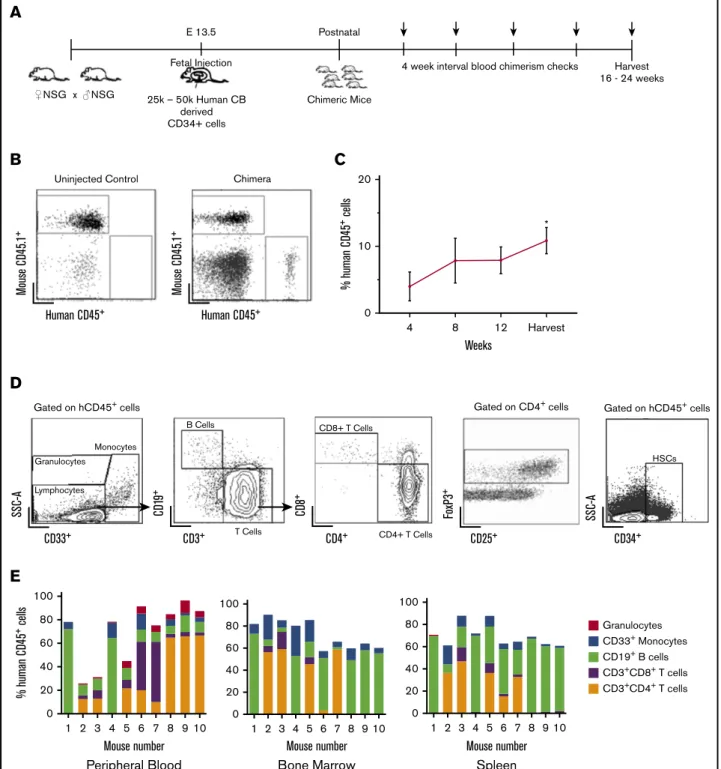
Figure 1.Stable, multilineage chimerism in humanized mice after in utero transplantation of human cord blood CD341cells.(A) Experimental design for in utero transplantation timing and measurements of blood chimerism levels. Arrows indicate time points for peripheral blood chimerism checks. (B) Representative gating strategy of
peripheral blood to detect human CD451cells. (C) Percentage of human CD451cells in peripheral blood over time (n525, *P,.05, **P,.01 by Studentttest comparing chimerism levels each week to the initial 4-week level). (D) Representative gating strategy for lineage-specific chimerism, FoxP31cells, and confirmation of the presence of CD341cells within the bone marrow. Chimeric mice demonstrated different relative proportions of granulocytes, monocytes, B cells, and T cells. (E) Compiled lineage data in 10 individual mice with evidence of CD341cells in the bone marrow (n510). CB, cord blood; SSC-A, side scatter.
B
A
0 50 100
****
**** ***
*
****
% human C
D4
5
+ cells
LN Spleen BM
PB
H
Brain
G
V
agina
F
Colon
Duodenum
E
Lung
CD45 CD3 CD4 CD8 CD68 CD20
0 20 40 60 80
IUHCT ACK2
% human C
D4
5
+ cells
D
C
R
ate of engraftment
IUHCT ACK2
* 100
80
60
40
20
0
levels of human cells in peripheral blood (chimerism) were de-termined by flow cytometry every 4 weeks for 16 to 24 weeks (Figure 1A-B). Peripheral blood chimerism increased over time in 23 of 25 mice from the initial analysis at the time of 4 weeks after birth to the time of harvest (Figure 1C).
Flow cytometry of bone marrow, peripheral blood, and spleen demonstrated the presence of different hematopoietic lineages in individual litters (Figure 1D). The transplanted stem cells gave rise to both human B cells (CD191) and T cells (both CD4 and CD8). Foxp31 regulatory T cells were also detected in chimeric mice, particularly in the bone marrow (Figure 1D). Additionally, we established the presence of human CD341 cells in the bone marrow of chimeric animals (Figure 1D). Compiled lineage data from 10 individual mice with bone marrow CD341cell engraftment demonstrate the range of human lineage distribution in this model (Figure 1E). We looked for the presence of human red blood cells in our mice but did not detect any. This finding is consistent with other reports in humanized mice, and the reason is thought to be secondary to destruction of human red blood cells by host murine macrophages and murine complement.25,26
Circulating levels of human cells were lower than those seen in the lymphoid organs of chimeric animals (Figure 2A). The engraftment results determined by flow cytometry were supported by histolog-ical analysis. In the spleens, we detected T cells in organized structures similar to lymphoid follicles and a smaller portion of B cells in structures similar to germinal centers (Figure 2B). We found higher percentage of T-cell engraftment than typically noted in adult transplanted NSG mice, which may reflect homeostatic proliferation of T cells, as our CD341 human samples were not completely pure and did contain a percentage of human T cells (3% to 17%). Earlier transplantation of human cells into NSG mice results in higher percentage of T cells when compared with adult transplantation,27 which may be taking place within our model. Although chronic graft-versus-host disease (GVHD) has been implicated in high T-cell numbers in NSG mice that receive human CD341cells,28our mice showed no physical evidence of GVHD at 24 weeks. Our mice also had B cell percentages as high as 50% to 65%, which is further evidence of true engraftment rather than chronic GVHD. Our experience with in utero injections have also demonstrated that the fetal host is remarkably resistant to GVHD.29
Fetal host conditioning increases engraftment rate
Although the in utero transplantation model allows transplantation of allogeneic or xenogeneic cells without host conditioning, the levels of engraftment could be improved by using a nontoxic strategy to increase space in the hematopoietic niche. One such strategy is in utero treatment with ACK2, a monoclonal antibody that depletes hematopoietic stem cells (HSCs) by blocking the function of the murine c-Kit receptor.16We have previously demonstrated
that this antibody can be used in the fetal transplantation setting to improve engraftment of mouse fetal liver–derived HSCs.17Because murine ACK2 does not inhibit human HSC survival, we next coinjected 2.5 mg/pup of ACK2 along with human cord blood– derived CD341cells at E14.5. Based on our previous work, human HSCs are only inhibited at doses 103 this dose, and this is the optimum dose for murine fetal HSC depletion.17Survival of injected pups was 33% (16/49) to birth and 24% (12/49) survival to wean, with no significant difference in the survival outcomes compared with injection of cells alone. Ten of the 12 surviving pups were chimeric (83%), representing a significant increase in the rates of engraftment compared with no ACK2 treatment (P,.05) (Figure 2C). The overall levels of chimerism were not significantly different (Figure 2D).
We did not observe the significant human myeloid cell engraftment seen in other models of NSG mice that use irradiation. We believe this may be secondary to the less space created in the niche with ACK2 relative to irradiation. Additionally, stem cell factor expression is increased with irradiation. In a transgenic NSG model that expressed membrane-bound human stem cell factor, human myeloid cell engraftment is increased in both irradiated and nonirradiated hosts.27,30 Our levels of granulomonocytic hematopoiesis are comparable to those found in models of NSG mice in which there is no irradiation for preconditioning.27 However, because ACK2 increases the rate of chimerism, this strategy can be used as an adjunct to improve the efficiency of in utero transplantation.
Engraftment of human lymphocytes in critical mucosal sites after in utero transplantation
Given the importance of hematopoietic reconstitution of mucosal surfaces for the study of human disease, we assessed for human cells in respiratory, reproductive, and gastrointestinal mucosa using immunohistochemistry and flow cytometry. This analysis revealed a relatively high frequency of human cells in all examined mucosal sites. Local tissue environment appeared to guide HSC differentiation, as we found different ratios of human leukocytes in different tissues. In the lungs, the majority of human (CD451) hematopoietic cells were hCD31, hCD41and hCD81T lymphocytes (Figure 2E), with fewer macrophages. In the gastrointestinal tract, we observed human CD451cells in both the small and large intestine (Figure 2F). Donor-derived CD451 human cells were also observed throughout the tissues of the female reproductive tract (including the vagina, cervix, and uterus) (Figures 1 and 2G). We did not observe any significant GVHD in transplanted mice (supplemental Figure 2).
Engraftment of human lymphocytes in the brain after in utero transplantation
The ability to transplant human cells prior to formation of the blood–brain barrier, which occurs at;E15.5 in mice,31may present an opportunity to engraft human cells within the brain. We observed
human hematopoietic cells (CD451) in the brainstem and cerebral cortex of chimeric mice; serial sectioning revealed these cells to be T cells (hCD31, hCD41, and hCD81) and hCD681macrophages (Figure 2H). The presence of human cells in the brain raises the possibility of using in utero transplantation to deliver cells into the brain for the treatment of neurological disorders.
In this study, we have demonstrated efficient systemic engraftment of human hematopoietic stem cells in NSG mice after in utero trans-plantation without the need for host irradiation. In future experiments, it will be important to determine whether repeated transplantation of human CD341further boosts engraftment and to investigate the ability of the stem cells in these mice to repopulate secondary recipients. This model will be useful for analysis of human stem cell plasticity, in vivo repopulation, and tissue-specific immunity during infection.
Acknowledgments
The authors thank the members of the T.C.M., T. D. Burt, and J.V.G. labs for helpful discussions and technical assistance with different aspects of this work.
This work was supported by the Eli and Edythe Broad Stem Cell Fellowship Grant TG2-01153 (R.G.W.), California
Institute of Regenerative Medicine New Physician Scientist Translational Research Award RN3-06532 (T.C.M), and Na-tional Institutes of Health (NIH), NaNa-tional Institute of Allergy and Infectious Diseases grants AI111899, AI073146, AI111899, and National Institute of Mental Health grant MH108179 (all J.V. G.). L.B.B. was supported by NIH, National Institute of Allergy and Infectious Diseases grant T32AI007151.
Authorship
Contribution: T.C.M., S.C.D., R.G.W., and E.M.K. designed the experiments; R.G.W., S.C.D., C.B., and P.W.M. performed the in utero injections, tissue harvesting, and flow cytometry; L.B.B., R.C., P.T.H., P.T., N.S., and J.V.C. performed immunohistochem-istry of harvested tissues and data analysis; and R.G.W. wrote the manuscript with assistance from L.B.B., J.V.G., and T.C.M.
Conflict-of-interest disclosure: The authors declare no compet-ing financial interests.
Correspondence: Tippi C. MacKenzie, 35 Medical Center Way, Box 0665, Room 903D, San Francisco, CA 94143-0665; e-mail: [email protected].
References
1. Nijagal A, Flake AW, MacKenzie TC. In utero hematopoietic cell transplantation for the treatment of congenital anomalies.Clin Perinatol. 2012;39(2): 301-310.
2. Nijagal A, Wegorzewska M, Jarvis E, Le T, Tang Q, MacKenzie TC. Maternal T cells limit engraftment after in utero hematopoietic cell transplantation in mice.J Clin Invest. 2011;121(2):582-592.
3. Pallavicini MG, Flake AW, Madden D, et al. Hemopoietic chimerism in rodents transplanted in utero with fetal human hemopoietic cells.Transplant Proc. 1992;24(2):542-543.
4. Durkin ET, Jones KA, Rajesh D, Shaaban AF. Early chimerism threshold predicts sustained engraftment and NK-cell tolerance in prenatal allogeneic chimeras.Blood. 2008;112(13):5245-5253.
5. Carrier E, Lee TH, Busch MP, Cowan MJ. Induction of tolerance in nondefective mice after in utero transplantation of major histocompatibility complex-mismatched fetal hematopoietic stem cells.Blood. 1995;86(12):4681-4690.
6. Vrecenak JD, Pearson EG, Santore MT, et al. Stable long-term mixed chimerism achieved in a canine model of allogeneic in utero hematopoietic cell transplantation.Blood. 2014;124(12):1987-1995.
7. Peranteau WH, Heaton TE, Gu YC, et al. Haploidentical in utero hematopoietic cell transplantation improves phenotype and can induce tolerance for postnatal same-donor transplants in the canine leukocyte adhesion deficiency model.Biol Blood Marrow Transplant. 2009;15(3):293-305.
8. Lee PW, Cina RA, Randolph MA, et al. In utero bone marrow transplantation induces kidney allograft tolerance across a full major histocompatibility complex barrier in Swine.Transplantation. 2005;79(9):1084-1090.
9. Fujiki Y, Fukawa K, Kameyama K, et al. Successful multilineage engraftment of human cord blood cells in pigs after in utero transplantation.
Transplantation. 2003;75(7):916-922.
10. Flake AW, Harrison MR, Adzick NS, Zanjani ED. Transplantation of fetal hematopoietic stem cells in utero: the creation of hematopoietic chimeras.
Science. 1986;233(4765):776-778.
11. Liechty KW, MacKenzie TC, Shaaban AF, et al. Human mesenchymal stem cells engraft and demonstrate site-specific differentiation after in utero transplantation in sheep.Nat Med. 2000;6(11):1282-1286.
12. Almeida-Porada G, Zanjani ED. A large animal noninjury model for study of human stem cell plasticity.Blood Cells Mol Dis. 2004;32(1):77-81.
13. Flake AW, Roncarolo MG, Puck JM, et al. Treatment of X-linked severe combined immunodeficiency by in utero transplantation of paternal bone marrow.
N Engl J Med. 1996;335(24):1806-1810.
14. Wengler GS, Lanfranchi A, Frusca T, et al. In-utero transplantation of parental CD34 haematopoietic progenitor cells in a patient with X-linked severe combined immunodeficiency (SCIDXI).Lancet. 1996;348(9040):1484-1487.
15. Vrecenak JD, Flake AW. In utero hematopoietic cell transplantation–recent progress and the potential for clinical application.Cytotherapy. 2013;15(5): 525-535.
17. Derderian SC, Togarrati PP, King C, et al. In utero depletion of fetal hematopoietic stem cells improves engraftment after neonatal transplantation in mice.
Blood. 2014;124(6):973-980.
18. Merianos DJ, Tiblad E, Santore MT, et al. Maternal alloantibodies induce a postnatal immune response that limits engraftment following in utero hematopoietic cell transplantation in mice.J Clin Invest. 2009;119(9):2590-2600.
19. Ishikawa F, Yasukawa M, Lyons B, et al. Development of functional human blood and immune systems in NOD/SCID/IL2 receptor gamma chain(null) mice.Blood. 2005;106(5):1565-1573.
20. Legrand N, Ploss A, Balling R, et al. Humanized mice for modeling human infectious disease: challenges, progress, and outlook.Cell Host Microbe. 2009; 6(1):5-9.
21. Denton PW, Nochi T, Lim A, et al. IL-2 receptorg-chain molecule is critical for intestinal T-cell reconstitution in humanized mice.Mucosal Immunol. 2012; 5(5):555-566.
22. Shultz LD, Brehm MA, Garcia-Martinez JV, Greiner DL. Humanized mice for immune system investigation: progress, promise and challenges.Nat Rev Immunol. 2012;12(11):786-798.
23. Nijagal A, Le T, Wegorzewska M, and Mackenzie TC. A mouse model of in utero transplantation.J Vis Exp. 2011(47):2303.
24. Nijagal A, Derderian C, Le T, et al. Direct and indirect antigen presentation lead to deletion of donor-specific T cells after in utero hematopoietic cell transplantation in mice.Blood. 2013;121(22):4595-4602.
25. Chen B, Fan W, Zou J, et al. Complement depletion improves human red blood cell reconstitution in immunodeficient mice.Stem Cell Reports. 2017; 9(4):1034-1042.
26. Hu Z, Van Rooijen N, Yang YG. Macrophages prevent human red blood cell reconstitution in immunodeficient mice.Blood. 2011;118(22):5938-5946.
27. Brehm MA, Racki WJ, Leif J, et al. Engraftment of human HSCs in nonirradiated newborn NOD-scid IL2rgnull mice is enhanced by transgenic expression of membrane-bound human SCF.Blood. 2012;119(12):2778-2788.
28. Sonntag K, Eckert F, Welker C, et al. Chronic graft-versus-host-disease in CD34(1)-humanized NSG mice is associated with human susceptibility HLA haplotypes for autoimmune disease.J Autoimmun. 2015;62:55-66.
29. Lee PW, Cina RA, Randolph MA, et al. Stable multilineage chimerism across full MHC barriers without graft-versus-host disease following in utero bone marrow transplantation in pigs.Exp Hematol. 2005;33(3):371-379.
30. Takagi S, Saito Y, Hijikata A, et al. Membrane-bound human SCF/KL promotes in vivo human hematopoietic engraftment and myeloid differentiation.
Blood. 2012;119(12):2768-2777.