IS SICKLE CELL TRAIT A RISK FACTOR FOR STROKE AND CEREBRAL SMALL VESSEL DISEASE?
Melissa Champlin Caughey
A dissertation submitted to the faculty at the University of North Carolina at Chapel Hill in partial fulfillment of the requirements for the degree of Doctor of Philosophy in the Department
of Epidemiology in the Gillings School of Global Public Health.
Chapel Hill 2014
Approved by: Laura Loehr Kari North
Rebecca Gottesman Nigel Key
ii ©2014
iii ABSTRACT
Melissa Champlin Caughey: Is sickle cell trait a risk factor for stroke and cerebral small vessel disease?
(Under the direction of Laura Loehr and Kari North)
We have recently shown an association between sickle cell trait (SCT) and ischemic stroke in the
Atherosclerosis Risk in Communities (ARIC) study. The etiology of stroke in this population is
unclear, however. Though not considered a hematological disorder, the SCT phenotype is
nonetheless associated with hypercoagulability, vasculopathy, and possibly hypoperfusion. To
further understand the cerebrovascular pathophysiology, we examined a subset of African
Americans in the ARIC study (N=844, mean age=62, female=64%) who were prospectively
imaged by cerebral MRI in 1993-1995, and 470 (56%) who returned for a follow up MRI in
2004-2006. White matter lesions (WML) and subclinical brain infarctions (SBI) in participants
with no prior history of stroke were detected by cerebral MRI. Associations between SCT and
WML prevalence and severity were analyzed using ordinal logistic and linear regression.
Similarly, associations between SCT and the prevalence and progression of SBI were analyzed
using logistic regression. Models were adjusted for age, sex, cerebrovascular risk factors, and 10
principal components of ancestry. SCT was identified in 56 (6.6%) participants at the first MRI.
Individuals with SCT had more prevalent (86% vs. 79%), and more severe (mean score 1.5 vs.
1.3) WML than individuals without SCT. SCT was also associated with a 20% increased odds of
WML prevalence (POR 1.2, 95% CI: 0.7 – 2.0), and an adjusted mean severity score that was 0.2
(-0.1 – 0.5) points higher; however neither of these estimates was statistically significant.
iv
incidence of new infarctions by the follow up exam (OR = 1.4; 95% CI: 0.6 – 3.1). In
conclusion, we observed no statistically significant associations between SCT and cerebral small
vessel disease. There was a trend for greater WML prevalence and severity among those with
SCT, as well as a higher 11-year incidence of SBI; however, the estimates were imprecise and
v
ACKNOWLEDGEMENTS
I am very grateful to the participants and staff of the Atherosclerosis Risk in Communities Study,
without whom none of this would be possible. I am also thankful to Vimal Derebail and Abhi
Kshirsagar, for permitting me the use of genotype data from their ancillary study. I am indebted
to Misa Graff for guiding me through the GWAS and Exome chip data from the ARIC study.
Last but not least, I would like to acknowledge my dissertation committee: Laura Loehr, Kari
North, Nigel Key, Rebecca Gottesman, and Gerardo Heiss, for their valuable insight and
vi
TABLE OF CONTENTS
LIST OF TABLES ... xi
LIST OF FIGURES ... xii
LIST OF ABBREVIATIONS ... xiii
Chapter I. LITERATURE REVIEW ...1
1. Introduction ...1
2. Sickle Cell Trait ...1
2.1 Overview ...1
2.2 The Sickle Mutation ...2
2.3 Erythrocyte Sickling ...3
2.4 Prevalence of Sickle Cell Trait ...4
2.5 Sickle Haplotypes ...5
2.5a Fetal Hemoglobin and Sickle Haplotypes ...6
2.6 Sickle Mutation and Hypercoagulability ...7
2.7 Sickle Mutation and Vasculopathy ...8
2.7a Sickle Mutation and Endothelial Dysfunction ...9
vii
2.7c Sickle Mutation and Dolichoectasia ...11
2.8 Sickle Cell Trait and Other Hemoglobinopathies ...12
2.8a Alpha Thalassemia ...14
2.8b Beta Thalassemia ...15
2.8c Hemoglobin C ...16
2.9 Summary of Sickle Cell Trait ...17
3. Stroke ...18
3.1 Overview ...18
3.2 Ischemic Stroke ...18
3.3 Hemorrhagic Stroke ...19
3.4 Stroke Symptoms ...20
3.5 Stroke Diagnosis ...20
3.5a. Stroke Diagnosis in Epidemiological Studies ...22
3.5b. Cerebral Magnetic Resonance Imaging ...23
3.5c. Subclinical Stroke Detection by MRI ...24
3.6 Stroke Prevalence...25
3.7 Stroke Risk Factors ...26
3.7a Genetic Risk Factors for Stroke ...27
3.8 Sickle Cell Anemia and Stroke ...27
3.8a Genetic Modifiers of SCA and Stroke ...28
3.8b Modification by Sickle Haplotype ...28
viii
3.8d Modification by Candidate Genes ...29
3.9 Stroke Burden ...30
3.10 Summary of Stroke ...31
4. Subclinical Brain Infarctions ...32
4.1 Overview ...32
4.2 Etiology of Subclinical Brain Infarctions ...33
4.3. Prevalence of Subclinical Brain Infarctions ...33
4.4 Risk Factors for Subclinical Brain Infarctions ...34
4.5 Sickle Cell Anemia and Subclinical Brain Infarctions ...34
4.6 Burden Associated with Subclinical Brain Infarctions ...35
4.7 Summary of Subclinical Brain Infarctions ...36
5. White Matter Lesions ...36
5.1 Overview ...36
5.2 Etiology of White Matter Lesions...37
5.3 Prevalence of White Matter Lesions ...37
5.4 Risk Factors of White Matter Lesions ...38
5.5 Sickle Mutation and White Matter Lesions ...39
5.6 Burden Associated with White Matter Lesions ...39
5.7 Summary of White Matter Lesions ...40
6. Sickle Cell Trait and Cerebrovascular Disease ...41
ix
6.2 Case Reports ...41
6.3 Retrospective Studies ...42
6.4 Prospective Studies ...43
7. Public Health Implications ...44
II. STUDY QUESTIONS AND RATIONALE ...46
III. METHODS ...48
1. Introduction ...48
2. Study Design ...48
3. Sickle Cell Trait Genotyping ...49
4. Genetic Modifiers ...50
5. Stroke and TIA Diagnosis ...55
6. Magnetic Resonance Imaging Protocol ...58
7. Subclinical Brain Infarction Detection ...59
8. White Matter Lesion Detection ...59
9. Covariate Assessment and Analysis ...60
9.1 Ancestry ...62
9.2 Smoking ...63
9.3 Blood Pressure ...64
9.4 Cholesterol ...64
9.5 Diabetes...65
9.6 Coronary Heart Disease ...65
9.7 Atrial Fibrillation ...66
x
10.1 White Matter Lesion Prevalence ...67
10.1.a Alternative Approach ...68
10.2 White Matter Lesion Severity ...68
10.3 White Matter Lesion Progression ...68
10.3.a Alternative Approach ...68
10.4 Subclinical Brain Infarction Prevalence ...68
10.4.a Alternative Approach ...70
10.5 Subclinical Brain Infarction Incidence ...70
10.5.a Alternative Approach ...71
11. Genetic Modification ...73
12. Power Calculations ...74
IV. MANUSCRIPT 1: SCT AND WML ...77
V. MANUSCRIPT 2: SCT AND SBI ...92
VI. DISCUSSION ...105
1. Introduction ...105
2. Key Findings ...105
3. Public Health Implications ...108
4. Strengths and Limitations ...108
5. Future Directions ...109
APPENDIX I. ...111
APPENDIX II. ...121
xi
LIST OF TABLES
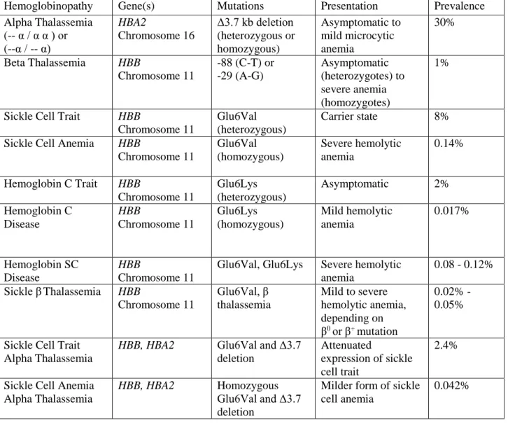
TABLE 1: Hemoglobinopathies and their prevalence in African Americans ...13
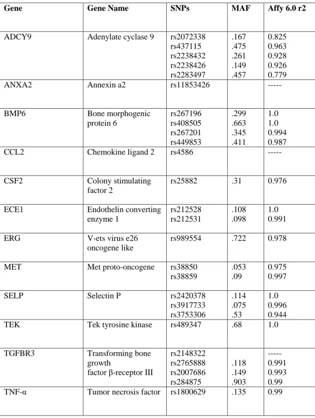
TABLE 2: Imputation quality scores for hemoglobinopathy modifier genes ...52
TABLE 3: Imputation quality scores for SCA stroke risk candidate genes ...53
TABLE 4: Imputation quality scores for stroke genes with genome-wide significance ...54
TABLE 5: Imputation quality scores for Senegal haplotype proxies ...55
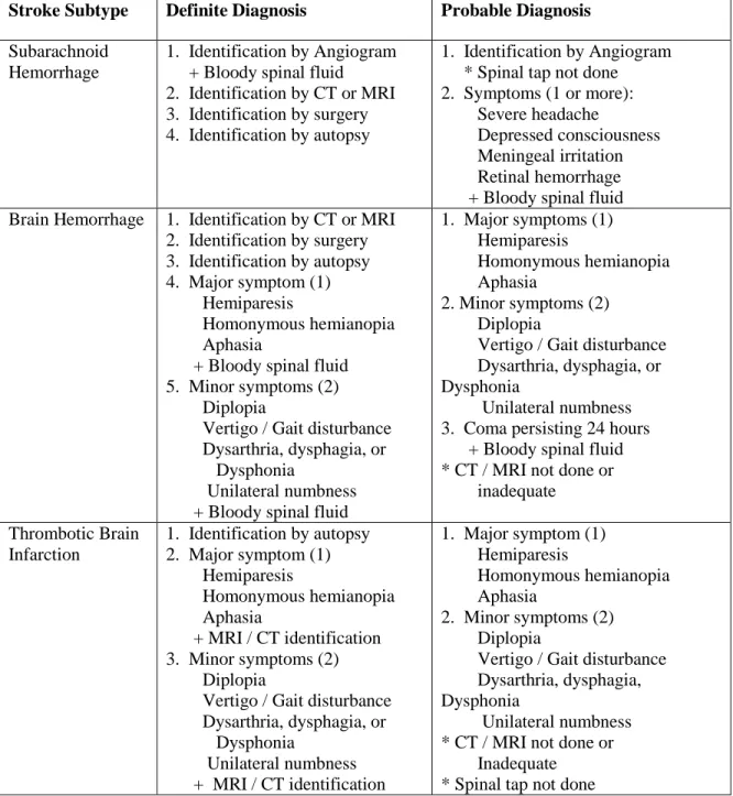
TABLE 6: Definite and probable stroke subtype classification ...57
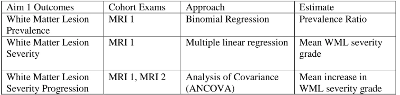
TABLE 7: Statistical plan for white matter lesion aim...67
xii
LIST OF FIGURES
FIGURE 1: The beta globin gene-like cluster ...2
FIGURE 2: The alpha globin gene cluster ...14
FIGURE 3: SNPs on 6th codon of beta globin gene ...17
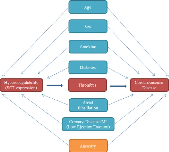
FIGURE 4: Directed acyclic graph: SCT, hypercoagulability, and stroke ...61
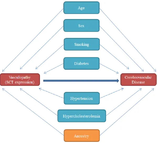
FIGURE 5: Directed acyclic graph: SCT, vasculopathy, and stroke ...62
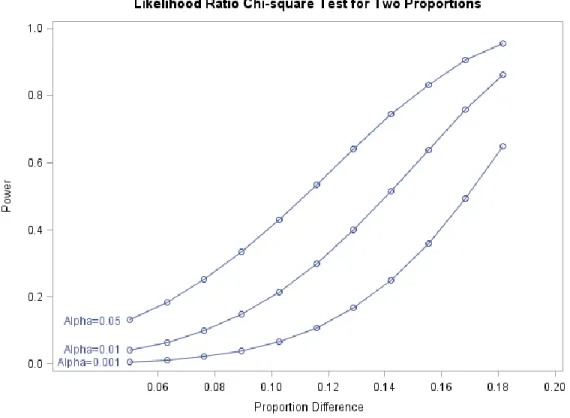
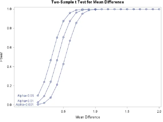
FIGURE 6: Power calculations for WML prevalence difference ...75
xiii
LIST OF ABBREVIATIONS AND SYMBOLS
ACAS: Asymptomatic Carotid Atherosclerosis Study ACPC: Anterior commissure / posterior commisure ARIC: Atherosclerosis Risk in Communities Study CSSCD: Cooperative Study of Sickle Cell Disease CHS: Cardiovascular Health Study
CT: Computed tomography
DNA: Deoxyribonucleic acid ECG: Electrocardiogram
FLAIR: Fluid attenuated inversion recovery FMD: Flow mediated dilation
GWAS: Genome-wide association study HWE: Hardy-Weinberg equilibrium
ICD-9: International classification of disease 9 LADIS: Leukoaraiosis and Disability Study MAF: Minor allele frequency
MRI: Magnetic resonance imaging
RFLP: Restriction fragment length polymorphism SBI: Subclinical brain infarction
SCA: Sickle cell anemia SCT: Sickle cell trait
SNP: Single nucleotide polymorphism
sVCAM: Soluble vascular cell adhesion molecule TIA: Transient ischemic attack
1
CHAPTER 1: LITERATURE REVIEW
1. INTRODUCTION
African Americans are disproportionately burdened by stroke, a leading cause of
disability and death in the United States. We hypothesize that sickle cell trait is a genetic risk
factor cerebrovascular disease in African Americans. The following literature review will
describe the genetics and pathophysiology of sickle cell trait, and its associations with
hypercoagulability and vasculopathy. Following this, the pathophysiology and epidemiology
of stroke, subclinical brain infarctions, and white matter lesions will be described,
highlighting the genetic risk factors and etiological associations with thrombosis and
vasculopathy. We will conclude by summarizing observational studies of sickle cell trait and
stroke, and the public health implications of the hypothesized association.
2. SICKLE CELL TRAIT
2.1 Overview
Sickle cell trait (SCT) is the heterozygous form of sickle cell anemia (SCA), a
Mendelian hemoglobinopathy characterized by misshapen red blood cells, acute chest
syndrome, and vaso-occlusive crises1. Unlike sickle cell anemia, SCT is generally
considered a benign carrier state, rather than a disease1. However, under certain conditions,
2
microcirculation and increasing coagulation. The expression of SCT is influenced by several
genetic factors and the coinheritance of other hemoglobinopathies, as described below.
2.2 The Sickle Mutation
Adult hemoglobin consists of 2 α-globin chains, 2 β-globin chains, and 4 heme
groups, which bind to and transport oxygen throughout the circulatory system2.
Hemoglobinopathies, genetic mutations affecting the hemoglobin, either decrease α or β
globin production, or generate mutated, functional variants of the β-globin chains1. Alpha
globin is encoded by 2 genes located on the 16th chromosome: hemoglobin, alpha 1 (HBA1),
and hemoglobin, alpha 2 (HBA2); however HBA2 produces 2-3 fold more protein than
HBA13. Beta globin is encoded by a single gene (hemoglobin, beta) located on the 11th
chromosome. The majority of hemoglobinopathies affecting African Americans arise from
mutations to the hemoglobin, beta (HBB) gene1.
Figure 1: The human beta globin gene like cluster. LCR = Locus Control Region; HBE1 = Hemoglobin Epsilon; HBG2 = Hemoglobin Gamma G; HBG1 = Hemoglobin Gamma A; HBBP1 = Hemoglobin Beta Pseudogene 1;
HBD = Hemoglobin Delta; HBB = Hemoglobin Beta
The HBB gene-like cluster (Figure 1) extends over 80,000 bases, and includes 6 genes:
hemoglobin, beta (HBB); hemoglobin, delta (HBD); hemoglobin, beta pseudogene 1
(HBBP1); hemoglobin, gamma A (HBG1); hemoglobin, gamma G (HBG2); and hemoglobin,
epsilon 1 (HBE1), which are regulated by a locus control region (LCR)4. Each gene is
3
invertebrates5. With the exception of the pseudogene HBBP1, each gene produces functional
globin proteins4. As the names imply, HBB, HBD, HBG1, HBG2, and HBE1, produce beta
globin, delta globin, gamma globin, gamma globin, and epsilon globin; respectively6. The
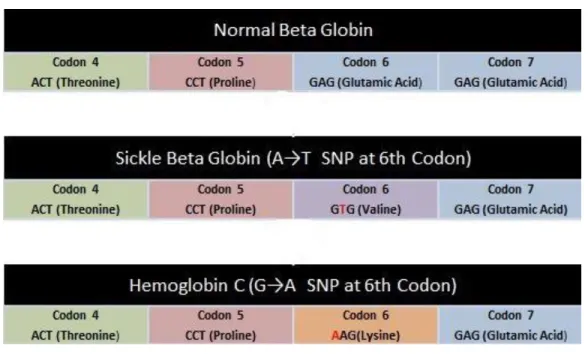
sickle mutation is an adenine to thiamine substitution occurring in the 6th codon of the human
beta globin gene (HBB)7. Transcription of the mutated DNA produces a functional variant of the β-globin protein, constructed with valine in the place of glutamic acid7. Sickle β-globin
combines with α-globin to form a protein product commonly referred to as hemoglobin S.
While the hemoglobin of individuals with sickle cell anemia is almost entirely hemoglobin S,
heterozygotes produce a combination of normal and mutated hemoglobin. The concentration
of hemoglobin S in SCT carriers varies considerably, ranging from 25-45%8. In both SCT
carriers and individuals with SCA, hemoglobin S production is known to be influenced by
genetic modifiers, described subsequently in sections 2.5 and 2.8.
2.3 Erythrocyte Sickling
With deoxygenation, the erythrocyes of individuals with SCA become sickled and
deformed1. To a lesser extent, sickling is also observed in SCT carriers1. Hemoglobin
transports oxygen throughout the systemic arterial circulation2. The oxygen is released in the
microcirculation, and supplied to the tissues by diffusion through the capillary walls2. When
hemoglobin S releases oxygen, the aberrant valine within the mutated beta globin chains
becomes exposed9. Unlike glutamic acid, found in normal beta globin, valine is a nonpolar,
hydrophobic amino acid9. Valine fits within a hydrophobic pocket formed by glutamic acid
and phenylalanine at the 88th and 85th residues, allowing it to bind to neighboring
hemoglobin molecules9. This activates a polymerization cascade, forming long chains of
4
sickle cell shape9. At rest, approximately 30-60% of the erythrocytes in the deoxygenated
venous circulation are sickled in individuals with sickle cell disease, compared to 1% in SCT
carriers10. However, under experimental conditions, the percentage of sickled red blood cells
in SCT carriers has been shown to increase from 1.0 ± 1.0% at rest, to 8.5 ± 7.1%, with
exercise at a simulated altitude of 4000 meters11. Even under controlled conditions, a wide
range of sickling (1% – 25%) among SCT carriers is observed11. In addition to oxygen
saturation, in vitro sickling is also influenced by temperature, pH, and the intracellular
concentration of hemoglobin S12-14. After several deoxygenation cycles, erythrocytes of
individuals with SCA eventually become permanently sickled1. However, sickling in SCT
carriers is generally reversible, as red blood cells regain normal morphololgy with
oxygenation in the arterial circulation1. Irreversible sickling, evidenced by sickled red blood
cells in the arterial circulation, is observed under experimental conditions in less than 1% of
red blood cells in SCT carriers subjected to exercise testing11.
2.4 Prevalence of Sickle Cell Trait
The sickled deformation of red blood cells is known to deter cytoadherance of
Plasmodium falciparum, the parasite responsible for malaria15. As a result, individuals
producing hemoglobin S have fewer episodes of malaria or high-density parasitemia, defined as an excess of 10,000 parasites per μL of blood16. While sickle cell anemia is a debilitating
disease with an average life span of 44 years in the United States17, the sickle mutation
confers a selective survival benefit to heterozygotes in areas with endemic malaria, even with
adequate access to antimalarial treatments. This is evidenced by a longitudinal study
following 1022 children from a Kenyan birth cohort, which reported a decreased risk of
5
when clinical immunity to malaria has not yet developed. Of note, all study participants with
a detected fever and positive blood film were treated with sulfadoxine-pyrimethamine, an
antimalarial drug16. Due to selective pressure, the sickle mutation, though deleterious in its
homozygous form, has a higher prevalence in the gene pools of populations arising from
tropical regions. In 2010, the world birth prevalence of SCT was estimated to be 5,476,000
(IQR: 5,291,000 – 5,679,000) 18. In support of the historic and possibly ongoing selective
advantage of SCT in areas with endemic malaria, high allelic frequencies (>10%) of the
sickle mutation were almost exclusively located in sub-Saharan Africa18. In the United
States, Americans with African ancestry have a much greater prevalence of SCT (8%) than
those with European ancestry (0.3%)19.
2.5 Sickle Haplotypes
Haplotype blocks, long stretches of alleles linked to one another on a single chromosome, are
often used to trace genetic ancestry. While the DNA of unrelated individuals will differ at
every 1000-2000 base pairs, identical polymorphisms within a haplotype block imply
common ancestry20. Five distinct haplotypes of the sickle mutation, Cameroon, Benin, Bantu
(Central African), Senegal, and Arab, have been discovered, indicating the sickle mutation
arose multiple times over the course of human history20. The sickle haplotypes are named for
their regions of discovery and geographical distribution. Sickle haplotypes are detected by
the presence or absence of restriction fragment length polymorphisms (RFLPs) around the
beta globin gene cluster20. When present, RFLPs are cleaved by the endonucleases Xmn I,
Hind III, Hinc II, and Hpa I21. While the Benin haplotype extends for 130 kilobase pairs, the
Senegal and Bantu haplotypes extend for 76 kilobase pairs21. In the United States, the most
6
most prevalent, found in 61% of African Americans with SCA.23 By comparison the Bantu
haplotype is found in 21%, and the Senegal haplotype in 10%23. These haplotypes are
associated with varying expression of the sickle mutation and hemoglobin S production, with
Bantu being the most severe, and Senegal the mildest21. Variability in severity is related to
hemoglobin F production, described below.
2.5a Fetal Hemoglobin and Sickle Haplotypes
Fetal hemoglobin, or hemoglobin F, suppresses the production and polymerization of
hemoglobin S, mitigating the sickle phenotype1. Hemoglobin F has a greater oxygen affinity
than adult hemoglobin, and is structurally distinct, composed of 2 alpha globin chains and 2
gamma globin chains (in the place of beta globin)1. At birth, the concentration of
hemoglobin F is 55-85%, which normally decreases to less than 1% in adults1. However,
multiple mutations, both within the HBB gene-like cluster and external to it, influence
hemoglobin F production24. Gamma globin is encoded by HBG2 and HBG1, both located on
chromosome 11, within the HBB gene like cluster24. Unlike the Bantu and Benin haplotypes,
the Senegal haplotype block contains a SNP (rs7482144), located 158 base pairs upstream
from HBG2, which is cleaved by endonuclease Xmn1 24. This polymorphism is thought to
be in linkage disequilibrium with functional elements increasing HBG2 expression; however
the exact mechanisms of genetic modification are unknown24. There is considerable
variability in hemoglobin F and hemoglobin S production among the sickle haplotypes24.
The Bantu haplotype is considered the most severe, with hemoglobin S comprising 90% of
the total hemoglobin in individuals with SCA21. The Benin haplotype is intermediate to the
Bantu and Senegal haplotypes, with hemoglobin S making up 85% of the total hemoglobin in
7
mildest phenotype of SCA, with hemoglobin S comprising 75% of the total hemoglobin, and
hemoglobin F concentrations approaching 20%21. However, hemoglobin F is not influenced
solely by sickle haplotypes. Quantitative trait loci within the HBS1L-MYB intergenic region
(rs9399137), and B-cell lymphoma gene (BCL11A, rs766432), also influence gamma globin
production, explaining 30-50% of the variation in hemoglobin F concentrations among
individuals with SCA24. The impact of sickle haplotypes and fetal hemoglobin expression in
SCT carriers has not been widely published in the literature.
2.6 Sickle Mutation and Hypercoagulability
Both SCA and SCT are associated with increased factors of coagulation, when
compared to African Americans with normal hemoglobin25. This is likely due to red blood
cell asymmetry and disordered cell membranes expressing phosphatidylserine25, 26. Under
hypoxic conditions, such as heavy exertion, dehydration, or high altitudes, the hemoglobin S
of SCT carriers polymerizes, causing erythrocytes to sickle11, 27, 28. As the red blood cells
sickle, phosphatidylserine, a cellular recognition signal, is pulled from the interior of the
phospholipid monolayer to the outside of the cell membrane surface29. Phosphatidylserine
serves as a docking site for enzymes which facilitate prothrombin activation, coagulation and
clot formation30. While red cell sickling is far less prevalent in SCT carriers than with SCA,
evidence for hypercoagulability in SCT carriers has been indicated by blood assays26.
Compared to African Americans with normal hemogloblin, SCT carriers have elevated levels
of prothrombin fragment 1+2, thrombin-antithrombin complexes, and d-dimer26. These
biomarkers signify coagulation activation. Prothrombin fragment 1+2 is a byproduct
released by the conversion of prothrombin to thrombin, an integral step in the coagulation
8
an indirect biomarker of hypercoagulability26. To some extent, the hypercoagulability in
SCT carriers may be modified by environment or behavior. This is evidenced by differing
levels of procoagulant activity in monozygotic twins with SCT31.
Hypercoagulability increases risk of thrombosis, and a greater prevalence of
thrombotic infarctions has been observed in individuals with SCT32-34. In an autopsy series
of 128 SCT carriers with a mean age of 47, obvious visceral infarcts were observed in 18%,
after excluding cases with cardiac thrombi, bacterial endocarditis, and atherosclerotic
emboli32. By comparison, visceral infarcts were observed in less than 1% of similarly aged
African Americans with normal hemoglobin. The spleen was the most common site of
infarction in SCT cases, followed by the kidneys, brain, and lung32. A higher prevalence of
thrombotic events has also been observed in SCT carriers in epidemiological studies. In an
observational study, 65,154 consecutively admitted African American males in 13 Veterans
Administration hospitals were tested for SCT33. Pulmonary embolism was noted in 2.2% of
SCT carriers, compared to 1.5% with normal hemoglobin33. Furthermore, in a case control
study of 1070 hospitalized African Americans, SCT carriers were observed to have twice the
risk of venous thromboembolism34. Certain exposures may influence the risk of thrombosis
in SCT carriers. For example, oral contraceptive use has been associated with deep vein
thrombosis in female SCT carriers of child bearing age35.
2.7 Sickle Mutation and Vasculopathy
Vasculopathy, a general term denoting disorders of the vasculature, has been
described in individuals with both SCA and SCT. The arterial vasculature is regulated by the
9
the arterial wall2. When injured or stimulated by irritants, the endothelium becomes
activated, releasing cellular signaling molecules that regulate vasomotor tone, inflammatory
responses, coagulation, and vascular remodeling36. Vascular remodeling may be either
dilatative, as is the case with dolichoectasia, or occlusive, the result of hyperplasia, or
thickening, or the intimal layer of the arterial wall. Endothelial dysfunction and vascular
remodeling associated with the sickle mutation will be described in greater detail next.
2.7a Sickle Mutation and Endothelial Dysfunction
Endothelial dysfunction is detectable by biomarkers and noninvasive testing37, and
has been observed in both individuals with SCA and SCT38. Hemolysis, bursting red blood
cells, creates endothelial dysfunction by depleting the bioavailability of nitric oxide, a potent
vasodilator produced by the endothelium, with anti-athrogenic and anti-thrombotic effects39.
Sickled red blood cells are particularly friable, and when ruptured, release free hemoglobin
and arginase into the blood plasma39. Free hemoglobin binds to and scavenges nitric oxide,
reducing its bioavailabiliy, while arginase converts L-arginine, a substrate required for nitric
oxide production, into ornithine, decreasing nitric oxide formation39. Among its many other
effects, nitric oxide represses the expression of vascular cell adhesion molecules on the
surface of endothelial cells40. Accordingly, elevated cellular adhesion molecules can be
considered biomarkers of endothelial dysfunction. In laboratory assays, patients with SCA
show elevated levels of soluble vascular cell adhesion molecules (sVCAM) compared to
African Americans with normal hemoglobin, an indication of endothelial dysfunction39.
In contrast to homozygous SCA, the erythrocytes of SCT carriers are less prone to
10
have demonstrated hemolysis in vitro.41 While biomarkers of endothelial dysfunction have
not been examined in SCT carriers, endothelial dysfunction in SCT carriers has been
suggested by noninvasive tests. One such test, flow mediated dilation (FMD) testing,
measures the brachial artery response to induced hyperemia. Briefly, the forearm blood flow
is occluded by a tourniquet, and reactive hyperemia, or turbulent, high velocity blood flow, is
induced upon release of the tourniquet. The sudden increase in arterial wall sheer stress,
caused by the hyperemic blood flow, stimulates the endothelium to release nitric oxide.
Endothelial function is gauged by the degree of arterial dilation, measured by high resolution
ultrasonography. FMD testing has demonstrated endothelial dysfunction in SCA patients,
and a dilation response in SCT carriers that is intermediate to SCA patients and African
American controls with normal hemoglobin38.
2.7b Sickle Mutation and Intimal Hyperplasia
Endothelial dysfunction and overexpression of vascular adhesion molecules induce intimal
thickening, or hyperplasia in individuals with sickle hemoglobinopathies. When expressed,
the cellular adhesion molecules on the endothelial cell surface recruit white blood cells, and
initiate their uptake into the vascular wall42. Leukocytes secrete cytokines and growth
factors, which induce smooth muscle cells to proliferate and migrate from the tunica media to
the intima43. The resulting vascular intimal hyperplasia can become stenotic, blocking the
passage of blood by protruding into the arterial lumen42. Additionally, the irregular surfaces
of intimal lesions facilitate thrombus formation, as activated platelets adhere to sites of
vascular injury44. Arterial intimal hyperplasia with superimposed thrombi have been noted in
deceased SCA patients, by post-mortem examinations. An autopsy series 12 SCA patients
11
and stenosis with occlusive thrombus in 645. Few post-mortem examinations of vasculopathy
in individuals with SCT have been published. However, in one report of 20 consecutive
patients who died of various causes (15 with SCA autopsied at mean age of 39, and 5 with
SCT autopsied at a mean age of 51), intimal hyperplasia and fibrosis was observed in the
pulmonary artery of all 2046. Thus, intimal hyperplasia may be a common finding with SCT,
at least in the pulmonary artery. Unfortunately, no comparisons can be made with African
Americans with normal hemoglobin, as none were autopsied in this case series.
2.7c Sickle Mutation and Dolichoectasia
Another form of vasculopathy, dilatative arteriopathy, has been detected by cerebral MRI in
both patients with SCA and SCT. Dolichoectasia, or dilatative arteriopathy, is characterized
by elongated cerebral vessels following a tortuous, or sinuous course, with marked thinning
of the arterial walls and dilation47. Bidirectional blood flow within ectatic, or dilated,
segments, results in blood stasis and intraluminal thrombus formation, which can lead to
downstream infarctions in the cerebral circulation47. In older individuals, dolichoectasia is
most commonly associated with hypertension and atherosclerosis47; however, wall shear
stress, induced by acute increases in blood volume and turbulent flow, has precipitated
dolichoectasia in experimental animals48. Chronic and uneven wall shear stress is thought to
denude the endothelium, and degrade the internal elastic lamina of the arterial wall by
stimulating the release of metalloproteinases49. Sickle cell anemia is characterized by high
cardiac output, as the heart compensates for anemia and oxygen demand by pumping a
greater blood volume1, which may be related to the increased prevalence of dolichoectasia
observed with SCA. In a retrospective study of 47 children with SCA who were imaged by
12
to 2% of controls without SCA, who were imaged to assess brain tumor50. While
heterozygous SCT is not characterized by anemia or high output states1, a prospective
imaging study comparing SCT children to normal hemoglobin siblings reported a
dolichoectasia prevalence of 19% in those with SCT, compared to none of the controls51.
Thus, both the homozygous and heterozygous sickle mutation may be associated with
dolichoectasia; however the mechanisms causing dolichoectasia in SCT carriers have not
been elucidated.
2.8 Sickle Cell Trait and Other Hemoglobinopathies
Several hemoglobinopathies, or mutations affecting alpha or beta globin production,
are observed in populations of African descent. These include alpha thalassemia, beta
thalassemia, and Hemoglobin C (Table 1). The SCT expression is influenced by co-inherited
hemoglobinopathies. While alpha thalassemia mitigates the phenotype, concomitant
heterozygosity for SCT and beta thalassemia or SCT and hemoglobin C produces a disease
state phenotypically similar to sickle cell anemia, exacerbating the impact of the SCT
variant1. The hemoglobinopathies affecting African Americans and their coinheritance with
13
Table 1: Hemoglobinopathies and their prevalence in African Americans1, 19, 52, 53
Hemoglobinopathy Gene(s) Mutations Presentation Prevalence
Alpha Thalassemia (-- α / α α ) or (--α / -- α)
HBA2
Chromosome 16
Δ3.7 kb deletion (heterozygous or homozygous) Asymptomatic to mild microcytic anemia 30%
Beta Thalassemia HBB
Chromosome 11
-88 (C-T) or -29 (A-G) Asymptomatic (heterozygotes) to severe anemia (homozygotes) 1%
Sickle Cell Trait HBB
Chromosome 11
Glu6Val (heterozygous)
Carrier state 8%
Sickle Cell Anemia HBB
Chromosome 11 Glu6Val (homozygous) Severe hemolytic anemia 0.14%
Hemoglobin C Trait HBB
Chromosome 11
Glu6Lys (heterozygous)
Asymptomatic 2%
Hemoglobin C Disease HBB Chromosome 11 Glu6Lys (homozygous) Mild hemolytic anemia 0.017% Hemoglobin SC Disease HBB Chromosome 11
Glu6Val, Glu6Lys Severe hemolytic anemia
0.08 - 0.12%
Sickle βThalassemia HBB
Chromosome 11
Glu6Val, β thalassemia
Mild to severe hemolytic anemia, depending on β0 or β+ mutation
0.02% -0.05%
Sickle Cell Trait Alpha Thalassemia
HBB, HBA2 Glu6Val and Δ3.7
deletion
Attenuated
expression of sickle cell trait
2.4%
Sickle Cell Anemia Alpha Thalassemia
HBB, HBA2 Homozygous Glu6Val and Δ3.7 deletion
Milder form of sickle cell anemia
0.042%
(-- α / α α ) = 3 functioning alpha globin genes, (-- α / -- α ) = 2 functioning alpha globin genes Glu6Val = glutamic acid to valine mutation, Glu6Lys = glutamic acid to lysine mutation β0 =no beta globin production, β+ =diminished beta globin production
14 2.8a Alpha Thalassemia
Alpha thalassemias result from deletions to the alpha globin encoding genes (HBA2
or HBA1), or mutations affecting their expression. The alpha globin gene cluster spans 30
Kb, and contains 5 genes: hemoblobin, zeta (HBZ); hemoglobin zeta pseudogene 1
(HBZP1); hemoglobin, mu (HBM, also referred to as hemoglobin alpha pseudogene 2, or
HBAP2); hemoglobin alpha pseudogene 1 (HBAP1); hemoglobin alpha 2 (HBA2);
hemoglobin alpha 1 (HBA1); and hemoglobin theta 1 (HPQ1), as shown in Figure 7.
Over 120 mutations are associated with alpha thalassemia; however, the Δ3.7 deletion
(rs63751476) of the HBA2 gene is most common in African Americans. African Americans
have a high prevalence (30%) of alpha thalassemia, defined by either a single (-- α / α α ) or
double (-- α / -- α ) alpha globin gene deletion, denoting either heterozygosity or
homozygosity for the Δ3.7 mutation52. The clinical presentation of alpha thalassemia trait
ranges from asymptomatic to mild microcytic anemia, characterized by small red blood
cells1. The more severe forms of alpha thalassemia, Hemoglobin H (-- -- / -- α ) and Barts
Hydrops Fetalis (-- -- / -- --) are exceedingly rare in African Americans, and as a result the
prevalence has not been estimated52.
When coinherited with SCT, alpha thalassemia decreases hemoglobin S production1.
Because alpha thalassemia minimizes the available pool of alpha globin chains, normal and Figure 2: The alpha globin gene cluster contains 6 loci
HBZ = Hemoglobin Zeta; HBZP1 = Hemoglobin Zeta Pseudogene 1; HBM = Hemoglobin Mu; HBAP1 = Hemoglobin Alpha Pseudogene 1; HBA2 = Hemoglobin Alpha 2;
15
sickle beta globin must compete for available alpha chains to form hemoglobin tetramers1.
Normal beta globin dimerizes twice as effectively as sickle beta globin, leaving sickle beta
globin without available alpha chains to form hemoglobin S1. In fact, hemoglobin S
concentrations in SCT carriers depend on the number of functioning alpha globin genes.
SCT carriers with all 4 alpha globin genes have hemoglobin S concentrations of about 40%,
compared to 35% in SCT carriers with 3 functioning alpha globin genes, and 30% in those
with 2 alpha globin genes52.
The prevalence of alpha thalassemia in patients with SCA appears to be comparable
to the general population of African Americans52, 54, 55. Alpha thalassemia has been
estimated to have a prevalence of 30% in African Americans52. Similarly, a 1 or 2 alpha
gene deletion was detected in 33% of SCA patients enrolled in the Cooperative Study of
Sickle Cell Disease54, but was slightly higher (40%) in infants with SCA enrolled in the Baby
Hug trial55. Alpha thalassemia is inherited independently of sickle haplotype groups as
well21. Assuming a random coinheritance of alpha thalassemia with respect to sickle
mutations or haplogroups, the expected prevalence of SCT and alpha thalassemia would be
the product of each muation’s prevalence in African Americans, or 2.4%.
2.8b Beta Thalassemia
Beta thalassemias are hemoglobinopathies that diminish (β+) or abrogate (β0)
production of the beta globin chains. When coinherited with SCT, beta thalassemia increases
the concentration of hemoglobin S. This is because beta thalassemia decreases the number of
16
On the other hand, SCT that is coinherited with β0 thalassemia has a clinical presentation that
is almost indistinguishable from sickle cell anemia1. Over 200 mutations are known to cause beta thalassemia. In African Americans, the -88 (CT) and -29 (AG) mutations, affecting
promoter regulatory elements of the HBB gene, are the most frequent cause of beta
thalassemia56. In African American birth cohorts, the prevalence of beta thalassemia is 1%53,
while the prevalence of coinherited SCT and beta thalassemia is 0.02 – 0.05%19, 53.
2.8c Hemoglobin C
Similar to hemoglobin S, hemoglobin C is a functional variant of the beta globin gene
(rs33930165), caused by a single nucleotide polymorphism of the 6th codon of the
hemoglobin gene1 (Figure 8). While sickle beta globin contains a glutamic acid to valine
substitution, hemoglobin C produces a mutated beta globin protein with lysine in the place of
glutamic acid1. The heterozygous genotype, or hemoglobin C trait, is asymptomatic. The
homozygous genotype, known as hemoglobin C disease, is characterized by mild hemolytic
anemia. In African Americans, the prevalence of hemoglobin C trait is 2%, while the
prevalence of hemoglobin C disease is much rarer, at 0.017%19. Concomitant heterozygosity
of SCT and hemoglobin C, however, results in hemoglobin SC disease, a hemolytic anemia
with symptoms of vaso-occlusive crises and acute chest syndrome57. In African American
birth cohorts, the reported prevalence of hemoglobin SC disease in ranges from 0.008 –
17
Figure 3: Single nucleotide polymorphisms on 6th codon of beta globin gene
2.9 Summary of Primary Exposure: SCT
Sickle cell trait, the heterozygous form of sickle cell anemia, affects approximately
8% of African Americans. Laboratory assays and observational studies suggest greater risk
of adverse vascular events associated with SCT. Under hypoxic conditions, the hemoglobin
S polymerizes, increasing hypercoagulability and risk of venous thromboembolism in SCT
carriers. The sickle mutation is also associated with vasculopathies, and a greater degree of
endothelial dysfunction, intimal hyperplasia, and dolichoectasia. However, the sickle
mutation is affected by several genetic factors. Hemoglobin F production, influenced by
polymorphisms within the sickle haplotype blocks, and coinheritance of alpha thalassemia,
reduce the production of hemoglobin S, mitigating the severity of the SCT phenotype. On
18
anemia. Thus, the hypothesized associations between SCT and cerebrovascular disease may
be influenced by concomitant hemoglobinopathies and haplotype.
3. STROKE
3.1 Overview
Stroke, or cerebrovascular accident, is an acute neurologic syndrome provoked by
interrupted blood supply to the brain58. The cerebral neurons lack glycogen; as a result,
blood flow cessation precipitates rapid energy failure followed by brain tissue death within 4 – 10 minutes58. Blood flow disturbances originate from vascular obstructions and ruptured
blood vessels, and occur in both the large and small cerebral vessels58. The large intracranial
arteries include the internal carotid arteries, vertebral arteries, and the middle, anterior, and
posterior cerebral arteries, which make up the Circle of Willis. The small vessels, which
typically range from 400 – 900 μ in diameter, branch from the large vessels deep into the
interior of the brain58. While most cerebral vascular accidents involve the large vessels, the
small vessels are implicated in 20%58. Stroke more often involves the arterial circulation;
however, cerebral vascular accident is also provoked by disrupted blood flow in the cerebral
veins and sinuses58.
3.2 Ischemic Stroke
The majority of strokes are ischemic, accounting for 87% of all cerebrovascular
accidents59. Ischemic strokes are caused by vascular obstructions from clot or atheroma,
with origins that are more often embolic than in-situ58. Common sources of emboli include
the heart and proximal extracranial arteries, and to a lesser extent, the venous circulation58.
19
for 20% of all ischemic strokes58. Clotting is induced by stasis of the left atrium, due to low
ejection fraction, atrial fibrillation, and left atrial dilation. Another source of embolic stroke
is the extracranial carotid arteries58. The bifurcation of the internal carotid is particularly
vulnerable to atherogenesis. Thrombus forming on the plaque surface can dislocate, or the
plaque itself can rupture and break loose, forming emboli that eventually become lodged in
the intracranial vessels. A third source of emboli is the venous circulation, causing ischemic
stroke by so-called paradoxical embolization58. Under normal circumstances, blood from the
systemic venous circulation is returned to the right atrium, and ejected by the right ventricle
to the lungs. However, in 15% of the general population, blood communicates from the right
to left atrium, through a small opening in the atrial wall, or atrial septal defect58. This
passage allows embolized clot from the venous circulation, often from deep vein thrombosis,
to cross the cardiac circulation and become ejected into the systemic arterial circulation.
Compared to embolic stroke, vascular obstruction by in-situ thrombosis is less frequently
observed, and usually occurs in either the small deep penetrating arteries, or the cerebral
veins and sinuses58. Stenotic plaques, typically in the carotid or vertebral arteries58, also
provoke ischemic stroke by inducing hypoperfusion to the downstream cerebral circulation.
In the United States, the 30-day case fatality rate of ischemic stroke is 7.8%60
3.3 Hemorrhagic Stroke
Cerebrovascular accidents that arise from ruptured blood vessels, rather than vascular
obstructions, are termed hemorrhagic strokes. Hemorrhagic strokes are particularly deadly,
with a case fatality rate of 50%58. The most common type of hemorrhagic stroke is
hypertensive intraparenchymal hemorrhage, invoked by spontaneous rupture of the small,
20
of the brain, causing hydrocephalus and increasing intracranial pressure perniciously58.
Hemorrhagic stroke is sometimes characterized by bleeding into the subarachnoid space
between the brain tissue and the pia mater membrane surrounding the brain58. Subarachnoid
hemorrhage may result from ruptured aneuryms, weakened areas of the arterial wall, and
generally arise from the large vessels comprising the Circle of Willis58. Other causes of
subarachnoid hemorrhage include trauma, and congenital arteriovenous malformations58.
Arteriovenous malformations allow abnormal communication between the arteries and veins,
bypassing the capillary circulation, and are prone to rupture58.
3.4 Stroke Symptoms
Symptoms and severity of stroke depend on type of obstruction, size of vessel
impacted, extent of collateralization, and brain region supplied by the arterial distribution58.
Symptoms are almost always contralateral to the brain hemisphere affected, with functional
disability reflecting the brain region impacted58. Limb paralysis (hemiplegia), limb weakness
(hemiparesis), impaired gait (ataxia), and clumsy hand suggest damage to the sensory and
motor cortex58. Aphasia (language deficit) implies damage to Broca’s or Wernicke’s area of
the cerebral cortex, while homonymous hemianopia (visual field loss) indicates injury to the
cerebral visual cortex58. The stroke syndrome may also present with severe memory loss,
stupor, coma, loss of consciousness, and seizure58.
Infarction of the small, deep penetrating arteries, or lacunar stroke, is typically less
severe than stroke related to the large vessels. In fact, small vessel infarcts are almost always
ruled out by presentation of severe memory loss, stupor, coma, aphasia, monoplegia,
21
the small vessels are less extensive than the large vessels, minimizing areas affected by
infarction. Lacunar infarcts are also more often due to thrombi, which occasionally lyse,
restoring the passage of blood flow58. Episodes of stroke symptoms lasting less than 24
hours, considered transient ischemic attacks (TIA), are often provoked by blood clots of the
small vessels that quickly lyse, allowing functional recovery58. However, TIAs are not
without risk; in 10-15% of TIA cases, major stroke occurs within 3 months58.
3.5 Stroke Diagnosis
Acute stroke is suspected by abrupt changes in mental status with presentation of
neurological symptoms, and diagnosed by imaging studies and assessment of cerebral spinal
fluid by lumbar puncture58. Hemorrhagic stroke is distinguished from ischemic stroke by the
presence of blood in the cerebral spinal fluid, and hemorrhaging in the absence of infarction.
Computed tomography (CT) is generally preferred for acute stroke diagnosis, due to its speed
and lower cost than magnetic resonance imaging (MRI)58. CT imaging reliably detects
intracranial hemorrhaging, but may not visualize infarctions until 24-48 hours after the
event58. Small infarctions along the cortical surface of the brain or within the posterior fossa
may also be missed, due to bone artifact obstructing the image58. As an alternative to CT,
cerebrovascular accident may be diagnosed with MRI. While MRI has less sensitivity
visualizing hemorrhagic bleeding, it is a superior imaging modality for the detection of
infarctions in all regions of the brain58. One limitation to stroke diagnosis, either by CT or by MRI, is the patient’s ability to seek medical care. Stroke symptoms are frequently painless,
or cause anosagnosia, the lack of perception that anything is wrong58. As a result, patients
22 3.5a. Stroke Diagnosis in Epidemiological Studies
In large observational studies, stroke cases are often classified by administrative
claims records, hospital surveillance, chart reviews, or diagnostic algorithms. The 9th
International Classification of Disease (ICD-9) codes, submitted to insurance carriers for
billing purposes, provide a facile but imperfect record of patients discharged with stroke. To
ascertain stroke diagnoses, epidemiological studies sometimes examine abstracted medical
records, including the physician notes and diagnostic imaging reports60. One disadvantage of
retrospective data collection after hospital discharge is the possibility of missing data relevant
to the study aims. Unlike prospective study designs, which rigorously record clinical data
related to pre-specified outcomes, retrospective surveillance, or “cold pursuit”, is limited to recorded data deemed relevant to the patients’ clinical care62. For this reason, stroke
diagnoses by chart reviews often include measures of uncertainty. For example, stroke may be considered “definite” when classified by clinical symptoms, imaging studies, and lumbar
puncture. On the other hand, stroke may be classified as “probable” when the clinical picture
suggests stroke, but the diagnosis does not meet certain criteria for the epidemiological
definition, for example if imaging studies are inconclusive, or lumbar puncture is not
performed60. Agreement between ICD-9 diagnoses and chart reviews varies, based on the
stroke subtype and the number of ICD-9 codes analyzed. In a study of 206 patients
hospitalized with stroke, ischemic stroke was more accurately classified by considering all
ICD-9 codes in the discharge record, while intracerebral and subarachnoid hemorrhage were
better classified by considering only the primary ICD-9 discharge code63.
Physician-reviewer validation of ICD-9 codes has also been ascertained in the ARIC study, which
23
ICD-9 codes and ischemic stroke (78%)60. In some epidemiological studies, reviewing the
medical record may be too labor intensive or cost-prohibitive. As an alternative, a validated
computer algorithm may be utilized to diagnose stroke cases, based on neurological signs and
symptoms64. In the ARIC study, which examined 538 stroke hospitalizations, agreement
between computer algorithm and physician reviewer diagnoses was 78%60.
3.5b. Cerebral Magnetic Resonance Imaging
Cerebral small vessel disease is often detected in clinical settings and observational,
prospective screenings by magnetic resonance imaging. MRI scanners use magnetic field
sequences to manipulate hydrogen protons, and convert the signals from protons returning to
baseline energy states into images. Hydrogen atoms are present in 99.98% of all human
tissues65. Positive subatomic particles, or protons, continuously rotate within the nuclei of
atoms, creating a magnetic field. When atoms are subjected to an external magnetic field, the
protons start to rotate around the axis of the magnetic field, in a direction that is either
parallel or antiparallel to the field65. This interaction creates magnetic resonance. After the
MRI scanner aligns atoms within a magnetic field, a second magnetic field, or excitation
pulse, is applied, pulling the atoms to a specified angle that is transverse to the first magnetic
field. The excitation pulse is then switched off, and protons are monitored as they return to
the original alignment65. The return time is dependent on the tissue. For example, the
relaxation time for hydrogen protons in fat is very brief, while the relaxation time in fluids is
very long65. These return time signals are converted into voxels, or 3D pixels, by fast Fourier
transform, allowing a visual representation of the tissues. For image optimization, the
magnetic resonance signal can be filtered, or weighted, by manipulating the repetition time
24
used, depending on the structures being visualized. When both the T1 and T2 weights are
removed, MRI is considered proton density weighted, meaning the magnetic resonance
signals are proportional to the water concentration65. Brain imaging is also optimized by
fluid attenuated inversion recovery, or FLAIR, which filters signals arising from the cerebral
spinal fluid65.
3.5c. Subclinical Stroke Detection by MRI
Silent stroke is unaccompanied by neurological symptoms, but is detectable by a
presentation of white matter lesions or subclinical brain infarctions on MRI.66 Severity of
white matter lesions is either quantified volumetrically, or graded qualitatively from
2-dimensional images67. Subclinical brain infarctions, which present on MRI as fluid-filled
lacunes, are quantified by measuring the largest diameters68. In highly selected populations,
such as patients with Alzheimers69 or Binswangers disease70, MRI-detected WML and
lacunes have been validated by autopsy. However, silent stroke detected by MRI in the
general population is not likely to be validated by autopsy, and few cohorts incorporate
post-mortem examination with prospective MRI. Despite this limitation to validity, the detection
of subclinical cerebrovascular disease by MRI is reproducible (discussed further in sections
4.1 and 5.1). Reliability is typically tested among randomly selected duplicate images, to
determine intra- and inter-reader reproducibility67, 71, 72. An advantage of MRI is its
suitability for prospective imaging in observational studies58. Unlike CT, which exposes a
patient to 3-5 mGy of radiation for a routine brain exam, MRI does not utilize ionizing
radiation58. However, MRI is time consuming and sensitive to motion, and children under
the age of 10 typically require conscious sedation to remain immobile for the duration of the
25
participants with metallic implants, such as aneurysm clips, hearing aids, cardiac
pacemakers, spinal cord stimulators or other internal electrical devices must be excluded.
Other considerations include extreme obesity and claustrophobia. As the MRI scanning
involves bodily insertion into the magnet bore, patients unable to fit, or tolerate, confined
spaces may need to be excluded.
3.6 Stroke Prevalence
In the United States, approximately 7 million people over the age of 20 have had a
stroke59. The Southeast has the greatest density of stroke cases, clustered within the so-called
stroke belt: Alabama, Arkansas, Georgia, Indiana, Kentucky, Louisiana, Mississippi, North
Carolina, South Carolina, Tennessee, and Virginia73. Stroke occurrence increases with age,
and is more prevalent in women than men. At the ARIC study baseline, the prevalence of
TIA or stroke symptoms in women was 7%, compared to 5% in men (p<.0001)64. However,
the greater prevalence of stroke in women appears to be predominantly due to the longer
lifespans of women74. African Americans are disproportionately burdened by
cerebrovascular disease, and experience first stroke at a younger mean age than European
Americans75. The prevalence of stroke in African Americans18 years or older is nearly twice
that of than non-Hispanic whites (4.0% vs. 2.3%)76. Annually, the incidence of stroke in the
United States has been estimated to be about 800,000 cases per year59, and has been
decreasing since the 1990s for European Americans. However, this trend has not been
observed for African Americans77. Over 8 years of follow up, a longitudinal analysis from
the ARIC study reported a 62% higher stroke incidence rate in African Americans than
26
age, gender, hypertension, diabetes, center, education, smoking, and history of coronary
disease60.
3.7 Stroke Risk Factors
Risk factors for stroke vary by stroke type; however, hypertension is a strong
determinant of both ischemic and hemorrhagic stroke58. Additional risk factors associated
with ischemic stroke include older age, diabetes, smoking, hyperlipidemia, atrial fibrillation,
and recent myocardial infarction. On the other hand, hemorrhagic stroke is correlated with
aging, hypertension, trauma, congenital arteriovenous malformation, and amyloid
angiopathy, the deposition of amyloid proteins within the brain and arterial walls58.
Endothelial dysfunction and dolichoectasia may increase the risk of lacunar stroke.
While endothelial heterogeneity is known to exist among the various vascular distributions36,
there is evidence that systemic endothelial dysfunction correlates with cerebral small vessel
disease and lacunar stroke. In a cross sectional study of 45 Korean patients with
symptomatic lacunar stroke undergoing flow mediated dilation testing, greater endothelial
dysfunction of the brachial artery was noted in the stroke patients than the age and sex
matched controls with hypertension78. Lacunar stroke has also been cross sectionally
associated with intracellular adhesion molecule (ICAM-1), tissue factor, and
thrombomodulin, all biomarkers of endothelial dysfunction79. Dolichoectasia appears to be a
risk factor for lacunar stroke, as well. In a population-based study of 387 patients with first
cerebral infarction, 42% with dolichoectasia were diagnosed with lacunar infarct, compared
27 3.7a. Genetic Risk Factors of Stroke
Family history and twin studies have predicted both ischemic and hemorrhagic stroke,
implying a genetic component of stroke risk59. Of the few studies that have reported
genome-wide associations between SNPs and stroke, none have been conducted in
populations of African ancestry. However, in European populations, variants of the histone
deacetylase (HDAC9) gene81, 82, paired-like homeodomain (PITX2) gene82, and Ninjurin
(NINJ2) gene83 have been shown to have validated associations with large vessel stroke, at
genome-wide statistical thresholds (p-value of 10-8 or smaller).
3.8 Sickle Cell Anemia and Stroke
Sickle cell anemia is a well-established genetic risk factor for stroke. The
Cooperative Study of Sickle Cell Disease, a longitudinal cohort of 4,082 patients with sickle
cell disease (sickle cell anemia, sickle hemoglobin C, and sickle thalassemia), observed that
by the age of 45, 24% of patients with sickle cell anemia had experienced a first stroke,
defined ischemic stroke, hemorrhagic stroke, or TIA84. This association is confirmed by
administrative claims data for stroke. The incidence of ischemic stroke in patients with
sickle cell anemia aged 35-64 is 740 per 100,000 person years85, much higher than 270 per
100,000 person years for 35-64 year old African Americans overall86. Unlike the general
population, ischemic stroke in patients with SCA is often due to arterial narrowing with
thrombus that is in-situ, rather than embolic. In an autopsy series of children with SCA and
fatal stroke, intimal hyperplasia with superimposed thrombosis were common findings,
particularly in the middle and anterior cerebral arteries45. In-situ thrombosis of the small
28
brain, are also observed with SCA58. Stroke risk in children with SCA is determined
noninvasively by transcranial Doppler, used to detect stenotic lesions and increased blood
flow velocities within the Circle of Willis87.
3.8a Genetic Modifiers of Sickle Cell Anemia and Stroke Risk
The stroke risk associated with sickle cell anemia is heterogenous, and may be
influenced by coinheritance of modifier genes. Support for this hypothesis comes from a
pediatric sibship study, examining siblings who were all affected by sickle cell anemia88. In
42 of the 207 sibships, at least one child experienced clinical stroke, and in 10 sibships, 2
siblings had a stroke. The number of families with 2 children with SCA and stroke exceeded
expectations derived by permutation analysis, leading the authors to conclude that
coinheritance of genetic modifiers shared among siblings with SCA increased stroke risk88.
The sickle haplogroups, alpha thalassemia, and candidate genes associated with stroke in the
general population have been investigated as potential genetic modifiers of the association
between sickle cell anemia and stroke, and are described next.
3.8b Sickle Cell Anemia and Stroke Risk Modification by Haplotype
Stroke risk modification by haplogroup polymorphisms remains uncertain. In a cross
sectional study of 221 patients with sickle cell anemia, the Bantu haplotype, the most severe
SCA phenotype with the greatest concentration of hemoglobin S, was associated with a
3-fold increase in obliterative sickle vasculopathy, an outcome that included stroke, renal
failure, chronic lung disease with cor pulmonale, leg ulcers, and young adult death21.
However, a similarly sized case-control study that examined stroke as an explicit, rather than
29
groups89. While statistical power calculations were not described, this study included 130
children with SCA and stroke, and 103 SCA controls without stroke89. Stroke risk
modification by sickle haplogroups remains an interesting hypothesis, but at the current state
of science, is far from established.
3.8c Sickle Mutation and Stroke Risk Modification by Alpha Thalassemia
Concomitant alpha thalassemia may also modify stroke risk in patients with SCA, and
appears to protective effect21, 89, 90. In a cross sectional study of 300 children with SCA, the
prevalence of confirmed alpha thalassemia in children experiencing stroke (21%) was lower
than those who were stroke free (38%)90. These findings were later replicated by a pediatric
case-control study including 233 children with SCA and alpha thalassemia detecting by
genotyping89. In SCT carriers, stroke risk modification has been investigated less
extensively. In a single study examining 355 hospitalized black men with SCT, no
differences in stroke risk were observed among quartiles of hemoglobin S concentrations91.
While alpha thalassemia was not ascertained by genotyping, it was inferred by hemoglobin S
concentrations and mean corpuscular volume. However, these biomarkers may be
confounded by iron-deficiency anemia91, leaving the results inconclusive.
3.8d Sickle Cell Anemia Stroke Risk Modification by Candidate Genes
Several candidate genes known to influence the development of vascular disease have
been validated as stroke risk modifiers in children with sickle cell anemia. In the
Cooperative Study of Sickle Cell Disease (CSSCD), which included 1,398 African
Americans with SCA, 108 SNPs in 80 candidate genes were analyzed by Bayesian networks
30
(ADCY9), annexin (ANXA2), bone morphogenic protein (BMP6), chemokine ligand (CCL2),
colony stimulating factor (CSF2), endothelin converting enzyme (ECE1), v-ets virus
oncogene (ERG), met-proto oncogene (MET), selectin p (SELP), Tek tyrosine kinase (TEK),
transforming bone growth factor beta receptor (TGFBR3) and tumor necrosis factor (TNF-α)
genes were found to be associated with stroke92. Genetic associations were then validated in
114 African Americans with SCA not included in the original Bayesian analysis, and were
found to predict stroke over a 5 year follow up with an accuracy of 98.2%92.
Candidate genes have also been associated with large and small vessel stroke
subtypes, classified by MRI. In a validation study involving the CSSCD cohort and Stroke
Prevention Trial in Sickle Cell Anemia (STOP), variants of tumor necrosis factor (TNF -308
G/A) and interleukin 4 (IL4R 503 S/P) were found to protect against and increase the risk of
large vessel disease, respectively93. Genetic polymorphisms have also been associated with
small vessel disease, in children with SCA prospectively screened by MRI. In the CSSCD
study, variants of the vascular cellular adhesion molecule (VCAM1 -1594) and the major
histocompatibility complex, class II (HLA-DPB1) were associated with small vessel
infarctions and silent stroke94; however, replication studies are needed to confirm these
associations.
3.9 Stroke Burden
Stroke is a leading cause of both long-term disability and death in the United States,
and African Americans shoulder the greatest burden. Severe to moderate disability has been
reported in 43% of elderly stroke survivors participating in the Framingham Study, which
31
study did not include African Americans. The National Health and Interview Survey, a more
representative sample of 1,613 stroke survivors, revealed greater activity limitations in
African Americans than European Americans, after adjusting for age, sex, and income.
African Americans were more likely to report difficulty standing for more than 2 hours,
pushing or pulling large objects, walking a quarter of a mile, stooping, bending, or kneeling,
and walking up 10 steps without resting96. While stroke is the fourth overall leading cause of
death in the United States97, this too is disproportionately high for African Americans. In
2009, the age-adjusted stroke death rate per 100,000 was 62 for African American men,
compared to 38 for non-Hispanic White men97. Racial disparities were also noted for
women; the adjusted death rate for African American women was 51, compared to 37 for
their non-Hispanic White counterparts97.
Stroke is not only twice as prevalent in African Americans compared to European
Americans, it occurs at a younger age, resulting in substantial loss of potential earnings75.
When direct and indirect costs are considered, including ambulance services, hospitalization,
rehabilitation, outpatient clinics, prescription drugs, nursing homes, informal caregiving, and
potential lost earnings, the projected cost of stroke from 2005-2050 is $1.52 trillion for
non-Hispanic whites, and $379 billion for African Americans75. However, the per capita cost of
stroke is projected to be $25,782 for African Americans, substantially higher than $15,597
for European Americans75.
3.10 Summary of Stroke
Stroke is a leading cause of disability and death in the United States, and African
32
from the large vessels, small deep penetrating arteries, or veins and sinuses. Stenotic
atherosclerosis and embolized blood clots from the heart and venous circulation increase
stroke risk, while in-situ thrombosis of the small vessels and vasculopathies contribute to
lacunar stroke and TIA. Sickle cell anemia is an established genetic risk factor for stroke,
associated with intimal hyperplasia and narrowing of the large cerebral vessels, with
superimposed in-situ thrombosis. However, the stroke risk associated with SCA may be
modified by haplotype group, concomitant alpha thalassemia, and candidate genes associated
with vascular disease in the general population. The associations between SCT and stroke,
and modification by candidate genes, alpha thalassemia, and haplotypes are largely
unexplored.
4. SUBCLINICAL BRAIN INFARCTIONS
4.1 Overview
Lacunar infarcts, or blockages of the small, deep, penetrating arteries of the brain, are
often considered subclinical brain infarctions (SBI) when asymptomatic. Upon healing,
subclinical brain infarctions leave behind small fluid-filled cavities, or lacunes, of necrosed
cerebral tissue. Lacunes typically measure 3 – 20 mm in diameter. In epidemiological
studies, SBI are detected by prospective MRI, using T1 and T2 weighted imaging in the gray
matter, and T1 weighted imaging in the white matter. While MRI-detected SBI are seldom
validated by post-mortem examination, image reliability has been reported, with intra- and
33 4.2 Etiology of Subclinical Brain Infarctions
The etiology of SBI appears to be heterogeneous, and related to vascular obstruction
by atheroma, blood clots, and small artery stenosis98. Brain autopsies identifying the source
of lacunar infarctions are infrequently done, as this requires uninterrupted serial sections of
the basal ganglia, tracing the infarcted area proximally until an occlusion is found98.
However, from the few published autopsy reports, obstructions of the penetrating arteries
appear to be due to embolic particles of atheroma, or to hyalinosis, an adaption to
hypertension that results in hypertrophic growth of the arterial walls that eventually occludes
the lumen61. In lacunar infarcts with no obstructions identified by autopsy, thrombus has
been implicated, as blood clots lyse and disappear within days of an embolic event98.
Occlusion by thrombus is further supported by an association between subclinical brain
infarctions and factors of coagulation (d-dimer and von Willebrand factor) in a cross
sectional sample of older adults from the ARIC study99.
4.3 Prevalence of Subclinical Brain Infarctions
The prevalence of SBI appears to increase with advanced age, and may be higher in
African Americans than European Americans71, 100. During the baseline MRI exam of the
ARIC study, 1890 participants aged 55-72 were prospectively imaged by brain MRI. The
overall prevalence of SBI was 15%, and increased with age from 8% in 55-59 year olds to
23% in 65-72 year olds71. The SBI prevalence was also shown to be higher in African
Americans (21%), compared to European Americans (10%). Similar trends were observed in
the Cardiovascular Health Study, which prospectively imaged 3,658 participants aged 65 and